J Neurol (2004) 251 : 1408–1410 DOI 10.1007/s00415-004-0544-7 Stephan J. Rüegg Manuela Bühlmann Susanne Renaud Andreas J. Steck Ludwig Kappos Peter Fuhr
Cervical dystonia as first
manifestation of multiple
sclerosis
Received: 24 February 2004
Received in revised form: 10 May 2004 Accepted: 17 May 2004
Sirs: Cervical dystonia is very rare in patients with multiple sclerosis. A case is reported in which cervical dystonia was the initial manifesta-tion of multiple sclerosis. Although a simple coincidence of multiple sclerosis and cervical dystonia can-not be excluded, epidemiological data and pathophysiological con-siderations make a causal correla-tion more likely. Multiple sclerosis as the cause of secondary cervical dystonia may probably be under-recognized.
Typical signs and symptoms of the first manifestation of multiple sclerosis (MS) include sensory disturbances, motor paresis, optic neuritis, and eye motility disorders [5]. Sustained dystonia, unlike paroxysmal movement disorders [1], is considered an extremely rare manifestation of MS [6, 9, 10, 13, 15]. We describe a patient in whom cervical dystonia was the
present-ing feature of MS.
A 38-year old woman of Italian origin presented with a ten month history of cervical dystonia with combined leftward rotation of 70 degrees and rightward tilt of 40 degrees, partially relieved by touching the left cheek, but persist-ing durpersist-ing sleep. Transitory im-provement had been achieved with tiapride and biperiden. Three days
before admission she noted loss of right retroauricular sensation. On admission, she was taking biperi-den 12 mg/d, tizanidine 6 mg/d, and clonazepam 1.5 mg/d for the dysto-nia, as well as citalopram 20 mg/d for panic attacks. She had no his-tory of exposure to neuroleptic drugs prior to onset of dystonia, drug or alcohol addiction, perinatal asphyxia, trauma, arterial hyper-tension or diabetes. Her family his-tory did not reveal any patients with movement disorders or psy-chiatric diseases.
Neurological examination showed 12 points on the Tsui rating scale [14] for cervical dystonia, and hypesthesia behind and below the right ear, most likely corresponding to segments C2 and C3, but no other pathological signs. Slit-lamp examination excluded Kayser-Fleischer rings. Blood tests showed normal results for cell counts, electrolytes, liver and renal func-tion, thyroid stimulating hormone, vitamin B 12, and ceruloplasmin. Enzyme-immuno-assays for herpes simplex virus, tick borne ence-phalitis, treponema, and borrelia were negative. Cell count (3.0 x 10⁹
(reference < 4.8 x 10⁹)) and total
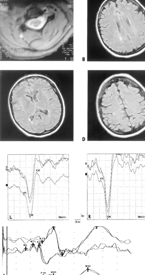
protein content (354 mg/l (refer-ence < 480 mg/l)) of the cere-brospinal fluid were normal, but intrathecal IgG production was shown by the presence of four oligoclonal bands and an elevated IgG Index (13.6, reference < 7.0). MRI of the cervical spine showed two non-enhancing lesions right dorso-laterally at levels C3/4 and C2/3 (Fig. 1A). Cranial MRI re-vealed two right-hemispheric ovoid lesions in periventricular (Fig. 1B and C) and juxtacortical areas (Fig. 1D). Visual evoked potentials of both eyes were within normal limits, but the side difference was abnormal (Fig. 2A). Somato-sen-sory evoked potentials recorded after stimulation of the left arm showed a markedly reduced
ampli-tude of the potentials recorded from the scalp (Figs. 2B and C).
A diagnosis of laboratory sup-ported definite MS (according to Poser’s criteria) or of MS (accord-ing to the new diagnostic criteria by McDonald et al. [8], i. e., two attacks, clinical evidence of one lesion, three MRI-detected lesions consistent with MS plus positive CSF) was made. The patient im-proved after injection of botulinum toxin A into the neck muscles com-bined with pulsed high-dose intra-venous methylprednisolone (500 mg daily for five days). She received the last injection of botu-linum toxin 12 months after the first presentation. Fifteen and 27 months after the first presentation, she showed only minimal signs of cervical dystonia (both times 3 points on the Tsui scale). Her med-ication at the last consultation con-sisted of tizanidine 6 mg/d, alpra-zolam 1 mg/d, paroxetine 40 mg/d, and mianserine 30 mg/d. Except for tizanidine, the other medications were for psychiatric symptoms.
In most cases of focal dystonia, the cause remains unknown, lead-ing to a diagnosis of primary dystonia. In contrast, some clinical “red flags” were present in our case and pointed to secondary focal dystonia. Sensory deficits in the C2 and C3 dermatomes and persis-tence of dystonia during sleep were suggestive of secondary focal dystonia. Paraclinical examinations such as imaging, electrophysiologi-cal and laboratory testing sup-ported the diagnosis of MS.
We cannot exclude a coinci-dence of MS and focal dystonia in our case. However, the probability of such a coincidence is extremely low (1: 3.500.000) according to re-ported prevalences of MS in Italy of 50/100.000 [12], and of cervical dystonia of 57/100.000 [3]. Further-more, the persistence of cervical dystonia, also during sleep, is a strong argument for secondary,
LETTER TO THE EDITORS
1409
Fig. 1 A: MRI of the cervical spine: T2-weighted ax-ial section at level C3/C4. Hyperintense lesion is pre-sent right dorso-laterally. B–D: MRI of the head. FLAIR sequences. Right-hemispheric ovoid lesions in periventricular (B, C) and juxtacortical location (D). Note rightward head tilt due to dystonia. The lesions did not enhance with gadolinium in T1-weighted im-ages
Fig. 2 A: Visual evoked potentials. The latency of component P100 on both sides (right: 99.0 ms; left: 108.0 ms) is within the range of normal values, but the side difference (9.0 ms, normal < 6.0 ms) is pathological, with prolongation of recording after stimulation of the left eye. The upper two traces: av-eraged curves of 200 recordings each. Lower traces: average of the two curves. L denotes recording after stimulation of the left eye; R, after stimulation of the right eye. B and C: Somato-sensory evoked poten-tials: Recordings after stimulation of the right median nerve at the wrist (B); of the left median nerve at the wrist (C). The top traces represent the recordings from the scalp, the bottom traces, from the processus
spinosus C6. The reference was at Fz. Latencies of P15
and N20 are within normal limits, without patholog-ical side differences. Components N9, N11, and N13 after stimulation of the left arm can not be identified (probably for technical reasons in the presence of dystonia); the potentials recorded from the scalp show a markedly reduced amplitude (diminution of amplitude N20-P25 by 86 %; normal < 45 %) A B C A C D B
1410
i. e., in this case MS-related, dysto-nia [11]. Thus, we propose the alteration of central processing produced by an MS lesion as the more likely explanation for the cervical dystonia in the present case [7]. Anecdotal reports de-scribe torticollis resulting from different cervical lesions [2, 4] and the association of spinal MS plaques with torticollis [6] and hand dystonia [15]. However, dys-tonia was not the first manifesta-tion of MS in any of these reports.
To conclude, MS may probably be an underdiagnosed cause of secondary focal dystonia, and con-versely, secondary focal dystonia may herald multiple sclerosis and should be included in the differen-tial diagnosis of patients present-ing with this clinical feature.
References
1. Berger JR, Sheremata WA, Melamed E (1984) Paroxysmal dystonia as the ini-tial manifestation of multiple sclerosis. Arch Neurol 41:747–750
2. Cammarota A, Gershanik OS, Garcia S, Lera G (1995) Cervical dystonia due to spinal cord ependymoma: involvement of cervical cord segments in the patho-genesis of dystonia. Mov Disord 10: 500–503
3. Epidemiological Study of Dystonia in Europe (ESDE) Collaborative Group (2000) A prevalence study of primary dystonia in eight countries. J Neurol 247:787–792
4. Fujimoto N, Nabatame H, Nakamura K, Konishi T, Kamiya Y (1991) Spinal epidural abscess as the cause of torti-collis – diagnosis by magnetic reso-nance imaging, (article in Japanese). Rinsho Shinkeigaku 31:49–53 5. Keegan BM, Noseworthy JH (2002)
Multiple Sclerosis. Annu Rev Med 53:285–302
6. KlostermannW, Vieregge P, Kompf D (1993) spasmodic torticollis in multi-ple sclerosis: significance of an upper cervical spinal cord lesion. Mov Disord 8:234–236
7. LeDoux MS, Brady KA (2003) Sec-ondary cervical dystonia associated with structural lesions of the central nervous system. Mov Disord 18:60–69 8. McDonald WI, Compston A, Edan G,
et al. (2001) Recommended diagnostic criteria for multiple sclerosis: Guide-lines from the international panel on the diagnosis of multiple sclerosis. Ann Neurol 50:121–127
9. Rozza L, Bortolotti P, Sica A, Weronig S, Orrico D (1993) Kinesigenic dystonia as the first manifestation of multiple sclerosis with cervical and brainstem lesions. Eur Neurol 33:331–332 10. Svetel M, Sternic N, Filipovic S, Kostic
V (1997) Spasmodic torticollis associ-ated with multiple sclerosis: report of two cases. Mov Disord 12:1092–1094 11. Svetel M, Ivancovic N, Marinkovic J, Jovic J, Dragasevic N, Kostic VS (2004) Characteristics of dystonic movements in primary and symptomatic dysto-nias. J Neurol Neurosurg Psychiatry 75:329–330
12. Tassinari T, Parodi S, Badino S, Vercelli M (2001) Mortality trend for multiple sclerosis in Italy (1974–1993). Eur J Epidemiol 17:105–110
13. Tranchant C, Bhatia KP, Marsden CD (1995) Movement disorders in multi-ple sclerosis. Mov Disord 10:418–423 14. Tsui JKC, Eisen A, Stoessel AJ, Calne S
(1986) Double blind study of botu-linum toxin in spasmodic torticollis. Lancet 328:245–247
15. Uncini A, Di Muzio A, Thomas A, Lu-garesi A, Gambi D (1994) Hand dysto-nia secondary to cervical demyelinat-ing lesion. Acta Neurol Scand 90:51–55 16. Yücesan C, Tuncel D, Akbostanci MC,
Yücemen N, Mutluer N (2000) Hemidystonia secondary to cervical demyelinating lesions. Eur J Neurol 7:563–566 St. J. Rüegg, MD · M. Bühlmann, MD · S. Renaud, MD · A. J. Steck, MD · L. Kappos, MD · P. Fuhr, MD Department of Neurology University Hospital Petersgraben 4 4031 Basel, Switzerland Tel.: +41-61/265-4167 Fax: +41-61/265-5638 E-Mail: pfuhr@uhbs.ch
This case report was presented as a poster at the Annual Meeting of the Movement Disorders Society, held in Miami, Novem-ber 7–11, 2002.