MOLECULAR AND GENOMIC PHYSIOLOGY
Gene expression analysis defines the proximal tubule
as the compartment for endocytic receptor-mediated
uptake in the
Xenopus pronephric kidney
Erik I. Christensen&Daniela Raciti&Luca Reggiani&
Pierre J. Verroust&André W. Brändli
Received: 16 January 2008 / Accepted: 28 February 2008 / Published online: 13 June 2008
# Springer-Verlag 2008
Abstract Endocytic receptors in the proximal tubule of the mammalian kidney are responsible for the reuptake of numerous ligands, including lipoproteins, sterols, vitamin-binding proteins, and hormones, and they can mediate drug-induced nephrotoxicity. In this paper, we report the first evidence indicating that the pronephric kidneys of Xenopus tadpoles are capable of endocytic transport. We establish that the Xenopus genome harbors genes for the known three endocytic receptors megalin/LRP2, cubilin, and amnionless. The Xenopus endocytic receptor genes share extensive synteny with their mammalian counterparts. In situ hybridizations demonstrated that endocytic receptor expression is highly tissue specific, primarily in the pronephric kidney, and did not occur prior to neurulation. Expression was strictly confined to proximal tubules of the pronephric kidney, which closely resembles the situation reported in mammalian kidneys. By immunohistochemistry, we demonstrated that Xenopus pronephric tubule epithelia express high amounts of the endocytic receptors megalin/
lrp2 and cubilin in the apical plasma membrane. Further-more, functional aspects of the endocytic receptors were revealed by the vesicular localization of retinol-binding protein in the proximal tubules, probably representing endocytosed protein. In summary, we provide here the first comprehensive report of endocytic receptor expression, including amnionless, in a nonmammalian species. Re-markably, renal endocytic receptor expression and function in the Xenopus pronephric kidney closely mirrors the situation in the mammalian kidney. The Xenopus proneph-ric kidney therefore represents a novel, simple model for physiological studies on the molecular mechanisms under-lying renal tubular endocytosis.
Keywords Xenopus . Endocytosis . Proximal tubule . Renal function . Gene expression . Kidney . Morphology
Introduction
The development of the vertebrate kidney involves the progressive formation of three distinct kidneys: the pro-nephros, the mesopro-nephros, and the metanephros [1]. The pronephros or pronephric kidney, while vestigial in higher vertebrates, is the functional embryonic kidney of amphib-ians. It consists of a single nephron and therefore represents the simplest vertebrate excretory organ [2, 3]. Due to its simple organization and the amenability of Xenopus embryos to experimental manipulation, the Xenopus pro-nephric kidney has emerged as an attractive model for studying human kidney development and function [4, 5]. Using gain- and loss-of-function approaches, several genes involved in pronephric kidney development and differenti-ation have been identified and characterized including key players such as wnt4, bicaudal-c, fgf8, notch, and irx3 [6–
DOI 10.1007/s00424-008-0488-3
Electronic supplementary material The online version of this article (doi:10.1007/s00424-008-0488-3) contains supplementary material, which is available to authorized users.
E. I. Christensen (*)
Department of Cell Biology, Institute of Anatomy, University of Aarhus,
Aarhus, Denmark e-mail: [email protected]
D. Raciti
:
L. Reggiani:
A. W. Brändli (*) Institute of Pharmaceutical Sciences,Department of Chemistry and Applied Biosciences, ETH Zürich, Zürich, Switzerland
e-mail: [email protected] P. J. Verroust
INSERM U538, Centre Hospitalier Universitaire, Saint Antoine, Paris, France
10]. Despite the long-known fact that functional pronephric kidneys are essential for survival of amphibian larvae [11], the range of physiological functions performed by the pronephros and their analogies to metanephric renal physiology still remain poorly characterized. Renal endo-cytosis, for example, has to our knowledge not been studied in Xenopus to date.
Megalin/LRP2 and cubilin are large endocytic receptors, 600 and 460 kDa, respectively, that are highly expressed in the apical endocytic apparatus and in apical microvilli in the mammalian proximal tubule [12]. Both receptors have also been localized to the proximal part of the pronephros in zebrafish where also fluid phase and receptor-mediated endocytosis were shown to take place [13]. The two receptors appear to be responsible for proximal tubular uptake of the majority of proteins filtered in mammalian glomeruli [12] including the carrier proteins retinol-binding protein (RBP) [14], vitamin D-binding protein [15, 16], transcobalamin [17], transferrin [18], and albumin [19–21], which illustrate the importance of renal endocytic receptors for minimizing urinary losses of vitamins and other solutes. It is interesting to note that many ligands bind to both receptors [12]. Megalin/LRP2 appears also to be involved in aminoglycoside nephrotoxicity by its ability to bind and mediate uptake of different aminoglycosides including gentamicin [22, 23]. Cubilin is a peripheral membrane protein, and several data suggest that the internalization of cubilin and its ligands at least in part is carried out by megalin/LRP2 [18,24,25]. Finally, amnionless (AMN) is a 45–50-kDa transmembrane protein apparently involved in cellular processing of cubilin and endocytosis of cubilin and its ligands [26–29]. The importance of AMN for endocytosis of cubilin and its ligands in megalin/LRP2-expressing cells is, however, unknown.
Megalin/LRP2 knockout mice [30] and conditional renal megalin/LRP2 knockout mice [31, 32] have been invalu-able tools in revealing the megalin/LRP2 function in the kidney. Conditional knockout mice present with low-molecular-weight proteinuria, while the conventional knockouts display in addition developmental abnormalities including holoprosencephalic syndrome [30]. Recently, mutations in the megalin/LRP2 gene have been identified in families with Donnai–Barrow syndrome and facio-oculo-acoustico-renal syndrome [33].
A strain of dogs carrying an amnionless (amn) gene mutation have been instrumental in unraveling the function of cubilin [28, 34]. Cubilin is also responsible for the intestinal uptake of intrinsic factor-vitamin B12 complexes. The AMN deficiency results in a lack of apical expression of cubilin in the proximal tubule and small intestine, and as a consequence, the affected dogs develop megaloblastic anemia 1 and low-molecular-weight proteinuria [16, 35]. Human counterparts are represented by the Imerslund–
Gräsbeck syndrome, where Finnish families carry muta-tions in the cubilin gene [36], and families from Norway and Turkey have mutations in the AMN gene [37].
The Xenopus pronephros is a bilateral excretory organ that consists of a single nephron composed of three basic components: (1) the glomus or glomerulus, which is the site of blood filtration, (2) the tubules, where filtrate resorption occurs, and (3) the duct, which carries the urine to the cloaca [2,38]. Recent studies have demonstrated that the organiza-tion of the pronephric nephron is more complex than previously anticipated. Distinct domains and subdomains within the tubule and duct compartments have been defined based on the localized expression of selected membrane transporters and ion channels [39, 40]. On the basis of a large-scale gene expression pattern screen, a comprehensive model of the segmental organization of Xenopus pronephric kidney was recently reported that suggests remarkable functional correlations to the segments of the mammalian kidney [7]. For example, the proximal tubule is divided into three segments (PT1, PT2, and PT3), which largely correspond to the S1, S2, and S3 segments of the mam-malian proximal tubule. Taken together, the characteristic hallmarks of vertebrate nephron organization—the presence of distinct segmented tubular compartments—can be delin-eated already at the level of the Xenopus pronephric nephron. To date, it is not known whether endocytic receptors are expressed in Xenopus pronephric kidney and whether this simple excretory organ is capable of renal tubular endocy-tosis. In this paper, we examined the Xenopus tropicalis genome and screened expressed sequence tag (EST) data-bases for Xenopus laevis and X. tropicalis complementary deoxyribonucleic acids (cDNAs) encoding AMN, cubilin, and megalin/LRP2. We show that all three genes are coexpressed in the pronephric kidney, where their expression domains are confined to the proximal tubule. In immunocy-tochemical studies, we localized cubilin and megalin/LRP2 expression to the apical plasma membrane of proximal tubules, where they are likely to be engaged in active endocytic protein transport as evidenced by the presence of RBP localizing to intracellular vesicles. The highly con-served renal expression domains of Xenopus and mammalian endocytic receptor genes suggest that the Xenopus embryo could serve as an alternative, simple animal model to study the physiology of endocytic receptor function.
Materials and methods
Nomenclature for Xenopus genes
The standard gene nomenclature suggested by Xenbase (http://www.xenbase.org/gene/static/geneNomenclature.jsp) and adopted by the National Center for Biotechnology
Information (NCBI) for X. laevis and X. tropicalis genes is utilized rather than the original gene names to maximize compatibility with data available from other model systems. Xenopus gene names are written in lower case. Where possible, Xenopus gene names are the same as the human orthologs. When a gene is duplicated in Xenopus relative to mammals the duplicated genes are tagged with“.1” and “.2.” Genome analysis
Synteny maps of endocytic receptor genes found in the human, mouse, and X. tropicalis genomes were retrieved from the Ensembl genome browser (http://www.ensembl. org; release 45) by virtue of gene name search and basic local alignment search tool (BLAST) sequence similarities. Identification and sequencing of Xenopus cDNAs encoding endocytic receptors
The predicted X. tropicalis cubilin sequence (protein ID 178868) was retrieved from the X. tropicalis genome assembly (version 4.1) website (http://genome.jgi-psf.org/ Xentr4) of the Joint Genome Institute (JGI). None of transcripts predicted by Ensembl encoded the complete open reading frame (ORF) of cubilin. They lacked the sequences encoding the signal peptide and the CUB domains 25, 26, and 27. Predicted nucleotide sequences of X. tropicalis gene transcripts encoding AMN and megalin/ lrp2 were retrieved from the Ensembl X. tropicalis genome browser (http://www.ensembl.org/Xenopus_tropicalis; re-lease 45). In cases where multiple transcripts were predicted, the longest transcript was selected. The Ensembl transcript IDs of the X. tropicalis sequences are as follows: AMN (ENSXETT00000027872) and megalin/lrp2 (ENS XETT00000035400). X. tropicalis AMN transcript harbored, however, only a partial ORF. Similarly, the longest anno-tated X. tropicalis megalin/lrp2 transcript (ENSXET T00000035400) lacked the N terminus. The predicted megalin/lrp2 gene model (protein ID 353026), which was retrieved from JGI X. tropicalis genome assembly (version 4.1), contained the missing 12 N-terminal amino acids. They were identified as“MHFNDIQFTFAA.”
Screening of nonredundant and EST nucleotide data-bases for X. laevis cDNAs encoding endocytic receptors was performed at the NCBI BLAST website (http://www. ncbi.nlm.nih.gov/BLAST). The X. tropicalis amino acid sequences for AMN, cubilin, and megalin/lrp2 were used as protein queries in TBLASTN searches, which compare a protein sequence to the six-frame translations of a nucleo-tide database. X. laevis cDNAs encoding the complete ORF of AMN (GenBank acc. no. NM_001092600) and several X. laevis EST cDNAs encoding partial ORFs of megalin/ lrp2 (CF522099, CD302505) and cubilin (DT081913,
CB2008401) were identified. Only one (GenBank CB208401; IMAGE:6881221) of the two partial X. laevis cubilin cDNAs identified could be verified by sequencing. In addition, X. tropicalis EST cDNAs encoding partial ORFs of cubilin (CX902985, CX982421) were retrieved.
The corresponding Xenopus cDNAs were obtained from the RZPD German Resource Center for Genome Research/ imaGenes. The following cDNAs were verified by DNA sequencing: X. tropicalis cubilin (CX982421) and X. laevis AMN (NM_001092600), cubilin (CB208401), and megalin/ lrp2 (CD302505). Double-stranded sequencing of the X. tropicalis cubilin (CX982421) and X. laevis megalin/lrp2 (CD302505) EST cDNAs was performed by Microsynth AG (Balgach, Switzerland). The resulting 3,137-bp nucleotide sequence of X. tropicalis cubilin encoded 994 residues of the C terminus of the protein encompassing part of CUB domain 19 and all the CUB domains from 20 to 27. The nucleotide sequence of X. laevis megalin/lrp2 had a length of 1,976 bp and encoded the C-terminal 572 residues of megalin/lrp2 starting upstream of the LDL-R class B repeat 35 in the extracellular domain and encompassing the transmembrane and cytoplasmic domains. The partial X. laevis megalin/lrp2 protein shared greater than 90% amino acid identity with X. tropicalis megalin/lrp2. In contrast to the predicted X. tropicalis megalin/lrp2 transcript, the partial X. laevis megalin/lrp2 transcript contained also the EGF repeats 16 and 17. The nucleotide sequences were deposited with GenBank: X. laevis megalin/lrp2 (GenBank acc. no. EU124653) and X. tropicalis cubilin (EU127292).
Sequence and phylogenetic analysis
The references sequences of human, mouse, and chicken endocytic receptors were retrieved from GenBank. The deduced human AMN, cubilin, and megalin/LRP2 proteins were used as queries in TBLASTN database searches to identify the corresponding Drosophila homologs. Analysis of nucleotide and protein sequences was performed using the DNAStar Lasergene software package (version 6.0). Signal peptides and transmembrane domains were predicted using web-based software at the SignalP 3.0 Server (http:// www.cbs.dtu.dk/services/SignalP/) and the DAS-TMfilter server (http://mendel.imp.ac.at/sat/DAS/DAS.html), respec-tively. Amino acid sequence alignments were generated with MegAlign (DNAStar) using the Clustal W algorithm and the identity residue weight table. The alignments served as a basis to compute phylogenetic trees with the neighbor-joining algorithm [41].
The GenBank accession numbers of the sequences used for phylogenetic analysis are as follows: chicken AMN, XM_ 421379; chicken cubilin, XM_001235155; chicken megalin/ LRP2, XM_422014; Drosophila CG11592, NM_134671; Drosophila CG32702, NM_167193; Drosophila CG12139,
NM_132335; human AMN, NM_030943; human cubilin, NM_001081; human Megalin/LRP2, NM_004525; mouse Amn, NM_033603; mouse cubilin, NM_001081084; mouse megalin/Lrp2, NM_001081088; X. laevis amn, NM_ 001092600; X. laevis megalin/lrp2, EU124653; and X. tropicalis cubilin, EU127292. The predicted amino acid se-quences for X. tropicalis amn (Ensembl peptide ID: ENS XETP00000027872) and megalin/lrp2 (ENSXETP000000 35400) were retrieved from the Ensembl genome browser. The X. tropicalis megalin/lrp2 sequence was amended to contain the complete N terminus as described above. The X. tropicalis cubilin sequence (protein ID 178868) was retrieved from the X. tropicalis genome assembly version 4.1. Embryo manipulations, in situ hybridizations, and mapping of gene expression
In vitro fertilization, culture, and staging of X. laevis embryos were performed as previously described [42,43]. In situ probe synthesis, whole-mount in situ hybridization, and bleaching of Xenopus embryos were carried out according to [8, 43, 44]. The X. laevis cubilin cDNA (CB208401) was amplified by polymerase chain reaction (PCR) using the Expand High Fidelity PCR System (Roche Diagnostics), subcloned into the pGEM-TEasy (Promega) vector, and confirmed by DNA sequencing. Digoxigenin-labeled probes were generated from linearized plasmids encoding X. tropicalis cubilin (GenBank acc. no. EU127292), and X. laevis amn (NM_001092600), cubilin (pGEM-cubn), and megalin/lrp2 (EU124653). Sense con-trols were tested negative by in situ hybridization. Digital photographs of stained embryos were taken with an AxioCam Color camera mounted on a Zeiss SteREO Lumar.V12 stereoscopic microscope. Pronephric gene expression patterns of endocytic receptors were mapped onto the contour model of the stage 35/36 pronephric nephron using unambiguous morphological landmarks as described previously [7]. In brief, these included the nephrostomes, the characteristically broad proximal tubule domain known as PT3, and the looped part of the pronephric nephron, which consists of IT1, IT2, and DT1. Electron microscopy and immunohistochemistry
For electron microscopy (EM) and immunohistochemistry, Xenopus embryos (stage 35/36 and 40) were fixed in 3.5% paraformaldehyde (PFA) for 2 h and then transferred to 1% PFA. For cryosectioning, the embryos were subsequently frozen. For EM or paraffin immunohistochemistry, PFA-fixed embryos were either embedded in low-temperature lowicryl [45] or paraffin. For EM morphological studies, PFA-fixed embryos were postfixed in 1% glutaraldehyde, 1% OsO4, and
embedded in epon. Semithin (0.8 μm) or ultrathin (70–
90 nm) cryosections were obtained at−100°C with an FCS Reichert Ultracut S cryo-ultramicrotome (Leica). Ultrathin, 60-nm lowicryl sections were obtained with an FCS Reichert Ultracut S ultramicrotome. Paraffin sections were cut at 2 μm. For light microscopy immunolabeling, the sections were preincubated in phosphate-buffered saline containing 5 mM glycine and 1% bovine serum albumin and then incubated with the primary antibodies (Sheepα-rat megalin/ LRP2 [46], 1:5,000; rabbitα-rat cubilin [47], 1:1,000; rabbit α-RBP [Dako A/S, Glostrup, Denmark], 1:2,000) at room temperature for 1 h. The sections were subsequently incubated with the relevant peroxidase-conjugated secondary (rabbit α-sheep and goat α-rabbit) antibodies (Dako). Peroxidase was visualized with diaminobenzidine, and the sections were subsequently counter stained with Meier’s stain for 2 min. The sections were examined with a Leica DMR microscope equipped with a Leica DFC320 digital camera. For EM immunolabeling, the sections were incubated with primary antibodies (α-megalin/LRP2, 1:2,000; α-cubilin, 1:200) at 4°C overnight followed by incubation at room temperature for 1 h with 10-nm gold particles coupled to the relevant immunoglobulins (BioCell, Cardiff, UK). The cryosections were embedded in methylcellulose containing 0.3% uranyl acetate, and the lowicryl sections were stained with uranyl acetate and lead. The sections were studied with a FEI CM100 electron microscope.
Computer graphics
All digital images were processed in Adobe Photoshop 8.0. Composite figures were assembled and labeled either with Adobe Illustrator CS2 or Adobe InDesign CS2. Schematic figures were drawn using Adobe Illustrator CS2.
Results
Isolation of Xenopus sequences encoding endocytic receptors
We used the human reference sequences of AMN, cubilin, and megalin/LRP2 as baits to survey public repositories of non-redundant nucleotide, EST, and genomic data to identify X. laevis and X. tropicalis sequences. For both Xenopus species, we identified distinct sequences encoding the three endocytic receptors (Supplementary Figs. S1–S3), which will be discussed in detail below.
Xenopus amnionless
Querying the X. tropicalis genome assembly (Ensembl, release 45), we found that the amn gene (ENSEXT G00000012751) was located on scaffold 332. For X. laevis,
a full-length AMN cDNA (GenBank acc. no. NM_ 001092600) encoding a deduced protein of 483 amino acids was identified. The two Xenopus amn gene products shared 89% identity at the nucleotide level and 85% at the amino acid level. A comparison with other AMN proteins revealed that the X. laevis protein shared 44% amino acid identity with the chicken and 39% with its mammalian counterparts. Supplementary Fig. S1shows an alignment of the predicted amino acid sequences of human and the Xenopus AMN proteins. The structural features conserved among these species included the predicted signal peptide cleavage site, 12 cysteine residues in the mature extracel-lular domain with nine clustered in the cysteine-rich domain (CRD), a single transmembrane domain, and a cytoplasmic domain harboring two amino acid sequences conforming to a consensus motif (F/Y)XNPX(F/Y) for ligand-independent endocytosis via clathrin-coated pits [27]. The N-terminal portion, which includes the CRD, is the most conserved region of vertebrate AMN proteins. Xenopus cubilin
Homology searches of Ensembl’s X. tropicalis genome assembly resulted in the identification of a single cubilin gene (ENSXETG00000007453) located on scaffold 437. The predicted complete cubilin ORF (PID 178868) was retrieved from the X. tropicalis genome assembly (Version 4.1) of the JGI. The X. tropicalis cubilin ORF has a length of 10,617 bases and encodes an amino acid sequence of 3,538 residues. Comparison of the predicted full-length X. tropicalis cubilin protein with other vertebrate cubilin proteins revealed highest overall amino acid sequence identity with chicken cubilin (62%; 3,729 residues) and lower values with the human (57%; 3,624 residues) and mouse cubilin (53%; 3,624 residues) proteins.
As its human counterpart, X. tropicalis cubilin encodes a peripheral membrane protein with a signal peptide, a con-served cleavage site for the endopeptidase furin and a 97-amino acid domain containing a single cysteine residue. This is followed by a cluster of eight contiguous epidermal growth factor (EGF) repeats, each ~40 amino acids in length and harboring six conserved cysteine residues, and a large cluster of 27 CUB domains, each about 100–110 amino acids in length (Supplementary Fig. S2). Most CUB domains contain signature glycine and phenylalanine residues and four conserved cysteine residues, which probably form two disulfide bridges (C1–C2; C3–C4) [48,49]. The following exceptions were found (Supplementary Fig. S2). CUB domains 6 and 15 contained only three instead of the four cysteine residues, suggesting that they form only one disulfide bridge. It is interesting to note that the CUB domain 6 of human cubilin is also predicted to contain only one disulfide bridge [50]. CUB domain 13 lacks, as was
described for mammalian cubilin proteins, the first two cysteine residues that are suggested to form the upstream first disulfide bond. Finally, the predicted X. tropicalis cubilin ORF contains a large deletion in CUB domain 27.
Mutations in the human cubilin gene cause hereditary megalobastic aneamia 1 (OMIN 261100) of the Finnish type [36]. The majority of patients carry a missense mutation changing proline to leucine (P1297L) in CUB domain 8. It interesting to note that this proline residue and the flanking amino acids are highly conserved as they are also found in the CUB domain 8 of X. tropicalis cubilin (Supplementary Fig. S2).
Xenopus megalin/lrp2
We identified a single megalin/lrp2 gene (ENSXET G00000016214) located on scaffold 236 in the X. tropicalis genome assembly. The complete amino sequence of X. tropicalis megalin/lrp2 of 4,507 residues was assembled in silico (see “Materials and methods”). The overall amino
acid identity shared between X. tropicalis megalin/lrp2 with its chicken counterpart was 79%, whereas the values were slightly lower with human (69%) and mouse (68%) megalin/LRP2 proteins. Amino acid sequence alignments revealed that the overall domain structure of X. tropicalis megalin/lrp2 and its human counterpart was well conserved (Supplementary Fig. S3). The large, 4,285-residue extra-cellular domain is comprised of the characteristic fourfold duplicated low-density lipoprotein receptor (LDL-R) extra-cellular domain consisting of three types of proteins folds: the ligand-binding LDL-R class A repeats, the EGF repeats, and the LDL-R class B repeats containing the YWTD motif [51, 52]. In line with human megalin/LRP2, X. tropicalis megalin/lrp2 contained 36 LDL-R class A repeats and 37 LDL-R class B repeats. It is interesting to note, however, that only 15 out of the 17 EGF repeats were present in the predicted X. tropicalis megalin/lrp2 protein. Sequences encoding EGF repeats 16 and 17 were absent due to a small deletion in the predicted nucleotide sequence. A single transmembrane domain and a comparatively short cytoplasmic domain containing two (F/Y)XNPX(F/Y) motifs, which are important for endocytosis, followed the large extracellular domain.
Human megalin/LRP2 gene mutations were reported to cause Donnai–Barrow and facio-oculo-acoustico-renal syndromes [33]. Affected individuals showed nonsense, splice junction, frameshift, or missense mutations that are believed to be functionally null. The only missense mutation known to date leads to the substitution of a tyrosine for a histidine residue (Y2522H) in the LDL-R class B repeat 27. This tyrosine residue is evolutionary conserved and present also in X. tropicalis megalin/lrp2 (Supplementary Fig. S3).
Genomic synteny and molecular phylogeny of endocytic receptor genes
We examined next the synteny maps between human, mouse, and X. tropicalis to confirm the identity of the Xenopus endocytic receptor genes. The synteny maps were derived from the Ensembl genome browser (Release 45— June 2007). Diagrammatic representations of the synteny maps including the gene orientation are presented in Figs.1,
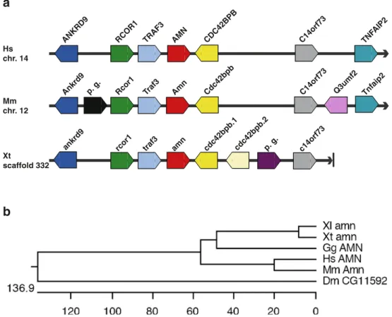
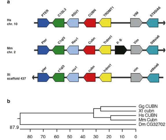
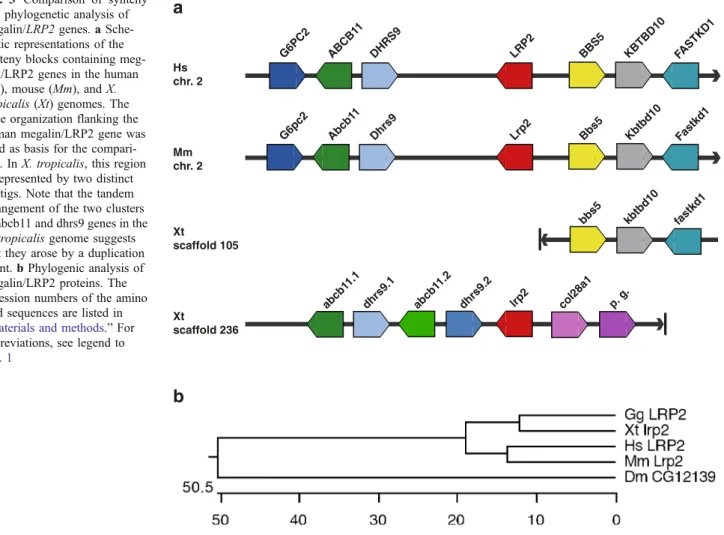
2, and 3. The flanking genes were remarkably conserved between the amn genes in all three genomes examined here (Fig.1a). The conserved syntenic regions consisted of the Rcor1, Traf3, Amn, and Cdc42bpb genes. A similar situation was observed for the cubilin genes, where the core of the syntenic region was comprised of the following genes: Pter, C1ql3, Rsu1, Cubn, and Trdmt1 (Fig. 2a). While the synteny blocks flanking the megalin/LRP2 genes were completely conserved in the human and mouse genomes, the situation for megalin/lrp2 in the X. tropicalis
genome was more complex (Fig. 3a). The Xenopus megalin/lrp2 gene is located on scaffold 236. The syntenic cluster of genes downstream of the mammalian megalin/ LRP2 genes comprised of Bbs5, Kbtbd10, and Fastkd1 was, however, not located on scaffold 236 but on scaffold 105. Furthermore, col28a1 is located downstream of megalin/lrp2 in the X. tropicalis genome. The synteny block immediately upstream of megalin/LRP2 consists in mammalian genomes of G6pc2, Abcb11, and Dhrs9. Similarly, the megalin/lrp2 gene is linked upstream to the abcb11 and dhrs9 genes, which are, however, present as a tandem duplicated clusters in the X. tropicalis genome.
We performed also phylogenetic analysis of endocytic receptor proteins to assess the orthology assignments by protein sequence conservation (Figs. 1b, 2b, and 3b). Besides human, mouse, chicken, and Xenopus proteins, we also included those Drosophila proteins showing the highest amino acid identities to human endocytic receptors proteins. Queries of the Drosophila genome identified the
b
a
AMN TRAF3 RCOR1 ANKRD9 CDC42BPB C14orf73 Hs chr. 14 Mm chr. 12 Xt scaffold 332 TNF AIP2p. g. Rcor1 Traf3 Amn
Ankrd9 Cdc42bpb C14orf73 Tnfaip2
amn traf3 rcor1
ankrd9 cdc42bpb.1cdc42bpb.2p. g. c14orf73
Q3umf2
Fig. 1 Comparison of synteny and phylogenetic analysis of AMN genes. a Schematic representations of the synteny blocks containing amn genes in the human (Hs), mouse (Mm), and X. tropicalis (Xt) genomes. The gene organization flanking the human AMN gene was used as basis for the comparison. The chromosomal localization is indicated on the left. For X. tropicalis, the scaffold number is given. Each gene is represented as a broad arrow indicating the orientation of the transcription unit. The relative spacing between the genes is not shown because it does not influence the assignment of synteny. Orthologous genes are shown in the same color. Predicted genes (p.g.)
that are species-specific are indicated. Note that the X. tropicalis genome contains to two cdc42 bpb genes (cdc42bpb.1, cdc42 bpb42.2), which probably arose by segmental duplication. b Phylogenic analysis of AMN proteins. The scale bar measures the distance between the sequences. Units indicate nucleotide substitution events. The accession numbers of the amino acid sequences are listed in“Materials and methods.” Abbreviations: Dm Drosophila melanogaster, Gg Gallus gallus, Hs Homo sapiens, Mm Mus musculus, Xl X. laevis, Xt X. tropicalis
following genes: Drosophila CG11592 represents an AMN homolog [53, 54] sharing amino acid identities ranging from 16% to 19% with its vertebrate counterparts. CG32702 encodes a Drosophila cubilin homolog with 28% overall amino acid identity to vertebrate cubilin pro-teins. CG12139 was identified as a Drosophila megalin/ LRP2 homolog sharing amino acid identities of 42–44% with vertebrate megalin/LRP2 proteins. The phylogenetic analysis revealed that the Xenopus endocytic receptor proteins are most closely related to their chicken orthologs. Overall, the calculated molecular phylogenies agreed well with the widely accepted evolutionary relationships be-tween Drosophila and vertebrate species [55, 56]. Taken together, the assigned orthology of the Xenopus endocytic receptor genes was confirmed by conserved gene synteny and molecular phylogeny.
Developmental expression of Xenopus amnionless, cubilin, and megalin/lrp2 genes
We performed whole-mount in situ hybridizations to examine the expression of endocytic receptor genes during embryogenesis of X. laevis. Embryos ranging from stage 18 (20 h postfertilization) to stage 40 (66 h postfertilization) were analyzed to cover the time period between neural plate stage to the establishment of functional cardiovascular and
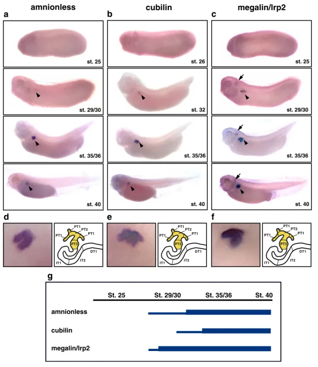
excretory organ systems (Fig. 4; Supplementary Fig. S4). Transcription of megalin/lrp2 was already evident at stage 18 in the developing nervous system with highest expres-sion in the brain (Supplementary Fig. S4). In contrast, no regionalized gene expression could be detected for amn and cubilin prior to embryos reaching tailbud stages. At stage 29/30, expression of amn and megalin/lrp2 became appar-ent in the developing pronephric anlage (Fig. 4a,c). This was followed by the onset of pronephric cubilin expression at stage 32 (Fig. 4b). Pronephric expression of all three endocytic receptor genes persisted at least until stage 40 (Fig. 4a–c,g). In addition, megalin/lrp2 expression also occurred in the developing brain, eyes, and otic vesicles (Fig. 4c). It is interesting to note that all three endocytic receptor genes were coexpressed in a proximal region of the nephron, whereas no expression could be detected in more distal parts. Using morphological landmarks (see“Materials and methods”), we mapped the proximal expression
domains in the stage 35/36 pronephric nephron to cover PT1, PT2, and PT3 (Fig. 4d–f).
Ultrastructural analysis of the proximal tubules of Xenopus laevis
Given that the expression of endocytic receptors was restricted to proximal tubules, we performed EM to study
a
CUBN RSU1 C1QL3 PTER TRDMT 1 VIM Hs chr. 10 Mm chr. 2 Xt scaffold 437 ST8SIA6 p. g. Cubn Rsu1 C1ql3 Pter Trdmt1 Vim St8sia6 cubn rsu1 c1ql3pter trdmt1 vim st8sia6
b
Fig. 2 Comparison of synteny and phylogenetic analysis of cubilin genes. a Schematic rep-resentations of the synteny blocks containing cubilin genes in the human (Hs), mouse (Mm), and X. tropicalis (Xt) genomes. The gene organization flanking the human CUBN gene was used as basis for the compari-son. b Phylogenic analysis of cubilin proteins. The accession numbers of the amino acid se-quences are listed in“Materials and methods.” For abbrevia-tions, see legend to Fig.1
the ultrastructure. The proximal tubules in the pronephric kidney at stage 40 presented with a very-well-developed brush border and an extensively developed apical endocytic apparatus including coated pits, endosomes, recycling dense apical tubules, and lysosomes (Fig. 5a–d). In
addition, most cells had large lipid droplets in the basal cytoplasm (Fig.5a,b). The cells were cuboidal and did not show the extensive interdigitating lateral processes contain-ing a large number of mitochondria that is characteristic for mammalian proximal tubules [57]. Instead, these cells have small folds or microvilli that project into the lateral intercellular spaces (Fig. 5a–c). This indicates that the
Xenopus proximal tubule in the stage 40 embryo has limited fluid absorption ability.
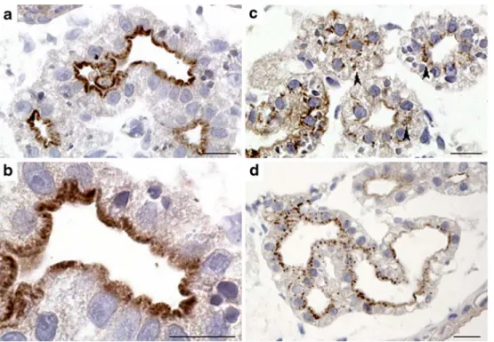
Subcellular expression of megalin/lrp2 and cubilin proteins Immunocytochemistry was performed to determine the subcellular localization of endocytic receptors in proximal tubule epithelia at the light microscope and electron microscope level. The analysis included cubilin and megalin/lrp2 proteins but not AMN for which no antibodies were available. The subcellular expression of megalin/lrp2
and cubilin proteins was examined in stage 35/36 and stage 40 embryos. Despite robust expression of megalin/lrp2 and cubilin transcripts at stage 35/36 (Fig.4a,c), only sporadic immunolabeling was observed (not shown). Both receptors were, however, clearly expressed in the proximal tubules at stage 40 (Fig. 6). Megalin/lrp2 expression was highly localized to the apical brush border membrane (Fig. 6a,b). Similarly, the expression of cubilin was found predomi-nantly apically in cross-sectioned proximal tubules (Fig. 6c). Robust cubilin labeling was also apparent in intracellular vesicles. Immuno-gold labeling demonstrated the presence of both receptors at apical microvilli, apical coated pits, coated vesicles, and small and large endosomes (Fig. 7). In addition, dense apical tubules, known to be responsible for receptor recycling in mammalian proximal tubule cells, were intensively labeled (Fig.7).
Presence of retinol-binding protein in proximal tubules In mammals, tubular uptake of RBP is mediated by megalin/LRP2 [58]. We therefore probed transverse sec-tions of stage 40 proximal tubules for the presence of RBP immunoreactivity. Labeling for RBP revealed intense
a
LRP2 DHRS9 ABCB1 1 G6PC2 BBS5 KBTBD10 Hs chr. 2 Mm chr. 2 Xt scaffold 105 FASTKD1 Lrp2 Dhrs9 Abcb1 1 G6pc2 Bbs5 Kbtbd10 Fastkd1 bbs5 kbtbd10 fastkd1 Xt scaffold 236 abcb1 1.2 dhrs9.1 abcb1 1.1 dhrs9.2 lrp2 col28a1 p. g.b
Fig. 3 Comparison of synteny and phylogenetic analysis of megalin/LRP2 genes. a Sche-matic representations of the synteny blocks containing meg-alin/LRP2 genes in the human (Hs), mouse (Mm), and X. tropicalis (Xt) genomes. The gene organization flanking the human megalin/LRP2 gene was used as basis for the compari-son. In X. tropicalis, this region is represented by two distinct contigs. Note that the tandem arrangement of the two clusters of abcb11 and dhrs9 genes in the X. tropicalis genome suggests that they arose by a duplication event. b Phylogenic analysis of megalin/LRP2 proteins. The accession numbers of the amino acid sequences are listed in “Materials and methods.” For
abbreviations, see legend to Fig.1
St. 25 St. 29/30 St. 35/36 St. 40 amnionless cubilin megalin/lrp2
g
d
e
f
amnionless
a
st. 25 st. 29/30 st. 35/36 st. 40megalin/lrp2
c
st. 25 st. 29/30 st. 35/36 st. 40b
cubilin
st. 26 st. 32 st. 35/36 st. 40 PT1 PT1 PT1 PT2 PT3 PT3 PT2 IT1 IT2 DT1 PT1 PT1 PT1 PT2 PT3 PT3 PT2 IT1 IT2 DT1 PT1 PT1 PT1 PT2 PT3 PT3 PT2 IT1 IT2 DT1Fig. 4 Pronephric expression of endocytic receptor genes is confined to proximal tubules. a–c Expression patterns of AMN (a), cubilin (b), and megalin/lrp2 (c) during X. laevis embryogenesis as determined by whole-mount in situ hybridization. Lateral views of whole embryos are shown. Arrowheads indicate pronephric expres-sion. Expression of megalin/lrp2 is also found in the developing otic vesicle (arrows). d–f Pronephric expression of AMN (d), cubilin (e), and megalin/lrp2 (f) in stage 35/36 Xenopus embryos. Enlarge-ments of the pronephric region (left panels) and color-coded schematic representations of the segment-restricted expression domains (right panels) are shown. The segmental organization of
the stage 35/36 pronephric nephron is drawn according to Reggiani et al. [7]. Note that expression of all three genes is confined to the proximal tubule (PT1, PT2, and PT3). Abbreviations: PT1 proximal tubule segment 1, PT2 proximal tubule segment 2, PT3 proximal tubule segment 3, IT1 intermediate tubule segment 1, IT2 interme-diate tubule segment 2, DT1 distal tubule segment 1. g Summary of the temporal expression profiles of endocytic receptor genes during pronephric kidney development. The embryonic stages of X. laevis are indicated. High and low levels of gene expression are illustrated with thick and thin lines, respectively
punctuate staining in proximal tubule cells (Fig.6d). This suggests that the Xenopus pronephric kidney is capable of endocytic uptake of endogenous proteins from the tubular lumen.
Discussion
We provide here first evidence on the basis of database searches and phylogenetic analysis that the X. tropicalis genome harbors distinct genes encoding the three endocytic receptors AMN, cubilin, and megalin/lrp2. Comparisons of the genome organization flanking the endocytic receptor genes demonstrated remarkable synteny between Xenopus and mammals. The predicted amino acid sequences suggest that the Xenopus endocytic receptors cubilin and megalin/ lrp2 genes encode for large cell surface proteins. It is interesting to note that we found that amino acid residues mutated in human patients with deficiencies in endocytic receptor function [33,36] were found to be invariant in the Xenopus proteins indicating that these residues are likely to be of functional importance also in amphibians.
To date, the expression and function of endocytic receptors during embryonic development has been best studied in rodents. In the mouse, expression of endocytic receptors is detected during the earliest stages of embryo-genesis, and targeted gene disruptions demonstrate distinct roles in the developing embryo. Homozygous mutant mice deficient for either amn or cubilin die during gastrulation due to impaired primitive streak assembly and functionally defective visceral endoderm [53,59]. AMN and cubilin are both expressed in the yolk sac visceral endoderm, where they appear to be required for endocytosis and/or trans-cytosis of high-density lipoproteins and possibly other factors necessary for proper growth of the embryo [29,
59]. In contrast, mutations in cubilin or AMN have no effect on embryonic development in humans or dogs [28,
36, 60], which suggests species-specific differences in visceral endoderm and yolk sac functions. The visceral endoderm also expresses megalin/Lrp2 [61], but its func-tion does not seem to be essential for early embryogenesis. Megalin/Lrp2-deficient mice die perinatally owing to respiratory insufficiency and suffer from malformations of the forebrain and face structures consistent with features
Fig. 5 Electron micrographs of proximal tubules in stage 40 Xenopus embryos. a Low magnification of proximal tubule cuboidal cells revealing an extensively developed apical brush border, an apical endocytic apparatus, and large lipid droplets in the basal cytoplasm. b Higher magnification illustrating the very large lipid droplets and the well-defined lateral border of the cells with small folds or microvilli projecting in the lateral intercellular space. c Micrograph
of apical cytoplasm demonstrating the microvilli of the brush border, a junctional complex (arrowhead), an apical coated pit, and endosomes. d Apical cytoplasm containing a large number of recycling dense apical tubules (arrowheads). The following abbreviations are used to indicate specific structures: BB brush borders, N nuclei, E endosomes, LD lipid droplets, Cap capillaries. Arrows are used to indicate coated pits. Scale bars: a, b, 5μm; c, 1 μm; d, 2 μm
observed in holoprosencephalic syndrome [30]. Similarly, fetuses carrying homozygous mutations in megalin/Lrp2 go to term but present with multiorgan defects resulting in high mortality [33]. In the postimplantation rodent embryo,
cubilin and megalin/Lrp2 share very similar expression patterns in sensory organs, the central nervous system, and various developing epithelia including those of the respira-tory system and kidney [30,62,63]. Finally, expression of
Fig. 7 Subcellular localization of megalin/lrp2 and cubilin by immunelectron microscopy. Lowicryl (a, c) sections or cryosections (b, d) of Xenopus embryos were cut at the level of the pronephric kidneys and treated with primary polyclonal antibodies to megalin/lrp2 (a, b) and cubilin (c, d) followed by treatment with secondary antibodies coupled to 10-nm gold particles. Arrows indicate dense apical tubular labeling. Endoso-mal (e) labeling is also shown. Labeling of microvilli is espe-cially seen in c and d. Scale bars: a–d, 0.5 μm
Fig. 6 Immunocytochemical localization of megalin/lrp2, cubilin, and RBP to proximal epithelia of the pronephric kidney. Transverse sections of Xenopus embryos cut at the level to the pronephric kidneys were treated with primary polyclonal antibodies to megalin/lrp2, cubilin, or RBP followed by treatment with peroxidase-coupled secondary antibodies. a, b Subcellular expression of megalin/lrp2.
Panel b is an enlargement of a illustrating in greater detail apically restricted expression of megalin/lrp2. c Subcellular expression of cubilin. Note that cubilin expression is to a larger extent present in intracellular vesicles (arrowheads). d Extensive labeling of RBP in proximal tubule. Scale bars=20μm
AMN during fetal development in the mouse is more restricted with transcripts only detected in the kidney and small intestine [29].
In zebrafish embryos, megalin/LRP2 expression was prominently detected in the developing nervous system (forebrain, midbrain, midbrain–hindbrain border, and neural tube), orofacial regions (frontonasal and maxillary pro-cesses), and selected sensory organs (optic and otic vesicles) [64]. Furthermore, expression of cubilin and megalin/LRP2 in the developing pronephric kidney was reported recently [13]. In Xenopus embryos, the expression of megalin/lrp2 in the developing nervous system, brain, sensory organs, and the pronephros mirrors the situation reported for zebrafish [64] and mouse embryos [62,63]. In contrast, AMN and cubilin were coexpressed with expres-sion domains confined to the developing pronephric kidney. While AMN expression in zebrafish has not been reported to date, the renal expression of Xenopus AMN was com-parable to the situation in mouse embryos [29]. Different to mouse, we could not detect any intestinal AMN expression in Xenopus. This apparent discrepancy is likely due to the fact that differentiation of the intestinal tract occurs only after stage 40 [65], which was the last stage analyzed in the present study. For cubilin, we also failed to observe any significant extrarenal expression in Xenopus embryos, which is a feature Xenopus shares with zebrafish [13]. These findings are in contrast with the broader range of developing tissues expressing cubilin in rodent embryos [62]. Coexpression of cubilin and AMN in Xenopus embryos is consistent with the role of AMN as a coreceptor of cubilin [27, 29]. Furthermore, it appears that the developing pronephros represents the primary organ system with endocytic receptor expression in the Xenopus embryo. Initiation of pronephric endocytic receptor expression starting first with AMN and megalin/lrp2 at stage 29/30 and followed by cubilin at stage 32 correlated well with the maturation phase of the pronephric development leading to a functional excretory organ [2]. In the mammalian metanephric kidney, all three endocytic receptors are coexpressed in epithelia of the proximal tubules [12, 27,
29]. Mapping of the AMN, cubilin, and megalin/lrp2 expression domains in Xenopus was performed on the basis of our model of pronephric nephron segmentation [7], which incorporates analogies to the mammalian metaneph-ric nephron. In the stage 35/36 pronephmetaneph-ric kidney, we detected that the Xenopus endocytic receptor genes were coexpressed in all three segments of the proximal tubule. At the subcellular level, megalin/lrp2 and cubilin were localized to in the apical plasma membrane of proximal tubule epithelia, and ultrastructural studies revealed an extensively developed endocytic apparatus, expressing the endocytic receptors megalin/lrp2 and cubilin. In zebrafish, megalin/LRP2 and cubilin were coexpressed in the distal
tubule and proximal duct epithelium [13]. It is interesting to note that it was recently shown that this domain of the zebrafish pronephros corresponds to the mammalian prox-imal tubule [66]. Uptake studies in zebrafish and the accumulation of the RBP in the Xenopus proximal tubules indicate that the pronephric kidneys in both species are capable of endocytic transport.
Most of our insights in endocytic receptor functions have been gained from studies in mammalian animal models and from patients carrying inherited mutations in endocytic receptor genes. Our studies in Xenopus demonstrate unequivocally that simple excretory organs such as the pronephric kidneys are endowed with a fully functional system of endocytic receptors that includes also AMN. Remarkably, endocytic receptor expression was compart-mentalized to the proximal part of the pronephric nephron highly reminiscent to the situation in the mammalian metanephric kidney. The renal tubular clearance mecha-nisms have therefore remained highly conserved during vertebrate evolution underscoring their fundamental impor-tance for excretory organ function and physiology. The framework of molecular tools established in the present study lays the foundations to dissect in detail endocytic receptor functions in Xenopus embryos taking advantage of the ability to perform rapidly gain- and loss-of-function approaches in this animal model. Given that endocytic receptors are generally not expressed prior to pronephric kidney development suggests that the renal functions of endocytic receptors could be studied more directly in Xenopus embryos than in mice, where the requirement of endocytic receptor functions in early embryogenesis has hampered the analysis of renal defects.
Acknowledgements We thank Hanne Sidelmann and Inger Kristof-fersen for skilful technical assistance. The work was supported in part by the Danish Medical Research Council, the University of Aarhus, the NOVO-Nordisk Foundation, and the Biomembrane Research Center to EIC, the ETH Zürich and the Swiss National Science Foundation (3100A0-101964) to AWB, and the European Community (EuReGene LSHG-CT-2004-005085) to EIC, PJV, and AWB.
References
1. Saxén L (1987) Organogenesis of the kidney. Cambridge University Press, Cambridge, UK
2. Brändli AW (1999) Towards a molecular anatomy of the Xenopus pronephric kidney. Int J Dev Biol 43:381–395
3. Vize PD, Seufert DW, Carroll TJ, Wallingford JB (1997) Model systems for the study of kidney development: use of the pronephros in the analysis of organ induction and patterning. Dev Biol 188:189–204
4. Brändli AW (2004) Prospects for the Xenopus embryo model in therapeutics technologies. Chimia 58:695–702
5. Jones EA (2005) Xenopus: a prince among models for pronephric kidney development. J Am Soc Nephrol 16:313–321
6. McLaughlin KA, Rones MS, Mercola M (2000) Notch regulates cell fate in the developing pronephros. Dev Biol 227:567–580 7. Reggiani L, Raciti D, Airik R, Kispert A, Brändli AW (2007) The
prepattern transcription factor Irx3 directs nephron segment identity. Genes Dev 21:2358–2370
8. Saulnier DM, Ghanbari H, Brändli AW (2002) Essential function of Wnt-4 for tubulogenesis in the Xenopus pronephric kidney. Dev Biol 248:13–28
9. Tran U, Pickney LM, Ozpolat BD, Wessely O (2007) Xenopus Bicaudal-C is required for the differentiation of the amphibian pronephros. Dev Biol 307:152–164
10. Urban AE, Zhou X, Ungos JM, Raible DW, Altmann CR, Vize PD (2006) FGF is essential for both condensation and mesenchymal– epithelial transition stages of pronephric kidney tubule develop-ment. Dev Biol 297:103–117
11. Howland RB (1916) On the effect of removal of the pronephros of the amphibian embryo. Proc Natl Acad Sci USA 2:231–234 12. Christensen EI, Birn H (2002) Megalin and cubilin:
multifunc-tional endocytic receptors. Nat Rev Mol Cell Biol 3:256–266 13. Anzenberger U, Bit-Avragim N, Rohr S, Rudolph F, Dehmel B,
Willnow TE, Abdelilah-Seyfried S (2006) Elucidation of megalin/ LRP2-dependent endocytic transport processes in the larval zebrafish pronephros. J Cell Sci 119:2127–2137
14. Christensen EI, Willnow TE (1999) Essential role of megalin in renal proximal tubule for vitamin homeostasis. J Am Soc Nephrol 10:2224–2236
15. Nykjaer A, Dragun D, Walther D, Vorum H, Jacobsen C, Herz J, Melsen F, Christensen EI, Willnow TE (1999) An endocytic pathway essential for renal uptake and activation of the steroid 25-(OH) vitamin D3. Cell 96:507–515
16. Nykjaer A, Fyfe JC, Kozyraki R, Leheste JR, Jacobsen C, Nielsen MS, Verroust PJ, Aminoff M, de la Chapelle A, Moestrup SK, Ray R, Gliemann J, Willnow TE, Christensen EI (2001) Cubilin dysfunction causes abnormal metabolism of the steroid hormone 25(OH) vitamin D(3). Proc Natl Acad Sci USA 98:13895–13900
17. Birn H, Verroust PJ, Nexo E, Hager H, Jacobsen C, Christensen EI, Moestrup SK (1997) Characterization of an epithelial approximately 460-kDa protein that facilitates endocytosis of intrinsic factor-vitamin B12 and binds receptor-associated protein. J Biol Chem 272:26497–26504
18. Kozyraki R, Fyfe J, Verroust PJ, Jacobsen C, Dautry-Varsat A, Gburek J, Willnow TE, Christensen EI, Moestrup SK (2001) Megalin-dependent cubilin-mediated endocytosis is a major pathway for the apical uptake of transferrin in polarized epithelia. Proc Natl Acad Sci USA 98:12491–12496
19. Birn H, Vorum H, Verroust PJ, Moestrup SK, Christensen EI (2000) Receptor-associated protein is important for normal processing of megalin in kidney proximal tubules. J Am Soc Nephrol 11:191–202
20. Cui S, Verroust PJ, Moestrup SK, Christensen EI (1996) Megalin/ gp330 mediates uptake of albumin in renal proximal tubule. Am J Physiol 271:F900–F907
21. Zhai XY, Nielsen R, Birn H, Drumm K, Mildenberger S, Freudinger R, Moestrup SK, Verroust PJ, Christensen EI, Gekle M (2000) Cubilin- and megalin-mediated uptake of albumin in cultured proximal tubule cells of opossum kidney. Kidney Int 58:1523–1533
22. Moestrup SK, Cui S, Vorum H, Bregengard C, Bjorn SE, Norris K, Gliemann J, Christensen EI (1995) Evidence that epithelial glycoprotein 330/megalin mediates uptake of polybasic drugs. J Clin Invest 96:1404–1413
23. Schmitz C, Hilpert J, Jacobsen C, Boensch C, Christensen EI, Luft FC, Willnow TE (2002) Megalin deficiency offers protection from renal aminoglycoside accumulation. J Biol Chem 277:618– 622
24. Hammad SM, Barth JL, Knaak C, Argraves WS (2000) Megalin acts in concert with cubilin to mediate endocytosis of high density lipoproteins. J Biol Chem 275:12003–12008
25. Moestrup SK, Kozyraki R, Kristiansen M, Kaysen JH, Rasmussen HH, Brault D, Pontillon F, Goda FO, Christensen EI, Hammond TG, Verroust PJ (1998) The intrinsic factor-vitamin B12 receptor and target of teratogenic antibodies is a megalin-binding periph-eral membrane protein with homology to developmental proteins. J Biol Chem 273:5235–5242
26. Coudroy G, Gburek J, Kozyraki R, Madsen M, Trugnan G, Moestrup SK, Verroust PJ, Maurice M (2005) Contribution of cubilin and amnionless to processing and membrane targeting of cubilin–amnionless complex. J Am Soc Nephrol 16:2330– 2337
27. Fyfe JC, Madsen M, Hojrup P, Christensen EI, Tanner SM, de la Chapelle A, He Q, Moestrup SK (2004) The functional cobalamin (vitamin B12)-intrinsic factor receptor is a novel complex of cubilin and amnionless. Blood 103:1573–1579
28. He Q, Madsen M, Kilkenney A, Gregory B, Christensen EI, Vorum H, Hojrup P, Schaffer AA, Kirkness EF, Tanner SM, de la Chapelle A, Giger U, Moestrup SK, Fyfe JC (2005) Amnionless function is required for cubilin brush-border expression and intrinsic factor-cobalamin (vitamin B12) absorption in vivo. Blood 106:1447–1453
29. Strope S, Rivi R, Metzger T, Manova K, Lacy E (2004) Mouse amnionless, which is required for primitive streak assembly, mediates cell-surface localization and endocytic function of cubilin on visceral endoderm and kidney proximal tubules. Development 131:4787–4795
30. Willnow TE, Hilpert J, Armstrong SA, Rohlmann A, Hammer RE, Burns DK, Herz J (1996) Defective forebrain development in mice lacking gp330/megalin. Proc Natl Acad Sci USA 93: 8460–8464
31. Gburek J, Birn H, Verroust PJ, Goj B, Jacobsen C, Moestrup SK, Willnow TE, Christensen EI (2003) Renal uptake of myoglobin is mediated by the endocytic receptors megalin and cubilin. Am J Physiol Renal Physiol 285:F451–458
32. Leheste JR, Melsen F, Wellner M, Jansen P, Schlichting U, Renner-Muller I, Andreassen TT, Wolf E, Bachmann S, Nykjaer A, Willnow TE (2003) Hypocalcemia and osteopathy in mice with kidney-specific megalin gene defect. FASEB J 17:247–249 33. Kantarci S, Al-Gazali L, Hill RS, Donnai D, Black GC, Bieth E,
Chassaing N, Lacombe D, Devriendt K, Teebi A, Loscertales M, Robson C, Liu T, MacLaughlin DT, Noonan KM, Russell MK, Walsh CA, Donahoe PK, Pober BR (2007) Mutations in LRP2, which encodes the multiligand receptor megalin, cause Donnai– Barrow and facio-oculo-acoustico-renal syndromes. Nat Genet 39:957–959
34. Fyfe JC, Giger U, Hall CA, Jezyk PF, Klumpp SA, Levine JS, Patterson DF (1991) Inherited selective intestinal cobalamin malabsorption and cobalamin deficiency in dogs. Pediatr Res 29:24–31
35. Birn H, Fyfe JC, Jacobsen C, Mounier F, Verroust PJ, Orskov H, Willnow TE, Moestrup SK, Christensen EI (2000) Cubilin is an albumin binding protein important for renal tubular albumin reabsorption. J Clin Invest 105:1353–1361
36. Aminoff M, Carter JE, Chadwick RB, Johnson C, Grasbeck R, Abdelaal MA, Broch H, Jenner LB, Verroust PJ, Moestrup SK, de la Chapelle A, Krahe R (1999) Mutations in CUBN, encoding the intrinsic factor-vitamin B12 receptor, cubilin, cause hereditary megaloblastic anaemia 1. Nat Genet 21:309–313
37. Tanner SM, Li Z, Bisson R, Acar C, Oner C, Oner R, Cetin M, Abdelaal MA, Ismail EA, Lissens W, Krahe R, Broch H, Grasbeck R, de la Chapelle A (2004) Genetically heterogeneous selective intestinal malabsorption of vitamin B12: founder effects, consanguinity, and high clinical awareness explain
aggre-gations in Scandinavia and the Middle East. Hum Mutat 23: 327–333
38. Vize PD, Woolf AS, Bard JBL (2003) The kidney: from normal development to congenital disease. Academic, San Diego 39. Eid SR, Terrettaz A, Nagata K, Brändli AW (2002) Embryonic
expression of Xenopus SGLT-1L, a novel member of the solute carrier family 5 (SLC5), is confined to tubules of the pronephric kidney. Int J Dev Biol 46:177–184
40. Zhou X, Vize PD (2004) Proximo-distal specialization of epithelial transport processes within the Xenopus pronephric kidney tubules. Dev Biol 271:322–338
41. Saitou N, Nei M (1987) The neighbor joining method: a new method for reconstructing phylogenetic trees. Mol Biol Evol 4:406–425
42. Brändli AW, Kirschner MW (1995) Molecular cloning of tyrosine kinases in the early Xenopus embryo: identification of Eck-related genes expressed in cranial neural crest cells of the second (hyoid) arch. Dev Dyn 203:119–140
43. Helbling PM, Tran CT, Brändli AW (1998) Requirement for EphA receptor signaling in the segregation of Xenopus third and fourth arch neural crest cells. Mech Dev 78:63–79
44. Helbling PM, Saulnier DM, Robinson V, Christiansen JH, Wilkinson DG, Brändli AW (1999) Comparative analysis of embryonic gene expression defines potential interaction sites for Xenopus EphB4 receptors with ephrin-B ligands. Dev Dyn 216:361–373
45. Christensen EI, Nielsen S, Moestrup SK, Borre C, Maunsbach AB, de Heer E, Ronco P, Hammond TG, Verroust P (1995) Segmental distribution of the endocytosis receptor gp330 in renal proximal tubules. Eur J Cell Biol 66:349–364
46. Moestrup SK, Nielsen S, Andreasen P, Jorgensen KE, Nykjaer A, Roigaard H, Gliemann J, Christensen EI (1993) Epithelial glycoprotein-330 mediates endocytosis of plasminogen activator– plasminogen activator inhibitor type-1 complexes. J Biol Chem 268:16564–16570
47. Sahali D, Mulliez N, Chatelet F, Laurent-Winter C, Citadelle D, Sabourin JC, Roux C, Ronco P, Verroust P (1993) Comparative immunochemistry and ontogeny of two closely related coated pit proteins. The 280-kD target of teratogenic antibodies and the 330-kD target of nephritogenic antibodies. Am J Pathol 142:1654–1667 48. Bork P, Beckmann G (1993) The CUB domain. A widespread module
in developmentally regulated proteins. J Mol Biol 231: 539–545 49. Romero A, Romao MJ, Varela PF, Kolln I, Dias JM, Carvalho
AL, Sanz L, Topfer-Petersen E, Calvete JJ (1997) The crystal structures of two spermadhesins reveal the CUB domain fold. Nat Struct Biol 4:783–788
50. Kozyraki R, Kristiansen M, Silahtaroglu A, Hansen C, Jacobsen C, Tommerup N, Verroust PJ, Moestrup SK (1998) The human intrinsic factor-vitamin B12 receptor, cubilin: molecular charac-terization and chromosomal mapping of the gene to 10p within the autosomal recessive megaloblastic anemia (MGA1) region. Blood 91:3593–3600
51. Hjalm G, Murray E, Crumley G, Harazim W, Lundgren S, Onyango I, Ek B, Larsson M, Juhlin C, Hellman P, Davis H, Akerstrom G, Rask L, Morse B (1996) Cloning and sequencing of human gp330, a Ca(2+)-binding receptor with potential intracel-lular signaling properties. Eur J Biochem 239:132–137
52. Saito A, Pietromonaco S, Loo AK, Farquhar MG (1994) Complete cloning and sequencing of rat gp330/“megalin,” a
distinctive member of the low density lipoprotein receptor gene family. Proc Natl Acad Sci USA 91:9725–9729
53. Kalantry S, Manning S, Haub O, Tomihara-Newberger C, Lee HG, Fangman J, Disteche CM, Manova K, Lacy E (2001) The amnionless gene, essential for mouse gastrulation, encodes a visceral-endoderm-specific protein with an extracellular cysteine-rich domain. Nat Genet 27:412–416
54. Kopczynski CC, Noordermeer JN, Serano TL, Chen WY, Pendleton JD, Lewis S, Goodman CS, Rubin GM (1998) A high throughput screen to identify secreted and transmembrane proteins involved in Drosophila embryogenesis. Proc Natl Acad Sci USA 95:9973–9978
55. Hedges SB, Kumar S (2002) Genomics vertebrate genomes compared. Science 297:1283–1285
56. Kumar S, Hedges SB (1998) A molecular timescale for vertebrate evolution. Nature 392:917–920
57. Maunsbach AB, Christensen EI (1992) Functional ultrastructure of the proximal tubule. In Handbook of Physiology. Renal Physiology. Section 8 (Renal Physiology, vol. I), edited by EE Windhager. Oxford University Press, New York pp. 41–107 58. Christensen EI, Moskaug JO, Vorum H, Jacobsen C, Gundersen
TE, Nykjaer A, Blomhoff R, Willnow TE, Moestrup SK (1999) Evidence for an essential role of megalin in transepithelial transport of retinol. J Am Soc Nephrol 10:685–695
59. Smith BT, Mussell JC, Fleming PA, Barth JL, Spyropoulos DD, Cooley MA, Drake CJ, Argraves WS (2006) Targeted disruption of cubilin reveals essential developmental roles in the structure and function of endoderm and in somite formation. BMC Dev Biol 6:30
60. Tanner SM, Aminoff M, Wright FA, Liyanarachchi S, Kuronen M, Saarinen A, Massika O, Mandel H, Broch H, de la Chapelle A (2003) Amnionless, essential for mouse gastrulation, is mutated in recessive hereditary megaloblastic anemia. Nat Genet 33:426–429
61. Drake CJ, Fleming PA, Larue AC, Barth JL, Chintalapudi MR, Argraves WS (2004) Differential distribution of cubilin and megalin expression in the mouse embryo. Anat Rec A Discov Mol Cell Evol Biol 277:163–170
62. Assemat E, Chatelet F, Chandellier J, Commo F, Cases O, Verroust P, Kozyraki R (2005) Overlapping expression patterns of the multiligand endocytic receptors cubilin and megalin in the CNS, sensory organs and developing epithelia of the rodent embryo. Gene Expr Patterns 6:69–78
63. Kounnas MZ, Haudenschild CC, Strickland DK, Argraves WS (1994) Immunological localization of glycoprotein 330, low density lipoprotein receptor related protein and 39 kDa receptor associated protein in embryonic mouse tissues. In Vivo 8:343– 351
64. McCarthy RA, Barth JL, Chintalapudi MR, Knaak C, Argraves WS (2002) Megalin functions as an endocytic sonic hedgehog receptor. J Biol Chem 277:25660–25667
65. Nieuwkoop PD, Faber J (1994) Normal table of Xenopus laevis (Daudin): a systematical and chronological survey of the development from the fertilized egg till the end of metamorphosis. Garland, New York
66. Wingert RA, Selleck R, Yu J, Song HD, Chen Z, Song A, Zhou Y, Thisse B, Thisse C, McMahon AP, Davidson AJ (2007) The cdx genes and retinoic acid control the positioning and segmentation of the zebrafish pronephros. PLoS Genet 3:1922–1938