ORIGINAL INVESTIGATION
Effects of MDMA alone and after pretreatment
with reboxetine, duloxetine, clonidine, carvedilol,
and doxazosin on pupillary light reflex
Cédric M. Hysek&Matthias E. Liechti
Received: 6 March 2012 / Accepted: 28 May 2012 / Published online: 15 June 2012 # Springer-Verlag 2012
Abstract
Rationale Pupillometry can be used to characterize auto-nomic drug effects.
Objective This study was conducted to determine the auto-nomic effects of 3,4-methylenedioxymethamphetamine (MDMA, ecstasy), administered alone and after pretreat-ment with reboxetine, duloxetine, clonidine, carvedilol, and doxazosin, on pupillary function.
Methods Infrared pupillometry was performed in five placebo-controlled randomized studies. Each study included 16 healthy subjects (eight men, eight women) who received
placebo–MDMA (125 mg), placebo–placebo, pretreatment–
placebo, or pretreatment–MDMA using a crossover design. Results MDMA produced mydriasis, prolonged the latency, reduced the response to light, and shortened the recovery time. The impaired reflex response was associated with subjective, cardiostimulant, and hyperthermic drug effects and returned to normal within 6 h after MDMA administra-tion when plasma MDMA levels were still high. Mydriasis was associated with changes in plasma MDMA concentra-tion over time and longer-lasting. Both reboxetine and duloxetine interacted with the effects of MDMA on pupil-lary function. Clonidine did not significantly reduce the mydriatic effects of MDMA, although it produced miosis when administered alone. Carvedilol and doxazosin did not alter the effects of MDMA on pupillary function.
Conclusions The MDMA-induced prolongation of the la-tency to and reduction of light-induced miosis indicate
indirect central parasympathetic inhibition, and the faster recovery time reflects an increased sympathomimetic action. Both norepinephrine and serotonin mediate the effects of MDMA on pupillary function. Although mydriasis is lasting and mirrors the plasma concentration–time curve of MDMA, the impairment in the reaction to light is associated with the subjective and other autonomic effects of MDMA and exhibits acute tolerance.
Keywords Pupil . Pupillary reflex . Pupillometry . MDMA . Norepinephrine . Serotonin
Introduction
3,4-Methylenedioxymethamphetamine (MDMA, ecstasy) induces the transporter-mediated release of serotonin and
norepinephrine (Liechti and Vollenweider 2001; Rothman
et al.2001; Verrico et al.2007) and produces
cardiostimu-lant and psychostimucardiostimu-lant effects in humans (Hysek et al.
2011). The autonomic sympathomimetic effects of MDMA
in humans include increases in blood pressure, heart rate,
body temperature, and pupil diameter (Farre et al. 2004,
2007; Hysek et al.2012c; Kolbrich et al. 2008; Liechti et
al.2001; Mas et al.1999). Pupil size and the response to a
flashlight stimulus are typically assessed in the evaluation of intoxicated patients. Mydriasis is a clinical hallmark of sympathomimetic toxicity in cases of ecstasy or cocaine use. Laboratory studies have also shown an increase in pupil
diameter after MDMA administration (Farre et al. 2004,
2007; Kolbrich et al. 2008; Mas et al. 1999). However,
whether MDMA alters the pupillary light reflex response and how pupillary changes are linked to MDMA exposure and other pharmacodynamic effects of the drug are unknown. Additionally, the pharmacological mechanism by which C. M. Hysek
:
M. E. Liechti (*)Division of Clinical Pharmacology and Toxicology, Departments of Biomedicine and Internal Medicine, University Hospital Basel and University of Basel,
Hebelstrasse 2,
CH-4031 Basel, Switzerland e-mail: mliechti@uhbs.ch
MDMA produces mydriasis and the potential changes in pupillary function are unclear. Mydriasis and alterations in the pupillary light reflex may result from increased
sympa-thetic activity, the release of norepinephrine, andα1
-adrener-gic receptor stimulation directly in the iris or from a decrease
in parasympathetic activity (Loewenfeld1999). At the level of
the iris, the latency to the light reflex and miotic response to light are thought to reflect parasympathetic activation (Heller
et al.1990; Loewenfeld1999), whereas redilation is
consid-ered to mainly reflect sympathetic activation (Loewenfeld
1999; Morley et al.1991). Notably, the parasympathetic input
to the pupil may also be inhibited centrally viaα2-adrenergic
receptors in the Edinger–Westphal nucleus by an increase in
sympathetic activity (Phillips et al.2000a; Siepmann et al.
2007; Szabadi and Bradshaw1996). Furthermore, the
seroto-nin system has been shown to indirectly influence pupillary function, possibly by enhancing sympathetic activity (Prow et
al.1996). Therefore, the MDMA-induced release of
norepi-nephrine in the periphery may stimulateα1-adrenergic
recep-tors in the iris or inhibit parasympathetic activity via central
α2-adrenergic receptors in the Edinger–Westphal nucleus. The
adrenergic mechanisms may be further enhanced by the potent MDMA-induced release of serotonin. To explore the mecha-nism of action of MDMA on pupillary function, we investi-gated the effects of five pretreatments on the response to MDMA. We used the norepinephrine transporter inhibitor reboxetine to block the transporter-mediated,
MDMA-induced release of norepinephrine (Hysek et al. 2011; i.e.,
the indirect sympathomimetic effect of MDMA). The seroto-nin and norepinephrine transporter inhibitor duloxetine was similarly used to block the MDMA-induced, transporter-mediated release of both serotonin and norepinephrine
(Simmler et al.2011). The α2-adrenergic agonist clonidine
was used as a sympathicolytic to inhibit the transporter-independent vesicular release of norepinephrine (Hysek et al. 2012a). Carvedilol and doxazosin were used to block
postsynapticα1β1–3- andα1-adrenergic receptors,
respective-ly (Hysek et al.2012c; i.e., to directly antagonize the effects of
norepinephrine in the iris, on the cardiovascular system, and on body temperature). The series of studies included
addition-al outcome measures presented elsewhere (Hysek et addition-al.2011,
2012a,b,d; Simmler et al.2011).
Material and methods Study design
This was a pooled analysis of five blind, double-dummy, placebo-controlled, randomized, crossover studies
(Hysek et al.2011, 2012a, b, d; Simmler et al. 2011). The
primary aim of the pooled analysis was to assess the effects of MDMA on pupil size and pupillary light reflex compared
with placebo in all 80 subjects and to explore associations with the pharmacokinetics of MDMA and other pharmacodynamic measures. All of the subjects included in the five studies received MDMA, placebo, one of five different pretreatments
prior to MDMA, or the pretreatment alone (Fig.1). Thus, the
four experiential conditions for all of the subjects were place-bo–placebo, pretreatment–placebo, placebo–MDMA, and pre-treatment–MDMA in a balanced order. Each of the five studies included 16 subjects (eight male, eight female). The pretreat-ments used in the five studies were reboxetine, duloxetine, clonidine, carvedilol, and doxazosin. The random allocation sequence was developed by a clinical pharmacist and concealed from all of the individuals involved in the study management.
The washout periods between sessions were ≥10 days. The
studies were conducted in accordance with the Declaration of Helsinki and International Conference on Harmonization Guidelines on Good Clinical Practice and approved by the Ethics Committee of the Canton of Basel, Switzerland. The use of MDMA in healthy subjects was authorized by the Swiss Federal Office of Public Health, Bern, Switzerland. The studies were registered at ClinicalTrials.gov (NCT00886886, NCT00990067, NCT01136278, NCT01270672, and NCT01386177).
Participants
Eighty healthy subjects (40 men and 40 women) aged 18 to 44 years (mean ± SD, 25±5 years) were recruited on the university campus. The exclusion criteria included the following: (1) age <18 or >45 years, (2) pregnancy determined by a urine test before each test session, (3)
body mass index <18.5 or >25 kg/m2, (4) personal or
family (first-degree relative) history of psychiatric disor-der [determined by the structured clinical interview for axis I and axis II disorders according to the Diagnostic and Statistical Manual of Mental Disorders, 4th edition
(Wittchen et al. 1997), supplemented by the SCL-90-R
Symptom Checklist (Derogatis et al. 1976; Schmitz et al.
2000)], (5) the regular use of medications, (6) chronic or
acute physical illness assessed by physical examination, electrocardiogram, standard hematology, and chemical blood analyses, (7) smoking more than 10 cigarettes per day, (8) a lifetime history of using illicit drugs more than five times, with the exception of cannabis, (9) illicit drug use within the last 2 months, and (10) illicit drug use during the study determined by urine tests conducted before the test sessions using TRIAGE 8 (Biosite, San Diego, CA, USA). The subjects were asked to abstain from excessive alcohol consumption between test ses-sions and limit alcohol use to one drink on the day before each test session. Eight of the 80 subjects had previously tried ecstasy (one to two times). Female sub-jects were investigated during the follicular phase (day
2–14) of their menstrual cycle to account for the poten-tial confounding effects of sex hormones and cyclic changes in the reactivity to amphetamines (White et al.
2002). All of the subjects provided their written informed
consent before participating in the study, and they were paid for their participation.
Measures Pupillometry
Pupillometry was performed 1 h before and 0, 0.33, 0.66, 1, 1.5, 2, 2.5, 3, 4, 5, and 6 h after MDMA or placebo administration. Pupil function was measured under stan-dardized dark–light conditions of 5.7±0.8 lx assessed by a Voltcraft MS-1300 lux meter (Voltcraft, Hirschau, Germany) following a dark adaption time of 1 min. Pupillometry was performed using a handheld PRL-200 infrared pupillometer
(NeurOptics, Irvine, CA, USA; Taylor et al. 2003). The
subjects were instructed to focus on a black dot on a white wall at a distance of 4 m. After a 10-s focusing period, measurements were taken for 5 s. During this time frame, the following parameters were assessed: dark-adapted pupil diameter (MAX), minimal pupil diameter after a light
stimulus (MIN), and latency to the pupillary light reflex
(Fig. 2). The constriction amplitude was calculated as
MAX−MIN. The time taken by the pupil to recover 75 % of the initial resting pupil size after it reached constriction Assessed for eligibility (n=92)
Screening
Excluded (n=12)
•Refused to participate (n=3) •Medical reason (n=8) •noncompliance (n=1) Randomized (n=80) assignment of order of
4 drug conditions for each subject
Placebo-Placebo (n=80) Placebo-MDMA (n=80) Reboxetine-Placebo (n=16) Reboxetine-MDMA (n=16)
All participants completed the study (n=80) Drop outs (n=0)
Data analyzed (n=80) Duloxetine-Placebo (n=16) Clonidine-Placebo (n=16) Duloxetine-MDMA (n=16) Clonidine-MDMA (n=16) Carvedilol-Placebo (n=16) Carvedilol-MDMA (n=16) Doxazosin-Placebo (n=16) Doxazosin-MDMA (n=16)
Fig. 1 Study diagram
0 1 2 3 4 5 6 4 5 6 7 8 Pu p il d ia m e te r (mm ) Time (s) MIN MAX latency Light stimulus 75% recovery time
Fig. 2 Schematic drawing of the light reflex response. MAX represents the dark-adapted resting pupil size before the light stimulus. Latency represents the time of the onset of constriction. MIN represents the minimal pupil size after the light stimulus. The constriction amplitude was calculated as MAX−MIN. The 75 % recovery time is the time to recover 75 % of the initial resting pupil size after reaching MIN
was also assessed. The dynamic pupil measurements
were triggered by a light impulse of 180 μW intensity
and duration of 167 ms. Measurements were performed on both eyes, and the average values were used for further analyses.
Subjective drug effect
Subjective drug effects were assessed using visual analog
scales (VAS) reported in detail elsewhere (Hysek et al.2011,
2012a). In the present report, we included only the VAS
rating of “any subjective drug effects,” measured using a
1000mm horizontal line marked “not at all” on the left and “extremely” on the right. The VAS was repeatedly admin-istered 1 h before and 0, 0.33, 0.66, 1, 1.5, 2, 2.5, 3, 4, 5, and 6 h after MDMA or placebo administration. The scale is very sensitive to the overall psychotropic effects of MDMA
(Farre et al.2007; Hysek et al. 2011). The comprehensive
assessments of different aspects of the psychotropic re-sponse to MDMA have been presented in the reports of
the individual studies (Hysek et al.2011,2012a,b,d).
Blood pressure, heart rate, and body temperature
Blood pressure and heart rate were assessed repeatedly before and 0, 0.33, 0.66, 1, 1.5, 2, 2.5, 3, 4, 5, and 6 h after MDMA or placebo administration using an OMRON M7 monitor (Omron Healthcare Europe, Hoofddorp, The Netherlands) in the dominant arm and after a resting time of 5 min. Measures were taken twice per time point with an interval of 1 min, and the average was used for analysis. Mean arterial pres-sure (MAP) was calculated from diastolic and systolic
blood pressure using the formula MAP0 diastolic blood
pressure + (systolic blood pressure−diastolic blood pres-sure)/3. Core (tympanic) temperature was assessed using a GENIUS 2 ear thermometer (Tyco Healthcare Group, Watertown, NY, USA).
Pharmacokinetics of MDMA
Blood samples were collected before and 0, 0.33, 0.66, 1, 1.5, 2, 2.5, 3, 4, and 6 h after MDMA or placebo adminis-tration, and plasma MDMA levels were determined as
pre-viously described (Hysek et al. 2012a). The data for the
plasma concentrations of MDMA were analyzed using non-compartmental methods. Maximal plasma concentration and the time to maximal plasma concentration were obtained directly from the concentration–time curves of the observed values. Plasma concentrations were only determined up to 6 h after MDMA administration because the aim of the study was to assess plasma exposure only during the time of the pharmacodynamic effects of MDMA.
Drugs
(±)-MDMA hydrochloride (Lipomed AG, Arlesheim, Swit-zerland) was prepared as gelatin capsules (100 and 25 mg). Identical placebo (mannitol) capsules were prepared. MDMA was administered in a single absolute oral dose of 125 mg. This dose of MDMA corresponds to a typical recreational dose or the dose of MDMA used as an adjunct to
psychother-apy (Mithoefer et al.2010). In the reboxetine–MDMA study,
reboxetine (Edronax; 8 mg; Pfizer, Zurich, Switzerland) or identical placebo was administered at 8:00 p.m. on the day before the test session and again at 7:00 a.m. on the test day. MDMA or placebo was administered at 8:00 a.m., 1 and 12 h after reboxetine. In the duloxetine–MDMA study, duloxetine (Cymbalta; 120 mg; Eli Lilly, Vernier, Switzerland) or identi-cal placebo was administered at 8:00 p.m. on the day before the test session and again at 8:00 a.m. on the test day. MDMA or placebo was administered at 12:00 p.m., 4 and 16 h after duloxetine. Reboxetine and duloxetine were administered twice at high doses to obtain peak plasma concentrations of (mean ± SD) 372±34 and 107±10 ng/ml, respectively, similar to the concentrations reached with chronic daily administra-tion of 4 and 60 mg of the drugs, respectively (Hysek et al.
2011; Simmler et al.2011), and as previously used to
manip-ulate noradrenergic function in healthy subjects (Roelands et
al.2008). Compliance with the first administration of
rebox-etine and duloxrebox-etine on the evening prior to the test day was
confirmed analytically in plasma (Hysek et al.2011,2012d).
In the clonidine–MDMA study, clonidine (Catapresan;
150μg; Boehringer Ingelheim, Basel, Switzerland) or
identi-cal placebo was administered at 8:00 a.m., 1 h before MDMA
or placebo (9:00 a.m.; Hysek et al. 2012a). Clonidine has
previously been shown to produce sympatholytic effects at
this dose in healthy subjects (Anavekar et al.1982; Bitsios et
al. 1996; Nieuwenhuis et al. 2007) and was expected to
produce peak plasma concentrations in the range of 0.6–
0.7 ng/ml (Anavekar et al.1982; Keranen et al.1978). In the
carvedilol–MDMA study, carvedilol (Dilatrend; 50 mg; Roche, Basel, Switzerland) or identical placebo was adminis-tered at 8:00 a.m., 1 h before MDMA or placebo (9:00 a.m.;
Hysek et al.2012c). The same dose of carvedilol has
previ-ously been shown to attenuate the smoked cocaine-induced increases in heart rate and blood pressure in humans (Sofuoglu
et al.2000) and was expected to produce peak plasma
con-centrations in the range of 120–180 mg/ml (Henderson et al.
2006; Morgan1994). At this dose, carvedilol is expected to
inhibit bothα1- andβ-adrenergic receptors (Sofuoglu et al.
2000; Tham et al. 1995), with fivefold to tenfold higher
activity atβ receptors (Tomlinson et al.1988,1992). In the
doxazosin–MDMA study, continued-release doxazosin
(Car-dura; 4 mg; Pfizer, Zurich, Switzerland) or identical placebo was used. A first dose of 4 mg of doxazosin was administered
second dose of 8 mg was administered 2 days before MDMA or placebo (−40 h) at 5:00 p.m., and a third dose of 8 mg was administered the day before MDMA or placebo administra-tion (−16 h) at 5:00 p.m. The subjects were reminded by a phone call or phone text message to ingest the capsules, and medication containers were checked to confirm that the first two doses of doxazosin were administered. The last adminis-tration was supervised by study personnel at the research facility. This administration schedule accounted for the long
tmaxof 8–10 h of the continuous-release formulation of
dox-azosin and reduced the risk of hypotension (Chung et al.
1999). Based on similar dosing regimes in healthy subjects
(Chung et al.1999; Shirai et al. 2010), the mean estimated
peak plasma concentration of doxazosin was 30±5 ng/ml, similar to the concentration with steady-state dosing of 4 mg
(Chung et al.1999). The pretreatment times for the
adminis-tration of the five pretreatments resulted in maximal plasma concentrations of the pretreatments at the time of or shortly before the maximal effect of MDMA, based on our analytical
results (Hysek et al. 2011, 2012a, c, d) or published data
(Anavekar et al.1982; Chung et al.1999; Henderson et al.
2006; Keranen et al.1978; Morgan1994; Shirai et al.2010).
Oral drug administration on the test days was supervised by study personnel.
Statistical analyses
Maximal effect values (Emax), minimal effect values
(Emin; only for clonidine), and areas under the effect–
time curves were determined with repeated measures. Values from the five studies were separately compared using two-way factorial general linear models repeated-measures analysis of variance (ANOVA), with the factors MDMA (MDMA vs. placebo) and pretreatment (pretreat-ment vs. placebo), using STATISTICA 6.0 software (Stat-Soft, Tulsa, OK, USA). Additionally, MDMA and placebo values from all of the studies were pooled and analyzed with MDMA as a single within-subjects factor. Tukey post hoc comparisons were performed based on significant main effects or interactions in the ANOVA. Analyses of the area under the effect–time curve data yielded identical results to those of the maximal values and are, therefore, not shown. Associations between the pharmacodynamic changes and plasma concentration of MDMA were analyzed using Spearman’s rank correla-tions. This first correlation analysis assessed the
associa-tions of the parameters between subjects (n080) for each
time point. The mean pharmacodynamic changes after MDMA administration for each time point were then plotted against the respective mean plasma concentrations of MDMA and graphed as hysteresis curves. Correlations between the pharmacodynamic–pharmacokinetic data
pairs over time (n09 time points) were then analyzed
using Spearman’s rank correlation. Associations between pupillary function parameters and cardiovascular or
sub-jective effects were similarly analyzed (n010 time
points). This second correlation analysis assessed the associations of mean parameter changes from baseline
over time within the 16 subjects (n09 or 10). The
criterion for significance was p<0.05.
Results
Parameters of pupillary function (placebo condition) Pupillary function parameters were measured 10 times in 80 subjects after placebo administration. Mean ± SEM values were as follows: pupil size06.23±0.09 mm, pupil size after
light04.34 ± 0.08 mm, constriction amplitude01.90 ±
0.01 mm, and recovery time02.46±0.06 s. Maximal values
are shown in Table 1. The diameter of the light-stimulated
pupil correlated with the resting pupil size prior to the light
stimulus (Rs00.94, p<0.001, n080).
Effects of MDMA on pupillary function
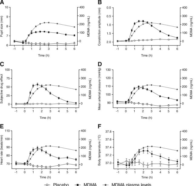
MDMA increased pupil size both at rest and after the light stimulus and lowered the constriction amplitude
compared with placebo (Fig. 3; Table 1). The effect of
MDMA on pupil size peaked (mean ± SEM) 2.3±0.2 h after drug administration at the time of the maximal plasma concentration of MDMA and remained high over 6 h in parallel with plasma levels that also remained high
over 6 h (Fig. 3a). The effect of MDMA on the
con-striction amplitude was maximal 1.7 ±0.1 h after drug administration and decreased to baseline levels over 6 h
(Fig. 3b) despite high plasma levels of MDMA. MDMA
also prolonged the latency to the pupillary light reflex and shortened the recovery time of the pupillary light
reflex response (Table 1).
Subjective effects of MDMA
MDMA produced significant subjective drug effects
com-pared with placebo (Table 1). The peak effect was
reached 1.5±0.1 h after MDMA administration (Fig. 3c).
The subjective effects of MDMA completely reverted to baseline within 6 h, although the plasma levels of
MDMA remained high (Fig. 3c). The effects of the
pre-treatments on the subjective response to MDMA are
reported in detail elsewhere (Hysek et al. 2011, 2012a,
b, d). Briefly, reboxetine and duloxetine reduced the
subjective effects of MDMA, whereas the other pretreat-ments overall had no effect on the subjective response to
Table 1 Effects of MDMA
n080 (values from all five stud-ies were pooled)
Placebo (mean ± SEM)
MDMA (mean ± SEM)
F1,79 p value
Pupil size (mm) Emax 6.60±0.09 7.58±0.07 288.1 <0.001
Pupil size after light (mm) Emax 4.76±0.09 6.86±0.09 646.9 <0.001 Constriction amplitude (mm) Emax 1.76±0.06 0.81±0.12 328.0 <0.001
Latency (s) Emax 0.25±0.00 0.33±0.02 16.2 <0.001
Recovery time (s) Emax 1.74±0.06 1.17±0.07 57.3 <0.001
Subjective drug effect (percent maximum) Emax 3.5±1.7 81.0±2.8 901.2 <0.001 Mean arterial pressure (mmHg) Emax 95.0±1.0 114.5±1.2 339.7 <0.001
Heart rate (bpm) Emax 76.0±1.2 96.2±1.9 138.1 <0.001
Body temperature (°C) Emax 37.3±0.1 37.6±0.1 26.2 <0.001
Placebo MDMA MDMA plasma levels
6 7 8 9 10 0 100 200 300 400 -1 0 1 2 3 4 5 6 Time (h) P upi l si ze ( m m ) M D M A ( ng/ m L ) 0 100 200 300 400 -1 0 1 2 3 4 5 6 2.0 1.5 1.0 0.5 0.0 Time (h) C onst ri c ti on a m pl it ud e ( m m ) MD MA ( n g /mL ) 0 25 50 75 100 0 100 200 300 400 -1 0 1 2 3 4 5 6 Time (h) S u bj ec ti ve dr ug e ff e c t MD MA ( n g /mL ) 90 100 110 120 130 0 100 200 300 400 -1 0 1 2 3 4 5 6 Time (h) M e an ar te ri al pr essur e ( m m H g) MD MA ( n g /mL ) 70 80 90 100 110 0 100 200 300 400 -1 0 1 2 3 4 5 6 Time (h) H e a rt r a te ( b e a ts /m in ) MD M A ( n g /m L ) 37.0 37.2 37.4 37.6 37.8 0 100 200 300 400 -1 0 1 2 3 4 5 6 Time (h) B o dy t e m p er at ur e (° C) MD M A ( n g /mL )
D
B
A
C
F
E
Fig. 3 Acute effects of MDMA on pupil function. Values are expressed as the mean ± SEM of 80 subjects. MDMA increased resting pupil size compared with placebo (a). The mydriatic effect of MDMA remained high in parallel with the plasma concentration of MDMA. MDMA reduced the pupil constriction amplitude compared with
placebo and this effect decreased more rapidly than the plasma con-centration of MDMA (b). The subjective (c), cardiovascular (d, e), and thermogenic (f) effects of MDMA also disappeared within 6 h when the plasma concentrations of MDMA were still high
Effects of MDMA on blood pressure, heart rate, and body temperature
MDMA significantly increased blood pressure, heart rate,
and body temperature compared with placebo (Table 1;
Fig. 3d–f). Similar to the subjective effects,
MDMA-induced increases in blood pressure and heart rate were short-lasting.
Pharmacokinetics of MDMA
Plasma MDMA concentrations are shown in Fig. 3. The
peak plasma MDMA concentration was (mean ± SEM) 243±6 ng/ml. The time to maximum plasma concentra-tion was 2.5±0.1 h.
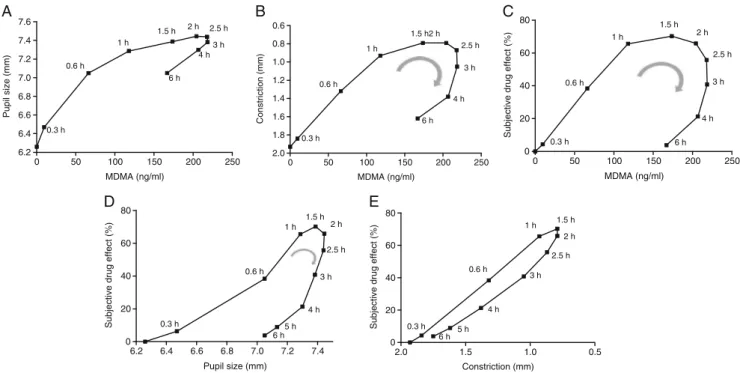
Pharmacokinetic–pharmacodynamic and
pharmacodynamic–pharmacodynamic associations
The relationships between the concentration of MDMA and
its pharmacodynamic effects are shown in Fig. 4a–c. The
average group pupil size was correlated with the average
plasma levels of MDMA over time (Rs00.77, p<0.01, n09),
with moderate clockwise hysteresis (Fig.4a). In contrast, the
MDMA-induced reduction in constriction amplitude was not
significantly associated with the plasma concentrations of
MDMA (Rs00.43, p00.24, n09), attributable to pronounced
clockwise hysteresis (Fig. 4b). There was a similar marked
hysteresis in the relationship between the concentration of
MDMA and the subjective drug effects (Fig. 4c) and no
correlation between the two (Rs00.48, p00.17, n09). The
association between the average subjective effect and pupil
size over time was relatively strong (Rs00.77, p<0.01, n010),
but hysteresis was observed in the relationship between
sub-jective effects and pupil size over time (Fig.4d), indicating
that the subjective effects decreased more rapidly than the mydriasis associated with MDMA. In contrast, little or no hysteresis was observed in the plot of the relationship of
subjective effects with constriction amplitude (Fig.4e),
indi-cating a closer association and more congruent subjective and dynamic pupillary effects of MDMA, also demonstrated by a very strong correlation between the means of these two effects
over time (Rs00.96, p<0.001, n010; Fig. 4e). There were
similar strong associations between MDMA-induced reduc-tions in constriction amplitude and changes in MAP, heart
rate, and body temperature (Rs00.98, 0.92, 0.87; all p<0.001,
n010). Between-subjects correlations further showed that
subjective effects were strongly correlated with reductions in
the light reflex but not with pupil size (Table2).
MDMA-induced increases in blood pressure and heart rate did not
0 50 100 150 200 250 6.2 6.4 6.6 6.8 7.0 7.2 7.4 7.6 0.3 h 0.6 h 1 h 1.5 h 2 h 2.5 h 3 h 4 h 6 h MDMA (ng/ml) Pupil size (mm) 0 50 100 150 200 250 0.6 0.8 1.0 1.2 1.4 1.6 1.8 2.0 0.3 h 0.6 h 1 h 1.5 h2 h 2.5 h 3 h 4 h 6 h MDMA (ng/ml) Constriction (mm) 0 50 100 150 200 250 0 20 40 60 80 0.3 h 0.6 h 1 h 1.5 h 2 h 2.5 h 3 h 4 h 6 h MDMA (ng/ml)
Subjective drug effect (%)
6.2 6.4 6.6 6.8 7.0 7.2 7.4 0 20 40 60 80 0.3 h 0.6 h 1 h 1.5 h 2 h 2.5 h 3 h 4 h 6 h5 h Pupil size (mm)
Subjective drug effect (%)
0.5 1.0 1.5 2.0 0 20 40 60 80 0.3 h 0.6 h 1 h 1.5 h 2 h 2.5 h 3 h 4 h 6 h 5 h Constriction (mm)
Subjective drug effect (%)
C B
A
E D
Fig. 4 Pharmacokinetic–pharmacodynamic relationship. MDMA effects plotted against the plasma concentrations of MDMA (a–c). The values are expressed as the means of 80 subjects, with SEM omitted for clarity. The times of pupillometry and blood sampling are noted next to each point in minutes or hours after MDMA administra-tion. While pupil size (a) remained high, constriction amplitude (b) and subjective effect (c) returned to baseline within 6 h when MDMA concentrations remained high. This clockwise hysteresis was moderate
for the mydriatic effect of MDMA, reflecting well the plasma concen-tration of MDMA (a), but pronounced for the impairment in the pupillary reflex response (b) and subjective effect of MDMA (c). The subjective effect of MDMA returned to baseline faster than the myd-riatic response to MDMA (d). In contrast, the time course of the subjective effect of MDMA was more congruent with the time course of the MDMA-induced impairment in constriction amplitude (e)
correlate with the plasma concentrations of MDMA over time, consistent with the reduced effect over time despite high
plasma concentrations of MDMA (Fig.3d, e).
The findings from the between-subjects analyses of the correlations between the plasma levels of MDMA and phar-macodynamic effects of MDMA for each time point (n080)
are shown in Table3. The MDMA-induced reductions in the
constriction amplitude, the pupil size after light, the increase in MAP, and the subjective effects were significantly and strongly
associated with the plasma levels of MDMA (Table3). Weaker
correlations were also found between plasma levels of MDMA
and the pupil diameter, latency, or heart rate (Table3).
How-ever, these associations were only observed at the beginning of the MDMA effect. Recovery time and body temperature after MDMA administration were not or only weakly and
inconsis-tently associated with plasma MDMA levels (Table3).
The MDMA-induced reduction in pupil constriction am-plitude was significantly greater in subjects with greater
MDMA-induced increases in MAP (Rs00.56, p<0.001,
n080) or more pronounced increases in heart rate (Rs00.30,
p<0.01, n080) as measured 1 h after MDMA administration. In contrast, MDMA-induced changes in the pupil size were not or only poorly associated with other autonomic changes across subjects.
Pupillary effects of reboxetine, duloxetine, clonidine, carvedilol, and doxazosin alone and on the pupillary response to MDMA
The peak effects of the pretreatments are shown in Table4.
The drug effects on pupil size over time for all five studies
are shown in Fig. 5. Both reboxetine and duloxetine
in-creased resting pupil size and pupil size after the light stimulus. Duloxetine also lowered the constriction
ampli-tude (Table 4). The effect of the two monoamine uptake
inhibitors on the static pupil diameter was similar in
mag-nitude to the effect of MDMA (Table 4; Fig. 5a, b). In
contrast, the effect of MDMA on the constriction amplitude was more pronounced. When duloxetine was administered together with MDMA, the drug effects on all static and dynamic parameters were nonadditive and showed negative synergism, reflected by a significant pretreatment × MDMA interaction in the factorial ANOVA. Thus, duloxetine pre-vented the effect of MDMA on pupil function, reflected by the absence of a mydriatic effect of MDMA compared with baseline in the duloxetine–MDMA condition and compared
with the duloxetine–placebo condition (Fig.5b). Duloxetine
also prevented the MDMA-induced impairment in the pu-pillary light reflex, although it had a similar effect when Table 2 Correlations between MDMA-induced changes in pupillary function and subjective drug effects
t00 t020 min t040 min t01 h t01.5 h t02 h t02.5 h t03 h t04 h t06 h
Pupil size (mm) NS NS 0.31 0.27 NS NS NS NS NS NS
Pupil size after light (mm) NS NS 0.62 0.51 0.42 0.26 0.27 NS NS NS
Constriction amplitude (mm) NS NS −0.74 −0.61 −0.41 −0.28 −0.28 −0.28 −0.23 NS
Latency (s) NS NS 0.46 0.29 NS NS NS NS NS NS
Recovery time (s) NS NS −0.42 −0.31 −0.32 −0.22 −0.38 −0.23 −0.28 −0.32
Values are Spearman correlation coefficients for significant correlations (p<0.05; p<0.001 in italics); n080 NS not significant
Table 3 Correlations between the effects of MDMA and plasma concentrations of MDMA
t00 t020 min t040 min t01 h t01.5 h t02 h t02.5 h t03 h t04 h t06 h
Pupil size (mm) NS 0.46 0.45 0.35 0.27 NS NS NS NS NS
Pupil size after light (mm) NS 0.50 0.64 0.57 0.54 0.53 0.49 0.47 0.34 NS
Constriction amplitude (mm) NS −0.28 −0.40 −0.55 −0.53 −0.60 −0.55 −0.65 −0.48 −0.28
Latency (s) NS 0.23 0.46 0.33 0.35 NS 0.25 0.25 NS 0.25
Recovery time (s) NS NS −0.32 NS −0.26 NS −0.37 −0.26 −0.26 NS
Subjective drug effect NS NS 0.68 0.56 0.37 0.31 0.44 0.34 0.46 0.31
Mean arterial pressure (mmHg) NS 0.22 0.69 0.60 0.47 0.35 0.33 0.29 0.39 NS
Heart rate (bpm) NS NS 0.61 0.47 0.46 0.32 NS NS NS NS
Body temperature (°C) NS −0.24 NS NS NS NS NS NS NS NS
Values are Spearman correlation coefficients for significant correlations (p<0.05; p<0.001 in italics) between MDMA-induced pharmacodynamic changes and plasma levels of MDMA; n080
T able 4 Ef fects of pretreatments, of MDMA, and of the combination on pupil function Mean ± SEM values Main ef fect of MDMA Main ef fect of pretreatment Pretreatment × MDMA Placebo Pretreatment MDMA Pretreatment –MDMA F1,15 p value F1,15 p value F1,15 p value Pupil size (mm) Reboxetine Emax 6.15 ± 0.19 7.22 ± 0.20*** 7.32 ± 0.17*** 8.08 ± 0.18*** ,### 124.8 <0.001 160.4 <0.001 10.7 <0.01 Duloxetine Emax 6.55 ± 0.21 7.30 ± 0.19*** 7.52 ± 0.16*** 7.65 ± 0.16*** 58.9 <0.001 37.0 <0.001 1 1.2 <0.01 Clonidine Emin 5.86 ± 0.20 4.93 ± 0.22*** ,### 7.06 ± 0.17*** 6.72 ± 0.15*** 87.9 <0.001 59.3 <0.001 7.8 <0.05 Emax 6.75 ± 0.16 6.35 ± 0.22*** ,### 7.65 ± 0.15*** 7.46 ± 0.16*** 68.7 <0.001 12.7 <0.01 2.8 NS Carvedilol Emax 6.88 ± 0.19 6.66 ± 0.16### 7.63 ± 0.13*** 7.66 ± 0.14*** 99.5 <0.001 8.0 <0.05 1.2 NS Doxazosin Emax 6.67 ± 0.20 6.69 ± 0.18### 7.78 ± 0.12*** 7.53 ± 0.14*** 58.2 <0.001 2.7 NS 2.2 NS Pupil size after light (mm) Reboxetine Emax 4.23 ± 0.17 5.50 ± 0.22*** , ### 6.65 ± 0.23*** 7.37 ± 0.21*** , ### 289.1 <0.001 129.9 <0.001 9.3 <0.01 Duloxetine Emax 4.78 ± 0.22 5.96 ± 0.26*** , ### 6.94 ± 0.19*** 6.38 ± 0.22*** , ## 108.0 <0.001 10.9 <0.01 84.2 <0.001 Clonidine Emin 3.84 ± 0.64 3.15 ± 0.63** , ### 5.31 ± 0.83*** 5.08 ± 0.97*** 93.1 <0.001 24.0 <0.001 5.2 <0.05 Emax 4.80 ± 0.16 4.47 ± 0.22### 7.01 ± 0.23*** 6.83 ± 0.24*** 21 1.2 <0.001 8.9 <0.01 1.2 NS Carvedilol Emax 5.10 ± 0.25 4.77 ± 0.17### 6.82 ± 0.17*** 7.04 ± 0.16*** 202.3 <0.001 0.4 NS 2.3 NS Doxazosin Emax 4.88 ± 0.19 4.88 ± 0.18### 6.86 ± 0.15*** 6.79 ± 0.16*** 169.4 <0.001 0.1 NS 0.2 NS Constriction amplitude (mm) Reboxetine Emin 1.74 ± 0.06 1.62 ± 0.07### 0.60 ± 0.12*** 0.67 ± 0.10*** 71.8 <0.001 0.7 NS 3.0 NS Duloxetine Emin 1.62 ± 0.05 1.27 ± 0.09** , ### 0.52 ± 0.1 1*** 1.18 ± 0.06*** , ### 63.9 <0.001 12.4 <0.01 71.2 <0.001 Clonidine Emin 1.81 ± 0.05 1.63 ± 0.08### 0.58 ± 0.1 1*** 0.50 ± 0.09*** 72.0 <0.001 7.6 <0.05 1.0 NS Carvedilol Emin 1.65 ± 0.10 1.75 ± 0.06### 0.68 ± 0.1 1*** 0.49 ± 0.08*** 99.5 <0.001 0.7 NS 4.2 NS Doxazosin Emin 1.76 ± 0.06 1.69 ± 0.05### 0.81 ± 0.12*** 0.78 ± 0.09*** 79.6 <0.001 1.2 NS 0.3 NS Latency (s) Reboxetine Emax 0.244 ± 0.006 0.254 ± 0.007## 0.303 ± 0.01 1*** 0.306 ± 0.015*** 14.8 <0.001 2.8 NS 0.0 NS Duloxetine Emax 0.245 ± 0.005 0.266 ± 0.007 0.423 ± 0.088* 0.275 ± 0.005 4.8 <0.05 2.2 NS 3.8 0.07 Clonidine Emax 0.252 ± 0.01 1 0.251 ± 0.023## 0.297 ± 0.033** 0.308 ± 0.061*** 30.9 <0.001 0.7 NS 0.4 NS Carvedilol Emax 0.260 ± 0.007 0.262 ± 0.009## 0.304 ± 0.016** 0.303 ± 0.010## 15.5 <0.001 0.0 NS 0.0 NS Doxazosin Emax 0.253 ± 0.008 0.249 ± 0.006# 0.325 ± 0.037* 0.291 ± 0.009 10.4 <0.01 1.1 NS 1.1 NS NS nonsignificant * p < 0.05, ** p < 0.01, *** p < 0.001, compared with placebo; # p < 0.05, ## p < 0.01, ### p < 0.001, compared with MDMA
administered alone compared with placebo. The effects of reboxetine and MDMA on pupil size were also
nonadditive (Table 4; Fig. 5b). However, resting pupil
size and pupil size after the light stimulus were signif-icantly larger after reboxetine plus MDMA compared with MDMA alone. Reboxetine also failed to prevent the effect of MDMA on the pupillary light reflex. In the present study, reboxetine also reduced the cardiostimu-lant and psychostimucardiostimu-lant effects of MDMA (Hysek et
al. 2011), and duloxetine nearly completely prevented
the cardiovascular, psychotropic, and neuroendocrine effects of MDMA as reported elsewhere (Hysek et al.
2012b, d; Simmler et al. 2011). Clonidine reduced
rest-ing pupil size and size after the light stimulus (Table 4;
Fig. 5c). This effect of clonidine was antagonistic and
overall additive with the effect of MDMA (Fig. 5c).
Specifically, clonidine did not significantly reduce the
effects of MDMA on any parameter of pupillary func-tion, although it had significant effects alone and re-duced the cardiovascular response to MDMA (Hysek et
al. 2012a). Clonidine did not significantly reduce the
mydriatic effects of MDMA, although it produced sig-nificant miosis. Clonidine also had no effects on the psychotropic response to MDMA as previously reported
(Hysek et al. 2012a). Carvedilol did not alter the effects
of MDMA on pupillary function. In contrast, carvedilol decreased the cardiostimulant and thermogenic effects of MDMA in the same subjects as reported elsewhere
(Hysek et al. 2012c). Carvedilol alone decreased pupil
size, reflected by a significant main effect of pretreat-ment in the ANOVA, but the reduction in pupil size after carvedilol–placebo treatment compared with the
placebo–placebo condition (Fig. 5d) was not significant
in the post hoc test. Doxazosin alone had no effect on Reboxetine-MDMA 5 6 7 8 9 pre 0 1 2 3 4 5 6 Time (h) P upi l si ze ( m m ) Duloxetine-MDMA 5 6 7 8 9 pre 0 1 2 3 4 5 6 Time (h) P u pi l si ze ( m m ) Clonidine-MDMA 5 6 7 8 9 pre 0 1 2 3 4 5 6 Time (h) P u p il s iz e ( mm) Carvedilol-MDMA 5 6 7 8 9 pre 0 1 2 3 4 5 6 Time (h) P upi l s ize ( m m ) Doxazosin-MDMA 5 6 7 8 9 pre 0 1 2 3 4 5 6 Time (h) P u p il si ze ( m m )
B
A
D
C
E
Pretreatment-MDMA Placebo-MDMA Pretreatment-placebo Placebo-placebo Fig. 5 Drug effects on pupilsize over time. MDMA increased pupil size compared with placebo (a–e). The pretreatment with reboxetine increased pupil size to a similar extent as MDMA alone (a). The effect of MDMA on pupil diameter after reboxetine pretreatment compared with reboxetine was significantly smaller than the effect of MDMA compared with placebo (a). Duloxetine increased pupil size similar to reboxetine and MDMA (b). Duloxetine pretreatment prevented the further increase in pupil size induced by MDMA administration (b). Clonidine significantly reduced pupil diameter (c). The effects of clonidine and MDMA on pupil size were additive (c). Carvedilol nonsignificantly decreased pupil size (d). Similar to the effects of clonidine and MDMA, the effects of carvedilol and MDMA on pupil size were additive (d). Doxazosin alone had no effect on pupil size compared with placebo, but it tended to nonsignificantly attenuate the mydriatic effect of MDMA (e). The data are expressed as the mean ± SEM values in 16 subjects per study
pupil size compared with placebo but slightly and non-significantly reduced the MDMA-induced increase in
pupil size (Fig. 5e).
Discussion
In the present study, we showed that MDMA impaired the pupillary reflex response to light, including inducing a lon-ger latency, reducing the constriction amplitude, and reduc-ing the recovery time. MDMA produced mydriasis as previously documented using nonautomated techniques
(Farre et al.2004, 2007; Kolbrich et al. 2008; Mas et al.
1999). MDMA also increased blood pressure, heart rate, and
body temperature and produced positive mood effects as
described in more detail elsewhere (Hysek et al. 2011,
2012a,b,d).
The analyses of the effects of MDMA over time showed a very strong correlation between the MDMA-induced re-duction in constriction amplitude and other autonomic or subjective effects of the drug. The MDMA-induced reduc-tion in the pupillary light reflex normalized over 6 h, similar to the cardiostimulant and subjective drug effects that also largely disappeared over 6 h, although the plasma levels of MDMA remained high. Thus, the reduced reactivity of the pupil to light is relatively short-lasting and subject to acute pharmacological tolerance, similar to the subjective and cardiostimulant effects of MDMA.
Clinical examination of pupil function in cases of drug intoxication typically includes both an estimation of static pupil size and an assessment of the reactivity to a flashlight stimulus. With regard to MDMA intoxication, our findings suggest that the impaired reactivity to light indicates MDMA exposure within the past 1–4 h and is a marker for the acute subjective and autonomic effects of the drug. In
contrast, mydriasis lasts at least 6–10 h (Farre et al. 2007;
Mas et al. 1999), correlates best with the plasma MDMA
concentration changes over time, and shows only moderate pharmacological tolerance. The mydriatic responses to two successive doses of MDMA separated by 24 h were similar, although the peak concentration after the second dose of MDMA increased by 29 %, indicating some degree of
tolerance (Farre et al. 2004). Although the mean group
changes in pupil size over time reflected the concentra-tion–time curve of MDMA, pupil size did not correlate well with the plasma concentrations of MDMA across subjects at various time points in our study or with MDMA plasma levels 1.25 h after drug administration in a previous study
(Kolbrich et al. 2008). This is not surprising because the
effects of MDMA on pupil size were maximal at single doses of 75 mg and did not further increase at 125 mg
(Mas et al.1999). Thus, the lack of an association is likely
attributable to a ceiling effect of the plasma MDMA
concentration–effect curve. In contrast, dynamic impair-ments of the pupil light reflex response were significantly associated with plasma MDMA levels or the cardiostimulant effects of MDMA across subjects. Evaluating the dynamic pupillary response to light may, therefore, be a better esti-mation of the time and amount of exposure to MDMA than static pupil size.
Both sympathetic and parasympathetic innervations con-tribute to the regulation of pupil size and the reflex response
(Loewenfeld 1999). At the level of the iris, the latency to
and amplitude of the reflex response are mainly determined
by parasympathetic activity (Heller et al. 1990), whereas
redilation is controlled by sympathetic inputs (Loewenfeld
1999; Morley et al. 1991). Additionally, parasympathetic
function is under tonic noradrenergic inhibition centrally at the level of the Edinger–Westphal nucleus where the
sym-pathetic stimulation of α2-adrenergic receptors may lower
parasympathetic output, resulting in
“pseudoanticholiner-gic” mydriasis (Phillips et al.2000a; Siepmann et al.2007;
Szabadi and Bradshaw 1996). Furthermore, the serotonin
system is implicated in pupillary function, possibly via
5-HT1A-mediated stimulation of the release of norepinephrine
and consequent activation ofα2-adrenergic receptors (Prow
et al.1996). MDMA mainly releases serotonin and
norepi-nephrine (Liechti and Vollenweider 2001; Rothman et al.
2001; Verrico et al. 2007). Because MDMA affected both
the parasympathetic and sympathetic aspects of the pupil-lary reflex response, all of the aforementioned mechanisms may be involved in the effects of MDMA on pupillary function.
The norepinephrine transporter inhibitor reboxetine sig-nificantly increased pupil diameter at rest and after light, consistent with previous studies (Theofilopoulos et al.
1995). Reboxetine did not reduce the mydriatic response
to MDMA, but the effects of the two drugs on pupil size were subadditive, indicating that MDMA produces part of its effects on pupil size through the transporter-mediated release of norepinephrine, which is inhibited by reboxetine
(Hysek et al. 2011). This finding is consistent with the
attenuation of the cardiostimulant and psychostimulant
effects of MDMA by reboxetine (Hysek et al. 2011) and
supports the view that norepinephrine is involved in the stimulant effects of MDMA.
The α1-adrenergic receptor inhibitor doxazosin did not
affect pupillary function when administered alone but non-significantly reduced the mydriatic response to MDMA. Prazosin did not antagonize mydriasis induced by norepi-nephrine or phenylephrine in anesthetized cats (Hey et al.
1988; Koss et al.1988). The data suggest thatα1-adrenergic
receptors in the iris may only minimally contribute to my-driasis induced by systemically administered sympathomi-metic drugs and that central parasympathetic inhibition may be more relevant.
Theα1β-adrenergic receptor inhibitor carvedilol had no
significant effect on pupil size compared to placebo,
consis-tent with earlier work (Hirohashi et al. 1990) and the
ab-sence of effects of the β-adrenergic receptor blocker
propranolol on pupillary function (Koudas et al. 2009).
Carvedilol did not affect the mydriatic response to MDMA, but it reduced other autonomic effects of MDMA, including increases in blood pressure and body temperature (Hysek et al.2012c).
Clonidine decreased pupil diameter and enhanced the pu-pillary reflex, consistent with its known sympatholytic effects
(Clifford et al.1982; Morley et al.1991; Phillips et al.2000b,
c). Clonidine also lowered the plasma concentrations of
nor-epinephrine and blood pressure in the subjects of the present
study (Hysek et al.2012a). The effect of clonidine on pupil
function is thought to involve the stimulation ofα2-adrenergic
receptors on central noradrenergic neurons, leading to de-creased sympathetic outflow to the iris. The enhancement of the parasympathetic light reflex is consistent with clonidine-induced disinhibition of the noradrenergic central control of
parasympathetic outflow (Phillips et al. 2000b). Despite its
significant sympathicolytic effects (Hysek et al.2012a),
clo-nidine failed to significantly reduce the effects of MDMA on pupillary function. Moreover, clonidine did not reduce the MDMA-induced increase in norepinephrine or blood pressure to the same extent as it reduced these parameters when
admin-istered alone (Hysek et al. 2012a). Thus, the sympatholytic
effects of clonidine and sympathomimetic effects of MDMA were antagonistic in an additive manner, without evidence of interactive effects of the two drugs. The findings indicate that
α2-adrenergic receptors and the vesicular release of
norepi-nephrine are not critically involved in the pharmacological effects of MDMA.
The dual serotonin and norepinephrine transporter inhib-itor duloxetine increased resting pupil diameter, prolonged the latency to the light reflex, and reduced the reaction to light. Identical effects on pupillary function have been reported for the serotonin and norepinephrine transporter
inhibitor venlafaxine (Bitsios et al.1999; Siepmann et al.
2007). Serotonin releasers, including fenfluramine (Kramer
et al.1973), meta-chlorophenylpiperazine (Benjamin et al.
1997), and MDMA, and serotonin transporter inhibitors
(Nielsen et al. 2010; Noehr-Jensen et al. 2009; Schmitt et
al. 2002) also cause mydriasis. Citalopram and paroxetine
have also been shown to reduce the constriction amplitude
(Nielsen et al.2010; Noehr-Jensen et al. 2009), similar to
previous observations with duloxetine. Duloxetine may, therefore, exert its effects on pupillary function via both noradrenergic and serotonergic mechanisms. Although both duloxetine and MDMA produced mydriasis, pupil size did not further increase after the administration of both drugs, suggesting interactive effects of the two drugs. Moreover, duloxetine almost completely prevented the effects of
MDMA on the light reflex. Duloxetine also markedly inhibited the cardiostimulant, psychotropic, and neuroendo-crine responses to MDMA in the same subjects (Hysek et al.
2012b,d; Simmler et al. 2011). Selective serotonin trans-porter inhibitors including citalopram, fluoxetine, and paroxetine have previously been shown to attenuate the physiological and psychological effects of MDMA in
humans (Farre et al.2007; Liechti et al. 2000; Liechti and
Vollenweider 2000; Tancer and Johanson 2007). Notably,
paroxetine also prevented the mydriatic effects of MDMA
(Farre et al. 2007). Together with the interactive effects of
duloxetine and MDMA in the present work, the findings provide strong support for a role of serotonin in the mech-anism of action of MDMA. The reduction of the effects of MDMA on the pupil light reflex by duloxetine but not reboxetine supports a central modulatory role of serotonin in the effects of MDMA on pupillary function, possibly involving central serotonergic potentiation of noradrenergic
outflow (Prow et al.1996).
In the present study, we assessed pupillary function under dark–light conditions, similar to other studies of the
autonom-ic effects of pharmaceutautonom-icals (Bitsios et al.1999; Nielsen et al.
2010; Noehr-Jensen et al.2009; Phillips et al.2000c). The
values of the latency to the light reflex and constriction am-plitude obtained in the present study were similar to those measured under daylight conditions with the same
pupillom-eter (Taylor et al.2003), indicating that these parameters may
not be critically affected by the light conditions. Overall, our data indicate that the constriction of the pupil represents a measure that is sensitive to pharmacological interventions and may be relatively insensitive to changes in light conditions compared with measures of pupil size.
In summary, MDMA increased pupil size and reduced the response to light. The MDMA-induced prolongation of the latency to the light reflex and reduction in light-induced miosis indicate indirect central parasympathetic inhibition. The faster recovery reflects increased direct sympathomi-metic action. Both reboxetine and duloxetine interacted with the effects of MDMA on static and dynamic measures of pupillary function, supporting a role for both norepinephrine and serotonin in the effects of MDMA on pupillary function. MDMA-induced mydriasis was associated with the plasma concentration–time curve of MDMA. The reduced miotic response to light was highly correlated with the cardiosti-mulant and subjective effects of MDMA and demonstrated acute pharmacological tolerance.
Acknowledgments We thank Y. Schmid, A. Rickli, A. Fink, R. Brugger, V. Nicola, C. Bläsi, L. Baselgia, S. Müller, and S. Purschke for their assistance in the study management, L. Simmler, M. Donzelli, and R. Brenneisen for the determination of plasma MDMA levels, and M. Arends for the editorial assistance. This work was supported by the Swiss National Science Foundation (grant no. 323230_126231) and University of Basel (grant no. DPH2037).
Conflict of interest The authors report no biomedical financial in-terest or potential conflict of inin-terest.
References
Anavekar SN, Jarrott B, Toscano M, Louis WJ (1982) Pharmacokinetic and pharmacodynamic studies of oral clonidine in normotensive subjects. Eur J Clin Pharmacol 23:1–5
Benjamin J, Nemetz H, Fux M, Bleichman I, Agam G (1997) Acute inositol does not attenuate m-CPP-induced anxiety, mydriasis and endocrine effects in panic disorder. J Psychiatr Res 31:489–495 Bitsios P, Langley RW, Szabadi E, Bradshaw CM (1996) Comparison
of the effects of clonidine on tyramine- and methoxamine-evoked mydriasis in man. Br J Clin Pharmacol 41:269–275
Bitsios P, Szabadi E, Bradshaw CM (1999) Comparison of the effects of venlafaxine, paroxetine and desipramine on the pupillary light reflex in man. Psychopharmacology (Berl) 143:286–292 Chung M, Vashi V, Puente J, Sweeney M, Meredith P (1999) Clinical
pharmacokinetics of doxazosin in a controlled-release gastroin-testinal therapeutic system (GITS) formulation. Br J Clin Phar-macol 48:678–687
Clifford JM, Day MD, Orwin JM (1982) Reversal of clonidine induced miosis by theα2-adrenoreceptor antagonist RX 781094. Br J Clin Pharmacol 14:99–101
Derogatis LR, Rickels K, Rock AF (1976) The SCL-90 and the MMPI: a step in the validation of a new self-report scale. Br J Psychiatry 128:280–289
Farre M, de la Torre R, Mathuna BO, Roset PN, Peiro AM, Torrens M, Ortuno J, Pujadas M, Cami J (2004) Repeated doses administra-tion of MDMA in humans: pharmacological effects and pharma-cokinetics. Psychopharmacology (Berl) 173:364–375
Farre M, Abanades S, Roset PN, Peiro AM, Torrens M, O’Mathuna B, Segura M, de la Torre R (2007) Pharmacological interaction between 3,4-methylenedioxymethamphetamine (ecstasy) and paroxetine: pharmacological effects and pharmacokinetics. J Pharmacol Exp Ther 323:954–962
Heller PH, Perry F, Jewett DL, Levine JD (1990) Autonomic compo-nents of the human pupillary light reflex. Invest Ophthalmol Vis Sci 31:156–162
Henderson LS, Tenero DM, Baidoo CA, Campanile AM, Harter AH, Boyle D, Danoff TM (2006) Pharmacokinetic and pharmacody-namic comparison of controlled-release carvedilol and immediate-release carvedilol at steady state in patients with hy-pertension. Am J Cardiol 98:17L–26L
Hey JA, Gherezghiher T, Koss MC (1988) Atypicalα-adrenoceptor mediates phenylephrine-induced mydriasis in anesthetized cats. J Ocul Pharmacol 4:303–310
Hirohashi M, Takasuna K, Tamura K, Yamaguchi K, Maekawa K, Yamada S, Iwasaki S, Yoshida M, Nomura M, Taguchi K (1990) General pharmacological profiles of the new beta-adrenoceptor antagonist carvedilol. Arzneimittelforschung 40:735–746
Hysek CM, Simmler LD, Ineichen M, Grouzmann E, Hoener MC, Brenneisen R, Huwyler J, Liechti ME (2011) The norepinephrine transporter inhibitor reboxetine reduces stimulant effects of MDMA (“ecstasy”) in humans. Clin Pharmacol Ther 90:246–255 Hysek CM, Brugger R, Simmler LD, Bruggisser M, Donzelli M, Grouzmann E, Hoener MC, Liechti ME (2012a) Effects of the α2-adrenergic agonist clonidine on the pharmacodynamics and pharmacokinetics of 3,4-methylenedioxymethamphetamine in healthy volunteers. J Pharmacol Exp Ther 340:286–294 Hysek CM, Domes G, Liechti ME (2012b) MDMA enhances“mind
reading” of positive emotions and impairs “mind reading” of
negative emotions. Psychopharmacology (Berl). doi:10.1007/ s00213-012-2645-9
Hysek CM, Schmid Y, Rickli A, Simmler L, Donzelli M, Grouzmann E, Liechti ME (2012c) Carvedilol inhibits the cardiostimulant and thermogenic effects of MDMA in humans. Br J Pharmacol. doi:10.1111/j.1476-5381.2012.01936.x
Hysek CM, Simmler LD, Nicola VG, Vischer N, Donzelli M, Krähenbühl S, Grouzmann E, Huwyler J, Hoener MC, Liechti ML (2012d) Duloxetine inhibits effects of MDMA (“Ecstasy”) in vitro and in humans in a randomized placebo-controlled laboratory study. PLoS ONE 7(5):e36476. doi:10.1371/journal.pone.0036476
Keranen A, Nykanen S, Taskinen J (1978) Pharmacokinetics and side-effects of clonidine. Eur J Clin Pharmacol 13:97–101
Kolbrich EA, Goodwin RS, Gorelick DA, Hayes RJ, Stein EA, Huestis MA (2008) Physiological and subjective responses to controlled oral 3,4-methylenedioxymethamphetamine administration. J Clin Psychopharmacol 28:432–440
Koss MC, Logan LG, Gherezghiher T (1988)α-Adrenoceptor activa-tion of nictitating membrane and iris in cats. Naunyn Schmiede-bergs Arch Pharmacol 337:519–524
Koudas V, Nikolaou A, Hourdaki E, Giakoumaki SG, Roussos P, Bitsios P (2009) Comparison of ketanserin, buspirone and pro-pranolol on arousal, pupil size and autonomic function in healthy volunteers. Psychopharmacology (Berl) 205:1–9
Kramer R, Rubicek M, Turner P (1973) The role of norfenfluramine in fenfluramine-induced mydriasis. J Pharm Pharmacol 25:575–576 Liechti ME, Vollenweider FX (2000) The serotonin uptake inhibitor citalopram reduces acute cardiovascular and vegetative effects of 3,4-methylenedioxymethamphetamine (‘Ecstasy’) in healthy vol-unteers. J Psychopharmacol 14:269–274
Liechti ME, Vollenweider FX (2001) Which neuroreceptors mediate the subjective effects of MDMA in humans? A summary of mechanistic studies. Hum Psychopharmacol 16:589–598 Liechti ME, Baumann C, Gamma A, Vollenweider FX (2000) Acute
psychological effects of 3,4-methylenedioxymethamphetamine (MDMA,“ecstasy”) are attenuated by the serotonin uptake inhib-itor citalopram. Neuropsychopharmacology 22:513–521 Liechti ME, Gamma A, Vollenweider FX (2001) Gender differences in
the subjective effects of MDMA. Psychopharmacology (Berl) 154:161–168
Loewenfeld IE (1999) The pupil: anatomy, physiology, and clinical applications. Butterworth-Heinemann, Boston
Mas M, Farre M, de la Torre R, Roset PN, Ortuno J, Segura J, Cami J (1999) Cardiovascular and neuroendocrine effects and pharmaco-kinetics of 3,4-methylenedioxymethamphetamine in humans. J Pharmacol Exp Ther 290:136–145
Mithoefer MC, Wagner MT, Mithoefer AT, Jerome I, Doblin R (2010) The safety and efficacy of ±3,4-methylenedioxymethamphet-amine-assisted psychotherapy in subjects with chronic, treatment-resistant posttraumatic stress disorder: the first random-ized controlled pilot study. J Psychopharmacol 25:439–452 Morgan T (1994) Clinical pharmacokinetics and pharmacodynamics of
carvedilol. Clin Pharmacokinet 26:335–346
Morley MJ, Bradshaw CM, Szabadi E (1991) Effects of clonidine and yohimbine on the pupillary light reflex and carbachol-evoked sweating in healthy volunteers. Br J Clin Pharmacol 31:99–101 Nielsen AG, Pedersen RS, Noehr-Jensen L, Damkier P, Brosen K
(2010) Two separate dose-dependent effects of paroxetine: my-driasis and inhibition of tramadol’s O-demethylation via CYP2D6. Eur J Clin Pharmacol 66:655–660
Nieuwenhuis S, van Nieuwpoort IC, Veltman DJ, Drent ML (2007) Effects of the noradrenergic agonist clonidine on temporal and spatial attention. Psychopharmacology (Berl) 193:261–269
Noehr-Jensen L, Zwisler ST, Larsen F, Sindrup SH, Damkier P, Nielsen F, Brosen K (2009) Impact of CYP2C19 phenotypes on
escitalopram metabolism and an evaluation of pupillometry as a serotonergic biomarker. Eur J Clin Pharmacol 65:887–894 Phillips MA, Bitsios P, Szabadi E, Bradshaw CM (2000a) Comparison
of the antidepressants reboxetine, fluvoxamine and amitriptyline upon spontaneous pupillary fluctuations in healthy human volun-teers. Psychopharmacology (Berl) 149:72–76
Phillips MA, Szabadi E, Bradshaw CM (2000b) Comparison of the effects of clonidine and yohimbine on pupillary diameter at dif-ferent illumination levels. Br J Clin Pharmacol 50:65–68 Phillips MA, Szabadi E, Bradshaw CM (2000c) Comparison of the
effects of clonidine and yohimbine on spontaneous pupillary fluctuations in healthy human volunteers. Psychopharmacology (Berl) 150:85–89
Prow MR, Martin KF, Heal DJ (1996) 8-OH-DPAT-induced mydriasis in mice: a pharmacological characterisation. Eur J Pharmacol 317:21–28
Roelands B, Goekint M, Heyman E, Piacentini MF, Watson P, Hasegawa H, Buyse L, Pauwels F, De Schutter G, Meeusen R (2008) Acute norepinephrine reuptake inhibition decreases performance in normal and high ambient temperature. J Appl Physiol 105:206–212 Rothman RB, Baumann MH, Dersch CM, Romero DV, Rice KC,
Carroll FI, Partilla JS (2001) Amphetamine-type central nervous system stimulants release norepinephrine more potently than they release dopamine and serotonin. Synapse 39:32–41
Schmitt JA, Riedel WJ, Vuurman EF, Kruizinga M, Ramaekers JG (2002) Modulation of the critical flicker fusion effects of seroto-nin reuptake inhibitors by concomitant pupillary changes. Psy-chopharmacology (Berl) 160:381–386
Schmitz N, Hartkamp N, Kiuse J, Franke GH, Reister G, Tress W (2000) The Symptom Check-List-90-R (SCL-90-R): a German validation study. Qual Life Res 9:185–193
Shirai K, Song M, Suzuki J, Kurosu T, Oyama T, Nagayama D, Miyashita Y, Yamamura S, Takahashi M (2010) Contradictory effects of beta1- and alpha1-aderenergic receptor blockers on cardio-ankle vascular stiffness index (CAVI)—CAVI independent of blood pressure. J Atheroscler Thromb 18:49–55
Siepmann T, Ziemssen T, Mueck-Weymann M, Kirch W, Siepmann M (2007) The effects of venlafaxine on autonomic functions in healthy volunteers. J Clin Psychopharmacol 27:687–691
Simmler LD, Hysek CM, Liechti ME (2011) Sex differences in the effects of MDMA (ecstasy) on plasma copeptin in healthy sub-jects. J Clin Endocrinol Metab 96:2844–2850
Sofuoglu M, Brown S, Babb DA, Pentel PR, Hatsukami DK (2000) Carvedilol affects the physiological and behavioral response to smoked cocaine in humans. Drug Alcohol Depend 60:69–76 Szabadi E, Bradshaw CM (1996) Autonomic pharmacology of
α2-adrenoceptors. J Psychopharmacol 10(S3):S6–S18 Tancer M, Johanson CE (2007) The effects of fluoxetine on the
subjective and physiological effects of 3,4-methylenedioxyme-thamphetamine (MDMA) in humans. Psychopharmacology (Berl) 189:565–573
Taylor WR, Chen JW, Meltzer H, Gennarelli TA, Kelbch C, Knowlton S, Richardson J, Lutch MJ, Farin A, Hults KN, Marshall LF (2003) Quantitative pupillometry, a new technology: normative data and preliminary observations in patients with acute head injury: technical note. J Neurosurg 98:205–213
Tham TC, Guy S, McDermott BJ, Shanks RG, Riddell JG (1995) The dose dependency of the alpha- and beta-adrenoceptor antagonist activity of carvedilol in man. Br J Clin Pharmacol 40:19–23 Theofilopoulos N, McDade G, Szabadi E, Bradshaw CM (1995)
Effects of reboxetine and desipramine on the kinetics of the pupillary light reflex. Br J Clin Pharmacol 39:251–255
Tomlinson B, Bompart F, Graham BR, Liu JB, Prichard BN (1988) Vasodilating mechanism and response to physiological pressor stimuli of acute doses of carvedilol compared with labetalol, propranolol and hydralazine. Drugs 36(Suppl 6):37–47
Tomlinson B, Prichard BN, Graham BR, Walden RJ (1992) Clinical pharmacology of carvedilol. Clin Investig 70(Suppl 1):S27–S36 Verrico CD, Miller GM, Madras BK (2007) MDMA (ecstasy) and
human dopamine, norepinephrine, and serotonin transporters: implications for MDMA-induced neurotoxicity and treatment. Psychopharmacology (Berl) 189:489–503
White TL, Justice AJ, de Wit H (2002) Differential subjective effects of
D-amphetamine by gender, hormone levels and menstrual cycle phase. Pharmacol Biochem Behav 73:729–741
Wittchen HU, Wunderlich U, Gruschwitz S, Zaudig M (1997) SKID-I: Strukturiertes Klinisches Interview für DSM-IV. Hogrefe-Verlag, Göttingen