Electrocardiographic changes during continuous intravenous
application of bupivacaine in neonatal pigs
J. Mauch
1,2*, A. P. N. Kutter
4, C. Madjdpour
2, N. Spielmann
2, C. Balmer
3, A. Frotzler
2,
R. Bettschart-Wolfensberger
4and M. Weiss
21Department of Anaesthesia and Perioperative Medicine, Kantonsspital Aarau AG, 5001 Aarau, Switzerland 2Department of Anaesthesia and3Department of Cardiology, University Children’s Hospital Zurich, Switzerland 4Section Anaesthesiology, Equine Department, Vetsuisse Faculty, University of Zurich, Switzerland
* Corresponding author. E-mail: [email protected]
Key points
† T-elevation after intravascular injection of a small bupivacaine test dose are caused by epinephrine. † This pig study
demonstrated that higher doses of i.v. infused bupivacaine can cause T-wave elevation. † Intravascular injection of large amounts of bupivacaine can be detected by changes in ECG. † Bupivacaine-induced T-wave elevation is not a reliable indicator for early detection of inadvertent systemic injection. † Bupivacaine-induced
T-ware elevation is a sign of impeding cardiac toxicity, especially during fast injection.
Background. It is controversial as to whether T-wave elevation is caused by local anaesthetics, epinephrine, or their combination. It has been shown that T-elevation after intravascular injection of a small bupivacaine test dose is caused by epinephrine and not by bupivacaine. The aim of this study was to investigate ECG changes with higher doses of i.v. bupivacaine.
Methods. Thirty neonatal pigs were anaesthetized with sevoflurane and their tracheas intubated and artificially ventilated. Under steady-state conditions, bupivacaine was continuously infused (flow rate 3.2 ml kg21min21) by a syringe infusion pump through a central venous catheter. Group 1 received bupivacaine 0.125%, Group 2 bupivacaine 0.5%. The ECG was continuously printed and subsequently analysed for alterations in heart rate, ventricular de- and repolarization, and arrhythmias at 1.25, 2.5, and 5 mg kg21bupivacaine infused.
Results. Sinus rhythm persisted in all pigs. Heart rate decreased progressively in both groups, but this was significantly more pronounced in Group 1. T-wave elevation occurred in 40% and 0% (Groups 1 and 2) at 1.25 mg kg21, in 80% and 0% at 2.5 mg kg21, and in 93% and 80% at 5 mg kg21 bupivacaine infused. There were significant differences between the two groups at 1.25 and 2.5 mg kg21infused.
Conclusions.Higher doses of i.v. infused bupivacaine can cause T-elevation. With slower injection technique, T-elevation can already be detected at lower bupivacaine doses administered.
Keywords: anaesthetics local, bupivacaine; heart, conduction; heart, heart rate; toxicity, local anaesthetics
Accepted for publication: 15 April 2010
Local anaesthetics (LA) for regional anaesthesia in children are usually administered under general anaesthesia.1Since neurological signs of LA toxicity in the event of inadvertent intravascular administration of LA are missing under general anaesthesia, ECG monitoring for the detection of LA-related changes, particularly the development of T-wave elevation, is recommended to detect inadvertent intravascu-lar administration of LA before severe cardiovascuintravascu-lar com-promise or collapse occurs.1–4
It is controversial as to whether T-wave elevation is caused by LA, epinephrine, or their combination.4 Recent research in neonatal pigs revealed that the formation of increased T-waves after injection of a common LA test
dose is mainly caused by epinephrine and not by bupiva-caine.5 However, there is evidence suggesting that higher doses of bupivacaine may result in similar alterations.5 6
The aim of this study was to investigate the course of ECG alterations during continuous i.v. infusion of bupivacaine in neonatal pigs with special regard to the development of T-wave elevation.
Methods
After approval from the local ethics committee for animal experiments, 30 healthy male and female neonatal pigs (up to 6 weeks of age) were included. The pigs had free
access to food until 1 h before induction of anaesthesia. Anaesthesia was induced by the inhalation route using sevo-flurane. After establishing venous access in an auricular vein, the trachea was intubated and the lungs were artificially ven-tilated using pressure-controlled ventilation. Anaesthesia was maintained using sevoflurane in oxygen/air 1:1.
Monitoring consisted of pulse oximetry, three-lead ECG, invasive arterial pressure measurement after cannulation of the carotid artery, end-tidal gas analysis (sevoflurane, O2, CO2), and rectal temperature control. Under steady-state conditions, bupivacaine was continuously infused (flow rate 3.2 ml kg21min21) through a central venous catheter at a dose rate of 4 mg kg21 min21 (Group 1: bupivacaine 0.125%) or 16 mg kg21min21(Group 2: bupivacaine 0.5%) using a syringe pump (Alaris TIVA, IVAC Corporation, Hamp-shire, UK).
The ECG lead with best representation of P-wave, QRS-complex, and T-wave was printed7and analysed after the experiment for alterations in heart rate, ventricular de-and repolarization, de-and arrhythmias at 1.25, 2.5, de-and 5 mg kg21bupivacaine infused by a blinded paediatric cardiologist and anaesthetist. Special attention was given to the appear-ance of T-wave elevation. T-wave elevation of≥25% baseline was considered a positive response.3 8
In each group, the percentage of heart rate change and the occurrence of positive T-wave elevation were compared between different amounts (1.25, 2.5, and 5 mg kg21) of bupivacaine administered. The Friedmann test followed by the Bonferroni corrected group comparisons using the Wil-coxon test was used for heart rate, and the x2 test was used to analyse the T-wave elevation data. Comparisons were made between slow and fast bupivacaine
administration (Groups 1 and 2). Percentage change in heart rate was analysed using the Mann –Whitney U-tests and the occurrence of positive T-wave elevations between the groups was analysed with x2 tests at corresponding doses of bupivacaine administered. A P-value of≤0.05 was considered to be significant. SPSS, version 16.0 (SPSS Inc., Chicago, IL, USA), was used for all analyses.
Results
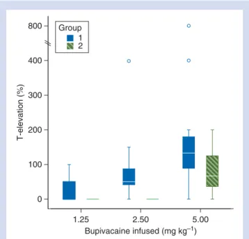
Thirty male and female piglets weighing 4.5 –6.2 kg (median 5.1 kg) were investigated. Changes in heart rate and T-wave elevation are shown in detail in Table1. T-wave increases as percentage above baseline demonstrated a huge variability in degree of T-wave elevation (Fig. 1). Examples of typical and extreme T-wave increases after the infusion of 1.25, 2.5, and 5 mg kg21bupivacaine are shown in Figures2and
3. T-wave elevation≥25% baseline was considered a positive response. This occurred in 40% and 0% (Groups 1 and 2) at 1.25 mg kg21, in 80% and 0% at 2.5 mg kg21, and in 93% and 80% at 5 mg kg21bupivacaine infused. There were sig-nificant differences between the two groups at 1.25 and 2.5 mg kg21infused (Fig.4). The probability of T-wave elevation was high at 2.5 and 5 mg kg21 bupivacaine infused in Group 1, but only at 5 mg kg21 in Group 2. Heart rate decreased significantly and progressively in both groups, but this was significantly more pronounced in Group 1 (Fig. 5). Sinus rhythm persisted in all animals. Neither a higher degree AV block, nor a complete bundle branch block, nor premature atrial, nor ventricular contractions were observed during bupivacaine infusion until 5 mg kg21.
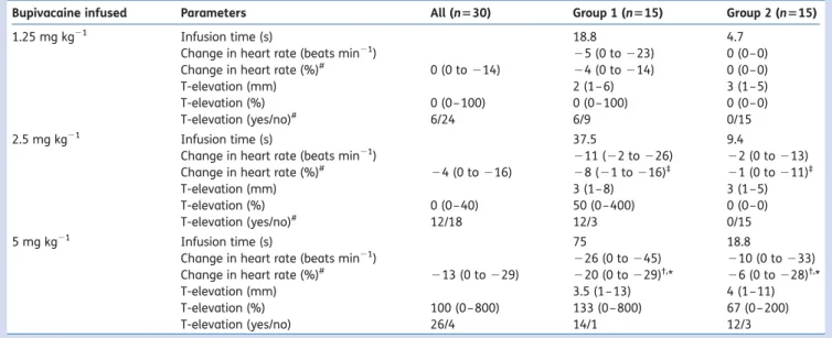
Table 1 ECG alterations after i.v. infusion of 1.25, 2.5, and 5 mg kg21bupivacaine (n¼30 piglets) at an infusion rate of 4 mg kg21min21(Group 1) or 16 mg kg21min21(Group 2). Results are given in median and range.‡Significant differences between 1.25 and 2.5 mg kg21bupivacaine
infused.†Significant differences between 2.5 and 5 mg kg21bupivacaine infused. *Significant differences between 1.25 and 5 mg kg21
bupivacaine infused.#Significant differences between Groups 1 and 2. Infusion time, the time needed to fill the catheter with bupivacaine was
1.3 s for both drug rates (same infusion rate) and is therefore neglected
Bupivacaine infused Parameters All (n530) Group 1 (n515) Group 2 (n515)
1.25 mg kg21 Infusion time (s) 18.8 4.7
Change in heart rate (beats min21) 25 (0 to 223) 0 (0–0)
Change in heart rate (%)# 0 (0 to 214) 24 (0 to 214) 0 (0–0)
T-elevation (mm) 2 (1– 6) 3 (1–5)
T-elevation (%) 0 (0–100) 0 (0– 100) 0 (0–0)
T-elevation (yes/no)# 6/24 6/9 0/15
2.5 mg kg21 Infusion time (s) 37.5 9.4
Change in heart rate (beats min21) 211 (22 to 226) 22 (0 to 213)
Change in heart rate (%)# 24 (0 to 216) 28 (21 to 216)‡ 21 (0 to 211)‡
T-elevation (mm) 3 (1– 8) 3 (1–5)
T-elevation (%) 0 (0–40) 50 (0 –400) 0 (0–0)
T-elevation (yes/no)# 12/18 12/3 0/15
5 mg kg21 Infusion time (s) 75 18.8
Change in heart rate (beats min21) 226 (0 to 245) 210 (0 to 233)
Change in heart rate (%)# 213 (0 to 229) 220 (0 to 229)†,* 26 (0 to 228)†,*
T-elevation (mm) 3.5 (1– 13) 4 (1–11)
T-elevation (%) 100 (0–800) 133 (0– 800) 67 (0 –200)
Discussion
This study investigated whether bupicavaine alone can cause T-wave alteration in the ECG when systemically adminis-tered. The main findings were that i.v. administered bupiva-caine alone causes T-wave elevation in a high percentage of subjects particularly at larger doses and with slower injec-tion, T-elevation can already be detected at lesser bupiva-caine doses.
Reliable signs of inadvertent systemic application of LA in anaesthetized children are of importance in order to avoid severe systemic LA toxicity, in particular haemodynamic col-lapse. The results of this study clearly indicate that not only systemically administered epinephrine5but also bupivacaine cause T-wave elevation, particularly at larger doses. This explains the confusion about origin and reported varying sen-sitivity of ECG for the detection of systemic administration of LA.4However, as shown by the current animal model, the sen-sitivity of bupivacaine-induced T-wave elevation for detecting inadvertent systemic LA administration is not 100%, demon-strates a large variability in T-wave elevation, and becomes mainly visible after doses corresponding to the maximum rec-ommended dose for humans.9 10 Therefore, bupivacaine-induced T-wave elevation is a sign for impending cardiac tox-icity and not helpful for early detection of inadvertent intra-vascular administration. In contrast, T-elevation caused by an epinephrine containing standard test dose of LA is the effect of epinephrine and has a sensitivity of 93%.5In this situ-ation, no adverse consequences have to be anticipated.5 11 12 The study results confirm the previously reported obser-vation that not only epinephrine in a normal test dose alone or in combination with bupivacaine but also bupiva-caine systemically administered in a twice fold test dose can evoke T-wave elevation.5 Nystro¨m and colleagues13 investigated acute bupivacaine overdose until near cardiac arrest in 12 adult male pigs (23– 25 kg) sedated with keta-mine and then anaesthetized with halothane. They also found significant increases in T-wave amplitude [+66% above baseline in maximum (mean)]. Tanaka and col-leagues6reported a 2-month-old healthy infant demonstrat-ing significantly increased T-wave amplitude after the
5.00 2.50 1.25 Bupivacaine infused (mg kg–1) 800 400 300 200 100 0 T-elevation (%) 2 1 Group
Fig 1 T-wave increase in percentage above baseline at different amounts of bupivacaine infused. n¼15 in both groups.
A
B
C
D
accidental systemic administration of a near full-dose lido-caine plus bupivalido-caine when performing a caudal block.
The QT interval is a measure from the body surface of the time required for ventricles of the heart to repolarize. Pro-longation or abbreviation of the QT interval occurs when the action potential of a significant number of cells in the ventricular myocardium is prolonged or abbreviated, as a result of an alteration in one or more ion channel currents. T-waves are in large part inscribed as a consequence of the voltage gradients that develop as a result of transmural differences in the time course of repolarization. When the T-wave is upright, the epicardial action potential is the
earliest to repolarize and the M cell (mid-myocardium, papil-lary muscles, interventricular septum) action potential is the last. Full repolarization of the epicardium coincides with the peak of the T-wave. The Tpeak–Tend interval provides an index of transmural dispersion of repolarization.14 The cardiac toxicity of LA is attributed to a blockade of sodium channels in the heart,15 leading to a prolonged conduction
A
B
C
D
Fig 3 ECG with extreme T-elevation caused by bupivacaine. Trace before (A), after 1.25 mg kg21(B), 2.5 mg kg21(C), and 5 mg kg21(D)
bupi-vacaine infused (Pig no. 2 – 9).
Bupivacaine infused (mg kg–1) 0 10 20 30 40 50 60 70 80 90 100 1.25 2.5 5 Positive T-elevation (%) Group 1 Group 2
Fig 4Percentage of pigs with positive T-wave elevation (≥25% baseline) at different doses bupivacaine infused. n¼15 in both groups. 5.00 2.50 1.25 Bupivacaine infused (mg kg–1) 0 –5 –10 –15 –20 –25 –30 Delta HR (%) 2 1 Group
Fig 5 Changes in heart rate (%) after i.v. infusion of 1.25, 2.5, and 5 mg kg21bupivacaine. n¼15 in both groups.
time with widening of QRS complexes, changes in QRS mor-phology, prolongation of PR interval, AV block, and arrhyth-mias. Owing to altered function of ion channels (mainly sodium channels, but also calcium and potassium chan-nels),16 17not only depolarization but also repolarization is affected. The exact mechanism for T-wave elevation caused by intravascular LA administration is not clear.4 6 Also, there is no clear explanation for the huge variability in the relative increase of T-wave in this study.
The current study included a slow (Group 1) and a fast dose rate (Group 2) of bupivacaine administration. One would expect that with slow injection of bupivacaine, a drug known to have a high first pass lung uptake,18 19ECG alterations are reduced, in contrast to fast injection, causing an excess in plasma concentration of bupivacaine and related toxicity.20 Interestingly, with slower infusion rates, ECG changes appeared at lower doses administered. We hypothesize that although the LA solution was given into a central vein, the circulation time needed to distribute the drug from the injection site into cardiac tissue is responsible for the delay in ECG alteration in the fast group. This reinforces the necessity that LA should be administered slowly. Not only to avoid high plasma peak levels but also not to miss LA-related ECG alterations for early cessation of LA injection.
On the basis of the fact that epinephrine early after a single LA test dose and bupivacaine alone only later and after higher doses leads to ECG alterations, for usual clinical practice, the test dose should contain epinephrine. Whether a change from an epinephrine containing LA solution after the test dose to a plain LA solution should be done is much discussed. Since regional anaesthesia needles or can-nulas may migrate into a vessel,21 our view is that the full LA dose administered should contain epinephrine to detect systemic injection at any period of injection.
In conclusion, this newborn animal model demonstrates that higher doses of systemically applied bupivacaine can cause T-elevation. However, bupivacaine-induced T-elevation is not a reliable indicator to early detect inadvertent systemic injection of LA and is rather a sign for impeding cardiac tox-icity, especially during fast injection of LA.
Conflict of interest
None declared.
Funding
The study was funded by a study grant from a donation made by UBS AG ‘by order of a client’ and by a study grant obtained from ‘Stiftung fu¨r wissenschaftliche Forschung’, University of Zurich, Switzerland.
References
1 Polaner DM, Suresh S, Cote´ CJ. Regional anesthesia. In: Cote´ CJ, Lerman J, Todres ID, eds. A Practice of Anesthesia for Infants and Children. Philadelphia: Saunders Elsevier, 2009; 867 2 Freid EB, Bailey AG, Valley RD. Electrocardiographic and
hemody-namic changes associated with unintentional intravascular
injection of bupivacaine with epinephrine in infants. Anesthesiol-ogy 1993; 79: 394 –8
3 Fisher QA, Shaffner DH, Yaster M. Detection of intravascular injection of regional anaesthetics in children. Can J Anaesth 1997; 44: 592–8 4 Tobias JD. Caudal epidural block: a review of test dosing and recognition of systemic injection in children. Anesth Analg 2001; 93: 1156– 61
5 Mauch J, Kutter APN, Madjdpour C, et al. Electrocardiographic alterations during intravascular application of three different test doses of bupivacaine and epinephrine: experimental study in neonatal pigs. Br J Anaesth 2010; 104: 94– 7
6 Tanaka M, Nitta R, Nishikawa T. Increased T-wave amplitude after accidental intravascular injection of lidocaine plus bupivacaine without epinephrine in sevoflurane-anesthetized child. Anesth Analg 2001; 92: 915– 7
7 Ogasawara K, Tanaka M, Nishikawa T. Choice of electrocardiogra-phy lead does not affect the usefulness of the T-wave criterion for detecting intravascular injection of an epinephrine test dose in anesthetized children. Anesth Analg 2003; 97: 372– 6
8 Tanaka M, Nishikawa T. Evaluating T-wave amplitude as guide for detecting intravascular injection of a test dose in anesthetized children. Anesth Analg 1999; 88: 754 –8
9 Rosenberg PH, Veering BT, Urmey WF. Maximum recommended doses of local anesthetics: a multifactorial concept. Reg Anesth Pain Med 2004; 29: 564 –75
10 Polaner DM, Suresh S, Cote´ CJ. Regional anesthesia. In: Cote´ CJ, Lerman J, Todres ID, eds. A Practice of Anesthesia for Infants and Children. Philadelphia: Saunders Elsevier, 2009; 871 11 Tanaka M, Nishikawa T. The efficacy of a simulated intravascular
test dose in sevoflurane-anesthetized children: a dose–response study. Anesth Analg 1999; 89: 632 –7
12 Sparks JW, Seefelder C. Neonatal T-wave elevation from a positive epidural test dose. Paediatr Anaesth 2005; 15: 706– 7 13 Nystro¨m EUM, Heavner JE, Buffington CW. Blood pressure is
maintained despite profound myocardial depression during acute bupivacaine overdose in pigs. Anesth Analg 1999; 88: 1143– 8
14 Antzelevitch C. Cellular basis and mechanism underlying normal and abnormal myocardial repolarization and arrhythmogensis. Ann Med 2004; 36: 5–14
15 Clarkson CW, Hondeghem LM. Mechanism for bupivacaine depression of cardiac conduction: fast block of sodium channels during the action potential with slow recovery from block during diastole. Anesthesiology 1985; 62: 396– 405
16 Berde C. Toxicity of local anesthetics in infants and children. J Pediatr 1993; 122: 14–20
17 Morrison SG, Dominguez JJ, Frascarolo P, Reiz S. A comparison of the electrocardiographic cardiotoxic effects of racemic bupiva-caine, levobupivabupiva-caine, and ropivacaine in anesthetized swine. Anesth Analg 2000; 90: 1308–14
18 Palazzo MG, Kalso E, Argiras E, Madgwick R, Sear J. The uptake of bupivacaine in an in situ isolated perfused rabbit lung prep-aration. Pharmacol Toxicol 1991; 69: 107 –11
19 Palazzo MG, Kalso EA, Argiras E, Madgwick R, Sear JW. First pass lung uptake of bupivacaine: effect of acidosis in an intact rabbit lung model. Br J Anaesth 1991; 67: 759– 63
20 Stoelting RK. Local anesthetics. Handbook of Pharmacology & Physiology in Anesthetic Practice. Philadelphia: Lippincott Wil-liams & Wilkins, 1995; 135
21 Shah S, Gopalakrishnan S, Apuya J, Shah S, Martin T. Use of intra-lipid in an infant with impending cardiovascular collapse due to local anesthetic toxicity. J Anesth 2009; 23: 439 –41