RESEARCH REPORT
Mitochondrial tRNA
Leu(UUR)
mutation m.3302A >G
presenting as childhood-onset severe myopathy:
threshold determination through segregation study
Diana Ballhausen&Frédéric Guerry&Dagmar Hahn&André Schaller&Jean-Marc Nuoffer&Luisa Bonafé&
Pierre-Yves Jeannet&Sebastien Jacquemont
Received: 11 January 2010 / Revised: 26 February 2010 / Accepted: 31 March 2010 # SSIEM and Springer 2010
Abstract Mitochondrial tRNALeu(UUR) mutation m.3302A> G is associated with respiratory chain complex I deficiency and has been described as a rare cause of mostly adult-onset slowly progressive myopathy. Five families with 11 patients have been described so far; 5 of them died young due to cardiorespiratory failure. Here, we report on a segregation study in a family with an index patient who already presented at the age of 18 months with proximal muscular hypotonia, abnormal fatigability, and lactic acidosis. This early-onset myopathy was rapidly progressive. At 8 years, the patient is wheel-chair bound, requires nocturnal assisted ventilation, and suffers from recurrent respiratory infections. Severe complex I
deficiency and nearly homoplasmy for m.3302A>G were found in muscle. We collected blood, hair, buccal swabs and muscle biopsies from asymptomatic adults in this pedigree and determined heteroplasmy levels in these tissues as well as OXPHOS activities in muscle. All participating asymptomatic adults had normal OXPHOS activities. In contrast to earlier reports, we found surprisingly little variation of heteroplasmy levels in different tissues of the same individual. Up to 45% mutation load in muscle and up to 38% mutation load in other tissues were found in non-affected adults. The phenotypic spectrum of tRNALeu(UUR)m.3302A>G mutation seems to be wider than previously described. A threshold of more than 45% heteroplasmy in muscle seems to be necessary to alter complex I activity leading to clinical manifestation. The presented data may be helpful for prognostic considerations and counseling in affected families.
Abbreviations
ADP Adenosine diphosphate
bp Base paires
CVS Chorionic villus sampling
DNA Desoxynucleic acid
ECG Electrocardiogram
KCN Potassium cyanide
MRI Magnetic resonance imaging
mt Mitochondrial
OXPHOS Oxidative phosphorylation
PCR Polymerase chain reaction
RCR Respiratory control ratio rRNA Ribosomal ribonucleic acid
TMPD N,N,N′,N′-tetramethyl-p-phenylenediamine, Wurster’s blue
tRNA Transfer ribonucleoic acid
Communicated by: Shamima Rahman
References to electronic databases: tRNALeu(UUR): OMIM *590050 D. Ballhausen (*)
:
L. BonaféDivision of Molecular Pediatrics, Centre Hospitalier Universitaire Vaudois, CI 02–35, Av. P. Decker 2,
1011 Lausanne, Switzerland e-mail: [email protected] F. Guerry
:
S. JacquemontMedical Genetics, Centre Hospitalier Universitaire Vaudois, Lausanne, Switzerland
D. Hahn
:
J.-M. NuofferUniversity Institute of Clinical Chemistry, Inselspital Bern, Switzerland
A. Schaller
Department of Pediatrics, Division of Human Genetics, Inselspital Bern, Switzerland
P.-Y. Jeannet
Neuropediatrics, Centre Hospitalier Universitaire Vaudois, Lausanne, Switzerland
Introduction
Human mitochondrial function requires precise excision of tRNAs from the precursor transcript by specific endonucleases for the 3′- and 5′-terminal nucleotides. The CCA-terminus, which is required for aminoacylation and thus for protein synthesis, is then added by a tRNA nucleotidyl transferase (Schurer et al. 2001). In the transcript of the human mitochondrial genome, the gene MT-TL1 for tRNALeu(UUR)is located between the genes for 16S rRNA and ND1, which encode the subunit 1 of respiratory chain complex I.
Mitochondrial (mt) tRNALeu(UUR) is a hot spot for mtDNA mutations linked with inherited severe neuromuscu-lar diseases. Mutation m.3302A > G is located in the aminoacyl stem of tRNALeu(UUR), 2 nucleotides away from the 3′-terminal nucleotide (Wallace and Lott 2009). This mutation leads to reduced efficiency of 3′-end cleavage (Levinger et al.2004). A substantial accumulation of RNA19, an unproprocessed RNA intermediate including mt-16S rRNA, mt-tRNALeu(UUR)and MTND1, was found in patient muscle. Mutation m.3302A>G leads to increased steady-state levels, and increased stability of RNA19 and decreased mt-tRNALeu(UUR)levels with unchanged stability consistent with a defect in RNA19 processing (Maniura-Weber et al. 2006). The quality and/or quantity of ND1 mRNA seems to be affected, resulting in reduced OXPHOS complex I activity. Important qualitative differences in the processing of the precursor were found when comparing muscle to skin fibroblasts. In muscle, processing appears to occur first at the 5′-end of the tRNA, generating 16S rRNA plus a tRNA + ND1 intermediate. In fibroblasts, processing occurs at the 3′-end of the tRNA, generating a 16S rRNA + tRNA intermediate (Bindoff et al.1993).
The m.3302A>G mutation has so far been described in 11 individuals of 5 families (Table1) (Watmough et al. 1990; Bindoff et al.1993; Shoffner et al.1993; van den Bosch et al.2004; Hutchison et al.2005). Slowly progressive adult-onset myopathy and exercise intolerance are the main clinical symptoms. Five patients (4 of the same family) died at young age (10–35 years) due to cardiorespiratory failure. Van den Bosch et al. described two affected individuals in the same family (mother and son). The mother had a mutation load of 76% in muscle, whereas other tissues showed lower levels (58% in hair, 52% in fibroblasts and 33% in blood). The son had 96% of mutation in muscle, 80% in hair, 60% in fibroblasts and 62% in blood. After a stay at high altitude, the son died of cardiac arrhythmia. The authors concluded that the m.3302A>G mutation can lead to fatal cardiorespiratory failure, likely triggered by low environmental oxygen pressure and exercise and that patients with a higher mutation load have a higher risk for cardiorespiratory failure (van den Bosch et al. 2004).
Watmough et al. reported on a 22-year-old affected woman with positive family history of muscle disease. Her mother had died aged 35 from cardiorespiratory failure. Postmortem investigation had shown accumula-tion of neutral lipids and subsarcolemmal aggregaaccumula-tion of mitochondria in skeletal muscle. A sister, aged 15, and a brother, aged 10, had also died of cardiorespiratory failure secondary to undiagnosed muscle disease (Watmough et al. 1990). Hutchison et al. described 4 patients (1 sporadic case and 3 patients of the same family) with adult onset. Mutation loads in these patients varied from >95% in muscle to 17% in lymphocyte DNA. Besides the typical clinical features they observed hearing loss, recurrent headaches, ptosis, progressive external ophthalmoplegia and depression in their patients (Hutchison et al. 2005). The youngest patient so far described with the m.3302A > G mutation had a normal development until 5 years of age when, over 1 year, a rapidly progressive myopathy with proximal muscle weakness, shoulder girdle atrophy, and profound weak-ness of neck flexion and extension became evident (Shoffner et al. 1993).
Variable age of onset and lack of data on the correlation between mutation load and phenotype, as well as variations of heteroplasmy levels in different tissues, complicate prognostic predictions in relatives of affected patients. No reliable test for prenatal diagnosis can be offered to female carriers of this mutation. Thus, genetic and reproductive counseling is particularly difficult in these families.
In this article, we describe a girl carrying the tRNALeu(UUR) mutation m.3302A>G at high levels in different tissues with very early onset and rapidly progressive myopathy. In addition, we present a segregation study of mutation loads in different tissues and OXPHOS activities in M. quadriceps biopsies in family members.
Materials and methods Patients
Written informed consent was obtained from all partici-pating family members. Blood, hair and buccal swab were sampled in the index patient, her mother, aunt, uncle and grandmother. Biopsy of the M. quadriceps in the index patient and the three individuals of the parent generation enabled OXPHOS assays and determination of mutation load in muscle. Skin biopsy was only performed in the index patient for investigation of mutation load, OXPHOS assays and oxygen consumption in fibroblasts. Besides the index patient, all study participants were in good health and did not show any signs of myopathy.
Case report on index patient
The girl was first referred to our clinic at the age of 4 years with a history of increasing muscular weakness since the age of 18 months when she began to walk. On examination, she showed normal psychomotor development. She had proximal muscle weakness. Her walking distance was limited to 100 m. She had marked neck extensor weakness with a drop-head syndrome after holding her head up for a few minutes and a positive Gowers sign. She climbed stairs using the ramp. Deep tendon reflexes were present. She suffered from recurrent episodes of obstructive bronchitis and otitis media.
During the following years, the patient showed rapid progression of muscular hypotonia. By the age of 5 years, she wore a neck brace all day long but her walking distance was stable. At age 6 years, weakness progressed and she was only able to walk very short distances and otherwise used an electric wheel-chair. She was able to continue school part-time with special supports (computer, adapted desk). At 7 years, nocturnal assisted ventilation was started due to episodes of desaturation. She continued to have frequent bronchitis necessitating respiratory assistance during these infections.
She is the first child of non-consanguineous parents of Swiss (mother) and Algerian (father) origin. Her younger brother (presently 6 years old) is asymptomatic. There is no positive family history. Neurological examination of the mother at age 36 years was normal.
Laboratory investigation showed metabolic acidosis (pH 7,319, bicarbonate 16,9 mM, BE –8,0 mM), elevated fasting serum pyruvate (218 μM), lactate (5,8 mM) and
β-hydroxybutyrate (355 μM) that paradoxically increased in the postprandial state (pyruvate 325 μM, lactate 8,06 mM and β-hydroxybutyrate 607 μM). Organic acids analysis in urine revealed elevation of several Krebs cycle intermediates: fumarate, malate, 2-oxoglutarate, cis-aconitate and isocitrate. There was no hypoglycemia or biochemical signs of liver affection. ECG and echocardi-ography and brain MRI were normal, with no increased lactate at spectroscopy. Histology of M. quadriceps showed slight variation in fiber size, striking subsarcolemmal accumulation of mitochondria, ragged-red fibers, neutral lipid deposits and diffuse overexpression of succinate dehydrogenase in some fibers. The suspicion of a mito-chondrial disorder was confirmed by analysis of the oxidative phosphorylation (OXPHOS) in M. quadriceps showing complex I deficiency (see “Results”). Molecular analysis of mtDNA extracted from M. quadriceps revealed the tRNALeu(UUR) m.3302A>G mutation using revised Cambridge Reference Sequence (rCRS) of the Human Mitochondrial DNA as reference sequence (GenBank NC_012920 gi:251831106) (Andrews et al.1999).
A therapeutic trial with administration of coenzyme Q10 (6,6 mg/kg/day) and riboflavin (5,3 mg/kg/day) over a period of 3 months did not show any clinical effect and plasma lactate remained unchanged. Under ketogenic diet, stable ketosis was difficult to obtain due to repetitive severe acidosis. Our patient tolerated a maximal lipid to protein + carbohydrate ratio of 2:1. The effect of the ketogenic diet was monitored by standardized evaluation of muscle strength and no improvement was observed after 4 months. At present, she is 8 years old, on a
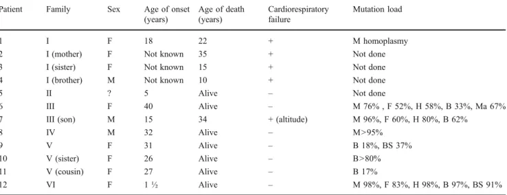
low-Table 1 Overview of patients with mutation m.3302A>G in tRNALeu (UUR). Family I: Patients 1–4 (Watmough et al.1990; Bindoff et al. 1993). Family II: Patient 5 (Shoffner et al.1993). Family III: Patients 6 and 7 (van den Bosch et al.2004). Family IV: Patient 8 (Hutchison et al.
2005). Family V: Patients 9–11 (Hutchison et al. 2005). Family VI: Patient 12 is the index patient described in this article. Known mutation loads are given for muscle (M), fibroblasts (F), hair (H), blood (B), breast tissue (Ma) and buccal swab (BS)
Patient Family Sex Age of onset (years) Age of death (years) Cardiorespiratory failure Mutation load 1 I F 18 22 + M homoplasmy
2 I (mother) F Not known 35 + Not done 3 I (sister) F Not known 15 + Not done 4 I (brother) M Not known 10 + Not done
5 II ? 5 Alive – Not done
6 III F 40 Alive – M 76% , F 52%, H 58%, B 33%, Ma 67% 7 III (son) M 15 34 + (altitude) M 96%, F 60%, H 80%, B 62%
8 IV M 32 Alive – M>95%
9 V F 31 Alive – B 18%, BS 37%
10 V (sister) F 26 Alive – B>80% 11 V (cousin) F 27 Alive – B 17%
carbohydrate diet, and takes high-dose bicarbonates in order to compensate the lactic acidosis.
DNA extraction
Total genomic DNA was extracted from buccal swabs and hairs (QIAamp DNA Mini Kit; Qiagen), from blood (Nucleon BACC Genomic DNA Extraction Kit; GE
Healthcare) and from muscle and fibroblasts (proteinase K and digestion buffer; Upadhyaya et al.1984).
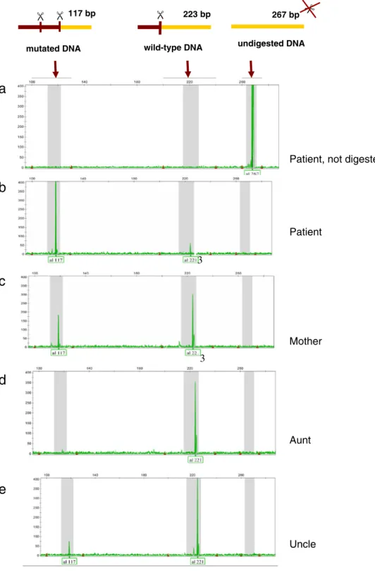
Fluorescent PCR and enzyme restriction
A fluorescent PCR (1,5 ng DNA, 22 cycles) of the region of interest was generated from the diverse tissues of the patient and relatives, using the primers 5′ TAC TTC ACA
3
3
267 bp 223 bp
117 bp
mutated DNA wild-type DNA undigested DNA
Patient, not digested
Patient Mother Aunt Uncle
a
b
c
d
e
Fig. 1 Results of capillary elec-trophoresis obtained after PCR amplification (a) and DdeI di-gestion of DNA extracted from blood (b–e). a Patient, undi-gested, shows a peak at 267 bp, corresponding to the size of the full amplicon. b Patient, digested, with a big 117 bp peak (mutated) and a small 223 bp peak (wt). The percentage of heteroplasmy is calculated by dividing the mutant peak area by the total wild-type and mutant peak areas, as forb : 3296=3296 þ 360 ¼ 0:9 ¼ 90%mutant heteroplasmy. c Mother. d Aunt. e Uncle
AAG CGC CTT CC 3′ (forward) and 5′ VIC-GGG CCT TTG CGT AGT TGT AT 3′ (reverse, fluorescent) (van den Bosch et al.2004). PCR amplification was performed using an Applied Biosystems Taq polymerase under the following conditions: 95°C/5′; 95°C/30″; 56°C/30″; 72°C/45″; 72°C/7′; 22 cycles. The mutation m.3302A>G creates a DdeI restriction site. The percentage heteroplasmy of the m.3302A>G mutation was determined by digestion with a mutation-specific restriction enzyme (DdeI, NEB). In the absence of the mutation, the digestion yields two fragments (44 and 223 bp), the largest being fluorescently labeled. Digestion in the presence of the mutation yields three fragments (44, 106, and 117 bp) of which the 117-bp fragment is fluorescently labeled.
Capillary electrophoresis
The fragments obtained were separated on capillary electrophoresis (ABI Prism 3100 Genetic analyzer) and analyzed using the Genemapper software version 4.0 (Applied Biosystems). The percentage of heteroplasmy was calculated by dividing the mutant peak area by the total wild-type and mutant peak areas. The small N + 1 peaks visible on some electropherograms are most likely the result of a residual activity of the Taq polymerase during the digestion, and the consequent addition of an A on some fragments (Fig. 1). Every sample was investigated at least in triplicates, except for hairs (duplicates).
Patient Normal range (n=26) Complex I 0.01/0.02 0.11–0.29
Complex II 0.14 0.14–0.36
Complex III 0.61 0.54–1.16
Complex IV 1.02 0.51–1.44
Complex V 0.60 0.17–0.55
Citrate synthase (mU/mg protein) 348 70–224 Table 2 OXPHOS assays in M.
quadriceps of the index patient. Respiratory chain complex ac-tivities are expressed as mU/mU citrate synthase B: 97% ± 1 BS: 91% ± 1 F: 83% ± 5 H: 98% ± 0 M : 98% ± 1
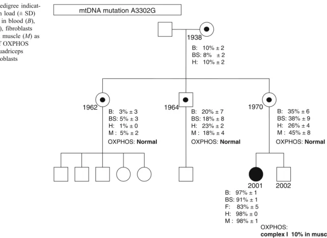
mtDNA mutation A3302G
OXPHOS: Normal 1938 1962 1964 1970 2001 2002 B: 3% ± 3 BS: 5% ± 3 H: 1% ± 0 M : 5% ± 2 B: 20% ± 7 BS: 18% ± 8 H: 23% ± 2 M : 18% ± 4 B: 35% ± 6 BS: 38% ± 9 H: 26% ± 4 M : 45% ± 8 B: 10% ± 2 BS: 8% ± 2 H: 10% ± 2
OXPHOS: Normal OXPHOS: Normal
OXPHOS:
complex I 10% in muscle 72% in fibroblastes
Fig. 2 Family pedigree indicat-ing age, mutation load (± SD) for m.3302A >G in blood (B), buccal swab (BS), fibroblasts (F), hair (H) and muscle (M) as well as results of OXPHOS analysis in M. quadriceps biopsies and fibroblasts
Cell culture
Primary fibroblast cultures were established from skin biopsy and cultured in minimal essential medium (MEM) supple-mented with 10% fetal calf serum, 4 mmol/L L-glutamine, 2 µmol/L uridine, 1 µmol/L sodium pyruvate, 50 U/ml penicillin, and 50 µg/ml streptomycin at 37°C under standard conditions.
OXPHOS assays
Isolation of mitochondria, preparation of skeletal muscle homogenates (600 g supernatants) were performed as described (Rustin et al.1994; Birch-Machin and Turnbull 2001). The activities of the individual respiratory chain complexes and the mitochondrial matrix marker enzyme citrate synthase were measured spectrophotometrically with a UV-1601 spectrophotometer (Shimadzu) using 1-ml sample cuvettes thermostatically maintained at 30° C in skeletal muscle homogenates and isolated mitochon-dria according to Birch-Machin and Turnbull (2001). Values were estimated by the difference in activity levels measured in the presence and absence of specific inhibitors and are expressed relative to the mitochondrial marker enzyme citrate synthase (mU/mU citrate synthase), that was determined as described (Shepherd and Garland 1969).
Oxygen consumption
Oxygen consumption in fibroblasts was determined at 37°C using the OROBOROS oxygraph (Innsbruck, Austria).
Fibroblasts were harvested, resuspended in Mitochondrial Respiration Medium MiR05 (Renner et al. 2003), and permeabilized by digitonin (10 μg/106cells). The rates of respiration (expressed in pmol O2 s−1 10−6 cells) were determined in the presence of 2 mM ADP and different combinations of substrates and inhibitors: 10 mM pyruvate/ 2 mM malate; 10 mM succinate/0.5 mM rotenone; 4 mM ascorbate/0.5 mM TMPD. Measurements of oxygen consumption by ascorbate/TMPD represent KCN (1 mM)-sensitive values in the presence of 12.5 mM antimycin A.
Results Mutation load
In the index patient, the tRNALeu(UUR) m.3302A > G mutation was found at levels >90% in blood, buccal swab, hair ,and muscle. Only in fibroblasts was the mutation load significantly lower with 83% (Fig. 2). In accordance with mitochondrial inheritance, the mutation was detectable in all tested individuals. The grandmother, mother, aunt, and uncle of the patient are asymptomatic and show a mean mutant load of around 10, 40, 5, and 20% respectively (Fig. 2). It is interesting to notice that we found little variation of heteroplasmy in different tissues of the same individual.
OXPHOS analysis
In skeletal muscle of the index patient, we found a marked deficiency of complex I activity (about 10% residual
Patient Normal range (n=23) Complex I 0.18/0.17 0.19–0.40
Complex II 0.29 0.10–0.45
Complex III 0.42 0.27–0.87
Complex IV 0.58 0.41–0.94
Complex V 0.10 0.10–0.34
Citrate synthase (mU/mg protein) 211 84–445 Table 3 OXPHOS assays in
skin fibroblasts of the index patient. Respiratory chain com-plex activities are expressed as mU/mU citrate synthase
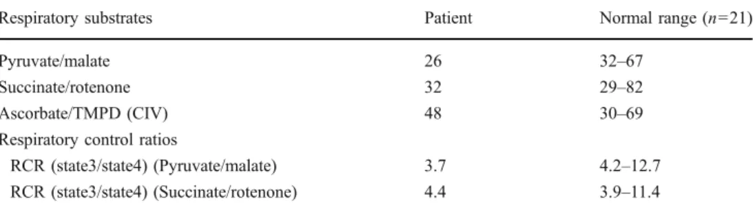
Respiratory substrates Patient Normal range (n=21)
Pyruvate/malate 26 32–67
Succinate/rotenone 32 29–82 Ascorbate/TMPD (CIV) 48 30–69 Respiratory control ratios
RCR (state3/state4) (Pyruvate/malate) 3.7 4.2–12.7 RCR (state3/state4) (Succinate/rotenone) 4.4 3.9–11.4 Table 4 Oxygen consumption
in permeabilized fibroblasts of the index patient. Respiratory rates are expressed in pmol O2
s−110−6cells. The respiratory control ratio (RCR) is defined as the ratio of state 3 (in the presence of ADP) to state 4 (in the absence of ADP) respiratory rates
activity compared to the lowest control value), while the other activities were in the range of the controls (Table2). In skin fibroblasts, the activity of complex I was only slightly below the control range (Table 3). The slightly reduced oxygen consumption with complex I substrates as well as the slightly reduced respiratory control ratio (RCR) with pyruvate and malate as substrates is in agreement with the results of the respiratory chain complex activities (Table4). OXPHOS analyses of M. quadriceps biopsies in the mother, aunt and uncle revealed normal results for all parameters (Fig.2).
Discussion
Mitochondriopathies are usually multi-system disorders (Zeviani and Carelli 2007). The fact that this mutation leads to an isolated muscle disease can probably be explained by its effect on 3′-end cleavage and different time courses of tRNA processing in different tissues (Bindoff et al. 1993; Levinger et al. 2004). As muscle is the principally affected organ, one would postulate that a higher mutation load in muscle should correlate with a more severe phenotype. This does not seem to be the case as several patients with very high mutation load in muscle have been described with variable degree of clinical severity (Bindoff et al. 1993; van den Bosch et al. 2004; Hutchison et al.2005). Thus, individual modifying factors may influence disease expression.
The diagnosis of an individual with the tRNALeu(UUR) m.3302A>G mutation confronts affected families, clinicians, and genetic counselors with a lot of unsolved questions. Due to variable age of onset (infancy to adulthood) and variable degree of symptoms (severe myopathy to mild exercise intolerance), genetic counseling and clinical prognosis is very hazardous. In relatives with mild muscle weakness, it might be difficult to distinguish between disease-related and psychological/subjective symptoms.
The determination of mutation load in muscle is a quite invasive diagnostic tool for family testing and does not lead to clear results. The cut-off between affected and non-affected is not yet known. In our family, we found mutation loads in muscle of clinically and biochemically unaffected individuals up to 45%. The value of mutation load of other, more easily available tissues, such as hair, buccal swab, and blood, are of questionable value as bad correlations with muscle have been described (van den Bosch et al.2004). In our study, however, we found less variability of mutation load in extra muscular tissues (blood, buccal swab, and hair) and skeletal muscle. In our index patient the mutation load was significantly lower in fibroblasts. Confirmation of this finding by other studies is necessary before it can be used as a predictive value for counseling.
The best existing diagnostic tool is probably OXPHOS analysis in M. quadriceps biopsy, as normal complex I activity seems to exclude the disease. However, it remains an invasive procedure offered in only a few specialized laboratories. Reliable results depend on correct sampling, optimal storage, and transport conditions, and laboratory analytical experience.
Prerequisites for prenatal diagnosis of mtDNA point mutations have been formulated by Poulton and Trunbull (2000). First, a close correlation between mutation load and disease manifestation. This seems to be the case for the tRNALeu(UUR) mutation m.3302A>G but we do not know yet which is the minimal mutation load to become symptomatic. Second, no significant time-dependent changes in mutation load. There is so far no knowledge about this point for the mutation m.3302A>G. And, third, a uniform distribution of mutation load in different tissues. In all reported cases, the mutation load for m.3302A >G was significantly lower in fibroblasts than in muscle leading to an approximately normal activity of complex I. This may be misleading, assuming that metabolic enzyme activities in amniocytes and chorionic villus sampling (CVS) show a similar expression as fibroblasts. Prenatal OXPHOS ana-lysis in CVS and amniocytes is commonly considered reliable only for mitochondrial defects in nuclear genes. Hence, prenatal diagnosis for tRNALeu(UUR) m.3302A>G mutation seems to remain very difficult. Pre-implantation genetic diagnosis may represent the best management option for many mtDNA diseases in the future but is nowadays only available in some countries and on a research basis. This approach, however, cannot guarantee that the fetus will be unaffected; it needs CVS and/or serial amniocentesis to confirm that the load of mutant mtDNA remains low or undetectable along the pregnancy, a condition that greatly improves the probability of an unaffected offspring (Poulton et al.2009).
Our results give an important insight into the correlation between mutant heteroplasmy, complex I activity, and clinical symptoms in the tRNALeu(UUR) m.3302A > G mutation. The early and severe manifestation of the disease in our index patient further broadens the clinical spectrum associated with the tRNALeu(UUR) muta-tion m.3302A>G. Mutamuta-tion loads higher than 45% in muscle are most probably necessary for complex I deficiency to develop, leading to clinical manifestation. These findings as well as heteroplasmy consistency through many tissue types represent valuable information for family members seeking accurate genetic, prognostic, and reproductive counseling.
Acknowledgment We thank Wassim Raffoul for performance of M. quadriceps biopsies in adults and Olivier Boulat for support on metabolic analyses.
References
Andrews RM, Kubacka I, Chinnery PF, Lightowlers RN, Turnbull DM, Howell N (1999) Reanalysis and revision of the Cambridge reference sequence for human mitochondrial DNA. Nat Genet 23 (2):147
Bindoff LA, Howell N, Poulton J et al (1993) Abnormal RNA processing associated with a novel tRNA mutation in mitochondrial DNA. A potential disease mechanism. J Biol Chem 268(26):19559–19564 Birch-Machin MA, Turnbull DM (2001) Assaying mitochondrial
respiratory complex activity in mitochondria isolated from human cells and tissues. Meth Cell Biol 65:97–117
Hutchison WM, Thyagarajan D, Poulton J et al (2005) Clinical and molecular features of encephalomyopathy due to the A3302G mutation in the mitochondrial tRNA(Leu(UUR)) gene. Arch Neurol 62(12):1920–1923
Levinger L, Oestreich I, Florentz C, Morl M (2004) A pathogenesis-associated mutation in human mitochondrial tRNALeu(UUR) leads to reduced 3′-end processing and CCA addition. J Mol Biol 337(3):535–544
Maniura-Weber K, Helm M, Engemann K et al (2006) Molecular dysfunction associated with the human mitochondrial 3302A>G mutation in the MTTL1 (mt-tRNALeu(UUR)) gene. Nucleic Acids Res 34(22):6404–6415
Poulton J, Turnbull DM (2000) 74th ENMC international workshop: mitochondrial diseases 19–20 November 1999, Naarden, The Netherlands. Neuromuscul Disord 10(6):460–462
Poulton J, Kennedy S, Oakeshott P, Wells D (2009) Preventing transmission of maternally inherited mitochondrial DNA diseases. BMJ 338:b94
Renner K, Amberger A, Konwalinka G, Kofler R, Gnaiger E (2003) Changes of mitochondrial respiration, mitochondrial content and cell size after induction of apoptosis in leukemia cells. Biochim Biophys Acta 1642(1–2):115–123
Rustin P, Chretien D, Bourgeron T et al (1994) Biochemical and molecular investigations in respiratory chain deficiencies. Clin Chim Acta 228(1):35–51
Schurer H, Schiffer S, Marchfelder A, Morl M (2001) This is the end: processing, editing and repair at the tRNA 3′-terminus. Biol Chem 382(8):1147–1156
Shepherd D, Garland PB (1969) The kinetic properties of citrate synthase from rat liver mitochondria. Biochem J 114(3):597– 610
Shoffner JM, Krawiecki N, Cabell MF, Torroni A, Wallace DC (1993) A novel tRNA leu(UUR) mutation in childhood mitochondrial myopathy. Am J Hum Genet 53(suppl):949
Upadhyaya M, Archer IM, Harper PS et al (1984) DNA and enzyme studies on chorionic villi for use in antenatal diagnosis. Clin Chim Acta 140(1):39–46
van den Bosch BJ, de Coo IF, Hendrickx AT et al (2004) Increased risk for cardiorespiratory failure associated with the A3302G mutation in the mitochondrial DNA encoded tRNALeu(UUR) gene. Neuromuscul Disord 14(10):683–688
Wallace DC, Lott MT (2009) MITOMAP: a human mitochondrial genome database.http://www.mitomap.org.
Watmough NJ, Bindoff LA, Birch-Machin MA et al (1990) Impaired mitochondrial beta-oxidation in a patient with an abnormality of the respiratory chain. Studies in skeletal muscle mitochondria. J Clin Invest 85(1):177–184
Zeviani M, Carelli V (2007) Mitochondrial disorders. Curr Opin Neurol 20(5):564–571