Received: 19 August 2004 Revised: 1 October 2004 Accepted: 7 October 2004
Published online: 24 November 2004 © Springer-Verlag 2004
Abstract The purpose of this study was to compare the diagnostic per-formance of axial and coronal views in multidetector CT enteroclysis (MDCTE). We retrospectively evalu-ated 48 patients with pathological correlation investigated by MDCTE for small bowel disorders. After na-sojejunal administration of 2 l of 5% methylcellulose axial arterial and ve-nous acquisition of MDCTE was fol-lowed by coronal reconstructions us-ing equal slice thicknesses of 2.5 mm with 2 mm increments. Spa-tial resolution of both planes was evaluated by phantom. Three radiol-ogists independently read axial and coronal images concerning 12 patho-logical features. The interobserver agreement and time of reading was calculated. Sensitivity and specifici-ty resulted from comparison with histopathology (n=39) or follow-up (n=9). Phantom study revealed high-er spatial resolution for axial than coronal views, whatever reconstruc-tion interval was used. However, spatial frequency always remained
high. Most pathological signs, such as bowel wall thickening (BWT), bowel wall enhancement (BWE) and intraperitoneal fluid (IPF), showed better interobserver agreement on axial than coronal views (BWT: 0.61 vs. 0.44; BWE: 0.56 vs. 0.5;
IPF:0.53 vs. 0.43). The Wilcoxon signed-rank test revealed significant-ly higher sensitivity for axial than coronal views (P=0.0453); the time of reading was significantly shorter for the latter (P=0.0146). The diag-nostic value of axial slices is superi-or to csuperi-oronal reconstructions despite the reduced data volume and display of the physiological course of bowel loops on the coronal plane.
Keywords Computed tomography multi-detector row · Intestines · Enteroclysis Sabine Schmidt Patrick Chevallier Marc Chalaron Bertrand Bessoud Francis R. Verdun Philippe Frascarolo Pierre Schnyder Alban Denys
Multidetector CT enteroclysis: comparison
of the reading performance for axial
and coronal views
Introduction
The technique of enteroclysis remains the gold standard for the radiological investigation of small bowel disor-ders [1–4], since only volume challenge results in homo-geneous luminal distension, which is mandatory for the detection of intestinal diseases. The additional inherent advantage of cross-sectional imaging modalities such as CT enteroclysis and MR enteroclysis is the simultaneous
depiction of intraluminal, mural and extraintestinal pathologies [2–6]. Spiral CT enteroclysis, which has evolved to an established modality for the investigation of various small bowel disorders [2, 3, 7–12], is now gradually being replaced by multidetector CT enterocly-sis (MDCTE) [13–15].
Development of multidetector row technology in 1998 [16], based on the helical CT that was introduced in 1989 [17], now allows data acquisition over the entire abdomen S. Schmidt (
✉
) · M. ChalaronP. Schnyder · A. Denys
Service de radiodiagnostic et radiologie interventionnelle,
Centre Hospitalier Universitaire Vaudois–CHUV,
Rue du Bugnon, 1011 Lausanne, Switzerland
e-mail: [email protected] Tel.: +41-21-3144444
Fax: +41-21-3144443 P. Chevallier
Department of Diagnostic Radiology, CHU,
Nice, France B. Bessoud
Department of General Radiology, BROCA,
Le Kremlin-Bicêtre, France F. R. Verdun
University Institute of Applied Radiophysics, Lausanne, Switzerland
P. Frascarolo
Department of Anesthesiology, Lausanne, Switzerland
in thin slices within one single breath-hold. This multi-slice acquisition not only increases spatial resolution, but also re-duces motion artifacts. Therefore, even the most fragile pa-tients can be investigated. Multiplanar reconstructions are secondarily realized within minutes without an increase of time. The coronal plane in particular is better suited to dis-play physiological bowel anatomy, especially the ileocecal valve. It corresponds to the view of the surgeon at laparoto-my and is of the same orientation as conventional examina-tions; therefore, it is well received by the referring clinician. Moreover, the entire abdominal cavity can be displayed in a lower number of slices than in the axial plane, potentially reducing the reading time of the radiologist. Axial slices of-ten demonstrate bowel loops in their short axis, so it is ofof-ten laborious to follow their course in order to determine the exact length of any mural pathology, especially in segmen-tary stenoses. The aim of this retrospective study is to com-pare the diagnostic performance of axial slices in MDCTE with coronal reconstructions.
Material and methods
Spatial resolution of axial slices and coronal reconstructions In order to compare the spatial resolution of axial and coronal slices, we performed an experimental study using a quality assur-ance phantom supplied by the manufacturer of the CT unit (QA phantom GE Medical Systems, Milwaukee, WI). This test object contains a polymethylmetacrylate (PMMA or Plexiglas) bloc with a resolution test pattern including the following spatial frequencies: 0.31 mm−1, 0.38 mm−1, 0.50 mm−1, 0.63 mm−1 and 0.83 mm−1. From the fact that these modulations make an angle of 45° with the x and y directions (Fig. 1a), their frequencies should be multiplied by cos (45°; i.e., 0.707) when dealing with their sampling in the x direction and y direction. This PMMA bloc is immersed in water in a cylindrical test object of 21 cm. To be representative of abdomi-nal acquisitions, transverse slices of this test pattern were first ac-quired using a pitch of 1.5 and a field of view of 36 cm. Matrix size always remained 512×512. To assess the spatial resolution of the
coronal reformatted images, the test object was rotated by 90°. A standard acquisition was then performed using a reconstructed slice thickness of 2.5 mm with an increment of 2.0 mm. From this set of data, coronal views, 2-mm thick, were reconstructed by means of the MPR algorithm available on the CT unit. This procedure is strictly equivalent to the one applied on the clinical images.
Based on the same acquisition parameters, a second coronal re-construction was performed using a slice increment of 1.2 mm. Spatial frequency of each of the two differently reconstructed co-ronal views was then compared with the one of axial slices.
Patients
Among 180 patients investigated by MDCTE within 31 months in our department, we retrospectively evaluated 48 patients (27 women, 21men, mean age =48 years) with pathological correla-tion. Clinical indications were the following: 30 patients (63%) presented with known or suspected inflammatory bowel disease, 10 (21%) with possible small bowel tumors or metastases, 5 (10%) with suspected postoperative complications, such as stric-tures of leakages, 2 (4%) with digestive vascular pathologies and 1 (2%) with diffuse abdominal complaints.
Technique
Patients had fasted for 8 h prior to MDCTE without other digestive preparation. After nasal intubation of the proximal jejunum under fluoroscopy, 2 l of 5% methylcellulose was introduced to the small bowel by a power-injector (Guerbet GmbH, Sulzbach/Taunus, Ger-many) at a flow rate of 200 ml/min immediately before MDCTE. All patients received 20 mg of scopolaminbutylbromid (Buscopan, Boehringer Ingelheim, Basel, Switzerland) i.v. or, if contraindicat-ed, 1 mg of glucagon (GlucaGen, Novo Nordisk, Bagsvaerd, Den-mark) to reduce peristalsis. After intravenous administration of the contrast medium iopentol (Imagopaque, Nycomed Imaging AS, Oslo, Norway) at a flow rate of 2.5 ml/s (volume in ml = body weight + 30 ml). Multidetector spiral-CT acquisition (four-detec-tor-array, Light Speed QX/i, GE Medical Systems, Milwaukee, WI) was performed within 3 min. We obtained slices in the arterial (at 30 s) and venous phase (at 60 s) from the diaphragm to pubic sym-physis (200–240 mA, voltage 120 kV, table speed: 15 mm/rotation, pitch 1.5, collimation 4×2.5 mm, axial and coronal slice
recon-struction:2.5 mm/2 mm). Advantage Windows 4.0 GE work station served for the postprocessing procedures (MPR).
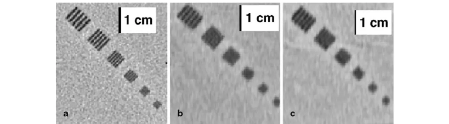
Fig. 1 a Transverse slice of the resolution test pattern of the test
object shows a spatial resolution of nearly 0.63 mm−1(i.e., modu-lations detectable up to the fourth line pairs group), their acquisi-tion parameters exactly corresponding to those of axial images of MDCTE (pitch = 1.5/slice thickness = 2.5 mm/slice increment = 2 mm) b Coronal view of the test object reformatted from axial
slices seen in a with an increment of 2 mm demonstrates that only one group of line pairs can be resolved (0.31 mm−1). c Coronal view of the test object using the same acquisition conditions as in
a, but a slice increment of 1.2 mm, leading to minimal change of
spatial resolution since still modulations up to 0.31 mm−1can be detected only
Proof of disease
The gold standard was defined as the number and anatomical exten-sion of pathological findings seen in consensus on axial slices by the two investigators (AD, SS), who were aware of all the data, always in correlation with patient outcome. Proof of disease was obtained by histopathological findings resulting from subsequent surgery or retro-grade endoscopy of the terminal ileum. Segment-by-segment correla-tion was made between imaging studies and histopathological proof. If there was no operation or simultaneous retrograde endoscopy per-formed, long-term clinical follow-up was taken into consideration.
For 39 (81.3%) patients, pathological proof of disease was available. The patients’ clinical outcomes had to be correlated with radiological features in the remaining nine. Median follow-up was 15 months for the latter.
Image analysis
Three radiologists (BB, MC, PC) with extensive experience in ab-dominal imaging resulting from working in a specialized university hospital independently evaluated the diagnostic findings of axial slices and coronal reconstructions separately in a blinded fashion in search of 12 pathological signs: bowel wall thickening (BWT), bowel stenosis (BST), pathological bowel wall enhancement (BWE) after intravenous injection of contrast medium, intra-/extraluminal mass (IEM), adjacent mesenteric fat infiltration (MFI), lymphadenopathy (ADP), intraperitoneal fluid (IPF), fistula (FIS), abscess (ABS), pari-etal vascular malformation (PVM), intestinal perforation (INP) and invagination (INV). Radiological features had to be attributed either to one of six bowel segments (jejunum, ileum, cecum/ascending co-lon, transverse coco-lon, descendent colon and sigmoid colon/rectum) or situated in one of six anatomical abdominal areas (left or right hypo-chondriac, left or right epigastric region and left or right iliac fossa).
We used a four-point scale to determine observer confidence: The diagnosis was certain (100%), probable (60%), possible (30%) or excluded (0%), receiving grades from one (certain) to four (excluded); the first two levels were considered as a positive, the last two levels as a negative result.
Image analysis was essentially filmless, done by cine-display on the work-station. Each radiologist independently and blindly reviewed axial slices in one session followed by reading of coro-nal reconstructions at a distance of at least 6 weeks in order not eventually to remember any cases, which had also been randomly mixed. Individual time of reading was taken into account for each technique and each patient.
Pathological features were exactly defined as follows: the bowel wall was considered as thickened if there was a parietal diameter of more than 3 mm. Bowel stenosis was present if the luminal narrow-ing was preceded by prestenotic dilatation. Pathological bowel wall enhancement and mesenteric fat infiltration were defined by their different density or attenuation compared to the surrounding equiva-lent structures. Mesenteric lymphadenopathy was defined as a lymph node whose smallest diameter was larger than 5 mm. Any in-traperitoneal fluid was considered as a pathological feature.
Statistical analysis
Interobserver agreement between the three radiologists was calcu-lated using kappa statistics concerning each of the 12 pathological features detected on MDCTE. Strength of agreement was charac-terized according to the proposed ratings by Fleiss [18]: kappa <0.4= poor, 0.40–0.75= good and >0.75= excellent agreement. By comparing the result of each of the three radiologists with histo-pathological results or patients’ clinical outcome, we calculated the mean sensitivity and mean specificity for each of the 12 patho-logical features. THe Wilcoxon signed rank test and paired Stu-dent’s t test were used to show statistical significance.
Results
Phantom study: results
In the standard acquisition conditions (FOV =36 cm/pitch =1.5/slice thickness =2.5 mm/slice increment =2 mm) of the transverse slices of the test object, the pixel size in plane resolution is equal to 0.7 mm, leading to an image sampling frequency of 1.4 mm−1. In such conditions the
Nyquist frequency (highest spatial frequency theoretical-ly detectable) is equal to 1.4/2=0.7 mm−1. Figure 1a
shows that modulations can be detected significantly un-der this value, i.e., modulations detectable up to 0.63 mm−1, which corresponds in fact to a frequency of
0.45 mm−1in the x or y direction.
Using a slice increment of 2 mm (FOV =36 cm), the sampling frequency of the reformatted coronal slices is equal to 0.5 mm−1, leading to a Nyquist frequency of
0.25 mm−1. As shown in Fig. 1b, the spatial frequency of
the reformatted images is much lower than the one shown on axial slices produced with a sampling of 1.4 mm−1 (Fig. 1a). The limiting spatial resolution of
Fig. 1b is now 0.31 mm−1 (only one group can be
re-solved), which corresponds to a frequency of 0.22 mm−1
in the x or y direction, a value compatible with a Nyquist frequency of 0.25 mm−1.
Secondly, without change of any other parameters, we used a slice increment of 1.2 mm, which thus increased the sampling frequency to 0.83 mm−1, leading to a
Nyquist frequency of 0.415 mm−1(Fig. 1c). However, in
spite of almost doubling the sampling frequency, the spa-tial resolution did not improve very much since it is still only possible to resolve modulations up to 0.31 mm−1, as
shown in Fig. 1c. Therefore, we applied the reconstruc-tion algorithm demonstrated in Fig. 1b on our coronal re-formatted images.
MDCTE: results
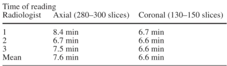
Time spent on reading by each of the three radiologists is displayed in Table 1. The mean time for axial slices was 7.6 min, which was statistically significantly longer than for coronal reconstructions with 6.6 min (P=0.0146). In general, the number of axial slices (280–300) was twice
Table 1 Time of reading per patient spent by each observer on
ax-ial slices and coronal reconstructions Time of reading
Radiologist Axial (280–300 slices) Coronal (130–150 slices)
1 8.4 min 6.7 min
2 6.7 min 6.6 min
3 7.5 min 6.6 min
Table 2 Interobserver agreement, sensitivity and specificity of the six most common pathological features resulting from independent,
random and blinded evaluation of axial slices and coronal reconstructions by the three radiologists: BWT bowel wall thickening, BST bowel stenosis, BWE pathological bowel wall enhancement, MFI adjacent mesenteric fat infiltration, ADP lymphadenopathy, IPF intra-peritoneal fluid
Pathological feature Interobserver agreement (kappa) Sensitivity (%) Specificity (%)
Technique Axial Coronal Axial Coronal Axial Coronal
BWT (n=49) 0.61 0.44 81.6 73.3 81.7 85 BST (n=24) 0.41 0.51 66.7 60.3 91.2 94.7 BWE (n=36) 0.56 0.5 75.9 73.7 88.9 90.6 MFI (n=37) 0.57 0.66 77.5 65.6 97.9 95.1 ADP (n=36) 0.69 0.65 85.2 79.4 96 93.5 IPF (n=22) 0.53 0.43 80.3 64.3 99.3 98.7 Average 0.56 0.52 77.8 (6.3a) 69.4 (7.2a) 92.4 (6.6a) 92.9 (4.7a) a Numbers in parentheses are standard deviation.
Fig. 2 a,b MDCTE in a 40-year-old woman with Peutz-Jeghers
disease: multiple pedunculated jejunal and ileal hamartomas (ar-rows) are equally detected on a axial and b coronal views
Fig. 3 a,b MDCTE in a 65-year-old man with known renal cell
carcinoma, now developing hypervascular metastasis within the proximal jejunum (arrow): optimal luminal distension with neutral contrast medium and thin-slice acquisition allows for clear visual-ization on a axial slice as well as on b coronal reconstruction
as important compared to those of coronal reconstruc-tions (130–150).
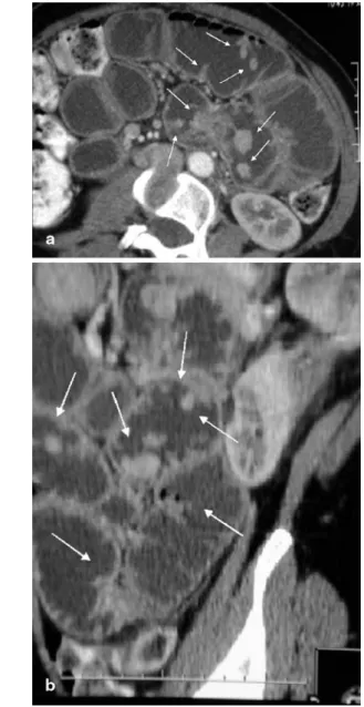
Overall incidence of the 12 pathological signs detect-ed in consensus on axial images by the two observers aware of the pathological and clinical context (see Ta-ble 2) was the following: 49 areas of bowel wall thicken-ing (BWT), 24 bowel wall stenoses (BWS) and 36 seg-ments of pathological bowel wall enhancement (BWE). In all these three pathological signs, the ileum was the predominant localization, accounting for 53% (n=26) in BWT, 68% (n=17) in BWS and 61% (n=22) in BWE. Five intraluminal masses, i.e., primary tumors or metas-tases (Fig. 2, 3), and four enteral fistulas could be seen equally on both techniques.
One of five abscesses detected on axial slices could not be detected on coronal reconstructions becauses of
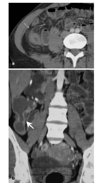
Fig. 4 a,b MDCTE in a 70-year-old man with acute inflammatory
bowel disease: small amounts of free pelvic fluid (arrow) seen on
a axial views are hardly detected on b coronal reconstructions
Fig. 5 a,b MDCTE in a 30-year-old woman with intermittent
Crohn’s disease after ileocaecal resection in the past: signs of acute inflammation, that is, hyperemia of mesenteric vessels and extension of adjacent mesenteric edematous infiltration (thin ar-row), are better seen on a axial slices, while the exact length of in-testinal stenosis (thick arrow) is better appreciated on b coronal reconstructions
its small size, the superficial location just beneath the ab-dominal wall and its extension mainly into the transverse plane, which was later confirmed by surgery. Axial slices revealed 36 areas of enlarged lymph nodes and 37 re-gions of mesenteric fat infiltration; among the latter, six were located in the pelvis without clear visualization on coronal reconstructions. Finally, there were 22 areas of intraperitoneal fluid hardly seen on coronal slices in eight patients probably because of the small amount of pelvic ascites (Fig. 4).
cent mesenteric fat infiltration” and “bowel stenosis” (MFI: 0.66 vs. 0.57, BST: 0.51 vs. 0.41). The average kappa on axial was slightly superior to coronal slices (0.56 vs. 0.52).
Sensitivity for each of the six signs was better on axi-al than coronaxi-al slices, ranging from 75.9% for BWE to 85.2% for ADP, so it was true for average sensitivity (77.8% vs. 69.4%). The largest discrepancy was seen in IPF (80.3% vs. 64.3%) followed by MFI (65.6% vs. 77.5%).
Three out of the most common six pathological fea-tures demonstrated better specificity on axial slices (MFI: 97.9% vs. 95.1%, ADP: 96% vs. 93.5% and IPF: 99.3% vs. 98.7%), whereas specificity was superior on coronal reconstructions for the other three signs (BWT: 85% vs. 81.7%, BST: 94.7% vs. 91.2% and BWE: 90.6% vs. 88.9%). Average specificity was nearly equal on both views (92.4% vs. 92.9%).
The Wilcoxon signed-rank test applied on the six most common pathological signs revealed significantly better sensitivity for axial slices than for coronal recon-structions (P=0.0453), whereas there were no significant differences in specificity.
According to Student’s t test, the three observers in-terpreted axial slices with a significantly higher level of confidence (P=0.0029) than the coronal reconstructions (mean ± standard error: 1.18±0.44 vs. 1.28±0.58).
Discussion
The technique of multi-detector row CT was developed 6 years ago, and it has been improving constantly [19]. The volume coverage speed of multidetector row helical CT is twice to three times as fast as in single-detector row helical CT, with diagnostically comparable image quality [20]. This shorter acquisition time and better spa-tial resolution regarding the z axis have improved the quality of multiplanar reconstructions, which is of partic-ular advantage for the investigation of intestinal patholo-gies [20, 21]. Therefore, we observed an extension of in-dications, such as virtual colonoscopy [22], intestinal ischemia [23], including simultaneous CT angiography [21] and entero-CT, respectively, MDCTE [13–15, 24]. By excluding all technically suboptimal MDCTE, we en-sured a good quality of imaging, especially complete small bowel opacification associated with good luminal distension in all patients.
We used dual-phase acquisition because parietal vas-cular malformations explaining subacute gastrointestinal bleeding are better seen in the arterial phase (at 30 s) of MDCTE. The venous phase (at 60 s) then allows detailed analysis of the intestinal wall, for example, inflammatory mucosal enhancement contrasting with adjacent submu-cosal edema [15, 24], provided that water or any other neutral enteral contrast medium is used, such as methyl-Interobserver agreement, sensitivity and specificity
resulting from the independent and blinded evaluation by the three radiologists are displayed in Table 2. It deals with the results of the six most common pathological signs, because the incidence of the other six was too small to be informative. Axial and coronal slices both showed good interobserver agreement in all six radiolog-ical features with kappa ranging from 0.41 to 0.69. Kap-pa values for axial slices were superior to coronal recon-structions in four out of six pathological signs (BWT: 0.61 vs. 0.44, BWE: 0.56 vs. 0.5, ADP: 0.69 vs. 0.65, IPF: 0.53 vs. 0.43). Coronal reformatted images only demonstrated better interobserver agreement for
“adja-Fig. 6 a,b MDCTE in a 22-year-old woman with acute Crohn’s
disease: important parietal thickening of the distal ileum (thin ar-row) and surrounding inflammation causes luminal stenosis com-plicated by an intraperitoneal abscess (thick arrow), both seen on
a axial and b coronal views; however, coronal reconstructions
bet-ter demonstrate the exact length of stenosis as well as the anatomi-cal abscess extension
cellulosis in our study. Opacified mesenteric vessels can be analyzed exactly on the coronal plane, sometimes completed by 3D angiography, without being obscured by high-density bowel content resulting from the administra-tion of positive enteral contrast agent [21, 25]. The detec-tion of small luminal subtracdetec-tion images such as polyps (Fig. 2) equally mostly enhancing with i.v. contrast medi-um [11] is also easier with the use of neutral enteral con-trast material. According to Orjollet et al., monodetector CT enteroclyis is not the best modality for the evaluation of certain polyps as they occur, for example, in familial polyadenomatosis, because these small and low-enhanc-ing adenomas are often mistaken for Kerckrlow-enhanc-ing folds on axial slices of 5 mm thickness [11]. MDCTE with thinner acquisition of axial slices and immediate coronal recon-structions now should allow us to overcome this problem. However, the exact role of multiplanar reconstructions in small bowel disease has not yet been evaluated on any large study population to our knowledge.
In a pictorial essay, Caoili et al. considered multiplanar reconstructions as a new perspective for the evaluation of small bowel obstruction: Using a helical single-detector CT and a collimation of 5 mm (pitch 1.5), axial images were reconstructed at 3-mm intervals in axial and coronal planes. According to the authors, coronal reformations in-crease diagnostic confidence and confirm the information revealed on axial slices, however, without proven efficacy and adding the own diagnostic value so far [26].
Raptopoulos et al. retrospectively evaluated the addi-tional diagnostic value of multiplanar and especially co-ronal projections to axial slices [27] in 22 patients. They equally used a pitch of 1.5- and 5-mm collimation with a slice increment of 3 mm. Additional coronal reconstruc-tions did not reveal further intestinal abnormalities, but significantly improved observers’ confidence in image interpretation, particularly in their assessment of compli-cated Crohn’s disease. In our study, radiologists’ confi-dence remained significantly higher in their interpreta-tion of axial slices than of coronal reconstrucinterpreta-tions alone, certainly due to the fact that radiologists are still more familiar with axial planes on CT. It remains the initial view in which images are displayed in the daily routine [28]. Certainly radiologists’ confidence will even be re-inforced by looking at both planes together [27], but this evaluation was beyond the aim of our study.
However, the higher diagnostic confidence obtained by Raptopoulos et al. also means a longer time of inter-pretation spent on one patient since its overall number of slices increases, including coronal reconstructions [27]. The use of work stations instead of film reading has meanwhile become mandatory in order to deal correctly with the large data sets radiologists have to face nowa-days [21, 29]. Our retrospective study showed a signifi-cantly shorter time of reading for coronal reconstructions than for axial slices, certainly because the former provid-ed fewer images, about half of the axial view. Therefore,
replacement of axial images by coronal reconstructions would be the ideal and time-appropriate method for im-age analysis for the future.
Prokesch et al. compared transverse images with curved coronal reformations for the staging and re-sectability of pancreatic carcinoma [28]. Average sensi-tivity and specificity between the two techniques con-cerning tumor detection and resectability were statisti-cally equivalent, average interpretation time being much shorter for reformatted images due to their limited num-ber compared to axial slices. These authors equally used a four-row multidetector CT and a dual phase i.v. injec-tion pancreatic protocol with thin collimainjec-tion of 4×1.2 mm during the pancreatic phase, from which the reformatted images were derived. This acquisition tech-nique allowed for more overlapping image reconstruc-tion than in our study, but the authors had to cover the pancreatic region only, therefore using a far shorter z-axis than in our case. Secondly, Prokesch et al. centerd the curved planar reformations on specific anatomical structures only, such as the mesenteric vessel and pan-creatic duct, etc., with the objective to complete and not to replace transverse images at the end, even if—for study purposes—both techniques were separately evalu-ated by three independent radiologists. Our coronal re-constructions, intended to be exhaustive, had to include a much larger volume of interest. They had to display the entire abdominal cavity from diaphragm to symphysis of 40–60 cm of length in order to include all the small bow-el loops and the adjacent mesentery. Besides, creation of curved planar images required trained full-time radiogra-phers under the guidance of experienced radiologists tak-ing about 20 min for each case, whereas our coronal re-formatted slices were semi-automatically reconstructed within seconds. Prokesch et al. point out the important clinical utility of these curved planar reformations, pro-viding an excellent and time-efficient overview of perti-nent findings especially for referring surgeons [28].
Our results demonstrate a good interobserver agree-ment, between 0.41 and 0.7 concerning the six most common pathological features on axial slices as well as on coronal reconstructions. However, sensitivity for all the six most common signs and interobserver agreement for four out of six features were inferior on coronal slices compared to the axial view.
At advanced stages of Crohn’s disease, coronal recon-structions are considered to be especially useful [27] to define the anatomical extension of the inflammatory pro-cess, to detect fistulae extending in the axial plane and to characterize the exact length of inflammatory or fibrotic segmental stenoses of the small bowel. The latter is con-firmed by the better interobserver agreement for bowel stenosis that we obtained for coronal slices compared to axial images and demonstrated in Fig. 5, 6. The patho-logical feature of adjacent mesenteric infiltration also showed better interobserver agreement on coronal
recon-structions, but a significantly lesser sensibility than on axial slices. Figure 5 demonstrates how coronal views can underestimate the extension of transmural inflamma-tion in Crohn’s disease, such as vascular hyperemia and mesenteric edema. This observation agrees with the re-sults of Raptopoulos et al., which showed inflammatory bowel involvement to be less extensive on coronal re-constructions than when assessed by axial slices alone [27]. The typical target or double halo appearance of in-flammed small bowel loops might more easily be detect-ed on the perpendicular view of axial images than on the coronal view where the bowel wall is mostly cut along its long axis. Therefore, our results indicates a better in-terobserver agreement and sensitivity for bowel wall thickening and pathological bowel wall enhancement on axial images than on coronal reconstructions.
In our study, the pathological sign lymphadenopathy showed altogether the best kappa and highest sensitivity of our six most common pathological features concern-ing the axial slices as well as the coronal reconstructions. The excellent spatial resolution with multidetector CT now allows the detection of lymph nodes of even 3 mm of smallest diameter in the abdominal cavity.
The largest statistical discrepancy between the two views concerns intraperitoneal fluid. In fact, Fig. 4 shows that small amounts of pelvic ascites were particularly dif-ficult to detect on coronal reconstructions, despite the bet-ter display of the abdominal cavity in the coronal plane.
Limitations of the study
We chose the axial view in combination with the histo-pathological result, respectively, long-term follow-up of patients as the gold standard. We had to rely on clinical evolution in nine patients (18.7%). Since non-invasive investigation of the small bowel is still difficult to per-form, there are only a few other imaging modalities that could serve as a comparison, such as MRI enteroclysis (MRE). In a recent prospective study, MDCTE revealed diagnostic superiority over MRE because of better sensi-tivity and interobserver agreement in detecting small bowel lesions [15]. MRE allows primary coronal views as a multiplanar modality, but the time of image acquisi-tion is still far longer and spatial resoluacquisi-tion still remains inferior compared to MDCTE.
As proven and explained by means of our quality as-surance test detailed above (Fig. 1a,b,c), our coronal re-constructions were of lower spatial resolution than axial slices. This difference in spatial resolution means that the coronal views obtained in our study should not be al-lowed to separate (in the worst case) two structures that are less than 4 mm apart (Nyquist frequency of 0.25 mm−1), whereas in the transverse view it should be
possible to discriminate structures that are 1.4 mm apart and above (i.e., Nyquist frequency of 0.7 mm−1). It
should be pointed out, however, that a lesion smaller than 2 mm, but highly contrasted, could be detected in the coronal views obtained in our conditions. However, its perceived contrast would be reduced due to pixel par-tial volume effects.
However, since the diameter of our smallest patholog-ical features was never below 2 mm, we accepted this drawback for practical reasons as being within the limi-tations of a retrospective study, since we could not change the applied acquisition parameters, especially the pitch factor, any more.
Furthermore, compared with other possibilities of im-age data reconstruction, multiplanar reformations still re-main a superior diagnostic tool, especially for small low contrast lesions (diameter of 3 mm and less), than vol-ume-rendering techniques, which are particularly suited for larger objects and data with high noise content [30].
The only possibility to improve the spatial resolution of the coronal views in our retrospective study was therefore the reduction of the slice increment to a value close to half of the acquisition slice thickness (i.e., lower than 1.25 mm). Figure 1c, however, shows that coronal reformatting with a slice increment of 1.2 mm does not significantly improve spatial frequency.
The narrower the axial collimation and the smaller the pitch of MDCTE chosen, the better the spatial resolution and also the coronal views will be due to sampling improve-ments [21]. The thinnest possible slice thickness on four-ar-ray-detector CT is 1.25 mm. This collimation, associated with a pitch value of 0.75 and a slice increment of 0.6 mm, would have allowed the improvement of the spatial resolu-tion in the coronal plane. From sampling properties, the lim-iting spatial frequency in the coronal plane would have been 0.8 mm−1(i.e., (1/0.6)×0.5 mm−1), but such a long image
ac-quisition would have significantly increased the risk of bow-el movement and respiration artifacts.
This inconvenience has now been overcome with the newer generation of 16-array-detector CT, since thinnest slices of 0.63 mm are possible. Using parameters of 16×0.63 mm at a pitch of 1.35, therefore covering a vol-ume of 13.6 mm per second, image acquisition then be-comes really isotropic, and multiplanar reformatting (MPR) with a slice increment smaller than half of the re-constructed slice thickness can easily be obtained.
Conclusion
In summary, our study confirms the present superior di-agnostic value of axial slices over coronal reconstruc-tions because of better sensibility in our six most com-mon pathological features and higher interobserver agreement in most of them. Diagnostic confidence is equally superior for axial images, each plane being ana-lyzed separately, certainly even increasing when looking at both views together.
References
1. Maglinte DD, Kelvin F, O’Connor K (1996) Current status of small bowel radiography. Abdom Imaging 2:247–257
2. Klöppel R, Thiele J, Bosse J (1992) The sellink CT method. RöFo 156:291–292
3. Bender GN, Maglinte DD, Klöppel R, Timmons JH (1999) CT-enteroclysis: a superflous diagnostic procedure or valuable when investigating small-bowel disease? Am J Roentgenol 172:373–378
4. Rieber A, Wruk D, Nüssle K, Aschoff AJ, Reinshagen M, Adler G et al (1998) MRT des Abdomens in Kombi-nation mit der Enteroklyse bei Morbus Crohn unter Verwendung von oralem und intravenösem Gd-DTPA. Radiologe 38:23–28
5. Maglinte DD, Siegelman ES, Kelvin FM (2000) MR Enteroclysis: the future of small-bowel imaging? Radiology 215:639–641
6. Maglinte DD, Bender GN, Heitkamp DE, Lappas JC, Kelvin FM (2003) Multidetector-row helical CT entero-clysis. Radiol Clin North Am 41(2):249–262
7. Bender GN, Timmons JH, Williard WC, Carter J (1996) Computed tomo-graphic enteroclysis—one methodolo-gy. Invest Radiol 31(1):43–39 8. Thiele J, Klöppel R, Schulz HG (1993)
CT-Sellink—eine neue Methode der Darmwandbeurteilung. RöFo 159(3):213–217
9. Schober E, Turetschek K, Schima W, Mostbeck GH (1997) Methylcellulose spiral CT in the preoperative assess-ment of Crohn’s disease: radiologic and pathologic correlation (abstract). Radiology 205(P):717
10. Walsh DW, Bender GN, Timmons JH (1998) Comparison of computed tomo-graphy-enteroclysis and traditional computed tomography in the setting of suspected partial small bowel obstruc-tion. Emerg Radiol 5:29–37
11. Orjollet-Lecoanet C, Menard Y, Martins A, Crombé-Ternamian A, Cotton F, Valette PJ (2000) L’En-teroscanner: Une nouvelle méthode d’exploration du grêle. J Radiol 81:618–627
12. Gaffke G, Stroszczynski C, Schlecht I, Reiche K, Wickede M von,
Ludwig WD, Felix R (2001) Diagnos-tik von Dünndarmtumoren mit Hilfe der CT-Sellink Methode (abstract). RöFo 173:S37
13. Rust GF, Holzknecht N, Olbrich D,Schöpf U, Brüning R, Reiser M (1999) Mehrschicht-Computertomo-graphie des Dünndarms—vorläufige Ergebnisse. Radiologe 39:965–970 14. Rust GF, Spiekermann A, Daum F, Schoepf UJ, Holzknecht N, Matz C, Staebler A, Reiser MF (2001) New de-velopments in imaging the small bowel with multislice computed tomography and negtive contrast medium. In: Reiser MF (ed) Multislice CT. Springer, Berlin Heidelberg New York, pp 49–60
15. Schmidt S, Lepori D, Meuwly JY, Duvoisin B, Meuli R, Michetti P et al (2002) Prospective comparison of MR-enteroclysis (MRE) with multidetector-spiral-CT-enteroclysis (MSCTE). Eur Radiol 13:1303–1311
16. Silverman PM, Kalender WA, Hazle JD (2001) Common terminology for single and multislice helical CT. Am J Roentgenol 165(5):1135–1136 17. Kalender WA, Seissler W, Klotz E,
Vock P (1990) Spiral volumetric CT with single-breath-hold technique, continuous transport, and continuous scanner rotation. Radiology
176(1):181–183
18. Fleiss JL (1985) Statistical methodes for rates and proportions. Wiley, New York
19. Kalender WA (2000) Computertomo-graphie. Publicis MCD Verlag, München
20. Hu H, He HD, Foley WE, Fox SH (2000) Four multidetector row helical CT: image quality and volume cover-age speed. Radiology 215:55–62
21. Ros PR, Ji H (2002) Multisection (multidetector) CT: applications in the abdomen. Radiographics 22:697–700 22. Luboldt W, Fletcher JG, Vogl TJ
(2002) Colonography: current status, research directions and challenges. Update 2002 (review). Eur Radiol 12:502–525
23. Horton KM, Fishman EK (2001) Multi-detector row CT of mesenteric ischemia: can it be done?
RadioGraphics 21:1463–1473 24. Bruel JM, Gallix B (2003) Scanner
multidétecteur face à l’IRM dans la pathologie du tube digestif. J Radiol 84:499–513
25. Horton KM, Fishman EK (2003) The current status of multidetector row CT and three-dimensional imaging of the small bowel. Radiol Clin North Am 41(2):199–212
26. Caoili EM, Paulson EK (2000) CT of small-bowel obstruction: another per-spective using multiplanar reforma-tions. Am J Roentgenol 174:993–998 27. Raptopoulos V, Schwartz RK,
McNicholas MMJ, Movson J, Pearlma J, Joffe N (1997) Multiplanar helical CT enterography in patients with Crohn’s disease. Am J Roentgenol 169:1545–1550
28. Prokesch RW, Chow LC, Beaulieu CF, Nino MM, Mindelzun RE, Huang J, Jeffrey RB (2002) Local staging of pancreatic carcinoma with multidetec-tor row CT: use of curved planar refor-mations—initial experience. Radiology 225:759–765
29. Foley WD (2002) Multidetector CT: abdominal visceral imaging. Radio-graphics 22:701–771
30. Shin HO, Falck CV, Galanski M (2004) Low-contrast detectability in volume rendering: a phantom study on multide-tector-row spiral CT data. Eur Radiol 14(2):341–349