O R I G I N A L A R T I C L E – G A S T R O I N T E S T I N A L O N C O L O G Y
Neoadjuvant Chemotherapy in Patients with Stage IV Colorectal
Cancer: A Comparison of Histological Response in Liver
Metastases, Primary Tumors, and Regional Lymph Nodes
Pascal Gervaz1, Laura Rubbia-Brandt3, Axel Andres1, Pietro Majno1,4, Arnaud Roth2, Philippe Morel1, and Gilles Mentha1,4
1Department of Surgery, University Hospital Geneva, Geneva, Switzerland;2Department of Oncology, University Hospital
Geneva, Geneva, Switzerland;3Department of Pathology, University Hospital Geneva, Geneva, Switzerland;4Service de Chirurgie Visce´rale et Transplantation, Hoˆpital Universitaire de Gene`ve, Geneva, Switzerland
ABSTRACT
Background. We report the histopathological results of a novel ‘‘inversed’’ strategy designed to manage patients with colorectal cancer (CRC) who have synchronous liver metastases by using chemotherapy first, liver surgery sec-ond, and resection of the primary tumor as a final step. This study was designed to compare the response to chemo-therapy in liver metastases, primary tumors, and locoregional lymph nodes.
Methods. Twenty-nine patients with stage IV CRC received a combination of oxaliplatin, irinotecan, 5-fluo-rouracil, and leucovorin (OCFL) for 3–4 months. Histological response to chemotherapy was assessed by using a tumor regression grading (TRG) score based on presence of residual tumor cells and extent of fibrosis. Results. Median age of patients was 56 (range, 37–69) years. Primary tumor location was right colon (n = 5), left colon (n = 7), and rectum (n = 17 patients). TRG scores correlated across disease sites (Spearman correlation coefficients for TRG in the primary tumor and lymph nodes was 0.59 [P = 0.005]; for the primary tumor and metas-tases 0.44 [P = 0.021]; and for lymph nodes and metastases 0.58 [P = 0.006]). Complete absence or poor tumor response (TRG4/5) was significantly more frequent in primary tumors (35.7%) and locoregional lymph nodes (38%) than in liver metastases (6.9%; McNemar test,
P = 0.02). Two patients had a complete pathologic response (pT0N0M0).
Conclusions. In patients with stage IV colorectal cancer, liver metastases exhibit a better histological response than primary tumors to OCFL neoadjuvant chemotherapy.
Standard management of colorectal cancer (CRC) cur-rently consists in resection of the primary tumor, followed by adjuvant chemotherapy for stage III and high-risk stage II patients. For patients with colon cancer, there is no controversy regarding the chronological order of these components. Surgery is performed first, and decision regarding adjuvant chemotherapy is based on histopathol-ogical examination of the surgical specimen.1It is therefore not surprising that by contrast with rectal cancer patients (who usually benefit from neoadjuvant chemoradiation), the response of colon cancer patients to neoadjuvant chemotherapy has rarely been reported. However, the development of novel multimodality strategies designed to curatively manage patients with stage IV CRC has recently provided an opportunity to address this issue.2,3
Since 1999, an inversed strategy for managing stage IV colorectal cancer patients presenting with synchronous liver metastases has been developed at our institution.4In this protocol, chemotherapy (a combination of oxaliplatin, irinotecan, 5-fluorouracil, and leucovorin [OCFL]) is administered first, followed by resection of liver metas-tases, and primary tumor resection is performed as the last step.5 The design of this protocol provides clinicians and pathologists with the opportunity to examine and compare the pathologic response to neoadjuvant chemotherapy in liver metastases, primary tumor, and regional lymph nodes.
Ó Society of Surgical Oncology 2010 First Received: 28 December 2008; Published Online: 20 April 2010 G. Mentha
e-mail: gilles.mentha@hcuge.ch DOI 10.1245/s10434-010-1056-6
Histological tumor response has more than a morpho-logical interest and is directly correlated with disease-free and overall survival in patients with breast carcinomas and osteosarcoma.6,7 It was recently demonstrated that pathologic response after neoadjuvant chemotherapy and resection of hepatic metastases is an independent predictor of 5-year overall survival in patients with stage IV CRC.8,9 Moreover, histological tumor response in liver metastases was directly affected by the chemotherapy regimen, with an increased response rate in patients who received oxa-liplatin and irinotecan in addition to 5-fluorouracil (5-FU) and leucovorin. This combination neoadjuvant chemo-therapy regimen has markedly improved the outcome of patients with CRC who have unresectable liver metastases, and it is tempting to hypothesize that its impact on primary tumors might be even more dramatic.10
From a biological standpoint, the current consensus is that secondary tumors result from nonrandom selection of a clonal population within the primary.11 It is expected that liver metastases demonstrate a more aggressive phenotype and are more resistant to treatment, because they remain the cause of 90% of deaths from solid tumors.12 Another aspect of response to chemotherapy is the role of organ microenvironment, which provides the secondary tumors with neovessels and might increase the delivery of che-motherapy to highly vascularised metastases.13 This study was designed to assess and compare the histological responses of primary CRC and liver metastases to a stan-dardized chemotherapy protocol. We postulated that primary and metastatic CRC—although both chemosensi-tive—would differ in their response to chemotherapy; our working hypothesis was that primary tumors would exhibit a better response to OCFL than liver metastases.
METHODS
The following data regarding 29 consecutive patients with histology-proven adenocarcinoma of the colon or rectum and synchronous liver metastases were prospec-tively entered into a computerized database: patients’ demographics; American Society of Anesthesiology (ASA) classification of operative risk; TNM classification and the site of the primary; presence of extrahepatic tumoral sites; clinical risk score (CRS) according to Fong’s classification; location and number of liver metastases; chemotherapy performed prior and after surgery; radiation therapy to the pelvis for rectal cancer; type and complications related to liver resection; and the details of resection of the primary tumor, including postoperative course and complications. All patients included in this study presented with advanced liver disease and underwent preoperative portal emboliza-tion and/or two-stage hepatectomy.
Neoadjuvant Chemotherapy
Twenty-eight of 29 patients received two to six cycles of chemotherapy before liver resection, using a combination of oxaliplatin [O], irinotecan (CPT-11[C]), 5-FU [F], and leucovorin [L], according to the following doses and schedule: oxaliplatin 70 mg/m2 on days 1 and 15, irino-tecan (CPT-11) 100 mg/m2on days 8 and 22, 5-FU 2 g/m2/ 24 h, and leucovorin (LV) 30 mg on days 1, 8, 15, and 22, repeated every 5 weeks, as described previously.5 Seven patients had a similar regimen but with 5-FU/LV replaced with capecitabine (the oral prodrug of 5-FU) 800 mg/m2 per day from day 1 to day 28. One patient received standard treatment with capecitabine and oxaliplatin (capecitabine 2 g/m2, oxaliplatin 130 mg/m2 day 1, days 1–14, repeated every 3 weeks). The response to chemo-therapy was assessed after two to three cycles. Further evaluations were obtained according to the clinical response and surgical pattern of disease. Additional cycles were given if a further response was likely to confer a surgical advantage. More recently, eight patients were treated with the addition of bevacizumab (AvastinÒ) and two other patients received cetuximab (ErbituxÒ) to com-plement OCFL regimen. All patients gave informed, written consent before receiving the chemotherapy proto-col, which was approved by the ethics committee of our institution.
Surgery
Simultaneous colonic and liver resections were per-formed in selected circumstances (right colon or \1–2 segments and left colon), but rectal resections and complex liver resections were not performed during the same operation. According to our protocol, when staged resec-tions were planned, liver resection was always performed first and colorectal resection second, with ideally a short (4–6 weeks) interval between the two procedures. For patients with rectal cancer, neoadjuvant radiation therapy to the pelvis, when indicated, was delivered for 5.5 weeks (28 9 1.5 Gy), and rectal surgery was performed 6 weeks later. The resection of all liver metastases was planned not only on the most recent CT scan or MRI but also on the CT scan before chemotherapy was started, to avoid leaving ‘‘missing metastases’’ behind.
Histopathological Assessment
Histological response to chemotherapy was assessed using a tumor regression grading (TRG) score based on the presence of residual tumor cells and the extent of fibrosis. Adequate assessment of the whole tumor (both primary and metastatic) specimen was performed by assessing TRG in
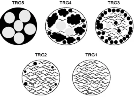
multiple tissues samples according to a previously descri-bed protocol.8 Briefly, hepatectomy specimens were sectioned in 5-mm–thick slices. In patients with multiple liver metastases, each lesion was extensively sampled from the center to the periphery to include multiple sections of tumor and liver parenchyma. More than one section per centimeter was reviewed from each tumor nodule. All tumors measuring \1 cm and/or exhibiting complete pathologic response were entirely submitted for micro-scopic examination. This grading system is similar to the Mandard protocol, which was initially elaborated for assessing response to neoadjuvant radiochemotherapy in esophageal carcinomas.14 More recently, this standardized protocol was extended to rectal carcinomas; TRG1 corre-sponded to absence of tumor cells replaced by abundant fibrosis, TRG2 to rare residual tumor cells scattered throughout abundant fibrosis, TRG3 to more residual tumor cells throughout a predominant fibrosis, TRG4 to large amount of tumor cells predominating over fibrosis, and TRG5 most exclusively to tumor cells without fibrosis15 (Fig.1).
Statistical Analysis
Statistical analyses were undertaken by means of the software package STATGRAPH 3.0 software for Windows (Statgraph Software Inc., San Diego, CA). Quantitative data were expressed as median (range). Groups’ compari-sons were made using chi-square or Fisher’s exact test for categorical variables, and Student’s t test, or Mann–Whit-ney U for continuous variables. To assess correlations between TRG scores across disease sites (primary site, lymph nodes, metastases), we obtained Spearman correla-tion coefficients. We also dichotomized TRG results as
good (scores 1–3) versus poor (scores 4 or 5) and compared probabilities of poor response across disease sites using McNemar tests. Two-sided P values B0.05 were consid-ered statistically significant.
RESULTS
Between January 2001 and July 2008, 29 patients with stage IV CRC completed our therapeutic protocol. Median age of patients was 56 (range, 37–69) years. Primary tumor location was right colon (n = 5), left colon (n = 7), and rectum (n = 17 patients). Median number of liver metas-tases was 5 (range, 1–21). Eight patients underwent a one-step procedure, with simultaneous resection of the primary tumor and liver metastases. Twenty-one patients underwent a staged procedure due to an extended hepatectomy or rectal location of the primary. In this group, median delay between liver surgery and resection of the primary tumor was 70 (range, 20–280) days. Characteristics of patients’ demographics, as well as primary and metastatic tumor locations, are summarized in Table1. Of 29 patients, 16 are still alive and 13 are free of recurrence; median survival for the entire group was 44 months, and overall 1-, 3-, and 5-year survival rates were 100%, 60%, and 31%, respectively.
TRG scores were available for all metastases, as well as 21 node-positive primary tumors. Data regarding primary tumor TRG were missing for one patient who underwent primary tumor surgery in another institution. TRG scores according to location are illustrated in Table2. TRG scores on a 1 to 5 scale were moderately correlated across disease sites: the Spearman correlation coefficient for TRG in primary tumor and lymph nodes was 0.59 (P = 0.005); for
FIG. 1 The grading system is based on a quantitative morphologic assessment of fibrosis versus residual tumor cells within tumor deposits. Grade 1 indicates complete response with maximal fibrosis, and no residual tumor cells, whereas grade 5 indicates a complete absence of fibrosis
primary tumor and metastases 0.44 (P = 0.021); and for lymph nodes and metastases 0.58 (P = 0.006).
Complete absence or poor tumor response (TRG4/5) was significantly more frequent in primary tumors (35.7%) and regional lymph nodes (38%) than liver metastases (6.9%) (McNemar test, P = 0.02; Fig.2). TRG5 (null response) was rare in liver metastases (3.4%) and primary tumors (0%) but was detected in 19% of N ? tumors. In two patients, the final diagnosis was pT0N1, indicating a better response in the primary tumors than in the locore-gional lymph nodes. Two patients (6.9%) had a complete pathologic response (pT0N0M0). Interestingly, both ini-tially presented with advanced rectal cancer, but one of them did not undergo preoperative radiation therapy and demonstrated complete response with chemotherapy alone.
DISCUSSION
The data presented here indicate that primary and met-astatic CRC differ in their response to neoadjuvant combination chemotherapy, consisting of oxaliplatin, iri-notecan, 5-fluorouracil and leucovorin. Poor tumor response (TRG4/5) was significantly more frequent in primary tumors (35.7%) and locoregional lymph nodes (38%) than liver metastases (6.9%). Of five tumor speci-mens that showed complete absence of response (TRG5), four (80%) were located within locoregional lymph nodes. Complete pathologic response (pT0N0M0) was reported in two (6.9%) patients.
The TRG score used in this series has been validated in CRC patients, not only as a morphologic variable to assess response to neoadjuvant chemo- or radiotherapy, but also as a prognostic factor for disease-free and overall sur-vival.16 The high histological response rates in this series are likely to result from the combination of oxaliplatin and
TABLE 1 Patient and tumor characteristics
Variable N = 29
Patients
Female 12
Male 17
Primary tumor location
Right colon 5 Left colon 7 Rectum 17 Metastases distribution Unilobar 10 Bilobar 19 Chemotherapy regimen Oxaliplatin 29 Irinotecan 28 5-fluorouracil 29 Leucovorin 23 Bevacizumab 8 Cetuximab 2
Pelvic irradiation for rectal cancer
Yes 8
No 9
Timing hepatectomy-colectomy
Synchronous 8
Staged 21
Final pathologic stage primary tumor
T0 4
T1 0
T2 4
T3 18
T4 3
Final pathologic stage metastases
M0 3
M1 26
Status at last follow-up
Dead 13
Alive with disease recurrence 3
Alive with no evidence of disease 13
TABLE 2 Tumor response according to location Site TRG Liver metastases Locoregional lymph nodesa Primary tumorb 1–2 major response 11 8 10 3 16 5 8 4–5 poor response 2 8 10
a 21 tumors only were N?
b One patient’s data missing (primary tumor surgery performed in
another institution) 30 25 20 15 10 5 p = 0.02 Poor tumor response Primary Tumors Locoregional lymph nodes Liver Metastases
FIG. 2 Poor tumor response (TRG4/5) was significantly more frequent in primary tumors (T = 35.7%) and locoregional lymph nodes (N = 38%) than liver metastases (M = 6.9%) (McNemar test, P = 0.02)
irinotecan; the vast majority (96.5%) of patients who received both drugs had TRG1-3 scores in their liver metastases. In addition, we report the results of a selected group of patients who responded to neoadjuvant chemo-therapy and eventually underwent liver and colorectal surgery with curative intent.17 This being considered, our data are in line with phase III trials, which have demon-strated clinical response rates in the 40–50% range for both oxaliplatin- and irinotecan-based regimens.18 Thus, path-ologic response to chemotherapy is progressively emerging, not only as a mere morphologic variable, but as a new prognostic parameter after surgery for CRC, whether or not it is metastatic.19
The better histological response rate in liver metastases than in primary tumors, as reported here, is intriguing, but probably results from the combination of two factors. First, a better drug delivery might be responsible for higher concentrations of cytotoxics drugs within the liver than in the colon and its mesentery. The histological demonstration of specific alterations in the hepatic parenchyma adjacent to metastases, such as oxaliplatin-related sinusoidal obstruction syndrome and irinotecan-related steatohepati-tis, provides indirect evidence that these drugs are present in high concentration within the liver.20,21In other words, the histological differences in response to OCFL are probably related more to the tumor microenvironment than to tumor cell chemosensitivity; the liver, with its dense vascular sinusoidal network, provides an excellent delivery access to metastases, which are dependent upon sinusoidal endothelial cells for their growth.22
The second explanation for these differences is related to the schedule of chemotherapy administration; after exclusion of 8 patients who underwent synchronous liver and colonic surgery, there was a median delay of 70 (range, 20–280) days between liver and colorectal surgery in 21 other patients. We hypothesize that this interval is a ther-apeutic window during which repopulation of cancer cells may occur within the primary tumor.23 Whereas little is known about tumor repopulation during chemotherapy, we have observed similar instances of tumor progression in patients who underwent two-step hepatectomy.24 We now try to reduce as much as possible the time interval between the two procedures, and we believe that colorectal surgery should ideally be performed 4 weeks after hepatectomy, except for rectal cancer patients who might benefit from neoadjuvant radiation therapy. In our series, seven patients had the two operations performed with an interval of 20– 40 days. Of note, a laparoscopic approach for colorectal surgery is feasible and in some patients might contribute to reducing the surgical trauma of the second operation.
Our results also demonstrate an identical poor response rate for primary tumors and locoregional lymph nodes (*36%). Approximately one-third of primary tumors were
classified as TRG4/5. Regional lymph nodes seem to har-bor the most resistant tumor deposits—a fact that, again, might reflect a lower concentration of cytotoxic drugs in the mesocolon. This finding is highly relevant and trans-lated clinically into the occasional occurrence (2 patients) of complete pathological response in the primary, but with residual tumor detected within regional lymph nodes (pT0N1).25 Interestingly, two studies that focused on patients with T3-T4 rectal cancer downstaged by neoad-juvant chemoradiation to pT0 reported a 17% rate of positive mesorectal lymph nodes.26,27 Finally, two other patients had a complete pathologic response (pT0N0M0)— a low incidence (6.9%) similar to what was observed in liver metastases by Adam et al. (4%).28
In conclusion, primary and secondary tumors differ in their response to neoadjuvant chemotherapy, but hepatic metastases exhibit better histological response rates despite being, in theory, phenotypically more aggressive. This unexpected finding, if confirmed by other series using similar chemotherapy regimen, warrants further investiga-tion. Until then, we postulate that high concentrations of oxaliplatin and irinotecan within the hepatic sinusoids are responsible for both positive (high rates of tumor response) and negative (sinusoidal obstruction syndrome) histologi-cal alterations within liver metastases and surrounding hepatic parenchyma, respectively.
REFERENCES
1. Meyerhardt JA, Mayer RJ. Systemic therapy for colorectal can-cer. N Engl J Med. 2005;352:476–87.
2. Mentha G, Majno PE, Andres A, Rubbia-Brandt L, Morel P, Roth AD. Neoadjuvant chemotherapy and resection of advanced syn-chronous liver metastases before treatment of the colorectal primary. Br J Surg. 2006;93:872–8.
3. Karoui M, Koubaa W, Delbaldo C, Charachon A, Laurent A, Piedbois P, Cherqui D, Tran Van Nhieu J. Chemotherapy has also an effect on primary tumor in colon carcinoma. Ann Surg Oncol. 2008,15:3440–6.
4. Mentha G, Majno P, Terraz S, Rubbia-Brandt L, Gervaz P, An-dres A, Allal S, et al. Treatment strategies for the management of advanced colorectal liver metastases detected synchronously with the primary tumour. Eur J Surg Oncol. 2007;33(Suppl 2):S76–83. 5. Seium Y, Stupp R, Ruhstaller T, Gervaz P, Mentha G, Philippe M, Allal A, et al. Oxaliplatin combined with irinotecan and 5-fluorouracil/leucovorin (OCFL) in metastatic colorectal cancer: a phase I-II study. Ann Oncol. 2005;16:762–6.
6. Bertheau P, Lerebours F, Mounier N, de Roquancourt A, Espie´ M, Clot P, Servant JM, et al. Prognostic significance of a com-bined clinicopathologic score for response to primary systemic therapy in locally advanced breast cancer. Oncol Rep. 2005; 14:513–20.
7. Bramwell VH. The role of chemotherapy in the management of non-metastatic operable extremity osteosarcoma. Semin Oncol. 1997;24:561–71.
8. Rubbia-Brandt L, Giostra E, Brezault C, Roth AD, Andres A, Audard D, Sartoretti P, et al. Importance of histological tumor response assessment in predicting the outcome in patients with
colorectal liver metastases treated with neo-adjuvant chemo-therapy followed by liver surgery. Ann Oncol. 2007;18:299–304. 9. Blazer DG, Kishi Y, Maru DM, Kopetz S, Chun YS, Overman MJ, Fogelman D, et al. Pathologic response to preoperative chemotherapy: a new outcome endpoint after resection of hepatic colorectal metastases. J Clin Oncol. 2008;25:5344–51.
10. Adam R, Delvart V, Pascal G, Valeanu A, Castaing D, Azoulay D, Giacchetti S, et al. Rescue surgery for unresectable colorectal liver metastases downstaged by chemotherapy: a model to predict long-term survival. Ann Surg. 2004;240:644–57.
11. Talmadge JE. Clonal selection of metastasis within the life his-tory of a tumor. Cancer Res. 2007;67:11471–5.
12. Gupta GP, Massague´ J. Cancer metastasis: building a framework. Cell. 2006;127:679–95.
13. Gervaz P, Scholl B, Padrun V, Gillet M. Growth inhibition of liver metastases by the anti-angiogenic drug TNP-470. Liver. 2000;20:108–13.
14. Mandard AM, Dalibard F, Mandard JC, Marnay J, Henry-Amar M, Petiot JF, Roussel A, et al. Pathologic assessment of tumor regression after preoperative chemoradiotherapy of esophageal carcinoma. Clinicopathologic correlations. Cancer. 1994;73: 2680–6.
15. Bouzourene H, Bosman FT, Seelentag W, Matter M, Coucke P. Importance of tumor regression assessment in predicting the outcome in patients with locally advanced rectal carcinoma who are treated with preoperative radiotherapy. Cancer. 2002;94: 1121–30.
16. Vecchio FM, Valentini V, Minsky BD, Padula GD, Venkatraman ES, Balducci M, Micchiche` F, et al. The relationship of patho-logic tumor regression grade (TRG) and outcomes after preoperative therapy in rectal cancer. Int J Radiat Oncol Biol Phys. 2005;62:752–60.
17. Mentha G, Roth AD, Terraz S, Giostra E, Gervaz P, Andres A, Morel P, et al. Liver first approach in the treatment of colorectal cancer with synchronous liver metastases. Dig Surg. 2008;25:430–5.
18. Leonard GD, Brenner B, Kemeny NE. Neoadjuvant chemother-apy before liver resection for patients with unresectable liver metastases from colorectal carcinoma. J Clin Oncol. 2005;23: 2038–48.
19. Suarez J, Vera R, Balen E, Gomez M, Arias F, Lera JM, Herrera J, Zazpe C. Pathologic response assessed by Mandard grade is a
better prognostic factor than down staging for disease-free sur-vival after preoperative radiochemotherapy for advanced rectal cancer. Colorectal Dis. 2008;10:563–8.
20. Rubbia-Brandt L, Mentha G, Terris B. Sinusoidal obstruction syndrome is a major feature of hepatic lesions associated with oxaliplatin neoadjuvant chemotherapy for liver colorectal metastases. J Am Coll Surg. 2006;202:199–200.
21. Morris-Stiff G, Tan YM, Vauthey JN. Hepatic complications following preoperative chemotherapy with oxaliplatin or irino-tecan for hepatic colorectal metastases. Eur J Surg Oncol. 2008;34:609–14.
22. Gervaz P, Scholl B, Mainguene C, Poitry S, Gillet M, Wexner S. Angiogenesis of liver metastases: role of sinusoidal endothelial cells. Dis Colon Rectum.
23. Kim JJ, Tannock IF. Repopulation of cancer cells during therapy: an important cause of treatment failure. Nat Rev Cancer. 2005;5:516–25.
24. Mentha G, Terraz S, Morel P, Andres A, Giostra E, Roth A, Rubbia-Brandt L, Majno P. Dangerous halo after neoadjuvant chemotherapy and two-step hepatectomy for colorectal liver metastases. Br J Surg. 2008;96:95–103.
25. Gervaz P, Morel P. T0N1 adenocarcinoma of the colon following oxaliplatin-based neoadjuvant chemotherapy. J Surg Oncol. 2008.
26. Hughes R, Glynne-Jones R, Grainger J, Richman P, Makris A, Harrison M, Ashford R, et al. Can pathological complete response of the primary tumour following pre-operative pelvic chemoradiotherapy for T3-T4 rectal cancer predict for sterilisa-tion of pelvic lymph nodes, a low risk of local recurrence and the appropriateness of local excision? Int J Colorectal Dis. 2006;21:11–7.
27. Bedrosian I, Rodriguez-Bigas MA, Feig B, Hunt KK, Ellis L, Curley SA, Vauthey JN, et al. Predicting the node-negative mesorectum after preoperative chemoradiation for locally advanced rectal carcinoma. J Gastrointest Surg. 2004;8:56–62. 28. Adam R, Wicherts DA, de Haas RJ, Aloia T, Levi F, Paule B,
Guettier C, et al. Complete pathologic response after preoperative chemotherapy for colorectal liver metastases: myth or reality? J Clin Oncol. 2008;26:1635–41.