HAL Id: inserm-00195972
https://www.hal.inserm.fr/inserm-00195972
Submitted on 11 Dec 2007
HAL is a multi-disciplinary open access
archive for the deposit and dissemination of
sci-entific research documents, whether they are
pub-lished or not. The documents may come from
teaching and research institutions in France or
abroad, or from public or private research centers.
L’archive ouverte pluridisciplinaire HAL, est
destinée au dépôt et à la diffusion de documents
scientifiques de niveau recherche, publiés ou non,
émanant des établissements d’enseignement et de
recherche français ou étrangers, des laboratoires
publics ou privés.
Chronic activation in presymptomatic amyotrophic
lateral sclerosis (ALS) mice of a feedback loop involving
Fas, Daxx, and FasL.
Cédric Raoul, Emmanuelle Buhler, Christel Sadeghi, Arnaud Jacquier, Patrick
Aebischer, Brigitte Pettmann, Christopher Henderson, Georg Haase
To cite this version:
Cédric Raoul, Emmanuelle Buhler, Christel Sadeghi, Arnaud Jacquier, Patrick Aebischer, et al..
Chronic activation in presymptomatic amyotrophic lateral sclerosis (ALS) mice of a feedback loop
involving Fas, Daxx, and FasL.. Proceedings of the National Academy of Sciences of the United States
of America , National Academy of Sciences, 2006, 103 (15), pp.6007-12. �10.1073/pnas.0508774103�.
�inserm-00195972�
Chronic activation in presymptomatic amyotrophic
lateral sclerosis (ALS) mice of a feedback loop
involving Fas, Daxx, and FasL
C. Raoul*, E. Buhler†‡, C. Sadeghi*, A. Jacquier†‡, P. Aebischer*, B. Pettmann‡§, C. E. Henderson‡§¶, and G. Haase†‡储
*Ecole Polytechnique Fe´de´rale de Lausanne (EPFL), Integrative Biosciences Institute, SV IBI LEN, AAB 1 32, CH-1015 Lausanne, Switzerland;†Institut de
Neurobiologie de la Me´diterrane´e (INMED), Institut National de la Sante´ et de la Recherche Me´dicale (INSERM), Equipe Avenir, F-13273 Marseille Cedex 09, France;‡Universite´ de la Me´diterrane´e, F-13288 Marseille, France; and§Institut de Biologie du De´veloppement de Marseille (IBDM), Institut National de la
Sante´ et de la Recherche Me´dicale (INSERM), Unite´ Mixte de Recherche 623, F-13288 Marseille Cedex 09, France
Edited by Fred H. Gage, The Salk Institute for Biological Studies, San Diego, CA, and approved February 14, 2006 (received for review October 10, 2005)
The reasons for the cellular specificity and slow progression of motoneuron diseases such as ALS are still poorly understood. We previously described a motoneuron-specific cell death pathway downstream of the Fas death receptor, in which synthesis of nitric oxide (NO) is an obligate step. Motoneurons from ALS model mice expressing mutant SOD1 showed increased susceptibility to exog-enous NO as compared with controls. Here, we report a signaling mechanism whereby NO leads to death of mutant, but not control, motoneurons. Unexpectedly, exogenous NO triggers expression of Fas ligand (FasL) in cultured motoneurons. In mutant SOD1G93Aand
SOD1G85R, but not in control motoneurons, this up-regulation
results in activation of Fas, leading through Daxx to phosphory-lation of p38 and further NO synthesis. This Fas兾NO feedback amplification loop is required for motoneuron death in vitro. In
vivo, mutant SOD1G93Aand SOD1G85Rmice show increased
num-bers of positive motoneurons and Daxx nuclear bodies weeks before disease onset. Moreover, FasL up-regulation is reduced in the presence of transgenic dominant-negative Daxx. We propose that chronic low-level activation of the Fas兾NO feedback loop may
underlie the motoneuron loss that characterizes familial ALS and may help to explain its slowly progressive nature.
cell death兩 motoneuron disease 兩 NO 兩 p38 kinase 兩 neurodegeneration
A
myotrophic lateral sclerosis (ALS) is the most frequent adult-onset motoneuron disease in humans. ALS is characterized by the selective degeneration of motoneurons in spinal cord, brain-stem, and cerebral cortex leading to muscle atrophy and paralysis and ultimately to death. About 1 to 2% of all human ALS forms are caused by dominantly inherited mutations in the Cu兾Zn superoxide dismutase (SOD1) gene. Mice transgenic for the ALS-linked SOD1 mutations G37R (1), H46R兾H48Q (2), G85R (3), and G93A (4) develop an adult-onset motoneuron disorder that remarkably re-sembles human ALS.Despite much intensive study, many questions remain concerning the mechanism(s) by which mutant SOD1 triggers specific mo-toneuron death. One unresolved issue is the cellular site of action of the gain-of-function mutations. Clement et al. (5) generated chimeric mice carrying a mixture of WT and mutant SOD1-expressing cells in the spinal cord. In these mice, WT motoneurons eventually showed stigmata of degeneration, whereas some mutant SOD1 motoneurons were protected from degeneration when sur-rounded by WT nonneuronal cells. These results suggest that mutant SOD1 in both motoneurons and surrounding cells may play a role in the disease process.
Our earlier studies using cultures of purified embryonic mo-toneurons reached similar conclusions. We found that motoneu-rons from SOD1G93A, SOD1G37R, and SOD1G85R mice survived
normally in the presence of optimal trophic support or when challenged by excitotoxic agonists. In marked contrast, compared with controls, they displayed a 10- to 100-fold increase in sensitivity to extracellular agonists of the Fas receptor or to exogenous nitric
oxide (6). Thus, motoneurons expressing mutant SOD1 have an intrinsic susceptibility that is only revealed when challenged with specific extrinsic agents. Only motoneurons, and no other cell type from mutant SOD1 mice, showed enhanced susceptibility. In motoneurons, we showed that the Fas receptor signals through two synergistic pathways involving Fadd兾Caspase-8 and Daxx兾Ask1兾 p38兾nNOS, respectively (6).
Another open question in ALS relates to the kinetics of the neurodegenerative process. Mutant SOD1 mice display multiple features of programmed cell death at presymptomatic stages. Examples are proteolytic activation of caspases-1, -3, -7, -8, and -9 in the spinal cord (7–9), mitochondrial release of cytochrome c (10), and translocation of Bax from the cytosol to mitochondria (11). These modifications are manifest weeks to months before there is significant loss of motoneuron cell bodies or axons. One aspect of Fas-triggered motoneuron death seemed relevant to these slow kinetics. Whereas Fas activation in vitro kills lymphocytes within hours, motoneuron death in the same conditions takes days (12). The first aim of this study was therefore to identify the molecular mechanisms that underlie this exceptionally protracted cell death. Our results linking Fas signaling to mutant SOD1 were obtained by using purified motoneurons in vitro. A second important aim of this study was therefore to investigate the significance of this signaling pathway in mutant SOD1 mice in vivo. Since our earlier publication (6), other groups studying mutant SOD1 mice have reported activation of certain inter-mediates in the Fas pathway we described. In the spinal cord of SOD1G93A mice, Tortarolo et al. (13), Hu et al. (14), and
Ackerley et al. (15) observed increased p38 activation, whereas Wengenack et al. (16) reported increased levels of Ask1. How-ever, these results concern intermediates that are common to several signaling mechanisms. We therefore focused our atten-tion on Fas and Daxx, which are specific to this pathway.
We report here that nitric oxide (NO), an end-product of the Fas pathway in motoneurons, unexpectedly induces expression of the endogenous Fas ligand (FasL), which in mutant SOD1 motoneu-rons then triggers further chronic activation of the pathway down-stream of Fas. This feedback loop is necessary for motoneuron death induced by NO or FasL in vitro. Furthermore, because all elements of this feedback loop show perturbed expression in mutant SOD1 mice at presymptomatic stages, it is possible that this
Conflict of interest statement: No conflicts declared. This paper was submitted directly (Track II) to the PNAS office. Abbreviations: DIV, day(s) in vitro; NB, nuclear bodies.
¶Present address: Departments of Pathology and Neurology, Columbia University, New
York, NY 10032.
储To whom correspondence should be addressed at: Institut National de la Sante´ et de la Recherche Me´dicale (INSERM), Institut de Neurobiologie de la Me´diterrane´e (INMED), Equipe Avenir, F-13273 Marseille Cedex 09, France. E-mail: haase@inmed.univ-mrs.fr. © 2006 by The National Academy of Sciences of the USA
pathway is linked to the slowly progressive nature of motoneuron loss in vivo.
Results
Identification of a Fas兾NO Feedback Loop in Mutant SOD1
Motoneu-rons.Because purified mutant SOD1 motoneurons show increased susceptibility to activation of the Fas death pathway, we asked whether expression of key signaling molecules differed between mutant SOD1 and control mice. Levels of Fas receptor and Fas ligand (FasL) were estimated by Western blotting. Levels of Fas were similar between C57BL兾6 and mutant SOD1 motoneurons and unchanged by the presence of NO donors such as Detanonoate {Z-1-[2-(2-aminoethyl)-N-(2-aminonioethyl)amino]diazen-1-ium-1,2-diolate} (data not shown). The levels of the 38-kDa form of FasL (17), however, were increased 10- to 15-fold within 6–12 h of addition of Detanonoate (20M) to cultures of SOD1G85Rand
SOD1G93Amotoneurons (Fig. 1 A and B). FasL was up-regulated
to a similar degree in motoneurons from C57BL兾6 mice (Fig. 1C), indicating that this event was independent of the presence of mutant SOD1. FasL up-regulation was concentration-dependent in the
range from 0 to 20 M Detanonoate and motoneuron-specific (Figs. 6 A–D and 7A, which are published as supporting information on the PNAS web site). FasL up-regulation was also observed after addition of the NO donor sodium nitroprusside (Fig. 6E). FasL can exist in both membrane-bound and soluble forms (18). However, by using a sensitive ELISA assay (detection limit 3.6 pg兾ml), no soluble FasL could be detected in media conditioned by Detanonoate-treated motoneurons. This result suggests that the FasL expressed after NO exposure is mostly membrane-bound, and therefore more likely to exert autocrine than paracrine effects under these exper-imental conditions.
We next asked whether the up-regulated FasL was capable of activating the Fas receptor expressed on the same motoneurons. As a reporter for Fas activation, we quantified phosphorylation of p38 kinase, a key event in the Fas pathway in cultured motoneurons (6), by immunolabeling of phospho-p38 (Tyr-180兾Tyr-182) followed by quantitative confocal microscopy. Phospho-p38 was only weakly detected in untreated motoneurons, irrespective of the genotype (mutant SOD1, SOD1WT, or nontransgenic). However, in mutant
SOD1 motoneurons treated with 20M Detanonoate, phospho-p38 kinase became clearly apparent in the nucleus and cytoplasm (Fig. 1D). Quantitative image analysis demonstrated that, after treatment with Detanonoate, immunoreactivity for phospho-p38 kinase increased by 2- and 2.8-fold in SOD1G93Aand SOD1G85R
motoneurons, respectively (P ⬍ 0.0005; Fig. 1E). In contrast, identical treatment of control motoneurons from C57BL兾6 or SOD1WT mice gave no significant increase in p38 activation
(Fig. 1E).
To confirm that p38 kinase activation was a result of FasL up-regulation, we preincubated motoneurons for 2 h with Fas-Fc (19), an extracellular decoy that competes with interactions be-tween Fas and FasL, before exposing them to NO donors. Fas-Fc inhibited the NO-induced phosphorylation of p38 kinase in both SOD1G93Aand SOD1G85Rmotoneurons by⬎70% (Fig. 1E). These
findings indicate that in both control and mutant SOD1 motoneu-rons, exogenous NO leads to up-regulation of FasL. However, only in the presence of G93A or G85R mutant forms of SOD1 does this mechanism lead to a Fas-dependent increase in phosphorylation of p38 kinase.
Functional Evidence for a Role of the NO兾Fas Feedback Loop in Mutant Motoneuron Death.To confirm that FasL-Fas interactions and p38 activation were functionally involved in NO-triggered death of mutant SOD1 motoneurons, we used dominant-negative constructs and pharmacological inhibitors. We previously reported that 20M Detanonoate triggers death of⬇45% of mutant SOD1G85Rand
SOD1G93A motoneurons, whereas it does not affect survival of
motoneurons from C57BL兾6 mice (6) or SOD1WTmice (Fig. 2E).
To detect potential involvement of Daxx in the NO兾Fas feedback loop, we electroporated purified SOD1 motoneurons with vectors encoding either a dominant-negative form of Daxx (Daxx-DN), WT Daxx, or a control vector. An EGFP vector was coelectroporated to monitor survival of transduced motoneurons (Fig. 2 A and B). After administration of 20M Detanonoate, the survival of SOD1G85R
and SOD1G93Amotoneurons transduced with WT Daxx or control
vector was reduced by 42% and 55%, respectively (Fig. 2 C and D). These figures are close to those for nonelectroporated cells, dem-onstrating that the transduced neurons are representative of the whole population. In contrast, expression of Daxx-DN almost completely protected mutant SOD1G85Rand mutant SOD1G93A
motoneurons against NO-induced cell death (Fig. 2 C and D). Mutant motoneuron death induced by exogenous NO was also strongly inhibited by Fas-Fc, SB203588, an inhibitor of p38 kinase, and L-VNIO, an inhibitor of nNOS (Fig. 3 A and B). Thus, in agreement with the expression data, FasL-Fas interactions, p38 kinase, and nNOS are all required for NO-triggered death.
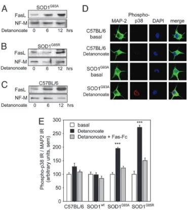
These results provided strong functional evidence for a feedback loop triggered by NO in mutant SOD1 motoneurons. However, it Fig. 1. Regulation of FasL and p38 kinase by NO in cultured motoneurons.
Motoneurons were cultured for 16 h and then treated for 6 or 12 h with the NO donor Detanonoate (20M). (A and B) Western blots of SOD1G85Rand
SOD1G93Amotoneuron protein extracts demonstrated that the 38 kDa form of
FasL protein was strongly up-regulated by NO within 6 –12 h. (C) Similar up-regulation of FasL protein was seen in C57BL兾6 motoneuron extracts after Detanonoate addition. Control protein extracts were prepared immediately before Detanonoate addition (0). Blots were reprobed with an antibody against neurofilament medium chain (NF-M). (D) Immunocytochemistry re-vealed a strong increase in phosphorylated p38 kinase in SOD1G93A
motoneu-rons exposed for 6 h to Detanonoate as compared with SOD1G93A
motoneu-rons cultured under basal conditions or motoneumotoneu-rons from C57BL兾6 mice. Motoneurons are colabeled with anti MAP-2 antibodies and DAPI. (E) Histo-grams showing that Detanonoate increased the phospho-p38 immunoreac-tivity (IR) in mutant SOD1G93Aand SOD1G85Rmotoneurons by 2- and 2.8-fold,
respectively, although having no significant effect on C57BL兾6 or SOD1WT
motoneurons. Detanonoate-induced phosphorylation of p38 kinase in mu-tant motoneurons was blocked by preincubation with Fas-Fc. Values for phospho-p38 kinase IR were normalized to MAP-2 IR; error bars represent SEM.
remained possible that NO from the exogenous donor was provided at levels in excess of those generated by the Fas兾NO pathway, and thereby led to nonphysiological activation of Fas. We therefore looked for evidence of the feedback loop in neurons triggered to die by another element of the pathway, the Fas receptor itself. Mutant SOD1 motoneurons were exposed to agonistic anti-Fas antibodies in the absence or presence of Fas-Fc (Fig. 3 C and D). Fas-Fc does not functionally interact with the Fas antibodies used to trigger death (Fig. 7C). Blockade of FasL兾Fas interactions by Fas-Fc gave nearly complete protection against cell death (Fig. 3 C and D). This result demonstrates that even when the pathway is triggered by the endogenous Fas receptor, further activation of Fas by FasL is required for cell death to occur (Fig. 3E).
FasL Is Up-Regulated in Mutant SOD1 Spinal Cord.Our in vitro results showed that, to kill a mutant SOD1 motoneuron, Fas or NO need to trigger a feedback loop. The involvement of such a loop, together with the requirement for transcriptional up-regulation of nNOS (6) may explain the relatively slow time course of motoneuron death when compared with other cellular models of Fas-triggered apo-ptosis. We reasoned that it might also be related to the late onset and prolonged time course of the neurodegenerative process in mutant SOD1 mice in vivo. We therefore asked whether the elements of the Fas兾NO feedback loop were expressed in the spinal cord and whether they showed alterations during the period leading up to the onset of clinical symptoms.
Fig. 2. Role of Daxx in mutant SOD1 motoneuron death. (A and B) Plasmid electroporation was used to coexpress a dominant negative FLAG-tagged form of Daxx (Daxx-DN) and EGFP in cultured motoneurons. (C and D) SOD1G85R or SOD1G93A motoneurons were electroporated with the EGFP
expression vector in combination with an empty control vector or vectors encoding either WT Daxx or Daxx-DN. At 1 day in vitro (DIV), motoneurons from each electroporation were either exposed to the NO donor Detanonoate or kept under basal conditions. Motoneuron survival at 3 DIV was expressed as the percentage of EGFP-positive cells in the presence versus absence of NO. Daxx-DN protected mutant SOD1G85Rand SOD1G93Amotoneurons from
NO-triggered cell death. Expression of Daxx-WT had no effect on survival. The differences between the effects of Daxx-DN and Daxx-WT were statistically significant: P⬍ 0.005 for SOD1G85R; P⬍ 0.01 for SOD1G93A, Student’s t test. (E)
Survival of SOD1WTmotoneurons transduced with control, Daxx-WT, or
Daxx-DN vectors with or without NO challenge. Histograms represent means and SEM from two independent experiments.
Fig. 3. Role of FasL, Fas, p38 kinase, and nNOS in mutant SOD1 motoneuron death. (A–D) Mutant SOD1 motoneurons were treated (or not treated) at 1 DIV with Detanonoate (A and B) or agonistic anti-Fas antibodies (C and D). Cell survival was quantified at 3 DIV by phase-contrast microscopy and expressed relative to the number of motoneurons alive under basal conditions (0). Addition of the extracellular part of Fas receptor, Fas-Fc, the p38 kinase inhibitor SB203580 (5M), or the nNOS inhibitor L-VNIO (10 M) significantly reduced the NO-triggered death of SOD1G85R(A) or SOD1G93A(B) motoneurons. Mutant SOD1
motoneuron death mediated by agonistic anti-Fas antibodies at 0.5 ng兾ml or 100 ng兾ml was prevented or significantly reduced by Fas-Fc. Error bars show SEM.*,
P⬍ 0.05;**, P⬍ 0.01;***, P⬍ 0.001, Student’s t test. (E) Model illustrating the Fas兾NO feedback loop in mutant SOD1 motoneurons.
Disease course varies between different lines of mutant SOD1 mice: SOD1G93A mice expressing a catalytically active form of
human SOD1 display an early disease onset⬇100 days of age (4), whereas SOD1G85Rmice express an inactive form of SOD1 leading
to late onset around day 200 (3). We therefore chose to analyze these mouse lines at comparable presymptomatic stages when the total number of motoneurons is still normal: day 75 for SOD1G93A
mice and day 120 for SOD1G85Rmice (7, 20) (Supporting Materials
and Methods, which is published as supporting information on the PNAS web site). We found that not only the Fas receptor, as shown in ref. 21, but also its endogenous ligand FasL and its signaling intermediate Daxx were expressed in control spinal cord (Fig. 4A). We therefore quantified the expression of different elements of the pathway in presymptomatic mutant mice.
In nontransgenic mice, 31⫾ 2% of all choline acetyl transferase (ChAT)-positive motoneurons in the lumbar spinal cord segment L4 expressed significant levels of FasL at day 75 (n⫽ 3 mice) and 29⫾ 4% were stained at day 120 (n ⫽ 2). In SOD1WTmice, the
number of FasL positive motoneurons (26.5⫾ 3.2%, n ⫽ 3) was close to that in nontransgenic mice. Strikingly, the proportion of FasL-positive motoneurons was at least two-fold higher in SOD1G93Amice at 75 days (70⫾ 1.9%, n ⫽ 4) and in SOD1G85R
mice at 120 days (60.2⫾ 6.6%, n ⫽ 4; Fig. 4B; P ⬍ 0.001, Student’s t test). We therefore asked whether FasL might be able to engage Fas receptor signaling in motoneurons in vivo. Double immuno-fluorescence labeling of presymptomatic SOD1G93A spinal cord
showed that all FasL-positive motoneurons coexpressed significant levels of Fas (Fig. 4C).
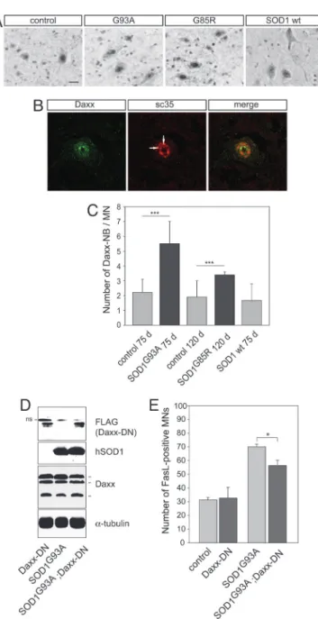
Daxx Accumulates in Nuclear Speckles in Motoneurons of Mutant SOD1 Mice.Immunostaining for Daxx in control and mutant SOD1 lumbar spinal cords revealed a diffuse cytoplasmic and nuclear localization in a broad range of neurons in the ventral and dorsal horn (Figs. 4A and 5A). Interestingly, in motoneurons, Daxx accumulated in discrete subnuclear domains (Fig. 5B) that stained positive for sc35, a general splicing factor and marker of nuclear speckles. Only a subpopulation of nuclear speckles were stained for Daxx. Colocalization of Daxx and sc35 was confirmed by Z-scan confocal analysis (data not shown). These nuclear domains (Daxx-NBs) are reminiscent of promyelocytic leukemia nuclear bodies (PML-NB) which, in other cell types, have been shown to contain sc35 (22–24). We quantified Daxx-NBs in control and mutant SOD1 motoneurons (Fig. 5C). Lumbar motoneurons contained 2.2 ⫾ 0.9 Daxx-NBs and 1.9 ⫾ 1.1 Daxx-NBs per section in C57BL兾6 mice aged 75 and 120 days, respectively, and 1.8 ⫾ 1.2 Daxx-NBs per section in SOD1WTmice aged 75 days. In contrast,
the number of Daxx-NBs was increased to 5.5⫾ 1.5 per section in SOD1G93Amice and 3.4⫾ 0.2 in SOD1G85Rmice (mean⫾ SD, n ⫽
3, P⬍ 0.001, Student’s t test). In conclusion, therefore, two key intermediates of the Fas兾NO signaling loop, FasL and Daxx, are activated in motoneurons of presymptomatic ALS mice.
A Dominant-Negative Form of Daxx Inhibits FasL Up-Regulationin Vivo.To address the functional relevance of Fas-Daxx signaling in mutant SOD1-linked motoneuron disease, we crossbred SOD1G93A
mice with transgenic mice expressing a dominant negative form of Daxx. These Daxx-DN mice (25) show a weaker phenotype than the Daxx null mutants, which are embryonically lethal (26). Western blot analysis demonstrated that SOD1 and Daxx-DN transgenes were expressed in lumbar spinal cord of double transgenic SOD1G93A;Daxx-DN mice at levels similar to those in the parental
strains (Fig. 5D). Endogenous Daxx expression was not influenced by Daxx-DN (Fig. 5D). Interestingly, in SOD1G93A;Daxx-DN mice,
only 56.4 ⫾ 3.9% of L4 motoneurons were FasL-positive, as compared with 70 ⫾ 1.9% in SOD1G93A and 32.6 ⫾ 7.9% in
Daxx-DN mice (mean⫾ SD, n ⫽ 3 each, P ⬍ 0.001; Fig. 5E). Thus, expression of Daxx-DN leads to a reduction of 36% in the mutant SOD1G93A-induced increase in FasL-positive motoneurons. These
data are consistent with a model in which chronic cycling of the mutant SOD1-dependent Fas feedback loop is required to build up signaling intermediates to levels at which they can trigger neuro-degeneration.
Discussion
Amplification mechanisms play an important role in intracellular signaling pathways. The best studied examples are posttranslational Fig. 4. Up-regulation of FasL in lumbar motoneurons of mutant SOD1 mice. (A) Transverse sections of L4 lumbar spinal cords of C57BL兾6 (control), 75-day-old SOD1G93A, and 120-day-old SOD1G85Rmice immunostained for Fas, Daxx,
FasL, and choline acetyl transferase (ChAT). Fas and Daxx displayed a scattered expression pattern in various cell types of the gray and white matter. In control mice, FasL was mainly detected in dorsal and ventral horn neurons, including a few motoneurons. In SOD1G93Aand SOD1G85Rmice, a high proportion of
motoneurons, as identified by ChAT staining on adjacent sections, was posi-tive for FasL. (Scale bar: 100m.) (B) Histograms showing the percentage of FasL-positive motoneurons in serial sections of L4 lumbar spinal cord. Note the increased percentage of FasL-positive motoneurons in SOD1G93A and
SOD1G85Rmice as compared with C57BL兾6 and SOD1WTmice. (C)
Immunola-beling reveals coexpression of FasL and Fas in spinal cord motoneurons in a 75-day-old SOD1G93Amouse. (Scale bar: 50m.)
modifications, e.g., protein phosphorylation and proteolysis, but signal amplification can also be achieved by induction of gene expression. We previously showed that Fas activation in cultured motoneurons results in transcriptional up-regulation of nNOS (6). Here we provide the evidence that this signal needs to be amplified by a positive feedback loop. In particular, we show that NO is able to up-regulate FasL, thereby retrogradely inducing Fas signaling. In mutant SOD1 motoneurons, this feedback loop leads to phosphor-ylation of p38 kinase and activation of Daxx and nNOS in the slowest cell death process yet reported downstream of the much-studied Fas receptor. Several key elements of this loop are activated
in vivo in mutant SOD1 mice. We show that SOD1G93A and
SOD1G85Rmice display presymptomatically an increased number
of FasL-positive motoneurons in comparison with control mice, as well as an increased accumulation of the Fas adaptor protein Daxx in nuclear bodies.
These findings extend reports concerning early activation of Ask-1 (16) and of several protein kinases including p38 (refs. 13–15 and 27; C.R., unpublished results) in SOD1G93A mice. Taken
together, these findings provide a molecular means for comparing the disease process in these mouse models of familial ALS with that in human patients with sporadic ALS unlinked to SOD1 mutations. Indeed, Bendotti et al. (27) report that sporadic ALS cases show the same characteristic skein-like staining pattern for phospho-p38 as that observed in mutant SOD1G93Amice.
A particularly striking aspect of the Fas兾NO feedback loop is that its activation is absolutely required for motoneuron cell death over the normal time span of motoneuron cultures. Even triggering the pathway by using agonistic Fas antibodies does not lead to death when interactions between FasL and Fas are blocked by using Fas-Fc. Therefore, the amplification provided by the loop is not simply a reinforcement mechanism. It is necessary to transform a subliminal activation of the pathway into an effective death stim-ulus. This result presumably reflects a low-intensity stimulation of the death-inducing signaling complex (DISC) complex in motoneu-rons compared with thymocytes or hepatocytes, perhaps because levels of Fas and its signaling intermediates are relatively low in these cells (12).
One prediction of our data is that blocking the Fas兾NO loop in mutant SOD1 mice should affect disease course. Indeed, inhibition of Daxx in mutant SOD1G93Amice by transgenic Daxx-DN
expres-sion blocked FasL up-regulation in about one-third of the sensitive motoneurons. This figure probably underestimates the role of the Fas兾NO loop because transgenic Daxx-DN mice are hypomorphs rather than complete nulls (25). The same is true for the majority of existing mouse mutants in which Fas signaling is still present at levels sufficient to trigger this loop: Fas knockout mice produce a truncated Fas protein that conserves part of its intracellular domain (28, 29) lpr mice are regulatory mutants of the Fas gene and express significant levels of Fas protein (ref. 30; C.R. and B.P., unpublished data), and gld mice bear a point mutation in FasL that does not completely inactivate the ligand (31). It will thus be critical to test the effects of complete inactivation of Fas or FasL on pathological motoneuron death.
In the past, crossing mutant SOD1 mice with mice carrying a loss-of-function allele of the nNOS gene did not prolong survival (32). Nevertheless, the nNOS enzyme can exist in three forms resulting from alternative splicing (33) and specific increases in the  and ␥ forms have been reported in spinal cords from sporadic ALS patients (34). The nNOS mutant mice used for the cross display residual NOS activity of⬇15% of control and, strikingly, produce the same and ␥ forms of nNOS as those found in ALS patients (32). In support of our model, administration of the nNOS inhibitor AR-R 17.477 significantly prolonged the lifespan of SOD1G93Amutant mice (32). In the future, it will be important to
determine the precise role of NO synthases in this process, because a requirement for NO also has been demonstrated in death of Fig. 5. Daxx activation in lumbar motoneurons of mutant SOD1 mice and its
functional relevance in the Fas兾NO loop. (A) Immunolabeling reveals that Daxx accumulates in the nuclei of motoneurons and forms nuclear bodies. SOD1G93A,C57BL兾6 littermates and SOD1WTmice were analyzed at age 75 days;
SOD1G85Rand C57BL兾6 litter mice (C.R., data not shown) were analyzed at age
120 days. (Scale bar: 20m.) (B) Daxx nuclear bodies (arrows) are associated with a subpopulation of nuclear speckles, as detected by confocal analysis of Daxx- and sc35-immunostained SOD1G93Alumbar spinal cords at age 75 days.
(C) The number of Daxx nuclear bodies (Daxx-NBs) in L4 motoneurons was higher in SOD1G93Aand SOD1G85Rmice than in C57BL兾6 or SOD1WTmice of the
same age. Values represent means⫾ SD from three mice per genotype;***,
P ⬍ 0.001, Student’s t test. (D and E) Analysis of double transgenic SOD1G93A;Daxx-DN mice. (D) Western blots of protein extracts from lumbar
spinal cords show that the dominant negative form of Daxx (Daxx-DN) and the human SOD1G93Aare expressed at similar levels in double transgenic mice and
in the parental mice at age 75 days. Daxx-DN was revealed with an anti-FLAG antibody that also detects a nonspecific (ns) upper band. Daxx-DN expression did not modify expression of endogenous Daxx because the three known Daxx isoforms of 70, 97, and 110 kDa (43) were detected at similar levels in all genotypes. (E) Transgenic Daxx-DN expression significantly reduced the per-centage of FasL-positive motor neurons in double transgenic mutant SOD1G93A;Daxx-DN mice as compared with values in age-matched 75-day-old
SOD1G93Amice. Values are means⫾ SD, n ⫽ 3 per genotype;
***, P⬍ 0.001, one-way ANOVA followed by Newman–Keuls post hoc analysis.
motoneurons induced by avulsion (35) or neurofilament gene mutations (36).
Feedback amplification loops involving other intermediates in the Fas signaling, such as caspase-8 and caspase-3 or Bid have been reported in acellular systems and nonneuronal cells (37, 38). In the context of ALS, the Fas兾NO feedback loop is of particular interest because it involves an extracellular step and diffusible factors. The cell death trigger NO is known to be produced not only by motoneurons but also by microglia and activated astrocytes (34, 39) and Fas agonists have been detected in sera of patients with sporadic ALS (40). ‘‘Community effects’’ may thus allow for cellular neighbors to accelerate or to inhibit motoneuron death (5) and also underlie the clinical finding that ALS often progresses locally, between adjacent muscles or motor pools. Further studies are required to better understand the molecular and cellular basis of these phenomena.
We believe that chronic cycling of feedback loops of the type described here may provide a general approach to understanding the delayed onset and relatively slow progression of many neuro-degenerative diseases. As has been proposed for nucleation of mutant proteins with polyglutamine expansions (41), the initial insult produced by the feedback loop may be subliminal and without phenotype. However, as levels of toxic intermediates and death signals build up, they may reach a threshold that can trigger the pathological process. If this model is correct, then therapeutic intervention at the level of ‘‘death receptors’’ and cell death pathways should be envisioned at much earlier stages in the disease process than is generally imagined.
Materials and Methods
See Supporting Materials and Methods for details.
Animals and Reagents.SOD1G85Rmice, line 148 (3), were
main-tained as homozygotes, SOD1G93Amice (4) and SOD1WTmice, line
76 (3) as hemizygotes. All mice were on a pure C57BL兾6 back-ground. The following reagents and antibodies were used: brain-derived neurotrophic factor and ciliary neurotrophic factor (R & D Systems), glial cell line-derived neurotrophic factor, sodium nitro-prusside (Sigma), Detanonoate, Fas-Fc, L-VNIO (Alexis, San Diego,), SB203580 (Calbiochem), anti-mouse Fas (JO2,
Pharmin-gen); anti Fas (M-20) and anti FasL (N-20 and N-20-G, Santa Cruz Biotechnology); anti-FLAG (M2, Sigma), anti-NF-M (Ab1987, Chemicon), anti-sc35 (S4045, Sigma), anti-phospho-p38 (Cell Sig-naling Technology, Beverly, MA), anti-MAP2 (Sternberger– Meyer, Jarrettsville, MD), and secondary antibodies (Jackson ImmunoResearch or Molecular Probes).
Immunohistochemistry of Spinal Cord.Spinal cord cryosections (16 m) were incubated with primary antibodies [1:100 for anti-FasL, N-20-G, and anti-Fas, M-20; 1:200 for anti-Daxx, M112; 1:500 for anti-sc35, S4045, and anti-choline acetyl transferase (ChAT)], re-vealed with the ABC staining kit (Vector Laboratories) or with fluorochrome-conjugated secondary antibodies and analyzed by confocal microscopy. FasL-positive motoneurons or fluorescently labeled Daxx nuclear bodies were counted on at least 20 different sections originating from three mice per genotype.
Motoneuron Culture and Analysis.Male transgenic SOD1 mice were mated with female C57BL兾6 mice, embryos harvested at embry-onic day 12.5, and genotyped by PCR (6). Motoneurons were purified from ventral spinal cords by using a metrizamide density gradient (42), cultured in the presence of neurotrophic factors and treated at 1 DIV with Detanonoate or anti-Fas antibodies. Elec-troporation was done as described (6). Cell survival was determined by fluorescence or phase-contrast microscopy. Immunohistochem-istry, quantitative confocal microscopy, and Western blot analysis were performed as described in Supporting Materials and Methods. Soluble FasL in conditioned media was measured by ELISA (R & D Systems). All experiments were performed in triplicate or quadruplicate and repeated at least twice.
We thank S. Corby for animal care and genotyping, Drs. A. O. Hueber (INSERM, Nice, France) for Daxx plasmids, I. Medina (INSERM, Marseille, France) for advice on quantitive imaging, S. Przedborski (Columbia University, New York) for providing SOD1WTmice, and G.
Tanackovic and T. Abbas-Terki (both of EPFL, Lausanne, Switzerland) for sc35 antibodies and helpful comments on the manuscript. This work was funded by grants from Institut National de la Sante´ et de la Recherche Me´dicale, Centre National de la Recherche Scientifique, Association Franc¸aise Contre les Myopathies, French Ministe`re de la Recherche et de la Technologie, American ALS Association, and Swiss National Scientific Foundation.
1. Wong, P. C., Pardo, C. A., Borchelt, D. R., Lee, M. K., Copeland, N. G., Jenkins, N. A., Sisodia, S. S., Cleveland, D. W. & Price, D. L. (1995) Neuron 14, 1105–1116. 2. Wang, J., Xu, G., Gonzales, V., Coonfield, M., Fromholt, D., Copeland, N. G., Jenkins, N. A.
& Borchelt, D. R. (2002) Neurobiol. Dis. 10, 128–138.
3. Bruijn, L. I., Becher, M. W., Lee, M. K., Anderson, K. L., Jenkins, N. A., Copeland, N. G., Sisodia, S. S., Rothstein, J. D., Borchelt, D. R., Price, D. L. & Cleveland, D. W. (1997) Neuron 18, 327–338.
4. Gurney, M. E., Pu, H., Chiu, A. Y., Dal Canto, M. C., Polchow, C. Y., Alexander, D. D., Caliendo, J., Hentati, A., Kwon, Y. W., Deng, H. X., et al. (1994) Science 264, 1772–1775. 5. Clement, A. M., Nguyen, M. D., Roberts, E. A., Garcia, M. L., Boillee, S., Rule, M., McMahon, A. P., Doucette, W., Siwek, D., Ferrante, R. J., et al. (2003) Science 302, 113–117. 6. Raoul, C., Estevez, A. G., Nishimune, H., Cleveland, D. W., deLapeyriere, O., Henderson,
C. E., Haase, G. & Pettmann, B. (2002) Neuron 35, 1067–1083.
7. Pasinelli, P., Houseweart, M. K., Brown, R. H., Jr., & Cleveland, D. W. (2000) Proc. Natl. Acad. Sci. USA 97, 13901–13906.
8. Li, M., Ona, V. O., Guegan, C., Chen, M., Jackson-Lewis, V., Andrews, L. J., Olszewski, A. J., Stieg, P. E., Lee, J. P., Przedborski, S. & Friedlander, R. M. (2000) Science 288, 335–339. 9. Inoue, H., Tsukita, K., Iwasato, T., Suzuki, Y., Tomioka, M., Tateno, M., Nagao, M., Kawata,
A., Saido, T. C., Miura, M., et al. (2003) EMBO J. 22, 6665–6674.
10. Zhu, S., Stavrovskaya, I. G., Drozda, M., Kim, B. Y., Ona, V., Li, M., Sarang, S., Liu, A. S., Hartley, D. M., Wu du, C., et al. (2002) Nature 417, 74–78.
11. Guegan, C., Vila, M., Rosoklija, G., Hays, A. P. & Przedborski, S. (2001) J. Neurosci. 21, 6569–6576. 12. Raoul, C., Henderson, C. E. & Pettmann, B. (1999) J. Cell Biol. 147, 1049–1062. 13. Tortarolo, M., Veglianese, P., Calvaresi, N., Botturi, A., Rossi, C., Giorgini, A., Migheli, A.
& Bendotti, C. (2003) Mol. Cell. Neurosci. 23, 180–192.
14. Hu, J. H., Chernoff, K., Pelech, S. & Krieger, C. (2003) J. Neurochem. 85, 422–431. 15. Ackerley, S., Grierson, A. J., Banner, S., Perkinton, M. S., Brownlees, J., Byers, H. L., Ward,
M., Thornhill, P., Hussain, K., Waby, J. S., et al. (2004) Mol. Cell. Neurosci. 26, 354–364. 16. Wengenack, T. M., Holasek, S. S., Montano, C. M., Gregor, D., Curran, G. L. & Poduslo,
J. F. (2004) Brain Res. 1027, 73–86.
17. Suda, T., Takahashi, T., Golstein, P. & Nagata, S. (1993) Cell 75, 1169–1178. 18. Tanaka, M., Suda, T., Takahashi, T. & Nagata, S. (1995) EMBO J. 14, 1129–1135. 19. Alderson, M. R., Tough, T. W., Davis-Smith, T., Braddy, S., Falk, B., Schooley, K. A.,
Goodwin, R. G., Smith, C. A., Ramsdell, F. & Lynch, D. H. (1995) J. Exp. Med. 181, 71–77. 20. Fischer, L. R., Culver, D. G., Tennant, P., Davis, A. A., Wang, M., Castellano-Sanchez, A.,
Khan, J., Polak, M. A. & Glass, J. D. (2004) Exp. Neurol. 185, 232–240.
21. Matsushita, K., Wu, Y., Qiu, J., Lang-Lazdunski, L., Hirt, L., Waeber, C., Hyman, B. T., Yuan, J. & Moskowitz, M. A. (2000) J. Neurosci. 20, 6879–6887.
22. Engelhardt, O. G., Boutell, C., Orr, A., Ullrich, E., Haller, O. & Everett, R. D. (2003) Exp. Cell Res. 283, 36–50.
23. Lamond, A. I. & Spector, D. L. (2003) Nat. Rev. Mol. Cell Biol. 4, 605–612.
24. von Mikecz, A., Zhang, S., Montminy, M., Tan, E. M. & Hemmerich, P. (2000) J. Cell Biol.
150,265–273.
25. Raoul, C., Barthelemy, C., Couzinet, A., Hancock, D., Pettmann, B. & Hueber, A. O. (2005) J. Neurobiol. 62, 178–188.
26. Michaelson, J. S., Bader, D., Kuo, F., Kozak, C. & Leder, P. (1999) Genes Dev. 13, 1918–1923. 27. Bendotti, C., Atzori, C., Piva, R., Tortarolo, M., Strong, M. J., DeBiasi, S. & Migheli, A.
(2004) J. Neuropathol. Exp. Neurol. 63, 113–119.
28. Adachi, M., Suematsu, S., Kondo, T., Ogasawara, J., Tanaka, T., Yoshida, N. & Nagata, S. (1995) Nat. Genet. 11, 294–300.
29. Yang, X., Khosravi-Far, R., Chang, H. Y. & Baltimore, D. (1997) Cell 89, 1067–1076. 30. Mariani, S. M., Matiba, B., Armandola, E. A. & Krammer, P. H. (1994) Eur. J. Immunol.
24,3119–3123.
31. Karray, S., Kress, C., Cuvellier, S., Hue-Beauvais, C., Damotte, D., Babinet, C. & Levi-Strauss, M. (2004) J. Immunol. 172, 2118–2125.
32. Facchinetti, F., Sasaki, M., Cutting, F. B., Zhai, P., MacDonald, J. E., Reif, D., Beal, M. F., Huang, P. L., Dawson, T. M., Gurney, M. E. & Dawson, V. L. (1999) Neuroscience 90, 1483–1492. 33. Wang, Y., Newton, D. C. & Marsden, P. A. (1999) Crit. Rev. Neurobiol. 13, 21–43. 34. Catania, M. V., Aronica, E., Yankaya, B. & Troost, D. (2001) J. Neurosci. 21, RC148. 35. Martin, L. J., Chen, K. & Liu, Z. (2005) J. Neurosci. 25, 6449–6459.
36. Strong, M., Sopper, M. & He, B. P. (2003) Amyotroph. Lateral Scler. Other Motor Neuron Disord. 4, 81–89.
37. Slee, E. A., Keogh, S. A. & Martin, S. J. (2000) Cell Death Differ. 7, 556–565. 38. Suhara, T., Kim, H. S., Kirshenbaum, L. A. & Walsh, K. (2002) Mol. Cell. Biol. 22, 680–691. 39. Almer, G., Vukosavic, S., Romero, N. & Przedborski, S. (1999) J. Neurochem. 72, 2415–2425. 40. Yi, F. H., Lautrette, C., Vermot-Desroches, C., Bordessoule, D., Couratier, P., Wijdenes, J.,
Preud’homme, J. L. & Jauberteau, M. O. (2000) J. Neuroimmunol. 109, 211–220. 41. Perutz, M. F. & Windle, A. H. (2001) Nature 412, 143–144.
42. Henderson, C. E., Bloch-Gallego, E. & Camu, W. (1995) in Nerve Cell Culture: A Practical Approach, eds. Cohen, J. & Wilkin, G. (Oxford Univ. Press, London), pp. 69–81. 43. Hollenbach, A. D., Sublett, J. E., McPherson, C. J. & Grosveld, G. (1999) EMBO J. 18,
3702–3711.