Design and Performance of Hemostatic Biomaterials for Managing Hemorrhaging by
Reginald Keith Avery B.S. Biocngineering
University of Maryland. College Park. 2012 Submitted to the Department of Biological Engineering in partial fulfillment of the requirements for the degree of
Doctor of Philosophy at the
Massachusetts Institute of Technology September 2018
C 2018 Massachusetts Institute of Technology. All rights reserved.
Signature redacted
Signature of Author:
Department of Biological Engineering June 28, 2018
Signature redacted
Certified by: oOBradley D. Olsen Paul M. Cook Career Development Associate Professor of Chemical Engineering Thesis Supervisor
Signature redacted
Accepted by:NOV
0
52018
LIBRARIES
Forest M. WhiteProfessor of Biological Engineering Chair of Graduate Program, Department of Biological Engineering
/0-, /z
The following committee has evaluated this doctoral thesis: Professor Bradley D. Olsen
Thesis Supervisor
Paul M. Cook Career Development Professor Associate Professor of Chemical Engineering Professor Darrell Irvine
Chairman, Thesis Committee Professor of Biological Engineering Professor of Materials Science Professor Krystyn Van Vliet Member, Thesis Committee
Professor of Biological Engineering
Professor of Materials Science and Engineering Professor Douglas Lauffenburger
Member, Thesis Committee Ford Foundation Professor
Head. Department of Biological Engineering Professor of Biological Engineering
Professor of Chemical Engineering Professor of Biology
Design and Performance of Hemostatic Biomaterials for Managing Hemorrhaging by
Reginald Keith Avery
Submitted to the Department of Biological Engineering on June 28, 2018 in partial fulfillment of the requirements for the degree of
Doctor of Philosophy in Biological Engineering Abstract
The high mortality rates associated with uncontrolled bleeding motivate hemostatic material development for traumatic injuries. Uncontrolled, or hemorrhagic, internal bleeding requires hemostatic materials that can be directly delivered to or target the bleeding locations. To address these needs, injectable systems are being developed that: (1) generate artificial clots independent of the coagulation cascade or (2) interact with blood components to accelerate or otherwise improve coagulation processes. Hemostatic materials designed for internal bleeding can save lives in the battlefield, en route to emergency rooms, and in the operating room.
This thesis first focuses on developing a shear-thinning hydrogel for injection onto bleeding surfaces and into ruptured vasculature. Based on in vitro assays of hydrogel performance, it was amenable to clinical delivery methods and reduced whole blood clotting times by 77%. In vivo bleeding models showed reduced blood loss and improved survival rates following a lethal liver injury. The hydrogel was also used as an embolic agent, where its occlusive potential in an anticoagulated model was demonstrated.
Next, recombinant protein-based hemostatic materials were expressed to modulate clotting kinetics and performance. By incorporating clot interacting peptide sequences (CIPs) into a protein scaffold, a family of multifunctional fibrinogen like proteins (MFLPs) was developed and assayed. Clot turbidity, an indication of fibrin clot formation, was increased among enzyme-interacting CIPs. Mimicking the polymerization mechanism of fibrinogen, knob sequences were shown to be procoagulant at low concentrations by increasing clot turbidity, reducing clotting times, and inhibiting plasmin lysis. Finally, to understand the impact of hemostats on clot structure, imaging procedures were developed to systematically assess hemostatic materials and their influence on clot architecture. Static and dynamic approaches were developed to quantify the activity of hemostats based on the spatial distribution of fibrinogen, red blood cells, and platelets around hemostat surfaces. Quantification of these hemostat-blood component interactions resulted in a unique pattern of interactions for each hemostat studied. These techniques could serve as a screening technique for hemostats and improve characterization prior to in vivo assays.
Taken together, the results highlight multiple approaches to address internal bleeding and opportunities to improve in vitro characterization of hemostats using microscopy.
Thesis Supervisor: Bradley D. Olsen
Acknowledgements
My time at MIT has been filled with unique experiences and opportunities to interact with diverse communities of researchers in the Boston area and from around the world. MIT is a place I never imaged myself being and am truly grateful for the lessons I was able to learn during my time there.
I would like to first thank the Biological Engineering and Chemical Engineering
Departments at MIT for giving me the freedom to try new things. Being a BE student interested in a ChemE lab involved in an interdisciplinary polymer program was a first for many of the departments, programs, and professors but I was supported throughout my time here from each and every group. I'd especially like to thank Prof. Lauffenburger, Prof. White, Prof. Irvine, and Prof. Olsen for supporting my unique journey navigating these environments. I'd also like to thank my colleagues in the PPSM program for their support during the first year and beyond as we all navigated graduate school together. Thanks to Aaron, Byungjin, Alina, Alice, Sunandini, and Mikhil for your companionship.
I'm very thankful to Ali Khademhosseini and his lab for welcoming me as a member and teaching me about biomaterials, tissue engineering, and animal studies. I appreciate the time Ali and other members of the group took to comment on my work and teach me about being a hard-working, motivated researcher. Akhilesh, Ali T., Alexander, Mohammad, Gaurav, Jeroen L., Jeroen R., Mehdi, Amir N., Hassan, Rahmi, Mehmet, Mario, Grissel, Nasim, Shrike, Su-Ryon, and Parastoo were mentors and colleagues that I would like to thank for their support during my time in lab.
I would also like to think the members of my committee who, at times, appeared to have more confidence in me than I did in myself. Prof. Van Vliet, Prof. Irvine and Prof. Olsen
provided a level of insight and support that elevated the quality of work I was able to perform. I would like to also thank those students that worked with me as undergrad or summer researchers, Rachel and Andriana. Your patience in me as I tried to teach you things I just learned the day before was commendable and the contributions you made to the research were key to many of the projects you were involved in. Thank you for the opportunity to work with you.
It has been a joy to work with Brad and all the members of the Olsen lab. Brad provided an atmosphere where I felt free to learn new skills without fear and collaborate with other lab members to further broaden my experiences. Brad brought excitement to the research that kept me motivated and eager to come to lab and try new things. Brad valued our growth outside of the lab as well I thank him for the opportunities to travel to beam lines, conferences, and even with him to Brazil for a conference and short course. I enjoyed all the collaborative projects I did in the Olsen lab and want to thank Matt, Michelle, Manos, Bruno, Rui, Chris, Minkyu, Danielle, Nari, Celestine, Carrie, Tom, and Yun Jung for letting me be involved in your exciting research projects and learn from you all. I want to thank all the lab members for making the lab
environment a supportive and positive one for me to spend my time in.
Lastly, I want to thank my family who has been a source of never ending support. My brother and I both took on the challenge of graduate school at the same time (he did finish before me though) and I can't thank him enough for his support and confidence in me. My parents, grandparents, aunts, uncles, and cousins have provided nothing but love and support and for that, I am grateful.
Table of Contents
Acknowledgements
---List of Figures --- 10
List of Tables 13 Chapter 1: Background on Hemostasis and Hemostat Development and Thesis Overview 1.1 Introduction and Motivation 15 1.2 Hemostasis Summary --- 19
1.2.1 Platelet Adhesion, Activation and Aggregation --- 21
1.2.2 Coagulation --- 24
1.3 Physiology of Hemorrhaging --- 29
1.3.1 Internal and External Bleeding --- 29
1.3.2 Altered Systemic Blood Properties --- 30
1.3.3 Altered Inhibitory Pathways --- 30
1.4 Managing Hemorrhaging --- 32
1.4.1 Treatment by First Responders --- 32
1.4.2 Treatment by Clinicians 32 1.5 Hemostatic Agents --- 34
1.5.1 Commercial Hemostatic Agents --- 34
1.5.2 Researched Hemostatic Agents --- 38
1.6 Assessment of Hemostatic Performance 40 1.7 Thesis Overview and Objectives --- 41
1.8 References 42 Chapter 2: Methods 2.1 Preparation of Gelatin-Silicate Nanocomposite Hydrogels --- 49
2.1.1 Materials and Synthesis of Nanocomposite --- 50
2.1.2 Nanocomposite Modifications for Imaging or In Vivo Assays --- 52
2.2 Preparation of Other Hemostats --- 58
2.2.1 QuikCotTM - - - - 58
2.2.2 Chitosan 59 2.3 Design and Synthesis of Multifunctional Fibrinogen Like Proteins (MFLPs) -- 59
2.3.1 Genetic Engineering --- 60
2.3.2 Protein Expression --- 71
2.3.3 Protein Purification 75 2.4 Mechanical Testing --- 78
2.4.1 Shear Rheology --- 78
2.4.2 Uniaxial Compression Measurements for Injection Force --- 81
2.5 Small Angle X-Ray Scattering --- 83
2.6 Biological Testing --- 84
2.6.1 In Vitro Tests 84 2.6.2 Ex Vivo 97 2.6.3 In Vivo 98 2.7 Microscopy --- 110
2.7.2 Static Clot Generation and Lysis 112
2.8 References 120
Chapter 3: Shear-Thinning Nanocomposite Hydrogels for the Treatment of Hemorrhage
3.1 Abstract 121
3.2 Introduction 122
3.3 A dditional Experim ental D etails ---124
3.4 Results and Discussion 126
3.4.1 Characterization of Nanocomposites --- 126 3.4.2 In Vitro Performance of Nanocomposite in Presence of Whole Blood - 130 3.4.3 In Vivo Hemostatic Potential of Nanocomposite in Liver Bleeding Model 134
3.5 Conclusion 137
3.6 Acknowledgements --- 138
3.7 References 138
Chapter 4: An Injectable Shear-Thinning Biomaterial for Endovascular Embolization
4.1 Abstract 143
4.2 Introduction 144
4.3 Additional Experimental Details --- 147
4.4 Results 152
4.4.1 STB Stability and Recoverability --- 152 4.4.2 STB Occlusion and Coagulation --- 158 4.4.3 STB Sterilization and Casting --- 160
4.4.4 In vivo Embolization 161
4.5 Discussion 168
4.6 Acknowledgements --- 171
4.7 References 171
Chapter 5: Modulation of Clot Architecture by Hemostatic Agents
5.1 Abstract 177
5.2 Introduction 178
5.3 Additional Experimental Details --- 181
5.4 Results 185
5.4.1 Fibrin Distribution 186
5.4.2 Fibrin and Cell Component Distributions in Platelet Rich Plasma --- 189
5.4.3 Dynamic Clot Generation --- 194 5.4.4 Plasmin-based Fibrinolysis --- 199
5.5 Discussion 201
5.6 Conclusion 205
5.7 Acknowledgements --- 206
Chapter 6: Developing Multifunctional Fibrinogen Like Proteins for Pro-Coagulant Applications
6.1 Introduction 211
6.2 Additional Experimental Details 214
6.3 Results and Discussion 217
6.3.1 Selection and Protein Engineering of MFLP Constructs 217
6.3.2 In vitro Coagulation Performance of MFLPs 218
6.3.3 MFLP Inhibition of Plasmin Lysis 227
6.4 Conclusion 230 6.5 References 232 Chapter 7: Conclusion 7.1 Summary 235 7.2 Future Directions 241 7.3 References 246 Appendix Appendix Appendix Appendix Appendix Appendix Appendix
A: Supporting Information for Chapter 2 247
B: Supporting Information for Chapter 3 251
C: Supporting Information for Chapter 4 263
D: Supporting Information for Chapter 5 281
E: Supporting Information for Chapter 6 297
F: Design of Inverted Microscope for Fluorescent Imaging --- 303 G: Protocols for Super Resolution Microscopy of Fibrin Networks --- 321
List of Figures Chapter 1
1-1. Platelet aggregation and coagulation processes --- 21
1-2. Fibrinogen and fibrin polymerization --- 26
Chapter 2 2-1. Nanocomposite design --- 50
2-2. Gelatin labeling for nanocomposite degradation assay --- 54
2-3. Nanocomposite phantom imaging --- 55
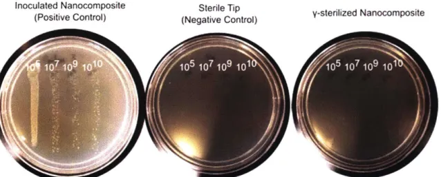
2-4. Sterility of nanocomposite after gamma sterilization --- 58
2-5. Multifunctional fibrinogen like proteins --- 60
2-6. Overview of genetic engineering plan to generate MFLPs 64 2-7. Example of digested vectors --- 68
2-8. Example of overnight culture of transformed ligation reaction --- 69
2-9. MFLP expression --- 72
2-10. Optimization of Ni-NTA resin for MFLP purification --- 76
2-11. Ni-NTA purification results for MFLPs 77 2-12. Nanocomposite recovery following high shear --- 79
2-13. Nanocomposite aging --- 81
2-14. Nanocomposite injectability assay design --- 82
2-15. Nanocomposite injection force profile --- 83
2-16. Degradation of nanocomposite --- 86
2-17. Whole blood coagulation --- 88
2-18. Plasma separation and coagulation --- 91
2-19. Platelet aggregation assay --- 93
2-20. Attempted clot lysis assays --- 95
2-21. Artery rupture setup --- 98
2-22. In vivo rodent nanocomposite occlusion --- 99
2-23. Subcutaneous nanocomposite implants in rodents 103 2-24. Images of liver bleeding assay --- 105
2-25. Example of hematoxylin and eosin stained tissue sample --- 106
2-26. Nanocomposite degradation under flow --- 111
2-27. Slide treatments impact on clot formation --- 113
2-28. Blood component labeling --- 116
Chapter 3 3-1. Structure, injectability, and self-healing characteristics of nanocomposite hydrogels --- 127
3-2. Effect of nanoplatelets on the clotting whole blood --- 133
3-3. In vivo evaluation of nanocomposite hydrogels as hemostats --- 135
Chapter 4 4-1. Shear-thinning biomaterial (STB) functions as an endovascular embolic system 153 4-2. STB is injectable through clinical catheters and is occlusive in ex vivo model - 157 4-3. STB is hemocompatible in vitro and demonstrates vessel occlusion ex vivo --- 159
4-4. Arterial injection of STB in mice occludes vasculature 162 4-5. Vascular embolization in porcine model suggests feasibility in porcine models 164
4-6. Histological assessment of vascular reorganization after STB injection --- 167
Chapter 5 5-1. Hemostat Effect on Clot Architecture 186 5-2. Fibrin Network Distributions 188 5-3. Plasma Distributions 191 5-4. Cell and Plasma Distributions 193 5-5. Microfluidic Design --- 195
5-6. Flow Profiles and Dynamic Clot Formation --- 197
5-7. Fibrin Profile of Clot under Flow 199 5-8. Hemostat-dependent Fibrinolysis Performance --- 200
Chapter 6 6-1. Multifunctional Fibrinogen Like Protein (MFLP) Design --- 219
6-2. Unfunctionalized MFLP Performance- --. 222
6-3. knob A functionalized MFLP Performance 225 6-4. knob A functionalized transglutaminase MFLP Performance --- 227
6-5. Inhibition of Plasmin Lysis by MFLPs --- 230
Appendix B B-1. Effect of silicate nanoplatelets on thermal stability of nanocomposite hydrogels -252 B-2. Linear viscoelastic range of nanocomposite hydrogels --- 253
B-3. Nanocomposite gel recovery and aging 254 B-4. Effect of nanocomposite on clot strength --- 255
B-5. Effect of silicate nanoplatelet on the stability of nanocomposite hydrogels in physiological solution --- 256
B-6. X-Ray Scattering indicates disk like scatterers --- 257
B-7. Injectable Gelatin/silicate hydrogel did not induce significant cytotoxic and pro-inflammatory effects in macrophages --- 258
B-8. Time series of clot formation 259 B-9. Nanocomposite surface initiates clotting through interactions with blood components --- 260
B-10. In vivo procedure showing the liver bleeding model --- 261
Appendix C C-1. Model Shear-Thinning --- 266
C-2. STB Thermal Stability --- 268
C-3. Zoomed Graph of Rapid STB Recoverability --- 269
C-4. STB Injectability Setup --- 270
C-5. STB Injection Forces --- 271
C-6. Ex Vivo Pressure Calibration 272 C-7. Creep Measurement of STB --- 273
C-8. STB Thrombosis Potential 274 C-9. Hemolysis Potential of Embolic Coils --- 275
C- 10. In Vitro Embolization Model 276
C-11. Impact of Sterilization on STB 277
C-12. STB Sterility by Colony Formation Count u-278
C-13. Imaging Phantom of STB 279
C-14. Histology Staining of STB Occluded and Patent Vessels 280 Appendix D
D-1. Bulk Clot Formation 284
D-2. Hemostat-Fibrin Network Interface 286
D-3. Fibrin Spatial Distribution --- 287
D-4. Antibody-labeled fibrin distribution --- 287
D-5. Blood component labeling --- 288
D-6. Hemostat-Red Blood Cell Interface 289 D-7. Hemostat-Platelet Interface 290 D-8. Laser cutting channels and mixers --- 291
D-9. Pathlines file format 292 D-10. Dynamic clot formation with gelatin hemostat --- 295
D-1 1. Volumetric clot lysis --- 296
Appendix E E-1. Coagulation Curve --- 297
E-2. Derivatives of clotting curve --- 298
E-3. Platelet Aggregation Curve --- 299
E-4. Biphasic coagulation observed in AP2x sample --- 299
E-5. Clotting Kinetics 300 E-6. Plasmin lysis in fibrinogen-thrombin solutions --- 301
E-7. Clot magnitude of knob A MFLPs --- 301
Appendix F F-1. Schematic of microscope and optical components required to align laser lines - 304 F-2. Front face of shutter controller 311 F-3. Wiring for triggered shutter control --- 313
F-4. Iris Use During Alignment --- 314
F-5. Perpendicular Laser Alignment --- 316
F-6. Epifluorescence and TIRF --- 318
Appendix G G-1. Thunder STORM Analysis --- 325
List of Tables Chapter 2
2-1. Nanocomposite compositions --- 51
2-2. Expression conditions for all multifunctional fibrinogen like proteins (MFLPs) 75 2-3. Buffers used during Ni-NTA purification --- 75
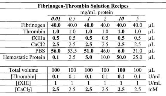
2-4. Table of stock solutions and standard fibrinogen solution components --- 92
2-5. Volumes of components used for fibrinogen-thrombin solutions --- 92
2-6. Table of component volumes in a PRP aggregation test including MFLP --- 94
2-7. Supplies for rodent in vivo surgeries --- 101
2-8. Table of fibrinogen solution compositions used for imaging studies for 0.5% labeled fibrinogen --- 118
Chapter 5 5-1. Characteristics of hemostats selected for study --- 185
Chapter 6 6-1. Clot Interacting Peptide (CIP) sequences, natural interactions, and expected interactions in MFLP constructs --- 217
6-2. MFLPs expressed in this study and theoretical molecular weights --- 218
Appendix B B-1. Weight percent (w/w) composition of each nanocomposite hydrogel --- 251
Appendix C C-1. Preliminary Injectability --- 267
Appendix D D-1. Clot compositions generated in this study --- 285
Appendix F F-1. Olympus Microscope Components --- 305
F-2. Lasers Supplies --- 307
F-3. Safety Equipment and Shutters --- 308
F-4. Alignment and Filter Equipment --- 309
F-5. Camera Trigger States --- 312
F-6. Voltages of Shutter Control Box 312 F-7. Standard Laser Conditions for Imaging --- 319
Appendix G G-1. Imaging Buffer Components --- 324
Chapter 1: Background on Hemostasis and Hemostat
Development and Thesis Overview
1.1 Introduction and Motivation 1.2 Hemostasis Summary 1.3 Physiology of Hemorrhaging 1.4 Managing Hemorrhaging
1.5 Hemostatic Agents
1.6 Assessment of Hemostatic Performance 1.7 Thesis Overview and Objectives
1.8 References
1.1. Introduction and Motivation
Hemostats are materials used to manage uncontrolled bleeding caused by trauma, 1-3
iatrogenic situations, such as during surgery,4-5 or pathological conditions, including
hemophilia.6 Hemostats function to promote blood clot formation or artificially provide a barrier to minimize continued blood loss, which can result in exsanguination and eventually death. To achieve these goals, a variety of materials and systems have been developed for use as
hemostatic agents. Most hemostatic agents, excluding tourniquet-based systems, can be classified under the following classes based on their mechanism of action:' (1) factor concentrators, (2) mucoadhesive agents, and (3) procoagulant supplementors. Tourniquets function by exerting sufficient pressure on ruptured vessels to physically occlude arterial and venous flow to and from an appendage.7 Tourniquets are best suited for bleeding from extremities (arms and legs) and fail when bleeding originates from the chest, abdomen, neck, head, groin, or truncal regions. External bleeding from the chest, abdomen, and other non-extremity regions requires the use of applied pressure first, accompanied by the use of hemostatic materials directly on the wound to stop bleeding.8
A multitude of hemostatic materials have been shown to achieve application-dependent performance8-9 during bleeding situations due to the complexity of hemostasis and the simplicity of the desired task. When hemostats are used, they utilize interactions with blood components or tissue interfaces exposed by the injury to promote the generation of a clot and stop bleeding, also known as hemostasis. The complexity of blood coagulation and hemostasis can allow materials that interact with particular biological processes to be used and all produce accelerated clot formation. These include adsorption of clotting biomolecules (QuikClot) 0 or activation and aggregation of blood cells (HemCon).1 In contrast, the simplicity of the desired outcome for any hemostat, stopping unwanted blood flow, results in the use of many biologically inert materials to simply serve as plugs to prevent continued blood loss. Examples include alginate gels,
cyanoacrylate-based systems, or absorption of excess fluid (cellulose and gelatin-based gauzes).4 Despite the wide array of hemostatic systems commercially and clinically available, research continues to find hemostats that address increasingly complex trauma environments or address sources of internal bleeding by minimally-invasive techniques. Battlefield injuries caused by improvised explosive devices (IEDs) and more traditional ordnance require rapid control of hemorrhaging at the front line, without the benefit of a surgical environment.7', 12 The ideal hemostat in these situations has been described by the US Army Institute of Surgical Research and Uniformed Services University of the Health Sciences as a material that satisfies 7 criteria.8 The hemostat must be (1) able to stop large vessel bleeding within 2 minutes of application, (2) ready to use immediately without any mixing or preparation, (3) simple to apply with minimal training, (4) lightweight and durable, (5) stable at extreme environment (-10 to 55 C), (6) safe to
use, with no risk of bacterial or viral transmission, and (7) inexpensive. Some systems, such as QuikClot, are able to address many of these criteria.1 3 However, when the wounds are not
externally accessible (e.g. blast-trauma resulting in a lacerated liver) solid hemostats are incapable of being delivered to the injury site and new solutions are required.
Hemostatic solutions for internal bleeding require stopping blood loss from a source that may be unknown, restricting the types of materials that can be used. Many sources of internal
bleeding are also non-compressible, meaning the application of external pressure either will not promote coagulation or limit blood loss, will cause damage to organs, or is impossible due to the bone/tissue structure of the wound site. Hemostats must either be targeted to the wound site or deliver a systemic stimulus that addresses the bleeding site while minimizing off-target effects. Foams have been developed that expand and generate the pressure internally, thereby controlling bleeding." Absent this breakthrough, there are no other systems that have been proposed to manage internal bleeding in a front-line environment. Within a clinic, there are a variety of materials and systems used to promote hemostasis with a process known as endovascular
embolization. The materials used, known as embolic agents, must be delivered through catheters, small diameter (< 1 mm) plastic tubing, directly to the bleeding or injured area." Computed tomography (CT) X-ray imaging is used to guide the radiologist or surgeon to the bleeding site. Additional requirements such as injectability, bioresorption, and stability against blood pressure or shear stresses are placed upon hemostatic materials used as embolic agents. Among embolic agents, time sensitive injections, complex delivery methods, and the reoccurrence of bleeding after application of an embolic motivate the development of new embolic agent.
Biomaterials are ideal for the development of next generation hemostats for all hemorrhaging situations, internal and external as well as among clinicians and civilian or military
first-responders. Modifications to known hemostatic materials, such as oxidation of cellulose,'6 have
activation of clotting pathways. Gauzes and hydrogels have been impregnated with biologically active molecules to be interact with blood or tissue components.17-8 Novel polymers and peptide sequences have also been used to serve as functionalization on inert scaffolds19 or serve as active scaffolds20 themselves to control sources of bleeding. Further biomaterial development can generate hemostats designed for specific environments (internal clinical bleeding vs. internal battlefield bleeding) that address the unique requirements presented for each bleeding situation.
The motivation to design and assess the performance of hemostatic materials in this thesis is centered upon developing new materials to treat sources of internal bleeding. Within this goal is the need to develop new materials as well as generate new assays of hemostatic materials that characterize their mechanisms of action and provide design principles upon which future hemostats can be based. The development of new hemostats for internal bleeding can be simple and serve as mechanical plugs against continuing blood loss. Physically associated shear-thinning hydrogels are excellent candidates for this purpose, allowing for injection into bleeding sites by needle or catheter, and conformation to irregular wound surfaces.2' Additionally, their
space-filling behavior is ideal for embolic agents that demands complete occlusion of a
hemorrhaging site, such as an aneurysm. 22-23 Alternatively, hemostats can be designed possessing more complex interactions with blood components and coagulation processes. Recombinant proteins are a framework from which particular interactions with coagulation processes can be encoded within the amino acid sequence at specific locations with high fidelity. Interactive amino acid sequences can be embedded within structural, reactive, or associative sequences to generate families of proteins with varying functionalities. Essential to the development of new hemostatic hydrogels and families of hemostatic proteins is the ability to assess their
kinetic, and morphological assessments of hemostat activity can all serve to identify mechanisms of action and generate a framework of assays from which future hemostats can be assessed and compared to characterize clot performance beyond final performance outcomes.
This chapter begins with an overview of hemostasis processes, with more focus afforded to the mechanisms that will be targeted in the subsequent hemostat designs ( 1.2). This is followed by the particular changes that occur in hemostasis when trauma occurs ( 1.3) followed by how hemorrhaging is addressed in the clinic and by first responders ( 1.4). The unique requirements of hemostats in each environment are outlined. Hemostatic systems that have been researched and presented in literature are reviewed and organized based on the particular process,
component, or mechanisms they target ( 1.5), followed by the standard techniques used to characterize hemostatic performance ( 1.6). Finally, the chapter concludes with an overview of the thesis objectives ( 1.7).
1.2. Hemostasis Summary
Hemostasis includes the overall process that stops bleeding, or hemorrhaging (Figure 1-1).24-25 Upon injury, a vessel wall is ruptured, exposing the previously contained blood components to external stimuli located in the vessel wall and beyond. The exposed substrates initiate two processes, aggregation of platelets which results in a platelet plug and coagulation which results in a fibrin network that finally will entrap the platelet plug and other blood cells. The solid mass of red blood cells, platelets, and a fibrin network are the predominate components of a natural blood clot. Platelets are anuclear cells sprouted from megakaryocytes in the bone marrow.26
When exposed to materials like collagen or von Willebrand factor (vWF), integrins on the platelet membrane will adhere to the exposed endothelial surface and initiate signaling
cascades to promote further platelet activation and aggregation. 6 Coagulation occurs on exposed endothelial cells and platelet surfaces, beginning on tissue factor-expressing endothelial cells.24
Coagulation occurs through a clotting cascade, where inactive enzymes, zymogens, in circulation are activated by previously activated enzymes in the clotting cascade. The cascade ends with the generation of a fibrin network in the area surrounding the injured site where thrombin levels, a central clotting factor, have been elevated. Aggregation and coagulation will be separated for the sake of discussion below but it should be noted that while platelet
aggregation and coagulation are commonly presented separately, both are occurring
simultaneously and in concert with one another. A significant amount of interaction is necessary between the two to achieve hemostasis. Indeed, it is the surfaces generated during platelet activation and aggregation that are the substrates on which clotting factors collect and complex to propagate the coagulation cascade.
(a)
PLATELET ACTION IN HEMOSTASISFlow Plalelt lranslocation Fbrinogen -aoActive GPIlb-Il a
Fmlowm adhsion and rolling -0 Mechanism Piinoe lNurediDenatured platelet and ibalon of the
Endothelial Cel acvation coagulation cascade
Active Platelet 0
~ ~ 00
Adhesion GPIba iAF Pla/lIa
Mechanisms (b)
(c)
(b) Initiation INTRINSIC EXTRINSIC PATHWAY PATHWAY ) J* Propagation COMMON PATHWAY prrmthmbnh thrombin V71 X prothrumhi 1 platelet VillAmplification activated platelet
Figure 1-1: Platelet aggregation and coagulation processes. (a) Platelet activation and aggregation. Reprinted with permission from Ref 69. Copyright 2012 American Chemical Society. (b) Classical coagulation pathway outlines clotting factor activation as a series of enzymatic reactions. Reprinted with permission from Ref 37. (c) Cell-based refined model of coagulation. Reprinted with permission from Ref 24.
1.2.1. Platelet Adhesion, Activation and Aggregation
Platelets in circulation are relatively inert and do not play a role in gas transfer or immune responses like other cells in circulation. However, upon contact with exposed proteins (P-selectin or vWF) in the subendothelial matrix or on endothelial cells, platelets can adhere to the injured vessel wall. Subsequently, the presence of enzymes (thrombin) or small molecules (adenosine diphosphate, ADP) in solution can activate platelets that will promote further platelet activation and contribute to aggregation.
Platelet adhesion occurs by interactions between platelet integrin and subendothelial matrix proteins (Figure 1-I a). The variety of interactions between platelets and exposed surfaces,
Active Platelet 0
-biomolecules, and themselves motivate their investigation as cells to mimic or attract when designing hemostatic systems. Interaction will be described separately below:
P-selectin binding to GPIb
P -selectin is released from a-granules upon activation of platelets. a-granules are
organelles within platelets containing an assortment of clotting factors, ADP, calcium fibrinogen, and P-selectin.2 7 After activation, the a-granules release their content to the extracellular space
and incorporate their membrane, which contains P-selectin, into the platelet membrane. Once exposed, P-selectin can interact with vWF on endothelial surfaces, P-selectin glycoprotein ligand-1 (PSGL-1) on leukocytes, and glycoprotein Iba (GP Iba) on platelets. Interactions between PSGL-I and P-selectin are generally seen as leukocytes are attracted to injury sites, which express P-selectin on exposed endothelial cells.28 On platelets it is predominately GP Iba,
a component of the GP-Ib-IX-V complex that allows platelets to roll and adhere to endothelial surfaces expressing P-selectin.26,2 8
vWF binding to GP Ib-IX-V
Similar to P-selectin, vWF is present in a-granules of platelets as well as within endothelial cells and in the subendothelial matrix. Upon injury, platelets and endothelial cells will release the large (500 kDa-20 MDa multimers29) protein, vWF will adhere to endothelial surfaces and the shear rates of blood flow will extend the multimeric protein to expose binding domains (Al domain) to GP Ib-IX-V complex (GP Ib). Under the correct shear rates, vWF-GP lb interactions serve as an initial adhesion step between platelets and the sub-endothelial surface.26
Adhered platelets are activated with proteins or enzymes like thrombin and small molecules like ADP or adenosine triphosphate (ATP). Activation results in release of small molecules that subsequently activate more platelets to eventually generate a platelet plug after platelet
aggregation. Among the activation mechanisms involved in platelet activation, collagen, thrombin, and ADP represent the major agonists in vivo. Each are described separately below:
Collagen activation
Following adhesion of platelets to subendothelial surfaces, exposed collagen will interact with glycoprotein VI (GP VI) and U2PI integrins. GP VI is the central receptor for collagen on platelets and activates downstream phosphorylation pathways within platelets. 3 0 (12pI integrins
also bind to collagen and through signaling pathways will activate glycoprotein Ilb/Ila (aIlbP3) which is essential for fibrinogen binding during platelet aggregation.
ADP activation
ADP initiates, through the P2YI and P2Y12 receptors on platelets, pathways that release calcium and inhibit ATP degradation by reducing adenylyl cyclase activity respectively.3' P2YJ and P2Yl 2 are G-protein coupled receptors that, in the presence of ADP, will induce platelet shape change that is observed during platelet aggregation. ADP activation also results in release of a-granules, which releases more ADP among other proteins into the extracellular space or into the platelet membrane.26
Thrombin activation
Thrombin interacts with platelets through a protease activated receptors (PAR) on the platelet surfaces.3 1 Thrombin cleavage of the receptor exposes new termini and initiates signaling
Platelet aggregation is accomplished predominately by interactions between fibrinogen and GP Ilb/II1a. A conformational change in the heterodimer orientation allow their main ligand, fibrinogen, to interact and serve as bridges between activated platelets. Release of calcium during activation results in a shape change due to cytoskeleton rearrangement that causes
platelets to change from discoid to spindled spheres with projections extending microns from the platelet center of mass.3 3 The extended shape allows for increased interactions between platelets
and binding of fibrinogen or vWF under high shear conditions.26 Continuing aggregation results
in the generation of a platelet plug in the injured area. Endogenous mechanisms including ADP-degrading enzymes, intact endothelial surfaces, and small molecules like nitric oxide,26 all serve to prevent continued growth of platelet plugs beyond an injury site.
1.2.2. Coagulation
Cell Mediated Clotting Cascade
Coagulation represents the second component of hemostasis, resulting in formation of a fibrin network containing the platelet plug and other blood cells trapped in the growing fibrin network. While coagulation and the coagulation cascade were hypothesized to be a series of enzymatic reactions culminating the activation of thrombin and cleavage of fibrinogen to fibrin (Figure 1-1 b), peculiarities in bleeding disorders suggested the influence of other factors.24 It is
now believed that, in addition to the enzymatic processes outlined in the coagulation cascade, the cell substrates upon which these processes occur are critical to a successful coagulation cascade (Figure 1-1 C).2 4 Coagulation initiates on tissue-factor (TF) containing endothelial cells where
factor VII complexes with TF. TF-VII complexes are activated into TF-VIIa complexes which can activate factor IX and X. Xa can activate Va, complexing into a prothrombinase complex (Xa/Va) to form small concentrations of thrombin near the TF-containing cell surface. fIXa is
able to move away from the TF surface without inactivation, allowing it to move onto platelet surfaces. The small levels of thrombin serve to activate adhered platelets as well as activating more factor V, released from platelet granules, VIII, and XI. vWF, in addition to binding platelets, interacts with inactive factor VIII. Thrombin can cleave VIII into VIlla and restrict VIla and Va onto a platelet surface. On the platelet surface, tenase (IXa/VIIa) and
prothrombinase (Xa/Va) complexes are formed. Tenase activates more factor X and
prothrombinase activates thrombin, resulting in the major propagation of thrombin generation for the conversion of fibrinogen into fibrin.2534-3 5
The above description of the coagulation cascade indicates that redundancies are embedded into the coagulation cascade, providing multiple pathways to achieve coagulation. However, thrombin is central to the cascade and any alteration in the cascade upstream of thrombin generation can impact the overall kinetics of clot formation. Additionally, the surfaces upon which these enzymatic reactions and cascades occur are as crucial to a successful clotting response as the activation of the enzymes themselves. Endothelial cell and platelet surfaces provide some of the only substrates upon which coagulation can proceed, considering the variety of anticoagulant small molecules and systems in process beyond an injured site and throughout the rest of the circulatory system.24, 36-37
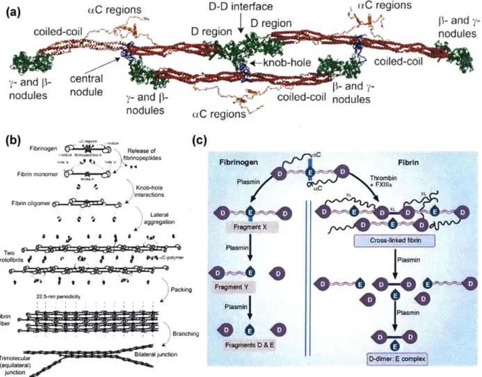
(a) (C regions D-D interface uC regions
D region s4 f- and
y-coiled-coil D region YI nodules
-rknob-hole coiled-coil
y- and p- central
nodules nodule y- and d nd
nodules (C region
(b) Fibrinogen Release of(C)
bnopeptides Fibrinogen Fibrin
Fibrin monomer A t NPlasrarn+FA Knob-hole C+ nteractions XL X Fibrin oligomer X Lateral1 FragmenlX Crow-linked fabrin prolofibrils * * CPIYMW $laymer
)Packing ragme t Y40i
Fii 22.5-nm pereid 1 Plasm 44 Pie0
Branching P0 s0n Fragments D & E Tnimolecular BiONlateraljunction r (equilateral) junction
Figure 1-2: Fibrinogen and fibrin polymerization. (a) Fibrinogen structure. Reprinted with permission from Ref 39. (b) Fibrin polymerization, from fibrinogen monomer to branched fibrin strand. Reprinted with permission from Ref 39. (c) Fibrinolysis of fibrinogen and fibrin
networks. Reprinted with permission from Ref 36.
Fibrinogen-Fibrin conversion
Fibrinogen conversion to fibrin is the final step that generates a fibrin network and completes clot formation. Fibrinogen is a large biomolecule composed of 6 distinct chains, 2 a chains, 2 [ chains, and 2 y chains, connected by disulfide bonds (Figure 1-2a). Schematically, fibrinogen in composed of 3 globular domains, one E domain, connected by coiled-coil
sequences to 2 D domains on either end of the 340 kDa molecule. D domains are composed of the C termini of the 0 and y chains while the E domain contains N-termini of the a,
p,
and yUpon generation of thrombin during the coagulation cascade, cleavage of the a and
P
N termini occurs, exposing new termini, termed knob A and knob B, and promoting fibrinpolymerization.38' 40-4 1 Fibrinopeptides A and B (FpA and FpB) are released from the fibrinogen molecule. Each fibrinogen contains 2 knob A and 2 knob B for each a and
P
chain. The exposed residues interact with complementary hole structures in the D domains, with exposed knob A residues interacting with the pocket generated in the y nodule and knob B residues interacting with the pocket generated in thep
nodule. Interactions between knobs and holes initiate fibrin polymerization, forming oligomeric chains. Oligomers expand laterally to form protofibrils, followed by packing into fibers that are the structures identified in fluorescence and electron microscopy images of clot structures. Fiber formation is assisted by interactions between C-terminal a chain domains (aC region), which associate with each other to increase crosslinks between protofibrils and further stabilize lateral aggregation of fibrin (Figure 1-2b).4 2 Finally, factor XIII (fXIII), also known as transglutaminase, generates crosslinks between glutamine and lysine residues on fibrin. These additional crosslinks assist in inhibiting clot lysis by fibrinolyticmechanisms.4 3
Viewing fibrin formation as a polymerization supports considering the stoichiometry of the monomers (fibrinogen) as well as the initiators (thrombin) and inhibitors involved. This
perspective has led to the development of models and simulations of fibrin formation.4 4-46 Manipulation of functionality, initiator activity, and other network factors can result in accelerated polymerization, altered network structures and mechanics.
Fibrinolysis Mechanism
Clots undergo a lifecycle, beginning with the formation of a platelet plug and coagulation, progressing to clot maturation and culminating in clot lysis and remodeling.
Depending on the age of the clot and the mechanism of degradation, clot lysis can initiate within minutes of activation, as highlighted by the use of tissue plasminogen activator (tPA) in cases of stroke.4 7 A balance between lysis and clotting is necessary to allow for clots to form at sites of
injury but prevent conditions such as disseminated intravascular coagulation or microthrombi formation from causing strokes. Significant inhibition or modulation of these pathways should be avoided to prevent systemic hemorrhaging. The activators (tPA or urokinase plasminogen
activator, uPA) of plasmin are expressed by cells and bind either to a cell surface (uPA) or the surface of a fibrin clot (tPA). A variety of inhibitors (plasminogen activator inhibitor -1: PA-1, plasminogen activator inhibitor -2: PAI-2, a2-antiplasmin: A2AP) are present in solution to inhibit fibrino(geno)lysis away from a fibrin clot or tissue surface.48-49 This allows fibrinolysis to be restricted to a thrombus surface or the extravascular space exposed to cells. Binding sites for plasminogen on fibrin and cell surfaces are physically close (45
A)
to the binding site for plasminogen activators and allow for complexation of the proteins to occur and activateplasminogen by cleavage of a single Arg-Val peptide bond.48 Plasmin is able to cleave tPA and increase activation activity, resulting in a positive feedback loop for plasminogen activation.50 Plasmin is able to cleave fibrin and expose C-terminal lysine residues, increasing binding sites for plasminogen and accelerating fibrinolysis (Figure 1-2c). Characteristic degradation products have been hypothesized and confirmed based on the fibrinogen sequence and experiments on plasmin lysed fibrin networks. Common fibrinogen degradation products include X (cleavage of A and B C-terminal regions), Y (E and D domains remaining after cleavage of coiled-coil polypeptides between D and E domains), D (remaining D domain after polypeptide cleavage), and E (cleavage of second coiled-coil region between D and E domain) while D-dimers are the common degradation product for fibrin networks.49
1.3. Physiology of Hemorrhaging
During traumatic bleeding, clotting kinetics, mechanics, and stability are modulated as a result of lost blood volume and activation of unique lytic and inhibitory pathways. Distinct physiological responses are evident during internal or external hemorrhaging. As a result of treatment with fluids, dilution effects can be observed on hemostasis in addition to a variety of local and systemic conditions that can inhibit clot generation. Coagulopathy is a term that
encompasses all conditions that impair clot generation during traumatic or non-traumatic injuries and is generally characterized by microvascular bleeding.36 Coagulopathy results in reduced generation of activated thrombin, modified platelet activity, and hyperfibrino(geno)lysis, increased degradation of fibrinogen or fibrin.36,51-53 First, distinctions between activities occurring during internal and external bleeding will be discussed. Next, coagulopathy will be separated into two broad categories: activation of alternative inhibitory pathways52 and systemic
changes to blood properties.36
1.3.1. Internal and External Bleeding
Internal and external hemorrhaging differ in their presentation to a first responder or clinician and their treatment of the wound ( 1.5). Physiologically, their origin and impact on the patient vary. External bleeding wounds predominately arise from some trauma and a puncture or laceration of the skin results in blood escaping the body. Damaged organs and tissues are within a defined path, even if is expansive. In contrast, internal bleeding can occur due to a disease, following a surgery,56 or a traumatic event.57 Blood is lost from circulation and accumulates in extravascular cavities in the abdomen, pelvis, or in tissues such as the brain. The collection of blood in cavities can generate hematomas, a collection of coagulated blood within a tissue. The injured vasculature may continue to bleed, increasing the size of the hematoma. Increased
pressure in regions like the brain can result in reduced cognitive abilities and precipitate a coma if the lost blood is not removed from the location and carefully monitored to alleviate pressure. In these situations, controlling blood loss is the first step of many necessary to stabilize a
hemorrhaging patient. Fluid accumulation in the abdomen can result in infections if tissue along the digestive tract is also damaged, exposing gastric and intestinal content to the excess blood and fluid. Blood is also an irritant to other tissues and can cause swelling and inflammation, both of which are secondary responses to traumatic bleeding.58
1.3.2. Altered Systemic Blood Properties
Traumatic bleeding can result in changes in blood pH (acidosis), reduced temperature (hypothermia), reduced concentrations of blood components (dilution), and exhaustion of blood component stored in the body (consumption).36 Lower levels of clotting components or
consumption of components during trauma inhibit future clot generation (coagulopathy). Changes to blood pH impact blood protease activity and temperature have been shown to severely impact bleeding at very low body temperatures (33 C).53 These responses
(hypothermia, acidosis, and coagulopathy) have been collectively termed the lethal or viscous triad of trauma, with higher mortality rates anticipated for patients presenting with the
symptoms.59 These changes are the classical characteristics that have described coagulopathy,
though their effects on impaired clot formation have been questioned or determined to be less impactful on coagulation than previously hypothesized.52, 59-60
1.3.3. Altered Inhibitory Pathways
Systemic pathways that result in inactivation of clotting proteases and increased fibrin(ogen) degradation have both grown in importance as mechanisms driving acute coagulopathy. Activated protein C (aPC) and plasmin pathways are two of the processes
involved in protease inactivation and fibrinolysis, respectively.12 Under normal conditions aPC
serves to contain clot formation to an injury site, preventing clots from spreading to uninjured endothelial locations. Its role in coagulopathy may be in response to hypoperfusion, the reduced blood flowrates that arise at hemorrhaging sites.5 1',61-62 aPC is capable of inactivating clotting
factors fVa and fVIIIa, components of the prothrombinase and tenase complexes necessary to activate thrombin.3 6 In addition, inhibitors to fibrinolysis are inhibited, thereby increasing the activity of fibrinolytic enzymes. Its mechanism involves binding of thrombin to
thrombomodulin, a transmembrane protein expressed on endothelial cells.
Thrombin-thrombomodulin complexes modify the function of thrombin and increase its interaction with and cleave of protein C to yield activated protein C. aPC and protein S are required to complex to yield high levels of fVa and fVIIIa inactivation.36 If this pathway is a critical component of coagulopathy, it discourages promoting increased thrombin generation to improve coagulation. Increased thrombin expression under traumatic conditions may serve to reduce the procoagulant activities of thrombin and increase anti-coagulant outcomes.52
Hyperfibrinolysis occurs in areas of low tissue perfusion and has shown strong correlations to hemorrhagic shock in rodent tests.36 Plasmin is the main enzyme that lyses fibrin clots under physiologic and traumatic conditions and misregulation of its activity results in increased fibrin lysis. It is unknown whether it is an increased expression of tPA or reductions in plasmin inhibitors (PAI-I and PAI-2). aPC, in addition to inactivation of clotting factors, is also able to bind and inactivate PAI-1, a primary inhibitor of plasmin. Regardless of the cause, higher levels of tPA allow for more fibrinogen and fibrin degradation by plasmin and impaired stability of fibrin networks in clots.
1.4. Managing Hemorrhaging
1.4.1. Treatment by First Responders
Civilian and military first responders are limited in their access to medical equipment, resulting in less insight into the most pressing needs for a patient than could be provided at a tertiary care facility. Triaging protocols help to address the most urgent things first. An example protocol is ABCDE, which represents the order in which medics and first responders should assess an injured patient. A (airway), B (breathing), C (circulation), D (disability), and E (environment) ensuring the medical team has fully assessed the patient.7 However, in combat situations, the protocol is commonly shifted to begin with circulation (C) and the threat of hemorrhage. Resuscitation fluids can be delivered and, less frequently blood products unless the patient has made it to a forward surgical team (FST).12 FSTs have the resources to perform some
surgical procedures and have blood products and other facilities to complete surgeries.7'12
Tourniquets and application of pressure are two of the oldest and first considered hemostatic techniques used. Sterile gauzes and a variety of powders and foams are commercially available and under investigation for use in topically exposed bleeding wounds. Some active hemostats have been developed containing clotting factors that can be stored in a lyophilized state, allowing for storage in a military medic's aid bag and immediate use without the need for a refrigeration chain.'7 Greater resources for first responders, in both military and civilian environments, are
limited due to a lack of significant reliable refrigeration resources and the need to have transportable, environmentally stable hemostats for use immediately without preparation. 1.4.2. Treatment by Clinicians
Traumatic and surgical situations in a clinical environment can both encounter uncontrolled bleeding. In a traumatic situation, the primary goal is to stabilize a patient prior to surgical
intervention if necessary. At their disposable are the same tools as first responders and the ability to utilize imaging modalities that can identify injured organs and bleeding sources. Treatments will begin with resuscitation and the delivery of fluids or blood components. Surface-based treatments include packing topical wounds with hemostatic gauzes or standard cotton gauze to remove any pooled blood and attempt to staunch blood loss by increasing pressure on bleeding sites. Invasive exposure of areas of the body (abdomen or chest) to search for sources of bleeding can be performed if less invasive approaches are not stabilizing the patient. Exploration of the pelvis, abdomen and chest can address many of the most urgent sources of bleeding. These sources include the liver and bleeding due to ruptured arteries or large vessels. Metallic or plastic clamps, also known as hemostats, can be used to physically close any damaged vessels or tissues that are hemorrhaging. Exposure of the bleeding site allows for the use of solid, gel, and liquid-based hemostatic materials.2,4,63 These include gauzes containing procoagulant materials, and
foams capable of absorbing lost blood volume and potentially releasing procoagulant molecules. Surgical management of bleeding similarly uses gauzes and, after surgeries, may use fibrin sealants and other sealant compounds to glue surgically cut areas on skin or organs back
together.63 A variety of gauzes and sealants are used that are capable of being applied and remaining at the site following closure of the patient and reabsorbing or degrading without impacting the natural biological processes involved in healing.5' 64-65
Access to imaging modalities, including CT X-ray imaging,66 ultrasound, and magnetic resonance imaging (MRI) for acute and chronic bleeds can allow for localized treatment of bleeding sites.67 Interventional radiology is a sub-specialty of radiology that uses minimally-invasive techniques to guide treatments to many organs in the body. Bleeding that is unable to be managed by trauma methods may progress to interventional radiologists to utilize catheters
guided by CT imaging and angiography to deliver embolic agents to a bleeding site identified by contrast dye injections. The hemostatic agents used in minimally invasive methods must be injected or pass through catheters. Solid hemostatic materials require a manual pusher while other particle or gel-based hemostats can be injected with a syringe through the catheter and delivered under imaging guidance. A majority of the embolic agents used by interventional radiologists function as mechanical occlusive agents.'5
1.5. Hemostatic Agents
Hemostatic materials function by a variety of mechanisms. Commercial hemostats have been separated into the categories of passive, active, flowable, sealant, and adhesives in reviews on hemostatic materials." 1 More recent advanced hemostatic materials under research may be
more appropriately separated into the blood products they are mimicking, modulating, or predominately interacting with to achieve their hemostatic impact. While some materials are continuing to be developed into absorbing gauzes or gauzes impregnated with activated components, a greater focus has been on active hemostats with mimicry or interactions with blood components or tissues.20',68-70 The following sections will introduce commercial hemostats according to the commonly used categories followed by introduction to the range of hemostats under research and development, organized by their most related blood/tissue involved in hemostasis.
1.5.1. Commercial Hemostatic Agents
Passive
Passive hemostats function predominately to absorb pooling blood and serve as a material that can be packed into or onto a wound.4' When applying pressure to a wound is possible and beneficial, passive hemostats are commonly placed between the hands of the medical
professional and the wound. Their composition is commonly in the form of a gauze, lyophilized foam, or lyophilized particle. The material composition itself is commonly secondary to its ability to absorb pooled blood and not cause any adverse inflammatory responses. Gelatin, collagen, cellulose, and polysaccharides have been used for and fashioned into a variety of delivery formats for use by first responders and surgeons. Some materials, such as chitosan, have been reported to have antimicrobial activity as well as affinity for negatively charged platelets through their positively charged backbone." Collagen is known in vivo to serve as an agonist for platelet activation,26,7' but this is not its primary purpose when used as a hemostatic sponge. Modifications are common to this core set of passive hemostats, including oxidation of cellulose72 and functionalization of chitosan,73 to improve absorption capacity or adhesion to tissues.
Clay-based hemostats can be considered as passive hemostats, as their mechanism involves rapid absorption of fluids and adsorption of clotting components. Powders and gauzes8'
13, 17 impregnated with kaolin clay are examples of clay hemostats that are able to successfully treat topical bleeding.9
Active
Commercial active hemostats are limited to including thrombin in injectable syringes.4 Due
to the potent activity of thrombin, active thrombin hemostats are used only in topical wounds with no risk of systemic delivery.7 4 Pooled blood products can similarly be considered as active
hemostatic agents.
Gelatin particles and thrombin have been combined in mixtures that were developed to combine the tamponade effect of gelatin with the coagulation quality of thrombin. These systems can easily conform to complex wound surfaces and flow into cavities that have been cleared of pooled blood.4 To maintain thrombin activity, the components require mixing prior to use,75 reducing their efficacy in trauma situations that require rapid application. Partially crosslinked gelatin particles have also been developed that, after mixing with sterile solutions of calcium chloride or saline, generate a paste that can be injected on the surface of a bleeding wound to promote contact activation. Other flowable hemostats include mixtures of the patient's plasma with biopolymers like collagen as well as thrombin. Though combination products including blood components from the patient can improve the performance of the hemostat, incorporating more of the necessary blood components for hemostasis (e.g. platelets), the added preparation precludes their use in most trauma environments and limits their use to surgical situations with extensive preparation time and refrigeration needs.5
Adhesives and Sealants
Hemostats classified as sealants and adhesives are capable of gluing or staunching additional blood loss independent of the coagulation cascade. The aforementioned hemostats all require the body to naturally generate a clot, acknowledging that some hemostats provide additional components or improve the potential for clot formation (e.g. applying pressure with passive hemostats). Commercial products combine thrombin and fibrinogen in a dual cartridge to inject as a spray onto the wound sites.63' 76 A fibrin network will form on the sprayed surface regardless of the state of the physiological clotting potential. Other systems combine albumin
and glutaraldehyde to seal small wounds and interact non-specifically with the exposed tissue.5' 77-78 Cyanoacrylate begins as a monomer that, upon exposure to air begins to polymerize and can
adhere surfaces non-specifically.79
It is the same components used in commercial super glues, highlighting its adhesive strength and broad adhesive potential. Sealants and adhesive are most commonly used in surgical situations due to the need for preparation. However, their
independence from the physiological coagulation process is essential in surgical environments where patients are maintained in an anticoagulated state to prevent unwanted clot formation.
Embolization
Embolization procedures require hemostats that are compatible with the unique
restrictions presented by delivery by a catheter. Some passive hemostats have been reformulated to allow them to be injected through a catheter locally to the bleeding site. Polymeric and gelatin microspheres are suspended in saline solutions or contrast dyes for visualization to occlude vasculature upstream of a bleeding site identified by CT imaging.15 Occlusion is also used to
deliver therapeutics to cancerous tissues while blocking blood flow to the tumor as well as blocking malformed vasculature. In all these scenarios, sphere sizes are critical for positive outcomes. The particle diameter must be correlated to the diameter of the vessel to ensure the particles do not occlude downstream vessels or get blocked too far upstream of the target site.80 Metallic coils are used to occlude vessels and fill aneurysms prior to rupture.81 Polymers
dissolved in solvents and cyanoacrylates are used to generate permanent blockages in vessels. As the solvent diffuses away after injection, the polymer becomes insoluble in the blood and
solidifies.82 Similarly, the polymerization of cyanoacrylate monomers can seal a wound by
adhering the vessel wall to itself However, these approaches are limited in application time and require extensive training and equipment to use.