Delineation of the Molecular Mechanisms Underlying DNA Replication Initiation and Changes in Gene Copy Number during Drosophila Development
by Brian L. Hua
B.S. Biochemistry and Molecular Biology, 2010 University of California, Davis
Davis, CA
Submitted to the Department of Biology
in Partial Fulfillment of the Requirements for the Degree of Doctor of Philosophy in Biology
at the
Massachusetts Institute of Technology Cambridge, MA February 2017 MASSACHUSETTS INSTITTE OF TECHNOLOGY
OCT
2
5
2016
LIBRARIES
ARC-E.S
@ 2017 Brian L. Hua. All rights reserved.
The author hereby grants to MIT permission to reproduce or distribute publicly paper and electronic copies of this thesis document in whole or in part.
Signature of Author Certified by ...
Signature redacted
Signature
redacted-... epartment of Biology October 15, 2016 . . ... .. Terry L. Orr-Weaver Professor of Biology Thesis Supervisor.Signature
redacted
The author hCrey grants to MT permission to Co-Chair, reproduce arG to distribute publiclv paper and
electronic copies of this thesis Cocument in
... Amy E. Keating Professor of Biology Biology Graduate Committee Accept
Delineation of the Molecular Mechanisms Underlying DNA Replication Initiation and Changes in Gene Copy Number during Drosophila Development
by Brian L. Hua
Submitted to the Department of Biology on October 21, 2016 in Partial Fulfillment of the Requirements for the Degree of
Doctor of Philosophy in Biology
ABSTRACT
The study of differential DNA replication programs in Drosophila has provided important insight into the molecular control of replication initiation and fork progression during development. We investigated the mechanisms by which binding of the origin recognition complex (ORC) and replication fork inhibition give rise to locally underreplicated regions in Drosophila polyploid tissues. We identified copy number changes genome-wide in two additional polyploid tissues and compared our results to three previously profiled larval tissues. These results revealed a high level of tissue-specificity in the number of underreplicated sites within a given tissue but also highlighted the conservation of the locations of many of these underreplicated regions across tissues. By mapping ORC binding sites in the larval fat body, we found that the repression of replication initiation is a common mechanism of underreplication in polytene tissues. Our ORC localization studies also suggest that underreplication zones are hard-wired across tissues and that differential underreplication of these zones is dependent upon variation in fork progression across these regions. We then utilized the Drosophila amplicons in follicle cells (DAFCs) as a model replication system to dissect the molecular mechanisms underlying the activation of individual replication origins. Repression of the DAFC-22B origin is not achieved through changes in subnuclear localization but rather through effects of the surrounding chromatin. We identified two novel genomic sites at which origin activity is modulated directly by the
surrounding chromatin environment. At one site, the surrounding chromatin promotes one additional round of origin firing at a specific developmental time point. At the other site, origin activity is repressed by the surrounding chromatin through inhibition of the localization of the MCM2-7 helicase complex. Origin repression at this site is not correlated with the establishment of heterochromatin, raising the possibility that the activity of individual replication origins are regulated by the chromatin environment on a greater, conformational level. Finally, we dissected the requirement of transcription in the activation of the DAFC-62D origin. Surprisingly,
transcription is not required in cis for origin activation. These results indicate the requirement of a trans-acting factor specifically at this site and highlight the diversity of mechanisms that control metazoan origin activation.
Thesis supervisor: Terry L. Orr-Weaver Title: Professor of Biology
Dedicated to my parents and brother, Toan, Chi, and Kevin Hua
Acknowledgments
This thesis is only a small product of the countless investments that people have made in me throughout my graduate career. The individual who took the greatest investment leap-of-faith is my graduate advisor, Terry. I should have been a tough investment decision, arriving with negligible experience in genetics (as exemplified by my less-than-stellar 7.52 grade) and a strong case of indecisiveness in choosing my graduate lab. But I remember clearly when I finally
decided to join Terry's lab. She immediately smiled at me and shook my hand without a second of hesitation. Terry has consistently been my example of an ideal scientist, mentor, and teacher, an image toward which I continually strive. Terry's mentorship has allowed me to develop the skills to tackle seemingly untackle-able problems at the lab bench and to conduct scientific research with the highest integrity. She has also taught me the importance of collaboration and the critical discussion of science with others. Finally, Terry has always been extremely generous in giving me her time, even when her time was much more valuable than I could imagine. I know I cannot ever truly return this to her, and for this reason I am eternally grateful for what she has given me.
My thesis committee members Stephen Bell and Laurie Boyer have been amazing guides throughout my graduate career. Steve has been a great mentor to me since my first year in the program when I rotated through his lab, fueling my indecisiveness in which lab to ultimately join. Laurie has always pushed me to be an innovative and creative thinker, a quality which I have learned to be one of most important of a great scientist. I am honored that these two great
scientists made investments in me over these years.
I am deeply thankful for the daily, recurring investments that the members of the Orr-Weaver Lab have made in me. I have had the honor of learning from, working with, and exploring life with the most amazing group of friends. I must emphasize my gratitude toward Jared Nordman and Masatoshi Hara, who continue to be exceptional scientific mentors and friends to me. I am also indebted to Belinda Pinto, Boryana Petrova, Jessica Alexander, Maiko Kitaoka, Laura Frawley, Emir Aviles Pagan, Jessica Von Stetina, Iva Kronja, and Helena Kashevsky, who have all made immense investments in me not only as a scientist, but as a human being as well.
My classmates have provided me with the best home these past six years. Quite literally, the members of the Biomansion have continued to welcome me into their home even after my departure from the apartment. My incredible classmates Lynne Chantranupong, Anthony Chiu, Courtney JnBaptiste, Jasmine De Cock, and Robert Erdmann have been rooting for me and celebrating every accomplishment with me the entire way. My great friends and fellow PhD-ers Sherry Lin, Cathy Su, and Linda Vo have taken me on the most amazing adventures, and I constantly think about how impossibly lucky my life must be to be part of theirs.
Finally, I would not have made it a single day in this PhD program without the
investments made by my family. My brother Kevin has been my best friend throughout my entire life, and there are relatively few memories that I have without him. He has been my constant source of support through school and life in general, and to this day I still aspire to be just like him. My parents, Chi and Toan, have been my biggest and most unconditional supporters. They have made the most valuable investment in me of all, and that has been more than enough to get me through even the most difficult of times. It is impossible to articulate how grateful I am for everything they have given and made possible for me. I hope that this thesis is just the beginning of the returns that have come of their investments in me.
TABLE OF CONTENTS Chapter One:
Introduction 9
DNA replication overview 10
Protein players at the origin of replication 11
Contributions from Drosophila to the DNA replication field 12 Hurdles for the molecular study of metazoan DNA replication 16 Developmentally-regulated cell cycle changes in Drosophila 17
Strategies involving changes in DNA copy number 18
Genome-level copy number changes - the endo cycle 21 Developmentally-programmed follicle cell gene amplification to
increase local copy number 34
Conclusions 50
Summary of thesis 53
References 54
Chapter Two:
Dynamic changes in DNA replication and ORC localization during
tissue differentiation 63
Abstract 64
Introduction 65
Results 67
Profiling of gene copy number in the adult Malpighian tubules 67 Changes in the underreplication profile through midgut development 72 Expression of underreplicated genes through fat body development 75 Repression of underreplicated genes in adult tissues 76 ORC binding is repressed in the fat body underreplicated regions 81 Replication ceases in the fat body at the end of larval development 84
Analysis of fat body ORC binding sites 84
Discussion 90
Materials and Methods 95
Acknowledgments 98
References 99
Chapter Three:
Investigating the molecular role of chromatin environment in DNA replication
origin activation 101
Abstract 102
Introduction 103
Results 106
DAFC-22B origin activity is not correlated with subnuclear
positioning in follicle cell nuclei 106
Sequence analysis reveal limited differences in the 22B regions
of OrRTow and OrRMOD 109
roles in ori22B repression 110 Identification of a transgenic origin activated by the
surrounding chromatin environment 115
Identification of a genomic site that inhibits origin activity 121 Repression of the RS 1 origin correlates with the repression
of MCM localization 121
Repression at RS 1 is not correlated with transcriptional activity 125 RS 1 origin repression is not correlated with changes in
replication-associated histone modifications 128
Discussion 131
Materials and Methods 137
Acknowledgements 142
References 142
Chapter Four:
The role of transcription in the activation of a Drosophila amplification origin 147
Abstract 148
Introduction 149
Results and Discussion 154
yellow-g2 is the sole transcription unit in the 10kb DAFC-62D amplicon 154
A tagged yellow-g2 transgene 154
Transcription in cis is not required for ori62 firing 159
Materials and Methods 163
Acknowledgements 165
References 166
Chapter Five:
Conclusions and Perspectives 169
Dynamics of ORC binding during tissue differentiation 170 Tissue-specificity of underreplication as a consequence
of differential fork progression 171
Regulation of origin activity by the surrounding chromatin environment 173 The role of 3D chromosome conformation in origin activation 174
Chapter One:
Introduction
Proper control of DNA replication is critical to ensure genomic integrity during cell proliferation. In addition, differential regulation of the DNA replication program during development can change gene copy number to influence cell size and gene expression.
Drosophila melanogaster serves as a powerful organism to study the developmental control of
the DNA replication program in various cell cycle contexts in a variety of differentiated cell and tissue types. Additionally, Drosophila has provided several developmentally-regulated
replication models to dissect the molecular mechanisms that underlie replication-based copy number changes in the genome, which include differential underreplication and gene
amplification. Here, I review key findings that have shed light on the developmental control of DNA replication in the contexts of the archetypal replication program as well as of
underreplication and differential gene amplification. We will focus on the use of these replication systems in delineating many of the molecular mechanisms that underlie the developmental
control of replication initiation and fork elongation.
DNA replication overview
Before cell division, the genome must be completely and accurately replicated to maintain the integrity of genetic information across cell generations. In metazoans, DNA replication initiates from thousands of DNA elements within the genome called origins of replication. Origins of replication direct the assembly of a large group of proteins and protein complexes to the site that ultimately allow for DNA unwinding and the establishment of two, bidirectional replication forks. These replication forks continue to progressively unwind upstream DNA, generating single-stranded DNA that serves as a template for the synthesis of new DNA (Costa et al. 2013). Through the molecular study of DNA replication initiation and elongation, it is clear that the mechanisms that regulate origin activity and replication fork
progression are diverse and complex, particularly in the context of development. Drosophila has provided powerful developmental systems to study both replication initiation and elongation at the cellular and molecular level (Nordman and Orr-Weaver 2012). Here, we summarize
important insights that the Drosophila system has shed upon the regulation of metazoan DNA replication. We then detail seminal studies that have led to critical understanding of the developmental control of replication origin activation and fork elongation. Finally, we address prevailing questions in DNA replication control and the outlook for the field.
Protein players at the origin of replication
DNA replication initiation requires the sequential recruitment and activation of a large number of replication protein components. Unlike in budding yeast, metazoan origins of replication are not defined by any known consensus sequence (Costa et al. 2013). However, protein factors required to establish the replication initiation complex and the replication fork are highly conserved in eukaryotes (Table 1). Replication initiation first requires that origins of replication are bound by the Origin Recognition Complex (ORC, composed of the six proteins ORC 1-6) in late M and G 1 phases of the cell cycle. The replication initiation factor Cell Division Cycle 6 (Cdc6) is then recruited to the origin to form a complex with ORC. ORC and Cdc6 work cooperatively to recruit the initiation factor Cdtl (Double parked, or DUP, in Drosophila) and finally the six-membered minichromosome maintenance (MCM)2-7 replicative helicase complex. In budding yeast, Cdtl and MCM2-7 form a stable complex in cell lysates and are recruited to origins of replication together (Tanaka and Diffley 2002; Kawasaki et al. 2006; Remus et al. 2009). However, in Xenopus extracts, Cdtl and MCM2-7 do not co-precipitate, suggesting that Cdtl and the MCM2-7 complex may be recruited sequentially to replication origins in metazoans (Maiorano et al. 2000).
Two hexamers of the MCM2-7 complex are loaded onto origin DNA in an inactive state before the onset of S-phase. Under the regulation of two kinases, S-phase cyclin-dependent kinase (CDK) and Dbf4-dependent kinase (DDK), the MCM2-7 complex is bound by CDC45
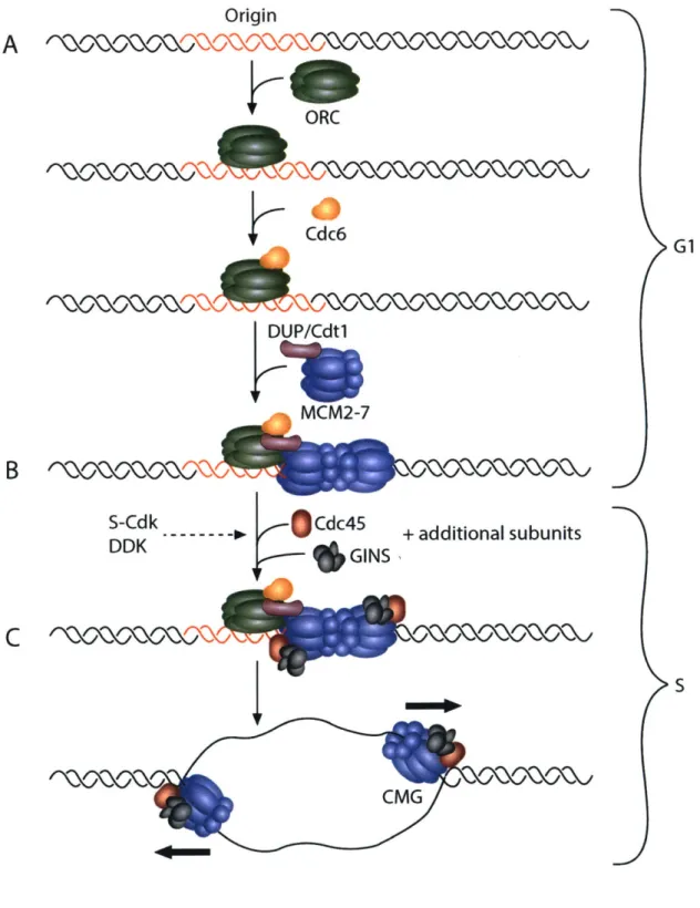
and the go-ichi-ni-san (GINS) complex, a four-membered complex composed of Sld5, Psf 1, Psf2, and Psf3. Together, the CDC45/MCM2-7/GINS (CMG) complex forms the functional replicative helicase (Costa et al. 2013). As two MCM2-7 hexamers are loaded onto a single origin of replication, two CMG complexes establish the independent, bidirectional replication forks after origin activation (Figure 1-1).
Contributions from Drosophila to the DNA replication field
Elegant genetic and biochemical studies initially performed in budding yeast allowed for the identification of the key, conserved protein factors that are involved in eukaryotic origin activation and fork elongation (Bell and Labib 2016). Importantly, the minimal set of protein factors required for DNA replication in budding yeast in vitro has been described (Yeeles et al. 2015). A high level of conservation of the protein players involved in replication initiation and fork elongation exists in eukaryotes, highlighting the importance of the DNA replication process in all organisms (Costa et al. 2013).
Whereas the budding yeast system has paved the way for the identification of DNA replication factors and the molecular events that are required for replication initiation and fork elongation, Drosophila has since emerged as an extremely powerful organism to study metazoan DNA replication at both the molecular and developmental levels. For example, the metazoan homologs of the key replication initiation factor Cdtl were first discovered in Drosophila (Whittaker et al. 2000) and Xenopus (Maiorano et al. 2000). Additionally, the functional helicase complex was first shown to exist as a large protein assembly consisting of CDC45,
Figure 1-1. Stepwise assembly and activation of the CMG helicase.
A) During G 1 phase of the cell cycle, an origin of replication is first recognized and bound by the ORC complex. Binding of the ORC complex promotes the recruitment of the Cdc6 and DUP/Cdtl initiation factors that work cooperatively to load the MCM2-7 helicase complex. B) S-CDK and DDK activity are required for the subsequent recruitment of the helicase components
Cdc45 and the GINS complex, along with additional subunits necessary for helicase function
(MuslOl/Dpbl 1, RecQ4/Sld2, and others). C) The replicative helicase, composed of MCM2-7, Cdc45, and GINS, is activated at the start of S-phase to begin replication. The separation of helicase loading and helicase activation in two distinct phases of the cell cycle ensures that an origin fires only once per cell cycle and prevents rereplication during S-phase.
Figure 1-1
OriginA
ORC Cdc6 G1 DUP/Cdt1 MCM2-7B
S-Cdk -- Cdc45 + additional subunits DDK GINS S CMGTable 1-1. Key proteins required for helicase loading and activation.
Drosophila Mammalian homolog Budding yeast homolog Function
ORC1 ORC1 Orci Helicase loading
ORC2 ORC2 Orc2 Helicase loading
Latheo ORC3 Orc3 Helicase loading
ORC4 ORC4 Orc4 Helicase loading
ORC5 ORC5 Orc5 Helicase loading
ORC6 ORC6 Orc6 Helicase loading
CDC6 CDC6 Cdc6 Helicase loading
Double Parked (DUP) CDT1 Cdtl Helicase loading
MCM2 MCM2 Mcm2 Helicase
MCM3 MCM3 Mcm3 Helicase
Disc Proliferation Abnormal (DPA) MCM4 Mcm4 Helicase
MCM5 MCM5 Mcm5 Helicase
MCM6 MCM6 Mcm6 Helicase
MCM7 MCM7 Mcm7 Helicase
MCM10 MCM10 Mcm10 Helicase activation
CDC45 CDC45 Cdc45 Helicase activation/helicase
SLD5 SLD5 SId5 Helicase activation/helicase
PSF1 PSF1 Psf 1 Helicase activation/helicase
PSF2 PSF2 Psf2 Helicase activation/helicase
PSF3 PSF3 Psf3 Helicase activation/helicase
MUS101 TopBP1 Dpb11 Helicase activation/helicase
RECQ4 RECQL4 Sld2 Helicase activation
MCM2-7, and GINS (CMG complex) through biochemical isolation from Drosophila embryo extracts (Moyer et al. 2006). Importantly, crucial structural insight into the regulation of metazoan DNA replication initiation is highlighted by extensive electron microscopy studies
(Clarey et al. 2006; Clarey et al. 2008) and the recent solving of the crystal structure of Drosophila ORC (Bleichert et al. 2015). Finally, Drosophila has long served as a metazoan model system to profile replication properties and dynamics genome-wide, beginning with the first genome-wide mapping of ORC in a differentiated metazoan cell type and tissue (MacAlpine
et al. 2010; Sher et al. 2012). These genome-wide approaches have allowed for more
comprehensive analysis of replication dynamics in the scope of the underlying chromatin landscape, developmental timing, and differentiation.
It also has become increasingly clear that the replication and developmental programs are tightly linked (Nordman and Orr-Weaver 2012). Comparison of the replication properties of various cell and tissue types of Drosophila undergoing different cell cycle programs has shed critical light on the role of development on origin specification, origin activation, fork
elongation, fork stability, and overall S-phase length. Drosophila also has emerged as an
important model to study the causes and consequences of DNA copy number changes in various developmental contexts. The study of differential replication programs in Drosophila have allowed for the dissection of the molecular mechanisms that underlie the activation of single origins of replication and the properties of replication fork progression in metazoans.
Hurdles for the molecular study of metazoan DNA replication
Although the requirements of the key proteins involved in DNA replication have been well studied, it is clear that origins of replication are regulated differentially within a single cell in all organisms studied. For example, within a single genome, only a subset of origins are
activated at a given time point in S-phase (Aparicio 2013). Furthermore, origins of replication are not uniformly distributed throughout the genome, resulting in large genomic regions that require the activity of single replication forks emanating from distant origins for their replication (Debatisse et al. 2012; Newman et al. 2013). It remains unclear what mechanisms exist to regulate the activation of single origins of replication and replication forks throughout the genome, especially in metazoan organisms. This is in large part due to the fact that detailed, molecular characterization of well-defined metazoan origins is lacking (Aladjem 2007). It has also been difficult to examine replication forks emanating from a single origin of replication. Finally, how developmental signals modulate the activity of replication origins and forks remains to be elucidated.
Developmentally-regulated cell cycle changes in Drosophila
Drosophila development is tightly-linked with changes in the cell cycle and DNA
replication programs. During early embryogenesis in the first two hours after fertilization, nuclei divide quickly with no defined gap phase, an S-phase length of about 3 minutes, and replication origins spaced less than 10kb apart (Blumenthal et al. 1974). This is in stark contrast to fully differentiated cell types in Drosophila that can exhibit S-phases lasting longer than 10 hours, with origins of replication spaced as much as 100kb apart (Spradling and Orr-Weaver 1987). After the thirteenth division cycle in embryogenesis, a gap phase is introduced and S-phase is dramatically lengthened to 40-50 minutes, highlighting a drastic change in both the cell cycle and DNA replication programs during development.
Most cells in the embryo cease mitotic divisions after the 1 6th division cycle and enter a
variant cell cycle called the endo cycle that continues through larval development (Smith and Orr-Weaver 1991). The neural and imaginal tissues are the only tissues to continue to divide
mitotically during embryonic and larval development. The endo cycle consists of alternating S-and G-phases without cell division (Figure 1-2B). During the endo cycle, DNA content is increased at the genomic level, leading to polyploidy. As organism size is greatly increased throughout larval development, polyploidy is thought to coordinate cell size and tissue growth by
generating large, highly metabolically active cells (Edgar et al. 2014; Orr-Weaver 2015). Indeed, blocking polyploidization inhibits cell and larval growth and normal tissue function (Edgar and
Orr-Weaver 2001).
Interestingly, increases in gene copy number in polyploid Drosophila cells are not uniform throughout the genome. Heterochromatin is repressed for replication in many
Drosophila polyploid cells, and in several larval tissues, defined eukaryotic genomic regions are underreplicated relative to overall ploidy of the cell. Additionally in the adult female, follicle cells complete endocycling and begin gene amplification at specific sites within the genome. The study of underreplication and differential gene amplification in Drosophila has provided important understanding about the developmental regulation of both origin activation and fork progression at the molecular level. In the following sections, we focus on the regulation of the endo cycle and summarize our current understanding of the molecular parameters of DNA replication from key studies in embryonically-derived cell lines, polyploid larval tissues, and the amplifying follicle cells.
Strategies involving changes in DNA copy number
Studies of the endocycling cells of Drosophila have revealed important mechanisms by which the cell cycle is coordinated with the DNA replication program and key molecular parameters of origin activity and fork progression. Here, we summarize the known regulatory
Figure 1-2. The oscillatory levels of key factors in endo cycle maintenance.
A) The canonical mitotic cell cycle is composed of four sequential phases: GI, S, G2, and M. This cell cycle gives rise to two identical daughter cells. B) The endo cycle is composed of two
sequential phases: G and S. This leads to increased DNA ploidy within and single cell. C) The endo cycle is driven by oscillations in key cell cycle factors. The E2FI transcription factor rises at the end of G-phase to turn on transcription of Cyclin E and is degraded during S-phase. Cyclin E/Cdk2 activity rises at the start of phase to initiate DNA replication and falls at the end of S-phase after the completion of replication. In addition, the activity of the E3 ubiquitin ligase CRL4-Cdt2 peaks in S-phase where it marks E2Fl and Cyclin E for degradation.
Figure 1-2
A Mitotic cycle
G1
K
JG2
C
.5 CB Endo cycle
s
L E2F1 Cycin E/Cdk2 CRL4'GI|
SI|
G
I
S
|G
elements that orchestrate the alternating S- and G-phases of the endo cycle. We then focus on how differential replication within the endo cycle has been exploited to uncover mechanistic insight into the regulation of origin selection, origin activation, fork stability, and fork progression.
Genome-level copy number changes - the endo cycle
The endo cycle is utilized throughout the plant and animal kingdoms, indicating the importance of this variant cell cycle during development across organisms (Orr-Weaver 2015). Importantly, our understanding of the regulation of the endo cycle and its coordination with the replication program has derived from seminal studies in Drosophila. Nearly all larval tissues and many adult tissues in Drosophila have increased ploidy that is achieved via the endo cycle. The replicated DNA duplex copies are held in register to produce polytene chromosomes with stereotypic banding patterns in most Drosophila endocycling tissues. The most well-studied of these polyploid tissues is the larval salivary gland, which undergoes about ten endo cycles during larval development to obtain a final ploidy of roughly 1024C (Hammond and Laird 1985b). During the endo cycle, cells must suppress the mitotic machinery to prevent entry into the mitotic program and subsequent cell division. One strategy that endocycling cells use to achieve this is to downregulate the activity of mitotic cyclins and mitotic CDKs at the transcriptional level. At the switch from the mitotic cell cycle to the endo cycle, cells in the embryo cease expression of the mitotic regulators Cyclin A, Cyclin B, Cyclin B3, String/Cdc25, and Cdkl (Sauer et al. 1995). However, the developmental signals that control transcription of these regulators at this switch is not well understood.
In Drosophila, Cyclin E/Cdk2 activity is the major driver of S-phase entry. Mutations in
1994). Importantly, continuous overexpression of Cyclin E in the salivary gland blocks endocycling, suggesting that oscillations in Cyclin E/Cdk2 activity are required for continued endocycling (Follette et al. 1998; Weiss et al. 1998). The oscillatory expression of CycE is mediated by oscillations in the levels of the transcription factor E2F1, which reaches high levels during G-phase and is degraded at the end of S-phase (Zielke et al. 2011). E2FI degradation is mediated by the E3 ubiquitin ligase CRL4-Cdt2 (Shibutani et al. 2007; Shibutani et al. 2008), whose activity peaks during S-phase (Zielke et al. 2011) (Figure 1-2C). Artificial stabilization of E2F1 prevents endocycling in the salivary gland, indicating that E2F1 degradation is required for continued endocycling (Zielke et al. 2011). At the end of S-phase, degradation of E2F1 is
followed by ubiquitin-dependent degradation of Cyclin E via the E3 ubiquitin ligase CRL1-Ago along with its activator Minus (Shcherbata et al. 2004; Szuplewski et al. 2009; Zielke et al. 2011). The degradation of Cyclin E allows for the completion of S-phase and the relicensing of replication origins in the subsequent G-phase. Additionally, oscillations of the Drosophila Cdk2 inhibitor Dacapo peak similarly to E2F1 during G-phase of the endo cycle (Hong et al. 2003; Hong et al. 2007). Dacapo contributes to the attenuation of Cyclin E/CDK2 activity during G-phase and is subsequently degraded during S-G-phase via its PIP degron (Swanson et al. 2015). Although Dacapo is not necessary for the endo cycle (Hong et al. 2003; Zielke et al. 2011), its overexpression inhibits the endo cycle, suggesting that Dacapo plays a role in establishing the Cyclin E/Cdk2 activity threshold necessary to trigger S-phase (Shcherbata et al. 2004; Hong et
al. 2007; Zielke et al. 2011; Swanson et al. 2015).
Much like during the archetypal cell cycle, endocycling cells must also prevent
rereplication during S-phase. In the mitotic cell cycle, helicase loading at origins is restricted to late M- through G1-phase. At the Gl/S transition, the activities of S-phase CDK and DDK
increase dramatically, allowing for the assembly and activation of the replicative helicase complex to begin DNA replication (Costa et al. 2013). After S-phase onset, high S-phase CDK activity prevents the reloading of the helicase complex at origins that have already fired by inhibiting the activity of several replication initiation proteins required to load the helicase onto origin DNA (Blow and Dutta 2005). Importantly, in Drosophila as well as in other metazoans, Geminin is an inhibitor of helicase loading and exhibits high levels during the S-phase in the archetypal cell cycle to prevent rereplication (Quinn et al. 2001). During M-phase, Geminin is targeted for degradation by the Anaphase Promoting Complex (APC)/Cyclosome, allowing for helicase loading in the subsequent G-phase (McGarry and Kirschner 1998). In a similar manner, Cyclin E/Cdk2 activity peaks during S-phase in the endo cycle (Figure 1-2C). Additionally, Geminin levels oscillate during the endo cycle, with low levels in G-phase to allow for helicase loading and high levels in S-phase to prevent reloading of helicases and rereplication. Geminin is targeted for degradation at end of the endo cycle S-phase by the APC/Cyclosome along with its activator Fzr/Cdhl (Narbonne-Reveau et al. 2008; Zielke et al. 2008). The oscillation of the level of Geminin is required for the endo cycle, as constitutive expression of Geminin inhibits endo cycle progression (Zielke et al. 2008). However, Geminin is not essential for salivary gland development (Zielke et al. 2011), suggesting that multiple overlapping mechanisms exist to prevent rereplication in endo cycling cells.
Parameters of origin licensing and activation
Genome-wide techniques have allowed for comprehensive profiling of replication initiation sites in several Drosophila cell culture systems (Cayrou et al. 2011; Comoglio et al. 2015). Upon replication initiation, two nascent leading DNA strands extend from RNA primers located at the replication origin. These leading nascent strands can be separated from smaller
RNA-primed Okazaki fragments on the lagging strand by size-selection and from non-RNA-primed DNA by lambda-exonuclease digestion (Gerbi and Bielinsky 1997). High-throughput sequencing of purified leading nascent strands allows for the identification of replication initiation sites genome-wide (Leonard and Mechali 2013). Comparison of the replication initiation sites in S2, BG3, and Kc cells revealed that 16-20% of initiation sites are common to all three cell types while 35-45% of activated origins are common to at least two cell types (Comoglio et al. 2015). These results highlight the cell type-specificity of origin sites, although an appreciable number of common origin sites exists as well.
Labeling of synchronized Drosophila cells in vitro with the nucleotide analogue 5-bromo-2'-deoxyuridine (BrdU) coupled to microarray analysis revealed that distinct regions of the genome are replicated at different times during S-phase. Most origins could be classified as early or late replicating origins with minimal overlap (MacAlpine et al. 2004; Eaton et al. 2011). Early replicating sites are correlated with increased chromatin accessibility (Bell et al. 2010;
MacAlpine et al. 2010; Comoglio et al. 2015). In a survey of Kc, S2, and BG3 cells, it was found that replication timing profiles, or the temporal program in which regions of the genome are replicated in S-phase, are largely correlated between these cell types, suggesting that replication timing is relatively conserved across different cell types (Lubelsky et al. 2014). Early replicating sequences are associated with activating chromatin marks such as H4K16ac, H3K79mel/2, H3K4mel/2/3, H3K27ac, and H3Kl8ac, ORC binding, high gene density, and high gene
expression. In contrast, late replicating sequences are associated with repressive chromatin marks such as H3K27me3 and H3K9me2/3 (Lubelsky et al. 2014). Furthermore, origins are generally enriched for several histone modifications, including H3K9me 1, H3K23me 1, and H4K20me 1 (Comoglio et al. 2015). Finally, origins are generally found to be enriched in GC content,
suggesting that DNA shape and structure may play an important role in origin specification (Cayrou et al. 2011; Comoglio et al. 2015).
ORC binding has served as a useful marker for potential origins, as its localization to chromatin is necessary ultimately to recruit the replication machinery to initiate replication. In S2 cells, tethering ORC to various chromosomal sites is sufficient to direct replication initiation (Crevel and Cotterill 2012). In budding yeast, ORC binding is directed to the autonomously replicating sequence (ARS) consensus sequence, a DNA sequence that is found at all origins of replication (Bell and Stillman 1992; Costa et al. 2013). However, in metazoans, ORC exhibits little to no sequence specificity both in vitro and in vivo (Vashee et al. 2003; Remus et al. 2004). ORC2 mapping in asynchronous Drosophila Kc167 cells revealed that ORC density is
significantly higher at sites that initiate replication early in S-phase, suggesting that replication timing is established in part at the level of ORC binding (MacAlpine et al. 2004; MacAlpine et
al. 2010). Additionally, ORC is significantly enriched at active promoters, suggesting the local
chromatin environment established at actively transcribed genes allows for ORC recruitment. ORC binding at transcription start sites is correlated with an enrichment for H3K9ac, H3K27ac, H3K4me2, and H3K4me3, histone modifications commonly found at active promoters.
Likewise, these ORC binding sites are anti-correlated with the presence of the heterochromatic histone marks H3K9me2/3 and H3K27me3 (Eaton et al. 2011). Furthermore, ORC binding sites are enriched in the histone variants H3.3 and H2Av and depleted of bulk nucleosomes at sites of active transcription as well as sites not associated with an active promoter, emphasizing the idea that ORC localization is largely dictated by an open and dynamic chromatin environment
(MacAlpine et al. 2010). Consistent with this idea, ORC binding sites are also highly enriched for ISWI, a member of the NURF chromatin remodeling complex (Eaton et al. 2011).
Methylation of H4K20 has been suggested to play important roles in replication initiation in mammalian cells by promoting the localization of ORC to replication origins (Jorgensen et al. 2007; Tardat et al. 2007; Houston et al. 2008; Tardat et al. 2010; Beck et al. 2012; Kuo et al. 2012). In Drosophila, decreased activity of PR-Set7, the methyltransferase responsible for H4K20 monomethylation, results in DNA damage checkpoint activation and a lengthened S-phase in neuroblasts (Sakaguchi and Steward 2007) and S2 cells (Sakaguchi et al. 2012).
Consistent with these findings, Kc cells inhibited for H4K20 methylation exhibit a perturbed cell cycle with gross DNA damage, suggesting a defect in DNA replication (Li et al. 2016).
Surprisingly, the inhibition of H4K20 methylation does not alter the genome-wide pattern of replication origin activation, but rather sensitizes late replicating domains to DNA damage. These results provide evidence that the primary role of H4K20 methylation in Drosophila is not to direct the recruitment of ORC to replication origins, but rather to ensure the integrity of late replicating domains during S-phase.
To date, the polytene larval salivary gland is the only differentiated tissue undergoing genomic replication in which genome-wide ORC localization has been reported (Sher et al. 2012). In a survey of ORC binding in Kc, S2, and BG3 cells, it was found that about a third of the identified ORC binding sites were shared between all three cell types (Eaton et al. 2011). Similarly, 31% of the ORC binding sites identified in the larval salivary gland are common with all three cell lines, indicating that a significant level of ORC binding site conservation may exist not only in cell culture lines but in differentiated tissues as well. Importantly, 28% of the salivary gland ORC binding sites are unique to this tissue compared to the three cell culture lines.
Consistent with cell culture studies, 73% of the salivary gland ORC binding sites are within a kilobase of a transcription start site. 57% of the salivary gland-specific ORC binding sites are
found near a transcription start site, but the genes controlled by these promoters are not uniquely expressed in the salivary gland. Thus, tissue-specific expression of genes does not correlate with tissue-specific ORC binding (Sher et al. 2012).
Underreplication and local copy number reduction
Although polytene cells have increased DNA content per cell, gene copy number is not uniform throughout the genome. It has long been known that the heterochromatic regions in polyploid salivary gland, follicle cell, and nurse cell chromatin are reduced in copy number relative to overall ploidy, a phenomenon known as underreplication (Figure 1-3A) (Hammond and Laird 1985a; Hammond and Laird 1985b). In addition to heterochromatin, comparative array-based genome hybridization (aCGH) and high-throughput genomic sequencing studies have revealed that larval salivary gland, mid gut, and fat body tissues contain precise
euchromatic regions that are underreplicated (UR) (Belyakin et al. 2005; Nordman et al. 2011; Sher et al. 2012; Yarosh and Spradling 2014). These euchromatic UR regions are large, ranging from 150-450kb in size. Only a third of identified UR regions are common to all the three tissues, highlighting the high degree of tissue specificity of underreplication (Nordman et al. 2011). Strikingly, full replication of the UR regions in all three tissues can be restored by mutation of a single factor, the SUUR protein (Nordman et al. 2011; Sher et al. 2012).
In addition to genome-wide profiling approaches in Drosophila cell culture, the study of underreplication in Drosophila polyploid tissues has uncovered important links between
differentiation, development, and the control of DNA replication. Notably, ORC is bound throughout most of the salivary gland genome but is specifically excluded within UR regions (Sher et al. 2012). This finding strongly suggests that replication initiation does not occur within
Figure 1-3. Differential DNA Replication.
A) Underreplication results from impaired replication fork progression through a 150-450kb domain (arrows indicate direction of fork progression). aCGH analysis of an underreplicated site indicates decreased copy number relative to overall ploidy. aCGH data modified from Sher et al. 2011. B) In endocycling follicle cells, developmentally-programmed gene amplification occurs through repeated replication origin firing followed by bidirectional fork progression away from the origin (arrows indicate direction of fork progression). Array-based comparative genome hybridization (aCGH) analysis of an amplified site indicates a gradient of increased copy number relative to overall ploidy spanning 100kb, with the highest copy number at the origin of
Figure 1-3
I Underreplicated DomainI
I
II
I
I Ik 4-- 300kb -- + IB
Amplified DomainI
3-I 0U -3-4-100kb
--- + I IA
I3-I
I Ithese regions, and thus replication of these regions is dependent upon replication forks emanating from outside the region. Interestingly, these UR regions are devoid of RNA polymerase II
localization, strongly inhibited for transcription, and are enriched for the heterochromatic
chromatin mark H3K27me3 (Sher et al. 2012). These results are consistent with the idea that UR regions in the salivary gland represent repressive chromatin domains that are inhibitory to both transcription and DNA replication initiation. Indeed, nearly all of the UR regions in the salivary gland correspond to domains of repressive chromatin as defined in genome-wide chromatin landscape studies (Filion et al. 2010; Kharchenko et al. 2011; Yarosh and Spradling 2014). UR regions in the larval fat body also are devoid of ORC binding, suggesting that inhibition of DNA binding of ORC in these domains may be a common feature of underreplication (B Hua, H Kashevsky, G Bell, J Von Stetina, and T Orr-Weaver, in prep).
Interestingly, orc] and orc2 null mutant salivary gland continue the endo cycle, though they reach ploidy levels two- to four-fold lower than wildtype salivary glands (Park and Asano 2008; Sher et al. 2012). These results indicate that the endo cycle can occur to a significant extent in the absence of newly synthesized ORC 1 and ORC2. However, orc] and orc2 mutants exhibited a marked change in the underreplication pattern in the salivary gland where all but the most pronounced UR regions become fully replicated (Sher et al. 2012). Thus, ORC plays an important role in the distribution of replication along polyploid chromosomes, and it is thought that replication in the orc] and orc2 mutants is allowed by maternal loading of ORC or by residual activity of ORC missing the ORCI or ORC2 subunits.
Inhibition of fork progression by SUUR
Underreplication in the salivary gland, fat body, and midgut are dependent upon the Suppressor of Underreplication protein, or SUUR, as all underreplicated regions become fully
replicated in the SuUR mutant (Nordman et al. 2011). Interestingly, SUUR is a chromatin protein that as of yet has not been identified outside of Drosophila. Loss of SUUR function does not restore ORC binding in the underreplicated regions of the salivary gland, indicating that SUUR
does not act at the level of replication initiation to inhibit replication. Instead, SuUR mutants exhibit enhanced rates of replication fork progression, suggesting that SUUR acts to inhibit
replication fork progression (Sher et al. 2012). These findings support a model in which
underreplicated domains are dependent upon replication forks emanating from origins outside of the region, and underreplication is achieved by the SUUR-mediated inhibition of fork
progression through these domains.
Subsequent studies revealed that SUUR co-immunoprecipitates with the sliding clamp PCNA and the replication fork factor CDC45 in embryonic nuclear extracts and tracks with the replication fork in follicle cells undergoing gene amplification (detailed in subsequent sections), further supporting the fact that SUUR is recruited to active replication forks (Kolesnikova et al. 2013; Nordman et al. 2014). Consistent with studies in endocycling tissues, SuUR mutants exhibit significantly enhanced fork progression in amplifying follicle cells, and overexpression of SUUR severely hampers fork progression (Nordman et al. 2014). Together, these results indicate that SUUR is a general inhibitor of fork progression and acts directly at the replication fork. However, the molecular mechanism of fork inhibition by SUUR remains to be elucidated.
Fork instability and DNA damage in UR regions
In the polytene chromosomes of the salivary gland, UR domains are cytologically enriched for a key marker of double-stranded DNA breaks, yH2Av (Andreyeva et al. 2008). Chromatin immunoprecipitation studies reveal that yH2Av is enriched throughout the entire region of each UR domain, indicating that UR domains are prone to DNA damage (Nordman et
al. 2014). Enrichment of yH2Av in these UR regions is dependent upon SUUR function,
suggesting that DNA damage in these regions is caused by fork instability mediated by SUUR. Additionally, high-throughput sequencing and analysis of read pairs generated from salivary gland DNA suggest that large deletions ranging 10-500kb in size may result from DNA damage and local repair in these regions (Yarosh and Spradling 2014).
What is the biological function of SUUR and underreplication? As SuUR mutants are viable and exhibit normal morphology and fertility (Belyaeva et al. 1998), it remains unclear to what extent SUUR is required in normal development. Given that SUUR is a general inhibitor of fork progression, it is possible that SUUR serves to provide an extra level of regulation to ensure proper replication timing in the genome. This function could allow prioritized replication of regions of the genome that are more susceptible to replication stress, thus maximizing genome
stability. Another way that SUUR could achieve this effect is to distribute termination events throughout the genome (Hawkins et al. 2013).
Gene expression in UR regions
Because UR regions become fully replicated in SuUR mutants, it is not clear what the biological role of underreplication is in development. The UR regions in the salivary gland are enriched in genes involved cell adhesion, segmentation, transcription factor activity,
programmed cell death, mesoderm development, and cell motility (Sher et al. 2012; Yarosh and Spradling 2014). Additional regions that are consistently underreplicated but to lower extents in the salivary gland are highly enriched in immunoglobulin superfamily genes and genes involved in the nervous system (Yarosh and Spradling 2014). Could underreplication regulate expression of these genes through decreased gene copy number? Interestingly, transcription of genes within UR regions is largely repressed in the larval salivary gland and midgut tissues, suggesting that
decreased copy number may cause lower gene expression (Nordman et al. 2011; Sher et al. 2012). Strikingly, in the SuUR mutant in which UR regions are fully replicated, gene expression within the UR regions remains repressed, demonstrating that underreplication is not required for transcriptional repression in these domains (Nordman et al. 2011; Sher et al. 2012). Additionally, many genes within the UR regions in the fat body are significantly transcribed, indicating that underreplication and the repression of transcription can be mechanistically uncoupled (Nordman
et al. 2011).
As deletions and rearrangements have been reported throughout UR regions, one proposed role for underreplication is to promote the somatic diversity of genes within these domains (Yarosh and Spradling 2014). This idea is especially interesting in the context of the immunoglobulin superfamily genes found in some UR sites in which gene rearrangements may be advantageous. Nevertheless, the biological role of underreplication has yet to be elucidated.
Underreplication as a model for common chromosomal fragile sites
In addition to its utility in understanding the mechanisms underlying differential replication inhibition, underreplication in Drosophila polyploid tissues serves as a promising model system for human common fragile sites. Common fragile sites (CFSs) are chromosomal locations characterized by recurrent breaks, gaps, and constrictions on metaphase chromosomes upon replication stress (Durkin and Glover 2007). CFSs often are found in euchromatin and extend over megabase-long regions of the chromosome (Schwartz et al. 2006; Smith et al. 2006). Interestingly, replication initiation does not occur within a 700kb region forming the core of the most active human CFS, FRA3B (Letessier et al. 2011). Thus, replication of this large region is dependent entirely upon replication forks emanating from origins of replication outside of this domain. A general challenge to replication forks results in incomplete replication of the FRA3B
domain, leading to chromosome instability and fragility. UR regions in the Drosophila salivary gland are also devoid of origins of replication and rely on forks coming from flanking regions for their replication (Figure 1-4). Additionally, UR regions are prone to DNA damage, a property common to CFSs (Andreyeva et al. 2008; Nordman et al. 2014). Combining the genetic and cell biological toolkits of the Drosophila system with genome-wide profiling techniques will allow for deeper understanding of the mechanisms that underlie replication initiation repression in these regions and the molecular properties of CFSs in human cells.
Developmentally-programmed follicle cell gene amplification to increase local copy number While the underreplication system has allowed study of replication properties and
dynamics across large, defined chromatin domains, the molecular dissection of the mechanisms that underlie origin activation requires the study of well-defined, single origins of replication. Additionally, it is necessary to know when single origins fire in order to study individual origin activation events. The study of Drosophila follicle cell gene amplification has allowed the
isolation and detailed molecular characterization of single metazoan origins of replication. In this section, we review the characteristics of follicle cell gene amplification and focus on key studies that have led to critical understanding of the molecular parameters that regulate origin activation and fork progression.
To date, aCGH analyses have been performed on seven distinct Drosophila tissues to assay differential DNA replication genome-wide (Kim et al. 2011; Nordman et al. 2011; Sher et
al. 2012; B Hua, H Kashevsky, G Bell, J Von Stetina, and T Orr-Weaver, in prep.). Of the
examined tissues, only the ovarian follicle cells have been found to exhibit gene amplification, or increased copy number of distinct genomic regions relative to overall ploidy of the cell.
Figure 1-4. Underreplication as a model for human common fragile sites.
A) In polyploid Drosophila larval tissues, underreplicated (UR) domains are largely devoid of replication origins and initiation events and depend on forks emanating from outside of the domain for their replication. Underreplication is dependent upon the SUUR protein, and loss of SUUR activity results in full replication of all UR regions. B) The human common fragile site FRA3B is also devoid of replication initiation events and depends on forks emanating from outside the 700kb region for its replication. Under replication stress, forks fail to enter the region and fully replicate the domain, leading to unreplicated DNA and chromosome fragility.
Common fragile site Early S-phase N Late S-phase c r Early S-phase Late S-phase *so @00 4 - 300kb-- *--
700kb-Figure 1-4
A
UR RegionB
Origins 00In the adult female fly, the somatic follicle cells form an epithelial cell layer around the developing oocyte in the egg chamber and are responsible for the production of egg shell
proteins that are important for the integrity of the chorion (Spradling 1993). The follicle cells are derived from the follicle cell stem cell population, which undergo mitotic divisions until stage 6 of development, resulting in roughly 1000 follicle cells on a single egg chamber. Follicle cells begin the endo cycle at stage 7, performing three asynchronous rounds until the end of stage 9. By stage 10A, all follicle cells have completed endocycling and nearly all have 16C genome content. At stage 10B, genome-wide replication shuts off, and specific origins in each follicle cell synchronously begin gene amplification (Calvi et al. 1998). During gene amplification,
amplicon origins undergo repeated firing through a rereplication-based mechanism, generating a series of bidirectional replication forks that progress 50kb to both sides of the origin (Claycomb
et al. 2002). This results in a gradient of amplified DNA, with the highest copy number at the
origin of replication (Figure 1-3B). Gene amplification continues until stage 13, and follicle cells are ultimately sloughed off the egg chamber at the end of oogenesis.
Most amplicon loci contain genes encoding critical protein components of the egg shell or proteins involved in the integrity of the chorion (Spradling 1981; Claycomb et al. 2004; Kim
et al. 2011; Kim and Orr-Weaver 2011) (Table 2). Gene amplification is used as a developmental
strategy to increase the template copy number for key chorion components whose protein products must be produced quickly and in a relatively short developmental time window (about 7.5 hours). Female-sterile alleles of essential replication factors demonstrate the requirement of ORC2 (Landis et al. 1997), MCM6 (Schwed et al. 2002), DUP/Cdtl (Whittaker et al. 2000), Chiffon/Dbf4 (Landis and Tower 1999), PCNA (Henderson et al. 2000), and MUS 101/TopBP1 (Komitopoulou et al. 1983; Orr et al. 1984; Yamamoto et al. 2000) during gene amplification
Table 1-2. Drosophila Amplicons in Follicle Cells Cytological Location 7F 22B 30B 34B 62D 66D
Max fold amplification 18-20 4 4 6 4 60-80
Stages of origin firing
10B-11 1OB-13 10B 10B, 13 10B, 13 1013-11 Genes involved in egg shell fuction
Cp7Fa, Cp7Fb, Cp7Fc, Cp36, Cp38 None None Vm34Ca yellow-g, yellow-g2 Cp18, Cp15, Cp19, Cp16
and egg development, indicating that gene amplification in the follicle cells likely uses the same components as those during normal S-phase.
Importantly, the gene amplification system has allowed the molecular characterization of single origins of replication and has shed critical light on the mechanisms that underlie origin activation. During gene amplification origin firing is tightly coordinated with follicle cell differentiation. Amplification is achieved by repeated rounds of origin firing that occur at defined developmental time points during follicle cell differentiation, providing powerful temporal and quantitative resolution or replication initiation events (Table 2). Furthermore, defined sets of replication forks are generated from these single origins of replication, allowing for both the cytological and molecular characterization of replication fork progression in these cells (Claycomb et al. 2002). In this section, we summarize the key findings regarding the
molecular mechanisms underlying origin activation and fork progression that have emerged from studying the gene amplification system.
Control of origin activation during gene amplification
Through aCGH analysis of 16C follicle cells, six distinct sites of amplification have been identified (Kim et al. 2011). These sites, termed Drosophila Amplicons in Follicle Cells, or
DAFCs, are located at distinct sites within the follicle cell genome and are referred to by their
cytological locations. The level of gene amplification varies, ranging from 60- to 80-fold amplification at DAFC-66D to 4-fold amplification at several amplicons (Spradling 1981; Claycomb et al. 2004; Kim et al. 2011) (Table 2).
Genome-wide ORC mapping from amplification-stage egg chambers revealed that ORC is enriched at all six amplification origins in broad domains ranging from 12-32kb in size (Kim
indicating that ORC binding alone is not sufficient for origin activation during gene
amplification. Interestingly, roughly two-thirds of the ORC binding sites identified overlapped with transcription units, consistent with ORC localization studies in cell culture. However, only a tenth of these ORC binding sites are associated with genes that are expressed at high levels (RPKM>3), in contrast to cell culture studies in which most ORC binding sites overlap with active promoters (MacAlpine et al. 2010).
Additionally, many studies have profiled the underlying chromatin signature at amplicon origins. The use of both cytological and molecular techniques have revealed that amplicon origin activity is correlated with a significant enrichment of histone acetylation marks, namely AcH3, H4K5ac, H4K8ac, H4KI2ac, and H4K16ac (Aggarwal and Calvi 2004; Hartl et al. 2007; Kim et
al. 2011; Liu et al. 2012; McConnell et al. 2012). Histone acetylation has been shown to play an
important role in origin activity, as tethering of the histone deacetylase Rpd3 to a transgenic amplicon origin significantly reduces its activity (Aggarwal and Calvi 2004; Kim et al. 2011). As histone acetylation also is correlated with transcriptional activity, it is thought that these histone modifications serve to establish an open chromatin environment that is conducive to the
recruitment and loading of the large protein complexes involved in transcription as well as DNA replication.
In Drosophila S2 cells, the histone variants H3.3 and H2Av are enriched at ORC binding sites (MacAlpine et al. 2010). In follicle cells, H3.3 is abundant at the amplicon sites before and during amplification, overlapping with ORC binding regions (Paranjape and Calvi 2016). However, H3.3 null mutant flies carry out genomic replication and gene amplification without detectable defects, indicating that H3.3 is not essential for origin activation in these cells. These results suggest that although H3.3 is not required for origin activation, it may serve as a marker,
possibly along with other histone variants and modifications, for chromatin attributes important for origin function and replication initiation.
Recently, nucleosome density and position have been investigated as regulators of ORC binding and replication initiation at the gene amplification loci. In budding yeast, nucleosomes
are strictly and reproducibly positioned around the ARS consensus sequence at origins across the genome (Eaton et al. 2011; Belsky et al. 2015). In follicle cells, ORC binding regions at the DAFC-66D origin correspond to nucleosome depleted regions (Liu et al. 2015). ORC binding
sites are generally depleted of nucleosomes in S2 cells as well (MacAlpine et al. 2010). However, nucleosome positioning in the follicle cells does not correlate with changes in
amplicon origin activity, and nucleosome positioning at DAFC-66D is remarkably similar to that in the equivalent region in non-amplifying S2 cells, indicating that nucleosome positioning does not independently govern the specificity of ORC binding and origin activity in Drosophila. Thus, nucleosome positioning may be a passive effect of origin specification to allow for the binding of downstream replication initiation factors.
Along these lines, ORC binding appears to be regulated in part by chromatin remodeling. In pupae and S2 cells, binding sites of the insulator protein Suppressor of Hairy wing, or
Su(Hw), are associated with the localization of members of the SAGA histone acetyltransferase complex as well as with OSA, a member of the Brahma (SWI/SNF) chromatin remodeling complex (Mazina et al. 2013; Vorobyeva et al. 2013). In su(Hw) mutants, enrichment of these factors is decreased at these insulator binding sites, concomitant with a higher enrichment of histone H3. Interestingly, ORC3 enrichment at these sites also is decreased in the su(Hw) mutant, posing the possibility that Su(Hw) may recruit these chromatin remodeling factors to create a platform for ORC binding. Similar associations are observed with the CTCF, GAF, and BEAF32
chromatin insulator proteins, suggesting that general chromatin remodeling is associated with ORC binding (Vorobyeva et al. 2013). Intriguingly, Su(Hw) co-immunoprecipitates with ORC3, and artificial tethering of Su(Hw) to an ectopic site is sufficient for the recruitment of chromatin remodeling factors as well as ORC, providing further support that the establishment of an open chromatin environment is important in specifying ORC binding in Drosophila.
Individual characterization of the follicle cell amplicons has revealed that the activation of metazoan origins is regulated by an extremely diverse set of mechanisms. First, it was found that the DAFC-66D origin, ori#, requires a 440bp enhancer element called A CE3 (Amplification Control Element for the third chromosome chorion cluster) for activity (Orr-Weaver and
Spradling 1986; Carminati et al. 1992). ACE3 directs ORC binding at orip, located 1.5kb away, to promote origin firing (Austin et al. 1999; Chesnokov et al. 1999). Additionally, normal DAFC-66D amplification requires the functions of Myb, Rb, and E2F1. E2F 1 and Myb are both
localized to A CE3, and an E2Fl-Rb-ORC complex can be identified in ovary extracts, suggesting a direct role of these factors in regulating ORC activity during DAFC-66D origin activation (Bosco et al. 2001; Beall et al. 2002; Beall et al. 2004). Second, it was found that DAFC-62D uniquely exhibits transcription-dependent origin firing. Interestingly, transcription is required at DAFC-62D in trans, though this trans-acting mechanism has yet to be elucidated
(Xie and Off-Weaver 2008; Hua et al. 2014). Third, DAFC-34B is unique in that it exhibits a round of origin firing in the absence of detectable ORC localization, suggesting the possibility of
ORC-independent origin firing (Kim and Orr-Weaver 2011). Finally, DAFC-22B exhibits strain-specific amplification. Strikingly, relocation of a 10kb fragment from the 22B locus from a 22B non-amplifying strain to an ectopic site restores DAFC-22B origin activity, indicating that the DAFC-22B origin is repressed in cis by an inhibitory chromosomal element at the endogenous