HAL Id: inserm-00696582
https://www.hal.inserm.fr/inserm-00696582
Submitted on 9 Jan 2013HAL is a multi-disciplinary open access archive for the deposit and dissemination of sci-entific research documents, whether they are pub-lished or not. The documents may come from teaching and research institutions in France or abroad, or from public or private research centers.
L’archive ouverte pluridisciplinaire HAL, est destinée au dépôt et à la diffusion de documents scientifiques de niveau recherche, publiés ou non, émanant des établissements d’enseignement et de recherche français ou étrangers, des laboratoires publics ou privés.
Evolution of the upper and lower landing site after
endovascular aortic aneurysm repair.
Adrien Kaladji, Alain Cardon, Bruno Laviolle, Jean-François Heautot,
Guillaume Pinel, Antoine Lucas
To cite this version:
Adrien Kaladji, Alain Cardon, Bruno Laviolle, Jean-François Heautot, Guillaume Pinel, et al.. Evo-lution of the upper and lower landing site after endovascular aortic aneurysm repair.. Journal of Vascular Surgery, Elsevier, 2012, 55 (1), pp.24-32. �10.1016/j.jvs.2011.07.067�. �inserm-00696582�
Evolution of the upper and lower landing
site after endovascular aortic aneurysm
repair
Adrien Kaladji1,2,3, Alain Cardon1, Bruno Laviolle,4,5,6, Jean-François Heautot7, Guillaume
Pinel1, and Antoine Lucas1,2,3
1. CHU Rennes, Vascular surgery unit, F-35033 Rennes, France
2. INSERM, U642, F-35000 Rennes, France
3. Université de Rennes 1, LTSI, F-35000 Rennes, France
4. CHU Rennes, Department of clinical pharmacology, F-35033 Rennes, France
5. INSERM, 0203, Clinical Research Center, F-35033 Rennes, France
6. University Rennes 1, Faculté de médecine, F-35043 Rennes, France
7. CHU Rennes, Department of medical imaging, F-35033 Rennes, France
Corresponding author:
Adrien Kaladji, Service de Chirurgie Vasculaire, CHU Hôpital Pontchaillou, 2 rue Henri Le
Guilloux, 35033 Rennes cedex 9, France
ABSTRACT
Background: To study the evolution and correlation between the aortic neck and
distally-located iliac necks following endovascular treatment of abdominal aortic aneurysms (AAA).
Methods: Of 179 patients who had undergone AAA repair between 2003 and 2007, 61 were
included in this retrospective study as they received the same radiological follow-up. In total,
61 aortic necks and 115 iliac arteries were analyzed using the data collected at the
preoperative scan, 1-month visit, and final follow-up, with a minimum follow-up period of 24
months and a mean of 39 months (±15.2). Three measurements were taken of the aortic neck
(subrenal D1a, 15mm below the lowest renal artery D1b, and at the origin of the aneurysm
D1c) and three at the level of the iliac arteries (origin Da, middle Db, and the iliac bifurcation
Dc). These measurements were analyzed using ANOVA and Spearman correlation
coefficient. The results were evaluated in terms of subsequent endoleaks, migrations, and
reinterventions. All diameters were compared between patients with a regression of more than
10% in the greatest diameter of AAA at last follow-up (Group A, n=35) and those without
(Group B, n=26).
Results: All diameters increased significantly over time at the level of both the proximal neck
(D1a=3.7±2.8, P=0.018; D1b=4.4±2.5, P=0.016; D1c=4.3±3.1, P=0.036) and iliac arteries
(Da=2.1±0.2, P=0.0006; Db=2.5±0.5, P=0.0006; Dc=3±0.7, P=0.007). The increase in
diameters at the proximal neck and iliac arteries evolved independently (insignificant
correlation) with the exception of D1b and Dc (P=0.006), which showed a weak correlation
(r=0.363). The patients of Group A presented increases in all diameters, although to a less
significant extent (P<0.05) than Group B. During follow-up, a proximal endoleak and a distal
Conclusion: Our results show a trend towards dilatation of the aortic neck and iliac arteries
with no correlation between the two levels, even in patients with a regression of the aneurysm
sac during follow-up. Although there was no correlation with the occurrence of endoleaks in
INTRODUCTION
The long-term results of endovascular treatment (EVAR) of abdominal aortic aneurysms
(AAA) in terms of morbidity and mortality are well-known1,2. However, certain
complications are directly related to the presence of endoprosthesis in native arteries. One
such complication is the dilatation of the aortic neck, which may be responsible for proximal
leaks and even endoprosthesis migration, requiring reintervention. This dilatation may relate
to the oversizing and radial force of the endoprosthesis, especially during the early months3.
In the long-term, dilatation may be due to the progression of artery wall degeneration4. At the
level of the distal iliac necks, there appears to be a dilatation, yet only a few studies have
treated the subject5. We do not know, however, if the dilatations develop in parallel, i.e.,
affecting the proximal and distal necks in the same proportions, or if they have two distinct
evolutions. In addition, we do not know whether the dilatation concerns only the anchor zone
based on which the endoprosthesis diameter was chosen, or if it encompasses the adjacent
vascular segments covered by the endoprosthesis. The aim of our study was to examine the
correlation between the diameter increases at the proximal and distal necks, while
investigating both the anchor zones and adjacent vascular segments, as well as observing
clinical events such as endoleaks.
METHODS
Of 179 patients having undergone AAA repair using EVAR in our clinic between 2003 and
2007, 61 (57 men, 4 women) with the same follow-up protocol and scan analysis undertaken
in the radiology department of our centre were included in this retrospective study, with a
mean follow-up of 39 months (standard deviation=15.2 months; minimum 24 months;
maximum 84 months; median 36 months). AAA repair was considered provided that the
growth was more than 10mm. During the studied period, endovascular treatment was
considered whenever the patient was not eligible for open surgery in accordance with the
criteria6 of the French National Agency of Health Accreditation and Evaluation. Patients who
had undergone emergency surgery, isolated iliac aneurysm or those with branched or
fenestrated endoprostheses were excluded from analysis.
Preoperative medical imaging
All patients were evaluated using spiral computed tomography angiography (CTA) prior to
EVAR. All imaging examinations were performed on a multislice CT scanner (General
Electric Medical Systems, Milwaukee, Wisconsin, LightSpeed16). Parameters for the
acquisitions were 1.25mm slice thickness, 120kVp, and 215-360mA tube current. Imaging
was initiated after administering 120mL of low-osmolar iodinated contrast agent (Hexabrix,
iodine concentration 320mg/ml). Soft tissue window settings with a width of 400HU and a
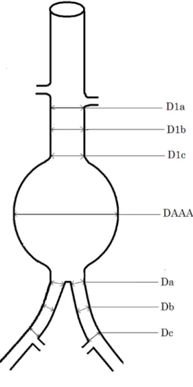
center of 40HU were applied. At the aortic neck, diameters were measured at the subrenal
aorta (D1a), 15mm below the lowest renal artery (D1b), origin of the aneurysm (D1c), as well
as the greatest diameter of the aneurysm (DAAA) (Fig 1). At the iliac artery level, the
diameters were take at the origin (Da), middle (Db), and bifurcation (Dc). For patients in
whom the end of the endograft did not correspond with the iliac bifurcation, a landmark was
positioned and then recorded on the preoperative CT, to ensure that the diameter Dc would be
measured at exactly the same position.
Intervention
The diameters of the implanted prosthesis conformed to the manufacturers' instructions with
16±9% oversizing at the aortic neck and 8±7% at the iliac arteries. An aorto-bi-iliac
endoprosthesis was implanted (54 patients) when the diameter of the aortic bifurcation
was used (seven patients). The proximal extremity of the endoprothesis was implanted close
to the renal arteries and its distal extremity as close to the iliac bifurcation as possible.
Different endoprostheses were used: 31 (51%) Talent Medtronic (World Medical/Medtronic,
Sunrise, FL, U.S.A), 23 (38%) Zenith Cook (William Cook Europe, Biaeverskow, Denmark),
6 (10%) Excluder Gore-Tex (WL Gore and Associates, Flagstaff, AZ, U.S.A), and one (1%)
Anaconda Sulzer-Vascutek (Edinburgh, U.K).
Follow-up
For the purpose of this study, CT scans taken prior to the intervention, at 1 month, and at last
follow-up were analyzed. The control scans followed the same procedure as the preoperative
scans, but in addition to the acquisition at the arterial phase, another at 60 seconds was
obtained in order to visualize late-phase, low-flow endoleaks. For the control scans, all
preoperative diameters were taken again, and where applicable, endoleaks noted and
migration length measured. In total, 61 proximal necks and 115 iliac arteries were analyzed.
CT analysis
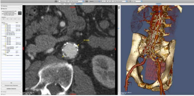
All pre- and post-operative imaging was analyzed using the dedicated program, Endosize
(Therenva©), which had previously been validated by our department (Fig.2) 7. All diameters
were measured perpendicularly to the central line, from adventitia to adventitia, by the same
person.
Statistical analysis
Statistical analysis was performed with SAS statistical software V9.2 (SAS Institute, Cary,
NC, USA). Data are presented as means±SD for quantitative variable unless otherwise noted,
and as numbers with corresponding percentages for qualitative variables. Evolution with time
CT scan taken as baseline values. Separate analyses according to the type of endoprostheses
were also performed. Correlations between growths of different aortic and iliac
measurements, and between growth of aortic neck and baseline characteristics, were
calculated by use of the Spearman correlation coefficient. Subgroup analyses were performed
between patients with aortic aneurysm regression >10% (group A) and those without (group
B). Comparisons of the evolution with time of mean aorto-iliac measures between the two
subgroups were performed by use of a two-way (time, group) ANOVA. For each of the
different endoprostheses, the evolution of each diameter was analyzed using the
Kriskal-Wallis and the Mann-Whitney test. The cumulative proportion of patients with a proximal
neck evolution greater than 20% was assessed by means of a Kaplan-Meier analysis. For all
analyses, a P value < 0.05 was considered to be significant.
RESULTS
Demographics
The general characteristics of the patients included in the study are shown in table 1. The
main risk factor in our patients was the coronary risk.
Type 1 endoleaks and secondary interventions
In our population, one patient (1.6%) with a distal endoleak was treated using iliac extension
because he had presented a 5mm progression of the anchor zone with a secondary retraction
at the bifurcation level, with a commune iliac artery measuring 16mm prior to the intervention
without associated iliac aneurysm. Another patient (1.6%) with proximal endoleak was
treated using an aortic cuff because he had presented a 10mm migration (endoprosthesis
Aortic neck
The three diameters taken at the proximal neck increased over time (Fig.3, Table 2), with a
mean increase of 3.7±2.8mm for D1a, 4.4±2.5mm for D1b, and 4.4±3.1 mm for D1c. This
increase was homogeneous across the three levels as there was a significant correlation
between the three levels, with P=0.001 between D1a and D1b, P<0.0001 between D1a and
D1c, and P<0.0001 between D1b and D1c (Fig.4). The increase in the proximal neck
appeared to be more marked at the level closest to the aneurysm than at the level of the renal
arteries (Table 2). When the first month post-implant CT scan was taken as a reference, the
observed dilatation of the aortic neck was also significant: D1a increased by 8.0±7.8%
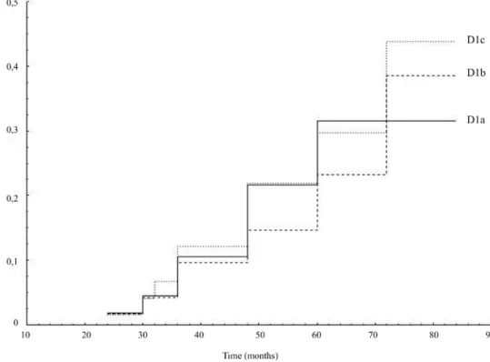
(P<0.0001), 10±8.7% for D1b (P<0.0001), and 10±8.9% for D1c (P<0.0001). The percentage
of patients with an increase in aortic neck diameter greater than 20% was 11.5% for D1a,
13.1% for D1b and 14.8% for D1c (Fig. 5). No baseline risk factor was found to be correlated
with an aortic neck evolution greater than 20%.
Iliac arteries
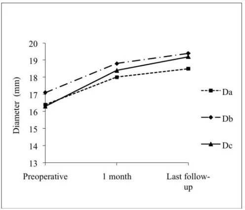
The three iliac artery diameters significantly increased over time (Fig.6, Table 2), with a mean
increase of 2.1±0.2mm for Da, 2.5±0.5mm for Db, and 3±0.7mm for Dc. Similarly, the
dilatation homogeneously affected the iliac artery as significant correlations were found
between the three levels, with P<0.0001 between Da and Db (Fig.7), P=0.004 between Da and
Dc, and P=0.001 between Db and Dc. The increase at the level of the iliac arteries was
distally more marked than at its origin (Table 2). When the first month post-implant CT scan
was taken as a reference, the observed dilatation of the iliac artery was still significant for all
diameters: Da increased by 6±10% (P<0.0001), 8±11% for Db (P<0.0001), and 12±13% for
Dc (P<0.0001). The percentage of patients with an increase in iliac artery diameter greater
Correlation between aortic neck and iliac arteries
The increase in the three measurements at the proximal neck was compared with that
observed at the iliac artery level (Fig.8, Table 3). No significant correlation was found
between the diameter increase at the proximal level and that at the iliac artery level, with the
exception of D1b and Dc (P=0.006), which showed a weak correlation (r=0.363).
Correlation between neck dilatation and baseline characteristics
Only D1a and Dc diameters, on which the choice of endoprosthesis diameter was based, were
found to be correlated with the oversizing. A significant but weak correlation was observed
between the progression of D1a and the oversizing (r=0.296, P=0.023), and between the
evolution of Dc and the oversizing (r=0.279, P=0.004). No correlation was found between the
evolution of D1a and the preoperative neck diameter (P=0.242), or the preoperative AAA sac
size (P=0.71).
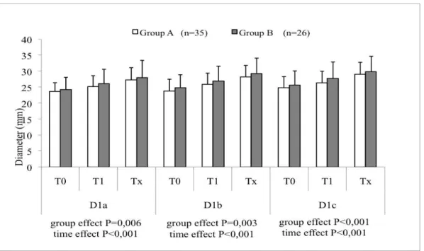
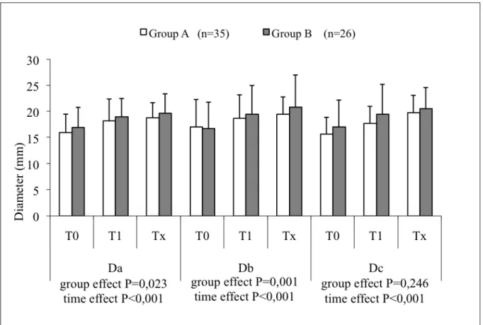
Subgroups analysis
In Group A (n=35), a significant increase (P<0.001) in all diameters was observed over time
at both the proximal neck and iliac artery necks (Fig. 9 and 10), which was also the case for
Group B (n=26) (P<0.001). When comparing the two groups, the increase was statistically
more marked in Group B for all diameters with the exception of the iliac bifurcation diameter.
Separate analysis of each type of endoprosthesis showed a significant difference at the aortic
neck (Fig. 11) for the three diameters (P=0.023 for D1a, P=0.021 for D1b and P=0.004 for
D1c). Although no difference was noted between the Talent and Zenith devices (P=0.164),
there was a moderate difference between the Talent and Excluder devices (P=0.022), and
between the Zenith and Excluder devices (P=0.042). At the iliac artery, no difference was
DISCUSSION
Currently, scarce data is available regarding the long-term progression of distal necks
following EVAR, whereas proximal necks have been extensively investigated in a number of
studies3,4,8-9. However, most studies conducted to date used different methodologies, and
differing results were observed. Badran et al.4 took the measurements 7.5mm below the
lowest renal artery using axial slices; therefore, in cases of iliac tortuosities, the diameter
taken on the image was smaller. We believe that this measuring method is not accurate, due to
an obvious parallax error, which cannot only be corrected by taking into account the smallest
diameter. For this reason, we measured all diameters perpendicular to the central line, which
is a reproducible10 and well-accepted method. In the study of Badran et al4, neck dilatation
during the first 2 years of follow-up was possibly linked to oversizing, after which, in their
opinion, further progression of parietal wall degeneration may come into play. Napoli et al.8
found no correlation between the suprarenal and infrarenal necks, while showing that neck
dilatation affected only 33% of the patients following EVAR. In contrast to this, our results
indicate that dilatation affects all patients, which is in line with the observation of Monahan et
al.11. Soberon et al.3 considered that dilation due to oversizing occurred mainly at 6 months.
Cao et al.12 identified the following factors predictive of neck dilatation: presence of
circumferential thrombus, preoperative neck diameter, and maximal AAA diameter. With
respect to this last parameter, the study by Dillavou et al.9 showed that the dilatation of the
neck was just as marked as the preoperative diameter was small (cut-off 25 mm). In our study,
the dilatation of the proximal neck seemed to homogenously affect the entire area rather than
just the zone immediately below the renal arteries. This is, in theory, the reference diameter
used to calculate the implemented prosthesis, and thus oversizing. Thus, the progression of
diameters D1b and D1c cannot be accounted for by oversizing. The heterogeneity of the
Diehm et al13, explain the origin of the highly variable results reported for AND. In order to
harmonize the clinical and morphologic outcomes following EVAR, the Society of Vascular
Surgery and the International Society of Vascular Surgery have published reporting
standards14, recommending the use of the first set of postoperative images. We thus compared
the first and last CT scan measurements, in addition to the ANOVA analysis. In both cases,
the dilatation of the necks was significant. To characterize AND, assessment of the full
proximal landing zone is necessary13. It is therefore recommended to use the AAA neck
volumetry for the assessment of AND15. Although the Endosize software has not been
designed to perform volumetric analyses of AAA, its algorithm could be used for this
purpose. We thus measured three different diameters, along the full proximal neck (and the
iliac artery). In our study, these diameters were shown to increase significantly over time, the
increase being more marked in the proximal zones of the aneurysm, as shown in Fig. 5. From
a physiological point of view, this kind of progression may point to a gradual extension of the
aneurysmal disease. This hypothesis was partially demonstrated by Diehm et al16, by means
of a histological and biochemical analysis. They determined in “seemingly non-diseased
infrarenal AAA neck” a number of histological signs of destruction and biochemical
disorders, which could explain the appearance of AND. This explanation would also apply to
patients presenting an aneurysm growth over time. Nevertheless, dilatation of the proximal
neck also affected patients exhibiting aneurysmal regressions, as shown by the results of
subgroup analyses. Therefore, while the difference between both groups was significant, more
relevant was the fact that in patients with aneurysm retraction on imaging, neck dilatation
could still be evidenced at all levels, suggesting that EVAR settles the mechanical17, but not
the biological aspects of AAA. In our series, there were not enough cases of proximal
endoleaks to draw any conclusions as to a potential correlation between both parameters,
the proximal neck, as shown in our own series. In line with this observation, Monahan et al.11
concluded that the dilatation of the proximal neck was not correlated to Type I endoleaks or
migrations.
Scientific literature on distal necks is rather scarce. For conventional AAA surgery, the
question has already been raised as to whether associated ectatic iliac arteries should be
treated simultaneously. In the retrospective study of Sala et al.18, the authors proposed to treat
routinely all patients with ectactic common iliac arteries larger than 18mm and a
life-expectancy of at least 7-8 years. Regarding endovascular AAA treatment, several studies have
attempted to demonstrate that patients with ectatic iliac arteries at the distal anchor zone could
be treated efficiently without further postoperative complications by using either the bell
bottom19 or standard endoprosthesis20, without loss of the hypogastric artery21. However, in
the medium-term, Mc Donnell et al22 found a 7% rate of distal endoleaks in patients with iliac
arteries larger than 16mm. It should further be mentioned that only a few articles reported
exclusively the evolution of normal and pathological iliac arteries over time. Falkensammer et
al.5 showed that dilatation of the distal anchor zone, while present in all patients, was more
marked in patients with concomitant iliac aneurysm, but was not associated with an increased
rate of endoleaks or reinterventions23,24, which is in contradiction with the findings of other
studies25,26. In addition, Adiseshiah et al.27 highlighted that long-term follow-up of these areas
was critical, as aneurysmal evolution was more likely to occur later in time in distal necks
than in proximal necks.
Our study yielded similar results, showing a significant increase over time in iliac artery
measurements at the three levels. Similarly to the proximal neck, there appears to be a
progression in all iliac artery diameters in patients presenting aneurysmal regression. This
trend, however, has to been put into perspective because, even if the analysis revealed a
insufficient number of distal endoleaks. As for the proximal neck, it may be assumed that the
parietal degeneration process is likely to extend to the iliac arteries progressively. However,
the correlation analysis revealed that diameter progressions of the distal and proximal necks
were an independent phenomenon and the increase at the level of the iliac arteries was distally
more marked than at its origin. A tentative explanation of these findings is based on
alterations in parietal hemodynamic constraints due to the endoprosthesis. In fact, the
increase in pressure was shown to be more marked at the level of the iliac bifurcation than at
the proximal neck28, 29, and this difference was more pronounced when the vessels were long
and tortuous30. Likewise, wall shear stress was shown to be more relevant at areas of
overlap28, as well as in the kinking zones of the endoprothesis. It seems likely that the
presence of the endoprosthesis, in addition to decreasing pressure in the aneurysmal sac, also
alters the constraints at the level of the iliac arteries with a more significant stress and
pressure as compared to the preoperative period. It should be noted, however, that this
hemodynamic modification alone is not sufficient to explain the results we observed with
respect to iliac artery dilatation.
The evolution with each endoprosthesis appears to be similar in our study. There was a
difference at the aortic neck only with the Excluder device, suggesting that AND is related to
supra or infra-renal fixation31. Nevertheless, in our study the number of patients treated with
the Excluder device is too small to draw any conclusions on the effects of supra or infra-renal
fixation. No difference was found between the Talent and Zenith devices, and in both cases
there was a dilatation at the aortic neck, in agreement with the findings of Badger et al32.
Overall, our results show a trend towards a neck dilatation incidence rate, which is greater
than that observed by other authors. Concerning the level of accuracy of the measurements,
the observed differences, which are only slightly greater, sometimes by only one or two
sufficient to affect the results of a statistical test. Although we used a 3D reconstruction based
on the use of spiral CT images, intra- or inter-observer variabilities could lead to difficulties,
especially with measurements requiring an accuracy of one millimeter. As most of the studies
investigating AND or iliac evolution do not make use of software with an automated
centerline extraction, we expected that by using the Endosize software, this type of variability
would be reduced. In order to reduce the measurement errors related to image quality, we
included only those patients of whom high quality images had been recorded in our hospital.
This was important, since the same acquisition parameters, in particular the slice thickness,
are not always used in other establishments. Moreover, Wever et al33 also showed that, for all
patients, the proximal neck demonstrates continued dilatation during follow-up, with a
median increase of 15.5% (cross-sectional area) at 12 months.
Our study was directly focused on the final status of necks without taking into account
intermediary scans except for the immediate postoperative scan. Our aim was not to
investigate the kinetics of progression but rather the potential correlations between the
progressions in the different anchor zones of the endoprosthesis. In spite of the retrospective
nature of the study design, this trend towards dilatation, which was even observed in
successfully-treated patients, is a new finding that must be taken into account as it raises the
question as to the modifications of native arteries caused by the endoprosthesis itself.
Presently, not enough time has passed, and there have been too few clinical events to allow us
to understand whether there is an implication on the occurrence of distal endoleaks and
aneurysms on the landing zones. This highlights the need for a sufficiently long follow-up for
recovered patients (young patients). To confirm these results, further long-term studies are
No competing interest declared
REFERENCES
1. Investigators UKET, Greenhalgh RM, Brown LC, Powell JT, Thompson SG, Epstein D,
et al. Endovascular versus open repair of abdominal aortic aneurysm. N Engl J Med
2010;362:1863-71.
2. Investigators UKET, Greenhalgh RM, Brown LC, Powell JT, Thompson SG, Epstein D.
Endovascular repair of aortic aneurysm in patients physically ineligible for open repair. N
Engl J Med 2010;362:1872-80.
3. Soberón AB, de Garcia MM, Möll GG, Vigil BR, Krauel MA, Alvarez-Sala Walter R.
Follow-up of aneurysm neck diameter after endovascular repair of abdominal aortic
aneurysms. Ann Vasc Surg 2008;22:559-63.
4. Badran MF, Gould DA, Raza I, McWilliams RG, Brown O, Harris PL, et al. Aneurysm
neck diameter after endovascular repair of abdominal aortic aneurysms. J Vasc Interv
Radiol 2002;13:887-92.
5. Falkensammer J, Hakaim AG, Andrew Oldenburg W, Neuhauser B, Paz-Fumagalli R,
McKinney JM, et al. Natural history of the iliac arteries after endovascular abdominal
aortic aneurysm repair and suitability of ectatic iliac arteries as a distal sealing zone. J
Endovasc Ther 2007;14:619-24.
6. Long A, Perez-Niddam K, Maisonneuve H. Abdominal aortic aneurysm repair treated with
endoprosthesis: technical and economic evaluation by ANAES (National Agency of
Health Accreditation and Evaluation). J Mal Vasc 2000;25:263-9
7. Kaladji A, Lucas A, Kervio G, Haigron P, Cardon A. Sizing for endovascular aneurysm
repair: clinical evaluation of a new automated three-dimensional software. Ann Vasc Surg
2010;24:912-20.
the proximal aortic neck enlargement following endovascular repair of abdominal aortic
aneurysm: 3-years experience. Eur Radiol 2003;13:1962-71.
9. Dillavou ED, Muluk S, Makaroun MS. Is neck dilatation after endovascular aneurysm
repair graft dependent? Results of 4 US Phase II trials. Vasc Endovascular Surg
2005;39:47-54.
10. Velazquez OC, Woo EY, Carpenter JP, Golden MA, Barker CF, Fairman RM. Decreased
use of iliac extensions and reduced graft junctions with software-assisted centerline
measurements in selection of endograft components for endovascular aneurysm repair. J
Vasc. Surg 2004;40:222-7.
11. Monhan TS, Chuter TAM, Reilly LM, Rapp JH, Hiramoto JS. Long-term follow-up of
neck expansion after endovascular aortic aneurysm repair. J Vasc Surg 2010;52:303-7
12. Cao P, Verzini F, Parlani G, Rango PD, Parente B, Giordano G, et al. Predictive factors
and clinical consequences of proximal aortic neck dilatation in 230 patients undergoing
abdominal aorta aneurysm repair with self-expandable stent-grafts. J Vasc Surg
2003;37:1200-05.
13. Diehm N, Dick F, Katzen BT, Schmidli J, Kalka C, Baumgartner I. Aortic neck dilatation
after endovascular abdominal aortic aneurysm repair : A word of caution. J Vasc Surg
2008;47:886-92
14. Chaikof EL, Blankensteijn JD, Harris PL, White GH, Zarins CK, Bernhard VM, et al.
Reporting standards for endovascular aortic aneurysm repair. J Vasc Surg
2002;35:1048-60
15. Diehm N, Kickuth R, Gahl B, Do DD, Schmidli J, Rattunde H, et al. Intraoberver and
interobserver variability of 64-row computed tomography abdominal aortic aneurysm
neck measurements. J Vasc Surg 2007;45:263-8
damage of the seemingly non-diseased infrarenal aortic aneurysm neck. J Vasc Surg
2008;48:425-34
17. Diehm N, Dick F, Katzen BT, Do DD, Baumgartner I. Endovascular repair of abdominal
aortic aneurysms: Only a mechanical solution to a biological problem ? J Endovasc Ther
2009;16(Suppl 1):119-26
18. Sala F, Hassen-Khodja R, Branchereau P, Berthet J, Batt M, Mary H, et al. Outcome of
common iliac arteries after aortoaortic graft placement during elective repair of infrarenal
abdominal aortic aneurysms. J Vasc Surg 2002;36:982-7.
19. Karch LA, Hodgson KJ, Mattos MA, Bohannon WT, Ramsey DE, McLafferty RB.
Management of ectatic, nonaneurysmal iliac arteries during endoluminal aortic aneurysm
repair. J Vasc Surg 2001;33:33-38.
20. Timaran CH, Lipsitz EC, Veith FJ, Chuter T, Greenberg RK, Ohki T, et al. Endovascular
aortic aneurysm repair with the Zenith endograft in patients with ectatic iliac arteries. Ann
Vasc Surg 2005;19:161-6.
21. England A, Butterfield JS, McCollum CN, Ashleigh RJ. Endovascular aortic aneurysm
repair with the talent stent-graft: outcomes in patients with large iliac arteries. Cardiovasc
Intervent Radiol 2008;31:723-7.
22. McDonnell CO, Semmens JB, Allen YB, Jansen SJ, Brooks DM, Lawrence-Brown
MMD. Large iliac arteries: a high-risk group for endovascular aortic aneurysm repair. J
Endovasc Ther 2007;14:625-9.
23. Parlani G, Zannetti S, Verzini F, De Rango P, Carlini G, Lenti M, et al. Does the presence
of an iliac aneurysm affect outcome of endoluminal AAA repair? An analysis of 336
cases. Eur J Vasc Endovasc Surg 2002;24:134-8.
24. Kirkwood ML, Saunders A, Jackson BM, Wang GJ, Fairman RM, Woo EY. Aneurysmal
aneurysm repair for abdominal aortic aneurysm. J Vasc Surg 2010;Available from:
http://www.ncbi.nlm.nih.gov/pubmed/21030200
25. Albertini J, Favre J, Bouziane Z, Haase C, Nourrissat G, Barral X. Aneurysmal extension
to the iliac bifurcation increases the risk of complications and secondary procedures after
endovascular repair of abdominal aortic aneurysms. Ann Vasc Surg 2010;24:663-9.
26. Hobo R, Sybrandy JEM, Harris PL, Buth J. Endovascular repair of abdominal aortic
aneurysms with concomitant common iliac artery aneurysm: outcome analysis of the
EUROSTAR Experience. J Endovasc Ther 2008;15:12-22.
27. Adiseshiah M, Boardley D, Raphael MJ. Late iliac artery aneurysm formation:
implications for the lower landing site after EVAR. J Endovasc Ther 2008;15:246-7.
28. Molony DS, Callanan A, Kavanagh EG, Walsh MT, McGloughlin TM. Fluid-structure
interaction of a patient-specific abdominal aortic aneurysm treated with an endovascular
stent-graft. Biomed Eng Online 2009;8:24.
29. Figueroa CA, Taylor CA, Yeh V, Chiou AJ, Zarins CK. Effect of curvature on
displacement forces acting on aortic endografts: a 3-dimensional computational analysis.
J Endovasc Ther 2009;16:284-94.
30. Frauenfelder T, Lotfey M, Boehm T, Wildermuth S. Computational fluid dynamics:
hemodynamic changes in abdominal aortic aneurysm after stent-graft implantation.
Cardiovasc Intervent Radiol 2006;29:613-23.
31. Leurs LJ, Stultiëns G, Kievit J, Buth J, EUROSTAR collaborators. Adverse events at the
aneurysmal neck identified at follow-up after endovascular abdominal aortic aneurysm
repair: how do they correlate ? Vasc 2005;13:261-7
32. Badger SA, O'Donnell ME, Loan W, Hannon RJ, Lau LL, Lee B et al. No difference in
medium-term outcome between Zenith and Talent stent-grafts in endovascular aneurysm
33. Wever JJ, de Nie AJ, Blankensteijn JD, Broeders IAMJ, Mali WPThM, Eikelboom BC.
Dilatation of the proximal neck of infrarenal aortic aneursyms after endovascular AAA
TABLES
Table 1. Demographic and clinical characteristics of the patients
Total Population (n=61)
Age (years; mean ± SD) 74,6 ± 8,3 Obesity (BMI>30) 8 (13,1%) Coronary artery lesions 26 (42,6%) Coronary artery by pass graft 10 (16,4%) Aortic valve replacement 3 (4,9%) Critical limb ischemia 2 (3,3%) Severe respiratory insufficiency 2 (3,3%) End-stage renal failure 1 (1,6%) Poorly-controlled dyslipidemia 13 (21,3%) Poorly-controlled arterial hypertension 5 (8,2%) Active smoker 8 (13,1%)
Diabetes 5 (8,2%)
Table 2. Aortic measures. Mean ± standard deviation (range) and P value from ANOVA analysis.
Diameter Preoperative 1 month Last follow-up Growth P value D1a (n=61) 23.9±3.3 (17; 26) 25.6±4 (19; 43) 27.6±4.6(20; 48 3.7±2.8(-2; 12) 0.018 D1b (n=56) 24.3±3.9 (18; 38) 26.3±4.1 (20; 43) 28.7±4.3(22; 44 4.4±2.5(-1; 12) 0.0156 D1c (n=61) 25±4 (18; 35) 27±4.4 (20; 46) 29.4±4.3(21; 41 4.4±3.1(-3; 12) 0.0358 Da (n=115) 16.4±3.6 (11; 30) 18±4 (13; 39) 18.5±3.3(11; 33 2.1±0.2(2; 3) 0.0006 Db (n=115) 16.9±5.2 (11; 48) 18,8±5 (12; 51) 19.4±4.8(10; 53 2.5±0.5(1-3) 0.0005 Dc (n=115) 16.2±4.2 (9; 48) 18.5±4.6 (12; 53) 19.2±3.6(11; 32 3±0.7(1; 4) 0.0007 DAAA (n=61) 55±7.7 (42; 83) 54.5±7 (40; 74) 49±12.6 (20; - 6±11(-34; 14)
Table 3. Correlation between proximal aortic neck growth and iliac growth. Spearman coefficient (r) and P value.
Diameters D1a D1b D1c Da r=0.086 r=0.095 r=0.221 P=0.515 P=0.489 P=0.09 Db r=0.051 r=0.231 r=0.237 P=0.699 P=0.09 P=0.068 Dc r=0.213 r=0.363 r=0.214 P=0.102 P=0.006 P=0.101
FIGURES
Fig 2. CTA analysis (Endosize, Therenva©). Outer-to-outer diameters were measured
perpendicularly to the center-line
Fig 4. Correlation between growth of D1b and D1c (aortic neck)
Fig 6. Evolution of distal landing site diameters
Fig 8. Correlation between growth of D1b and Da
Fig 9. Proximal neck: mean±standard deviation at each time point (T0: preoperative; T1: 1
month; Tx: last follow-up) for Group A and Group B. The p value for the variables “Group”
Fig 10. Illiac arteries: mean±standard deviation at each time point (T0: preoperative; T1: 1
month; Tx: last follow-up) for Group A and Group B. The p value for the variables “Group”
and “Time” (two-way ANOVA analysis)
Fig 11. Evolution (percentage) of diameters with respect to each endoprosthesis used