HAL Id: hal-01100888
https://hal.archives-ouvertes.fr/hal-01100888
Submitted on 27 May 2020
HAL is a multi-disciplinary open access
archive for the deposit and dissemination of
sci-entific research documents, whether they are
pub-lished or not. The documents may come from
teaching and research institutions in France or
abroad, or from public or private research centers.
L’archive ouverte pluridisciplinaire HAL, est
destinée au dépôt et à la diffusion de documents
scientifiques de niveau recherche, publiés ou non,
émanant des établissements d’enseignement et de
recherche français ou étrangers, des laboratoires
publics ou privés.
Distributed under a Creative Commons Attribution| 4.0 International License
Finding and identifying the viral needle in the
metagenomic haystack: Trends and Challenges
Hayssam Soueidan, Louise-Amélie Schmitt, Thierry Candresse, Macha
Nikolski
To cite this version:
Hayssam Soueidan, Louise-Amélie Schmitt, Thierry Candresse, Macha Nikolski. Finding and
identify-ing the viral needle in the metagenomic haystack: Trends and Challenges. Frontiers in Microbiology,
Frontiers Media, 2014, 5, pp.5:739. �10.3389/fmicb.2014.00739�. �hal-01100888�
Finding and identifying the viral needle in the
metagenomic haystack: trends and challenges
Hayssam Soueidan1,2, Louise-Amélie Schmitt1,3
, Thierry Candresse4,5
and Macha Nikolski1,3
*
1
Bordeaux Bioinformatics Center, Université de Bordeaux, Bordeaux, France
2INSERM U1035, Université de Bordeaux, Bordeaux, France 3
Centre National de la Recherche Scientifique/Laboratoire Bordelais de Recherche en Informatique, Université de Bordeaux, Talence, France
4
Institut National de la Recherche Agronomique, UMR 1332 Biologie du Fruit et Pathologie, Villenave d’Ornon, France
5
UMR 1332 Biologie du Fruit et Pathologie, Université de Bordeaux, Villenave d’Ornon, France
Edited by:
Bas E. Dutilh, Radboud University Medical Center, Netherlands Reviewed by:
Ivan Merelli, Institute for Biomedical Technologies, Italy
Simon Roux, University of Arizona, USA
*Correspondence: Macha Nikolski, Bordeaux Bioinformatics Center, Université de Bordeaux, 146 rue Léo Saignat, 33076 Bordeaux, Bordeaux, France e-mail: macha.nikolski@labri.fr
Collectively, viruses have the greatest genetic diversity on Earth, occupy extremely varied niches and are likely able to infect all living organisms. Viral infections are an important issue for human health and cause considerable economic losses when agriculturally important crops or husbandry animals are infected. The advent of metagenomics has provided a precious tool to study viruses by sampling them in natural environments and identifying the genomic composition of a sample. However, reaching a clear recognition and taxonomic assignment of the identified viruses has been hampered by the computational difficulty of these problems. In this perspective paper we examine the trends in current research for the identification of viral sequences in a metagenomic sample, pinpoint the intrinsic computational difficulties for the identification of novel viral sequences within metagenomic samples, and suggest possible avenues to overcome them.
Keywords: microbial metagenomics, NGS, virome, host—pathogen interactions, taxonomic assignment
INTRODUCTION
While genomics is the research field relative to the study of the genome of any organism, metagenomics is the term coined for the research that focuses on many genomes at the same time, as typical in some sections of environmental studies. The analysis of microbial communities has been until recently a complicated if not untractable task due to their high diversity and to the fact that many of these organisms cannot be cultured. Harnessing the major advances achieved in sequencing technologies, metage-nomics has emerged as the only currently available approach to extensively characterize these largely unculturable communities. Besides vastly enriching our knowledge of microbial diversity in a varied range of environments, and providing information on the dynamics and on the overall functioning of microbial communi-ties, metagenomics is also shedding light on many important bio-logical processes and, in particular, on the role of the microbiome in biological functions essential for the development of higher order organisms harboring it (Blottière et al., 2013; Manor et al., 2014), or in the development of pathological problems (Cénit et al., 2014; Vayssier-Taussat et al., 2014). In addition, metage-nomic efforts also vastly enrich the repertoire of genes available for biotechnological applications (Ni and Tokuda, 2013).
At the same time metagenomics extensively relies on bioin-formatics to tackle the huge amounts of sequence data involved, and recognizes the need to develop computational methods that maximize our understanding of the genetic composition and the biological activities expressed in communities so complex that they can only be sampled, never completely characterized. Computational analysis has become a genuine bottleneck for
metagenomics due not only to the large amount of sequence data, but also to the new questions such as, for example, the need for simultaneous assembly of multiple genomes or tran-scriptomes and the analysis of complex networks of host-microbe interactions (Wooley and Yuzhen, 2009).
In this context the analysis of viral communities presents par-ticular interest but also computational challenges. The ability to thoroughly analyze the viral composition of an environmental sample is of paramount importance, in particular because viruses have turned out to play a major role in the functioning of micro-bial communities by processes such as viral infection and selective killing of certain taxa or as vectors for horizontal gene transfer (Suttle, 2007). Consequently, the viral part of the microbiome has been shown in a number of situations to have a major impact on the dynamics and on the evolutionary processes of their host pop-ulations. The discovery and classification of novel viral species, but also of higher order taxa, is therefore of particular interest in this context (Rosario and Breitbart, 2011).
One of the main goals of metagenomic projects is to char-acterize the microbial communities in terms of the identity and diversity of species present (species richness) in a given environ-ment. When it comes to species identification, the task is called taxonomic assignment. Current NGS technologies have provided an opportunity for doing this analysis routinely (Petrosino et al., 2009). Software tools for automated taxonomic assignment for organisms such as bacteria and fungi have since become a mature technology and are now routinely used in many studies.
If bacterial or fungal applications have recently seen major advances, the problem of taxonomic assignment for
Soueidan et al. The viral needle in the metagenomic haystack
viruses—such as it arises in environmental studies—remains largely unsolved from the computational point of view, as exemplified by the difficulty of distinguishing viral genomes from eukaryotes and bacteria observed in some studies (Bazinet and Cummings, 2012). Indeed, ab-initio identification of a sequence as belonging to a cellular organism or to a virus remains a complicated task outside of the popular sequence-homology based approaches that rely on direct comparisons with already known viral sequences present in international databases.
We distinguish the task of deciding to which first-level domain (eukaryotes, bacteria, archaea, virus) a given sequence belongs—that we call first-level assignment—from a more fine-grained taxonomic assignment at, e.g., family, genus or species level. In virome studies the latter task is greatly facilitated when targeted sequencing of purified viral particles is per-formed (Hall et al., 2014), but the former is particularly dif-ficult for complex samples containing both eukaryotic and viral sequences and when, as is very frequently the case, unknown viral species are present. In this paper we examine reasons behind this difficulty and suggest possible avenues to overcome them.
FIRST-LEVEL CLASSIFICATION OF COMPLEX ENVIRONMENTAL SAMPLES
The first-level assignment of sequence data coming from a non-targeted sequencing of a metagenomic sample is a particularly challenging computational problem. The most blatant difficulty is in the recognition of novel viral sequences, for which no close homologs have been previously characterized. This question is however of paramount importance for the biologists. From the biodiversity point of view, the identification of unknown viruses representing novel higher order taxa (genera, families. . . ) is of clear interest as evidenced, for example, by the discovery of the Mimiviruses with genomes exceeding in size those of many bac-terial genomes (Claverie et al., 2009). But this question can also have important practical implications as when trying to identify novel viruses responsible for particular syndromes or diseases in humans, plants or husbandry animals (Roossinck, 2012; Lecuit and Eloit, 2014).
A number of bioinformatics methods efficiently per-form the first-level assignment of sequences from a sample mainly containing known species. Computational solu-tions can be broadly organized in two main categories: (1) sequence similarity methods and (2) sequence composition methods.
Methods that rely on sequence similarity can be themselves subdivided in alignment-based techniques (mostly attempting to improve BLAST accuracy) and index-based. Alignment-based methods suffer from two limitations: speed and lack of sensitivity (e.g.,Bazinet and Cummings, 2012; Wood and Salzberg, 2014). Recently, novel solutions have been suggested to overcome these limitations. These methods are based on long k-mers (words of size k) and conceptually rely on the fact that when k is sufficiently large, k-mers become very specific. Consequently, the idea is to index the databases by long k-mers. This is indeed the founda-tion of MegaBlast (a general-purpose sequence aligner using long
seeds), but also of a number of methods specific for taxonomic assignment such as LMAT (Ames et al., 2013) and Kraken (Wood and Salzberg, 2014). The downside of these approaches is over-specificity, which makes classification of unknown sequences problematic. This limitation can be particularly acute given the known very high intraspecific variability existing in some viral species or higher order taxa. For example, current criteria of the International Committee for the taxonomy of viruses tolerate up to 28% of nucleotide sequence divergence for the polymerase or capsid protein genes for isolates of a same species in the
Betaflexiviridae family and a similar level of divergence at the
whole genome level in the Potyviridae family (King et al., 2012). A complementary approach is based on sequence composition analysis. Such methods rely on the decomposition of sequences into frequencies of short k-mers and make use of machine learn-ing techniques (e.g., SVM, kNN, Naive Bayes, etc.) to train a clas-sifier on a reference database. The taxonomic assignment of novel sequences is then predicted by applying the pre-trained model. These methods theoretically are better suited to the task of novel species classification as short k-mers distributions are less prone to over-fitting. However, even these techniques fail to classify about 50% of species absent from the training set (Nalbantoglu et al., 2011). This is especially salient for viral sequences, as the vast majority of them fail to be uniquely assigned to any domain of life (Rosen et al., 2010).
Recent results show that contig-level assembly improves the strength of the taxonomic signal contained in individual short reads, even in the case of increased chimericity (Mende et al., 2012; Teeling and Glockner, 2012). This is why in our experi-mental evaluation (see Section Why is the First-level Assignment Problem Hard?) we work exclusively with sequence lengths that are comparable to contigs obtained by a standard metagenomic assembly step when the data originate from complex biological communities.
In summary, even the simple goal to provide a first-level description of a sample composition and be able to reveal if viral sequences are present, has been eluding a satisfactory solu-tion. Indeed, for viral (and also eukaryotic) sequences, none of the existing methods produces a taxonomic distribution that is even remotely close to the expected one (Bazinet and Cummings, 2012).
FINE-GRAINED CLASSIFICATION FOR BACTERIAL AND VIRAL COMMUNITIES
On the other side of the spectrum, the problem of fine-grained characterization of datasets produced by targeted sequencing has seen great progress in recent years. Contrary to the anal-ysis of non-selected and therefore more complex metagenomic samples, efficient methods have been developed for cases where certain components of microbial communities are experimentally targeted (bacterial or viral). This has been an effective way to cir-cumvent the difficulty of the first-level assignment, albeit without solving it.
For bacterial communities the most efficient solution is to per-form a tag survey, where only partial genomic inper-formation is used and the sequencing is performed for marker genes, such as 16S rRNA for prokaryotes and 18S rRNA for eukaryotes (fungi).
This simplifies the analysis for two reasons. First, the amount of data remains reasonable (for a high-throughput analysis) and second, known marker genes’ taxonomic classification is avail-able through reference taxonomies such as RDP (Cole et al., 2009) or Greengenes (McDonald et al., 2012). Sequence similar-ity techniques combined with reference taxonomies recapitulate the known distribution of bacterial phyla extremely well (Bazinet and Cummings, 2012). However, this type of analysis has one major pitfall: it does not provide a reliable method to quantify the identified species (Roux et al., 2011).
While this approach is feasible for bacterial populations, it is not applicable for the analysis of viral communities due to the absence of such marker genes (e.g., Edwards and Rohwer, 2005). Virome studies concentrate on the viral part of the envi-ronmental sample and isolate viral genomes encapsidated in viral particles that are purified by a combination of filtration and (utlra)centrifugation. This now popular approach drastically reduces the complexity of the community, which makes it pos-sible to assemble longer contigs routinely (10 kb and more), and even complete genomes from low-complexity samples (Coetzee et al., 2010; Minot et al., 2012). However, it does not really solve the problem of first-level assignment but merely sidesteps it: given the purification step, all generated sequences are gener-ally considered “by definition” as viral, unless proven otherwise by homology-based approaches. In addition, this strategy is not without some caveats (see for more detailsFancello et al., 2012). For example, the purified particles may contain cellular genome fragments rather than viral genomes, because of the presence of GTA (Lang and Beatty, 2007) or as a consequence of general-ized transduction (for a review seeFrost et al., 2005). Also, while 0.22 µ filtering avoids contamination by bacterial, archaeal or eukaryotic cells, other DNA-containing elements, such as bac-terial vesicles (Biller et al., 2014) may co-purifiy with virions. Such filtering-based purification also excludes the largest viruses and therefore results in an incomplete picture of viral diversity. Moreover, both LA (seeDuhaime et al., 2012) and MDA ampli-fications have their downfalls. For the former, adapter ligation is only possible for dsDNA viruses and hence ssDNA viral genomes are mostly absent in the sample. For the latter, the amplification is preferentially performed for circular ssDNA viruses rather than dsDNA. The effect of the presence of cellular genes on the bioin-formatics analysis of viral metagenomic data has been described and some approaches to detect their presence have been proposed (Roux et al., 2014).
Notwithstanding, virome studies have seen large success. In contrast with bacterial communities, alignment-based methods do not seem to be best suited for viral classification. Indeed, as mentioned by Suttle (2007) even for relatively long viral reads the homolog frequency between these reads and protein sequences within the Genbank database is only about 30%. The idea is to avoid the strong sequential constraint imposed by alignment methods on nucleotides’ similarity and to cap-ture a global similarity signal based on sequence composition (k-mers). Composition-based techniques seem to provide satis-factory results for fine-grained taxonomic classification of filtered viral samples (e.g., Yang et al., 2005; Trifonov and Rabadan, 2010).
WHY IS THE FIRST-LEVEL ASSIGNMENT PROBLEM HARD? As we observed in the previous sections, methods for first-level and fine-grained assignment of metagenomic samples co-exist, but exhibit drastically different performances. This naturally raises the question of reasons underlying this performance gap. Since the characterization of metagenomic samples can be for-mulated as a supervised machine-learning task, we propose here to employ data complexity and hardness measures to compare the intrinsic difficulty of classifying metagenomic samples at the first-level with that of fine-grained assignment.
We consider here three classification tasks whose goals are to assign a class label to each instance of a set of sequences. The three tasks we describe vary by the composition of the set of sequences and by the scope of the class labels to assign.
(1) Given a sample of bacterial sequences, to assign each of them to a phylum (e.g., Proteobacteria) or to a class (e.g.,
Gammaproteobacteria);
(2) Given a sample of viral sequences, to assign each of them to a group (e.g., dsDNA) or to a family (e.g., Plasmaviridae); and (3) Given a sample of sequences, to assign each of them to a
first-level domain (e.g., bacteria, archaea, eukaryota, or virus).
The classification tasks (1) and (2) are fine-grained assignment problems and mimic characterization of targeted metagenomic studies; while task (3) represents a first-level assignment and mimics the analysis of complex, untargeted environmental sam-ples. Since we are interested in the identification of novel species in large metagenomic samples, we adopted the representation of sequences as k-mer frequency vectors.
We analyzed these three classification tasks using an instance-level analysis of data complexity. In supervised machine learning, the performance of a classifier is dependent both on the learning algorithm (e.g., SVM or Naïve Bayes) and on the training data. While global metrics recapitulate overall performances of a clas-sifier, they fail to indicate whether moderate performances are a consequence of wrong parameter adjustments, biased resampling for training data or of the intrinsic difficulty of the classifica-tion task. However, recent literature on instance misclassificaclassifica-tions demonstrates that for a given classification task, some instances are intrinsically hard to classify and that their presence is indica-tive of the global difficulty (seeSmith et al. (2014)for a review). Most studies agree on the hardness of outlier instances or on instances belonging to a minority class, but Smith demonstrated that simple metrics can actually quantify the intrinsic hardness of an instance. One of these metrics is the k-Disagreeing Neighbors (kDN), which measures for a given instance the number of k nearest neighbors that do not share its class label. Smith demon-strated that the kDN measure is strongly positively correlated with the misclassification of an instance over a wide range of learning algorithms and of training data resampling.
To compare the classification hardness of the three tasks, we generated from a representative subset of sequenced organisms from Genbank (September 2014 download, 25,624 bioprojects, 100% of viruses, archaea and bacteria, 24 eukaryotes with 18 plants) 100 sets of 10,000 randomly chosen contiguous genomic
Soueidan et al. The viral needle in the metagenomic haystack
FIGURE 1 | Distribution of kDN by classes for each of three classification tasks. (A) Corresponds to Task 3—assignment of 500 nt
contigs to first-level domains; (B1,B2) to Task 2—assignment of 500 nt viral contigs to a group or to a family, respectively; (C1,C2) to Task 1—assignment of 500 nt bacterial contigs to a phylum or to a class, respectively. Each of the 300,000 randomly selected contigs sampled from different first-level domains were represented as vectors of 3-mer frequencies. Histograms indicate how many contigs (y-axis) per class (colors) have a certain number of neighbors (x-axis) not sharing their own
class label, within the closest 73 neighbors. Neighbors are determined w.r.t. euclidean distance in the space of 3-mer frequencies (cf. Section Why is the First-level Assignment Problem Hard? of main text). For example, there are more than 6000 different archaeal contigs (red bar) not having a single non-archeal contigs in their closest 73 neighbors (red bar corresponding to 0 kDN). The dashed line represents the boundary between contigs easy to classify correctly with a majority vote (to the left of the line) and hard to classify (to the right). Only the top 4 most abundant classes are shown for (B1,C1); and 6 for (B2,C2).
fragments of 500 nt average length (corresponding to the aver-age size of metaver-agenomic contigs to simulate an assembly step). For task (1), only bacterial genomes were considered, for task (2) only viral genomes were considered, while for task (3) a balanced
composition of viruses, archaea, bacteria and eukaryotes were considered. Each sequence was represented as a 3-mer frequency vector (i.e., the number of time each possible 3-nt sub-sequence appears in the contig) and we defined the distance between two
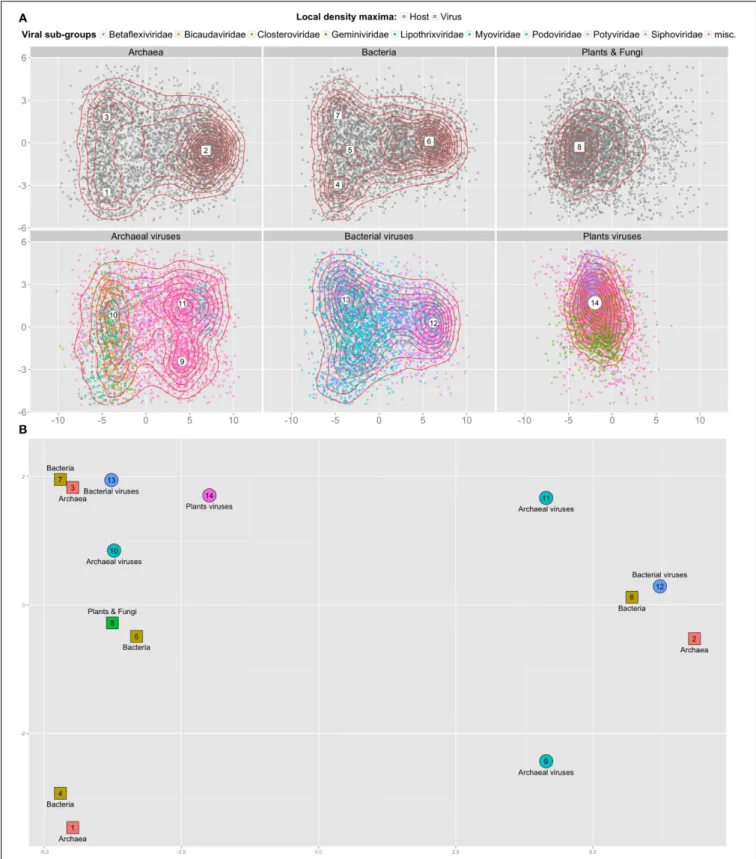
FIGURE 2 | 2D projection of 3-mer frequencies for cellular and viral contigs. (A) Top two dimensions from the PCA reduction of 28,134 contigs
(points) of average length 500 nt represented as frequency vectors of 3-mers; sampled equally from genomes originating from 3 top levels cellular domains (top row) and from 3 viral types known to infect them (bottom row). Dimension 1 (x-axis) accounts for 30% of the variance, dimension 2 (y-axis)
for 8% of the variance. For each sub-panel, 2 d kernel density estimation is represented using red contour lines and local density maxima are numbered within large white shapes. (B) Close up of (A) with all local density maxima. The principal components were computed once for the whole set of contigs of all genomes. Position, coordinates and axes from all sub-panels are comparable.
Soueidan et al. The viral needle in the metagenomic haystack
contig as the Euclidean distance between their respective 64 (43) dimensional vectors. For each contig, its kDN value is the num-ber of other contigs that do not share its class label among its closest 73 neighbors. The corresponding class hardness is then measured as the median kDN of all the contigs in a given class. We also determined whether an observed median kDN is significantly extreme (low value indicating easy classification, high value cor-responding to difficult classes), by estimating the distribution of the median kDN under the null hypothesis of no relation between class labels by random permutations.
We summarize in Figure 1 the distribution of kDN by class for each of three tasks. The upper panel shows that for the first-level classification task, archaeal and bacterial contigs can be easily assigned to their respective domain, and that this classification is hard for eukaryotic contigs and even harder for viral ones. When the classification task is restricted to bacteria only (panels C1 and C2), fine-grained classification is not hard at both phylum and class levels. For viruses (panels B1 and B2), fine-grained classi-fication to groups (ssDNA, dsRNA etc.) is hard, while assigning a viral sequence to a family level is easier, though less easy than for bacteria. Using a permutation scheme, we established that the observed kDN value is significantly different from the null kDN values for all but the virus fine-grained classification to groups (data not shown).
Consistent with previous work (Mende et al., 2012; Teeling and Glockner, 2012), we have verified that for contigs shorter than 500 nt, distributions are shifted to the right—which cor-responds to a harder classification problem (data not shown); conversely, for contigs longer than 500 nt, distributions are shifted to the left, corresponding to an easier classification problem (see Supplementary Figure 1).
It has been previously observed that viral 3-mer signatures are close to that of their hosts (Pride et al., 2006). However, evidence contradicting this observation has also been proposed, for exam-ple for large viruses (Mraìzek and Karlin, 2007) and for viruses of monocots and dicots (Adams and Antoniw, 2004). We inves-tigated whether the classification difficulty could be explained by overlapping k-mer distributions between different types of hosts and viruses that infect them. To this end we sampled 4689 con-tigs from each first level cellular groups (archaeal, bacterial, plant and fungal genomes); and of viruses known to infect them. Using principal component analysis (PCA), we projected the 3-mers fre-quencies vectors of these contigs on 2 dimensions. Figure 2 shows that viral and cellular contigs are spread uniformly in these 2 dimensions, with the exception of plants viruses that are more compact. Using local density analysis, we observed that contigs of bacterial viruses indeed are close to their hosts (points 12 and 6, 13, and 7), but that they are also as close to archaeal contigs (points 13 and 3). On the other hand, archaeal viruses are not close to their hosts; while plant viruses are closer to bacteria and archaea than to their hosts.
DISCUSSION
Distinguishing viral and cellular sequences in non-targeted envi-ronmental studies is a yet unresolved classification problem, especially for unknown viral species. We have shown that the rea-son why this problem has been eluding a satisfactory solution
lies in its intrinsic computational difficulty. The reason for this difficulty lies in the fact that viral sequences k-mer distributions overlap with cellular one’s almost indiscriminately. This is to be contrasted with the relative ease of the corresponding classifi-cation task for archaea and bacteria that certainly underlies the success of bacterial taxonomic assignment studies. The difficulty for viral sequence classification will be alleviated as the public sequence databases become further populated with acquired viral data but this will not provide a sufficient solution to the problem of novel species discovery.
We strongly believe that appropriate choice of computational methodology and further research efforts in this direction are key for the advancement of this field. In the current state of knowledge, we recommend adopting the strategy of contig-level assembly of reads combined with k-mer frequency-based analysis for the identification of viral sequences in metagenomic samples. As for the development of new methods, the promising avenue for the discovery of novel viral sequences seems to be the relaxation of the stringency of long k-mer indexing.
ACKNOWLEDGMENTS
This work was supported in part by the SIRIC BRIO (Site de Recherche Intégrée sur le Cancer-Bordeaux Recherche Intégrée Oncologie). The authors would like to thank Patricia Thébault for helpful discussions as well as the referees for their constructive comments.
SUPPLEMENTARY MATERIAL
The Supplementary Material for this article can be found online at: http://www.frontiersin.org/journal/10.3389/fmicb.2014. 00739/abstract
REFERENCES
Adams, M. J., and Antoniw, J. F. (2004). Codon usage bias amongst plant viruses. Arch. Virol. 149, 113–135. doi: 10.1007/s00705-003-0186-6
Ames, S. K., Hysom, D. A., Gardner, S. N., Lloyd, G. S., Gokhale, M. B., and Allen, J. E. (2013). Scalable metagenomic taxonomy classification using a reference genome database. Bioinformatics 29, 2253–2260. doi: 10.1093/bioinformat-ics/btt389
Bazinet, A., and Cummings, M. (2012). A comparative evaluation of sequence clas-sification programs. BMC Bioinformatics 13:92. doi: 10.1186/1471-2105-13-92 Biller, S. J., Schubotz, F., Roggensack, S. E., Thompson, A. W., Summons, R. E., and
Chisholm, S. W. (2014). Bacterial vesicles in marine ecosystems. Science 343, 183–186. doi: 10.1126/science.1243457
Blottière, H. M., de Vos, W. M., Ehrlich, S. D., and Doré, J. (2013). Human intestinal metagenomics: state of the art and future. Curr. Opin. Microbiol. 16, 232–239. doi: 10.1016/j.mib.2013.06.006
Cénit, M. C., Matzaraki, V., Tigchelaar, E. F., and Zhernakova, A. (2014). Rapidly expanding knowledge on the role of the gut microbiome in health and disease. Biochim. Biophys. Acta 1842, 1981–1992. doi: 10.1016/j.bbadis.2014.05.023 Claverie, J. M., Abergel, C., and Ogata, H. (2009). Mimivirus. Curr. Top. Microbiol.
Immunol. 328, 89–121. doi: 10.1007/978-3-540-68618-7_3
Coetzee, B., Freeborough, M.-J., Maree, H. J., Celton, J.-M., Rees, D. J. G., and Burger, J. T. (2010). Deep sequencing analysis of viruses infecting grapevines: virome of a vineyard. Virology 400, 157–163. doi: 10.1016/j.virol.2010.01.023 Cole, J. R., Wang, Q., Cardenas, E., Fish, J., Chai, B., Farris, R. J., et al. (2009).
The Ribosomal Database Project: improved alignments and new tools for rRNA analysis. Nucleic Acids Res. 37, D141–D145. doi: 10.1093/nar/gkn879 Duhaime, M. B., Deng, L., Poulos, B. T., and Sullivan, M. B. (2012). Towards
quantitative metagenomics of wild viruses and other ultra-low concentra-tion DNA samples: a rigorous assessment and optimizaconcentra-tion of the linker amplification method. Environ. Microbiol. 14, 2526–2537. doi: 10.1111/j.1462-2920.2012.02791.x
Edwards, R. A., and Rohwer, F. (2005). Viral metagenomics. Nat. Rev. Microbiol. 3, 504–510. doi: 10.1038/nrmicro1163
Fancello, L., Raoult, D., and Desnues, C. (2012). Computational tools for viral metagenomics and their application in clinical research. Virology 434, 162–174. doi: 10.1016/j.virol.2012.09.025
Frost, L. S., Leplae, R., Summers, A. O., and Toussaint, A. (2005). Mobile genetic elements: the agents of open source evolution. Nat. Rev. Microbiol. 3, 722–732. doi: 10.1038/nrmicro1235
Hall, R. J., Wang, J., Todd, A. K., Bissielo, A. B., Yen, S., Strydom, H., et al. (2014). Evaluation of rapid and simple techniques for the enrichment of viruses prior to metagenomic virus discovery. J. Virol. Methods 195, 194–204. doi: 10.1016/j.jviromet.2013.08.035
King, A. M. Q., Adams, M. J., Carstens, E. B., and Lefkowitz, E. J. (2012). Virus Taxonomy, Classification and Nomenclature of Viruses. Amsterdam: Elsevier Academic Press.
Lang, A. S., and Beatty, J. T. (2007). Importance of widespread gene trans-fer agent genes in alpha-proteobacteria. Trends Microbiol. 15, 54–62. doi: 10.1016/j.tim.2006.12.001
Lecuit, M., and Eloit, M. (2014). The human virome: new tools and concepts. Trends Microbiol. 21, 510–515. doi: 10.1016/j.tim.2013.07.001
Manor, O., Levy, R., and Borenstein, E. (2014). Mapping the inner workings of the microbiome: genomic- and metagenomic-based study of metabolism and metabolic interactions in the human microbiome. Cell Metab. 20, 742–752. doi: 10.1016/j.cmet.2014.07.021
McDonald, D., Price, M., Goodrich, J., Nawrocki, E., DeSantis, T., Probst, A., et al. (2012). An improved Greengenes taxonomy with explicit ranks for ecological and evolutionary analyses of bacteria and archaea. ISME J. 6, 610–618. doi: 10.1038/ismej.2011.139
Mende, D., Waller, A., Sunagawa, S., Jarvelin, A., Chan, M., Arumugam, M., et al. (2012). Assessment of metagenomic assembly using simulated next generation sequencing data. PLoS ONE 7:2. doi: 10.1371/journal.pone.0031386
Minot, S., Grunberg, S., Wu, G. D., Lewis, J. D., and Bushman, F. D. (2012). Hypervariable loci in the human gut virome. Proc. Natl. Acad. Sci. U.S.A. 109, 3962–3966. doi: 10.1073/pnas.1119061109
Mraìzek, J., and Karlin, S. (2007). Distinctive features of large complex virus genomes and proteomes. Proc. Natl. Acad. Sci. U.S.A. 104, 5127–5132. doi: 10.1073/pnas.0700429104
Nalbantoglu, O. U., Way, S. F., Hinrichs, S. H., and Sayood, K. (2011). RAIphy: phylogenetic classification of metagenomics samples using iterative refine-ment of relative abundance index profiles. BMC Bioinformatics 12:41. doi: 10.1186/1471-2105-12-41
Ni, J., and Tokuda, G. (2013). Lignocellulose-degrading enzymes from ter-mites and their symbiotic microbiota. Biotechnol. Adv. 31, 838–850. doi: 10.1016/j.biotechadv.2013.04.005
Petrosino, J., Highlander, S., Luna, R., Gibbs, R., and Versalovic, J. (2009). Metagenomic pyrosequencing and microbial identification. Clin. Chem. 55, 856–866. doi: 10.1373/clinchem.2008.107565
Pride, D. T., Wassenaar, T. M., Ghose, C., and Blaser, M. J. (2006). Evidence of host-virus co-evolution in tetranucleotide usage patterns of bacterio- phages and eukaryotic viruses. BMC Genomics 7:8. doi: 10.1186/1471-2164-7-8 Roossinck, M. J. (2012). Plant virus metagenomics: biodiversity and ecology. Annu.
Rev. Genet. 46, 359–369. doi: 10.1146/annurev-genet-110711-155600 Rosario, K., and Breitbart, M. (2011). Exploring the viral world through
metage-nomics. Curr. Opin. Virol. 1, 289–297. doi: 10.1016/j.coviro.2011.06.004
Rosen, G. L., Reichenberger, E. R., and Rosenfeld, A. M. (2010). NBC: the Naive Bayes Classification tool webserver for taxonomic classification of metagenomic reads. Bioinformatics 27, 127–129. doi: 10.1093/bioinformatics/ btq619
Roux, S., Enault, F., Bronner, G., and Debroas, D. (2011). Comparison of 16S rRNA and protein-coding genes as molecular markers for assessing microbial diversity (Bacteria and Archaea) in ecosystems. FEMS Microbiol. Ecol. 78, 617–628. doi: 10.1111/j.1574-6941.2011.01190.x
Roux, S., Tournayre, J., Mahui, A., Debroas, D., and Enault, F. (2014). Metavir 2: new tools for viral metagenome comparison and assembled virome analysis. BMC Bioinformatics 15:76. doi: 10.1186/1471-2105-15-76
Smith, M. R., Martinez, T., and Giraud-Carrier, C. (2014). An instance level anal-ysis of data complexity. Mach. Learn. 95, 225–256. doi: 10.1007/s10994-013-5422-z
Suttle, C. A. (2007). Marine viruses—major players in the global ecosystem. Nat. Rev. Microbiol. 5, 801–812. doi: 10.1038/nrmicro1750
Teeling, H., and Glockner, F. (2012). Current opportunities and chal-lenges in microbial metagenome analysisc—bioinformatic perspective. Brief. Bioinformatics 13, 728–742. doi: 10.1093/bib/bbs039
Trifonov, V., and Rabadan, R. (2010). Frequency analysis techniques for iden-tification of viral genetic data. MBio J. 1:e00156-10 . doi: 10.1128/mBio. 00156-10
Vayssier-Taussat, M., Albina, E., Citti, C., Cosson, J., Jacques, M. A., Lebrun, M. H., et al. (2014). Shifting the paradigm from pathogens to pathobiome: new concepts in the light of meta-omics. Front. Cell. Infect. Microbiol. 4:29. doi: 10.3389/fcimb.2014.00029
Wood, D. E., and Salzberg, S. L. (2014). Kraken: ultrafast metagenomic sequence classification using exact alignments. Genome Biol. 15:R46. doi: 10.1186/gb-2014-15-3-r46
Wooley, J., and Yuzhen, Y. (2009). Metagenomics: facts and artifacts, and computa-tional challenges. J. Comput. Sci. Technol. 25, 71–81. doi: 10.1007/s11390-010-9306-4
Yang, A., Goldberger, A., and Peng, C.-K. (2005). Genomic classification using an information-based similarity index: application to the sars coronavirus. J. Comput. Biol. 12, 1103–1116. doi: 10.1089/cmb.2005.12.1103
Conflict of Interest Statement: The authors declare that the research was
con-ducted in the absence of any commercial or financial relationships that could be construed as a potential conflict of interest.
Received: 10 October 2014; accepted: 05 December 2014; published online: 07 January 2015.
Citation: Soueidan H, Schmitt L-A, Candresse T and Nikolski M (2015) Finding and identifying the viral needle in the metagenomic haystack: trends and challenges. Front. Microbiol. 5:739. doi: 10.3389/fmicb.2014.00739
This article was submitted to Virology, a section of the journal Frontiers in Microbiology.
Copyright © 2015 Soueidan, Schmitt, Candresse and Nikolski. This is an open-access article distributed under the terms of the Creative Commons Attribution License (CC BY). The use, distribution or reproduction in other forums is permit-ted, provided the original author(s) or licensor are credited and that the original publication in this journal is cited, in accordance with accepted academic practice. No use, distribution or reproduction is permitted which does not comply with these terms.