HAL Id: hal-03060332
https://hal.archives-ouvertes.fr/hal-03060332
Submitted on 13 Dec 2020
HAL is a multi-disciplinary open access
archive for the deposit and dissemination of
sci-entific research documents, whether they are
pub-lished or not. The documents may come from
teaching and research institutions in France or
abroad, or from public or private research centers.
L’archive ouverte pluridisciplinaire HAL, est
destinée au dépôt et à la diffusion de documents
scientifiques de niveau recherche, publiés ou non,
émanant des établissements d’enseignement et de
recherche français ou étrangers, des laboratoires
publics ou privés.
Inhibited via the α2A Adrenoreceptor Signaling Pathway
Alexis Bavencoffe, Dimitra Gkika, Artem Kondratskyi, Benjamin Beck,
Anne-Sophie Borowiec, Gabriel Bidaux, Jérôme Busserolles, Alain Eschalier,
Yaroslav Shuba, Roman Skryma, et al.
To cite this version:
Alexis Bavencoffe, Dimitra Gkika, Artem Kondratskyi, Benjamin Beck, Anne-Sophie Borowiec, et al..
The Transient Receptor Potential Channel TRPM8 Is Inhibited via the α2A Adrenoreceptor Signaling
Pathway. Journal of Biological Chemistry, American Society for Biochemistry and Molecular Biology,
2010, 285 (13), pp.9410-9419. �10.1074/jbc.M109.069377�. �hal-03060332�
The Transient Receptor Potential Channel TRPM8 Is Inhibited
via the
␣2A Adrenoreceptor Signaling Pathway
*
□SReceived for publication, September 29, 2009, and in revised form, January 26, 2010Published, JBC Papers in Press, January 28, 2010, DOI 10.1074/jbc.M109.069377
Alexis Bavencoffe‡1, Dimitra Gkika‡1,2, Artem Kondratskyi§, Benjamin Beck‡, Anne-Sophie Borowiec‡, Gabriel Bidaux‡, Je´roˆme Busserolles¶, Alain Eschalier¶, Yaroslav Shuba§, Roman Skryma‡3,
and Natalia Prevarskaya‡3,4
From‡INSERM U800, Equipe Labellise´e par la Ligue Nationale contre le Cancer, Universite´ des Sciences et Technologies de Lille (USTL), F59655 Villeneuve d’Ascq, France, the§Bogomoletz Institute of Physiology and International Center of Molecular Physiology of the National Academy of Sciences of Ukraine, Kyiv, Ukraine, and¶INSERM, U766, Faculte´ de Me´decine, Universite´ d’Auvergne, 63001 Clermont-Ferrand, France
The transient receptor potential channel melastatin member 8 (TRPM8) is expressed in sensory neurons, where it constitutes the main receptor of environmental innocuous cold (10 –25 °C). Among several types of G protein-coupled receptors expressed in sensory neurons, Gi-coupled␣2A-adrenoreceptor (␣2A-AR), is known to be involved in thermoregulation; however, the underlying molecular mechanisms remain poorly understood. Here we demonstrated that stimulation of␣2A-AR inhibited TRPM8 in sensory neurons from rat dorsal root ganglia (DRG). In addition, using specific pharmacological and molecular tools combined with patch-clamp current recordings, we found that in heterologously expressed HEK-293 (human embryonic kid-ney) cells, TRPM8 channel is inhibited by the Gi protein/adeny-late cyclase (AC)/cAMP/protein kinase A (PKA) signaling cas-cade. We further identified the TRPM8 S9 and T17 as two key PKA phosphorylation sites regulating TRPM8 channel activity. We therefore propose that inhibition of TRPM8 through the
␣2A-AR signaling cascade could constitute a new mechanism of modulation of thermosensation in both physiological and path-ological conditions.
The members of the transient receptor potential (TRP)5
superfamily of cationic channels display diverse activation mechanisms and participate in a plethora of physiological and
pathological processes (1), which made them the focus of intense research over the last decades. A number of TRPs, dubbed thermo-TRPs, from TRPV (vanilloid), TRPM (melasta-tin), and TRPA (ankyrin) subfamilies can be activated by vari-ous ambient temperatures ranging from noxivari-ous cold to nox-ious heat. They also respond to chemical imitators of temperatures and to a number of chemical and environmental irritants (2). No wonder that with such activating stimuli, vir-tually all thermo-TRPs are implicated in nociception and pain transduction (3, 4).
So far, the best known and characterized temperature-gated
TRP channels are TRPV1, activated by noxious heat (⬎42 °C)
(2), and TRPM8, activated by innocuous cold (⬍25 °C) (5).
Cap-saicin, the active constituent of hot chili pepper, mimics the sensation of heat via TRPV1 activation, while the peppermint oil component, menthol, causes a cooling sensation, via TRPM8 gating (2). Except for the innocuous cold and menthol, TRPM8 can be also activated by some other cooling agents such as icilin and eucalyptol as well as by non-cooling compounds hydroxy-citronellal, geraniol, and linalool (6). It has been shown that the mechanism of TRPM8 activation by cold and menthol involves a negative shift in the channel voltage-depen-dent opening from very positive non-physiological membrane potentials toward physiological values (7, 8).
TRPM8 is expressed in the subset of dorsal root ganglion (DRG) and trigeminal sensory neurons in which it acts as a cold receptor (5, 9). Recently, using TRPM8 knock-out mice, three independent groups have established that TRPM8 is indeed the principal detector of environmental cold (10 –12). TRPM8-de-ficient mice have severe deficits in avoiding cold temperatures and in paw withdrawal responses to acetone and icilin, suggest-ing that TRPM8 activation mediates generation of an unpleas-ant signal sent to the brain. Moreover, the expression of TRPM8 is increased in neuropathic pain models. However, the consequences of such increases may depend on the nature of the pain and pain condition. Indeed, enhanced TRPM8 expres-sion in the rat model of chronic constriction injury of sensory nerves (CCI) induces hyperexcitability of menthol- and cold-sensitive neurons to innocuous cold, which underlies the mech-anism of cold allodynia (13, 14). On the other hand, TRPM8 activation also mediates analgesic effects to the more noxious stimuli, as TRPM8 agonists are known to suppress mechanical and heat nociception in CCI animals (15). The analgesic effect
*This work was supported by grants from INSERM, la Ligue Nationale contre le Cancer, le Ministe`re de l’E´ducation Nationale, la Re´gion Nord/Pas-de-Calais, and INTAS 05-1000008-8223.
□S The on-line version of this article (available at http://www.jbc.org) contains
supplemental Table S2 and Figs. S1 and S2.
1Equivalent first authors.
2Supported by a post-doctoral fellowship from the Institut National du
Can-cer (INCa).
3Equivalent senior authors.
4To whom correspondence should be addressed. Tel.: 33-3-20-33-60-18; Fax:
33-3-20-43-40-66; E-mail: natacha.prevarskaya@univ-lille1.fr.
5The abbreviations used are: TRP, transient receptor potential;AR,
␣2A-adrenoreceptor(s); AC, adenylate cyclase; PKA, cAMP-dependent kinase; ISO, isoproterenol; TRPM8, TRP melastatin 8; DRG, dorsal root ganglion; ITRPM8, current through TRPM8 channel; HEK-293M8, human embryonic
kid-ney 293 cells stably transfected with human TRPM8; HEK-293M8-␣2A-AR, HEK-293 cells transiently transfected with␣2A-AR construct; ER, endoplas-mic reticulum; PTX, pertussis toxin; GTP␥-S, guanosine 5⬘-3-O-(thio)tri-phosphate; GDP␥-S, guanosine 5⬘-3-O-(thio)diphosphate; GPCR, G pro-tein-coupled receptor; CCI, chronic constriction injury; 8Br-cAMP, 8-bromo-cAMP; db-cAMP, dibutyryl cAMP; IBMX, 3-isobutyl-1-methylxan-thine; PLC, phospholipase C.
by guest on December 21, 2020
http://www.jbc.org/
of TRPM8 activation was suggested, though, to involve central metabotropic glutamate receptors (mGluRs) and glutamate release from TRPM8-containing afferents exerting an inhibi-tory gate control over nociceptive inputs (15).
Among several types of G protein-coupled receptors (GPCR)
expressed in sensory neurons, Gi-coupled␣2A-adrenoreceptor
(␣2A-AR) is known to be involved in analgesia response after
nerve injury and in thermoregulation (16, 17). Moreover, ␣2A-AR is central to the antinociceptive action of the clinically
used ␣2A-AR agonist, clonidine. Antinociceptive effect of
␣2A-AR activation becomes much more pronounced following peripheral nerve injury (18). Given that the molecular target of ␣2A-AR-mediated antinociception is not well understood and that CCI animal model is associated with 1) up-regulated TRPM8 expression, 2) gain in TRPM8-mediated cold allodynia,
and 3) increased␣2A-AR-mediated antinociception, we
rea-soned that there might be a mechanistic link between␣2A-AR
and TRPM8, through which␣2A-AR can negatively control
TRPM8 function under conditions of its overexpression. Thus, the purpose of our work was to investigate whether ␣2A-AR can modulate TRPM8 activity, and if so, what signal-ing pathway is involved. Our results show that stimulation of ␣2A-AR inhibits TRPM8 in sensory neurons from rat DRG. Based on heterologous expression of various components of the ␣2A-AR-to-TRPM8 pathway in HEK-293 cells, employment of specific pharmacological and molecular tools combined with patch-clamp recording of the whole-cell TRPM8 currents, we
show that this effect is mediated via Giprotein coupled to the
inhibition of the AC/cAMP/PKA pathway. Resulting decreases in the PKA-dependent phosphorylation of TRPM8 reduce nor-mal channel activity. Thus, in this work we propose a novel physiological mechanism regulating TRPM8 channel activity. EXPERIMENTAL PROCEDURES
Cells and Electrophysiology—HEK-293 cells stably
trans-fected with human TRPM8 (HEK-293M8) were cultured as
described previously (19), and TRPM8 expression was induced with tetracycline 24 h before the start of the experiments (20). The composition of the normal extracellular solution used for
electrophysiological recordings was (in mM): 140 NaCl, 5 KCl, 2
CaCl2, 1 MgCl2, 0.3 Na2HPO4, 0.4 KH2PO4, 4 NaHCO3, 5
glu-cose, 10 HEPES (pH adjusted to 7.4 with NaOH). Patch-clamp pipettes were filled with an intracellular solution containing (in
mM): 140 KCl, 1 MgCl2, 2.5 CaCl2, 4 EGTA, 10 HEPES
(calcu-lated free Ca2⫹concentration: 150 nM, pH adjusted to 7.2 with
KOH).
Neurons were isolated from the lumbar DRG of adult Wistar rats (250 –300 g) using an enzymatic digestion procedure described elsewhere (21). The cell suspension was plated onto Petri dishes filled with Dulbecco’s modified Eagle’s medium (Invitrogen) supplemented with 10% fetal calf serum and 8 g/ml gentamicine and incubated for up to 18–24 h at 37 °C in
the 95% air 5% CO2atmosphere prior to using them in
electro-physiological experiments. During whole-cell patch-clamp recordings, DRG neurons were bathed in the standard
extracel-lular solution (in mM): 140 NaCl, 5 KCl, 10 glucose, 10 HEPES,
2 CaCl2, 1 MgCl2, pH 7.4, while dialyzed with Cs-based
intra-cellular patch-pipette solution to minimize background
out-ward currents (in mM): 140 CsCl, 5 EGTA, 10 HEPES, 2
Mg-ATP, 0.5 Li-GTP, pH adjusted to 7.3 with Cs(OH).
Whole-cell patch-clamp experiments on DRG neurons and
HEK-293M8cells were performed using Axopatch 200B
ampli-fier and pClamp 9.0 software (Molecular Devices, Union City, CA) for data acquisition and analysis. Patch pipettes for the whole-cell recordings were fabricated from borosilicate glass capillaries (World Precision Instr., Inc., Sarasota, FL) on hori-zontal puller (Sutter Instruments Co., Novato, CA) and had a
resistance of 3–5 M⍀ for HEK-293 cells or 2–3 M⍀ for DRG
neurons when filled with intracellular solutions.
In the course of patch-clamp recording, drugs, and solutions were applied to the cells using temperature-controlled microperfusion system (Cell MicroControls, Norfolk, VA) with common outflow of the multiple solution lines, which was
placed in close proximity (⬃200m) to the studied cell.
Mem-brane currents through TRPM8 channels (ITRPM8) were
acti-vated by a temperature drop from 33 to 20 °C (cold), icilin (10
M), or menthol (500Mor 100Min the event of DRG
neu-rons) and monitored by applying every 3 s, voltage-clamp pulses that consisted of an initial 200-ms depolarization to ⫹100 mV, enabling full current activation followed by
descend-ing ramp (rate 0.4 mV/ms) to⫺100 mV (see Fig. 6A). The ramp
portion of the current served to construct the I-V relationship. Plasmids and Transfections—␣2A-Adrenoreceptor subtype constructs are kind gifts from Prof. Lutz Hein (Institute of Phar-macology and Toxicology, University of Freiburg, Germany) and Prof. Stephen Lanier (Department of Pharmacology and Experimental Therapeutics, Louisiana State University Health Sciences Center, New Orleans, LA). Constructs of the wild type, constitutively activated (Q205L), and constitutively inactivated
(G204A) forms of Giare kind gifts from Dr. Sylvie Hermouet
(Laboratoire d’He´matologie, Institut de Biologie, CHU de Nantes, France).
HEK-293M8were co-transfected with 2g of each construct
and 0.4 g of pmax GFP using a NucleofectorTM (Amaxa,
Gaithersburg, MD). Cells were used for patch-clamp experi-ments 24 h after nucleofection.
Drugs and Chemicals—All chemicals were purchased from Sigma Aldrich except for icilin, which was from Tocris. The final concentration of ethanol and DMSO in the experimental solution did not exceed 0.1%.
Data Analysis—Data were analyzed with Clampfit 9.0 and Origin 5.0 (Microcal Software Inc., Northampton, MA). Data
are expressed as mean⫾ S.E. Overall statistical significance was
determined by analysis of variance (ANOVA). In the case of significance, differences between the means of two groups were analyzed by unpaired Student’s t test, while multiple compari-sons between groups were performed by ANOVA tests
fol-lowed by Dunnett tests unless otherwise indicated. p⬍ 0.05 was
considered significant. The statistical analyses were performed using the InStat v3.06 (GraphPad Software, Inc., San Diego, CA). All results presented in this article are representative of two or three experiments.
Mutagenesis—The five putative PKA phosphorylation sites predicted for the human TRPM8 sequence by the prediction tool pkaPS described (22) were mutated into “inactive” alanines and constitutively “active” aspartic acids. Ten mutants were
by guest on December 21, 2020
http://www.jbc.org/
therefore generated by in vitro mutagenesis in the hTRPM8pcDNA4 plasmid (19) using the primers shown in Table 1 (QuikChange Site-directed Mutagenesis kit, Agilent Technologies-Stratagene products).
RESULTS
Stimulation of ␣2A-Adrenoreceptor Inhibits TRPM8 Function—To test for functional coupling between ␣2A-adre-noreceptors and TRPM8 channel, both of which were impli-cated in antinociception and cold hypersensitivity, we used HEK-293 cell line stably expressing human TRPM8
(HEK-293M8), which was created in our laboratory (20), in addition
transiently transfected with ␣2A ␣2-adrenoreceptor subtype
(HEK-293M8-␣2A-AR). ␣2A-AR was activated by the agonist
clonidine, and TRPM8 functionality was assessed by the ability
to generate membrane current (ITRPM8) in response to cold,
menthol, or icilin.
As documented in Fig. 1, A–C, incubation of HEK-293M8-␣2A-AR cells with clonidine (10 M) for 10 –20 min
resulted in the decrease of ITRPM8density in response to the
three major TRPM8-activating stimuli, cold (temperature drop
from 33 to 20 °C), icilin (10M), and menthol (500M), by 70⫾
7%, 58⫾ 6% and 52 ⫾ 7%, respectively, compared with the cells,
which were not exposed to clonidine. Because the effect of clonidine appeared to be the same when TRPM8-activating stim-uli were applied independently (Fig. 1C) or in a successive way (supplemental Fig. S2C), for practical reasons we chose to perfuse cold, icilin, and menthol consecutively for the next experiments.
The Inhibitory Effect of␣2A-AR Stimulation on TRPM8 Does Not Involve the Gq/PLC Pathway—Because
␣2-adrenorecep-tors act through G proteins, we investigated possible regulation of TRPM8 activity by G proteins activators and inhibitors. To do so, we used non-hydrolyzable GTP analogues, GTP␥-S, which is a constitutive G protein activator, and GDP-S, which is G protein inhibitor. Predialysis of HEK-293M8cells with the
intracellular pipette solution supplemented with GTP␥-S (100 M) attenuated their responsiveness to icilin and menthol, respectively, by 72⫾ 5% and 55 ⫾ 9% (Fig. 2A). On the other hand, inclusion in the pipette solution of GDP-S (100 M) did not impair ITRPM8 activation by both icilin and menthol,
strongly suggesting the involvement of G proteins in TRPM8 inhibition.
␣2A-ARs are generally known to be coupled to the inhibitory Gi proteins (23) through which they inhibit AC activity, although there is evidence of the possibility of␣2A-AR cou-pling to the Gqproteins as well (24). In the latter case, one can
expect stimulation of catalytic activity of PLC, which hydro-lyzes phospholipids. Noteworthy, depletion of phosphatidyl-inositol bisphosphate (PIP2), which is an important TRPM8
FIGURE 1. Co-expression of␣2A-AR with TRPM8 channels in HEK-293
cells induces inhibition of TRPM8-carried current (ITRPM8) by the
␣2A-adrenergic agonist, clonidine. A, representative time courses of ITRPM8
(measured as current density at⫹100 mV) in response to TRPM8-activating stimuli (shown by horizontal bars): temperature drop from 33 to 20 °C (cold), icilin (10M), and menthol (500M) in HEK-293M8cells transiently transfected
with the␣2A subtype of ␣2-adrenoreceptor (HEK-293M8-␣2A-AR) under control conditions (black circles) or following treatment with clonidine (10M, open
circles). B, I-V plot of A. C, quantification of the inhibitory effect of clonidine in
cold-, icilin-, and menthol-activated ITRPM8in HEK-293M8-␣2A-ARcells (mean⫾
S.E., n⫽ 5–10 for each cell type and condition). Each TRPM8-activating stim-ulus was applied independently of each other. (**) and (***) denote statisti-cally significant differences with p⬍ 0.02 and p ⬍ 0.01, respectively. TABLE 1
Primers used for the in vitro mutagenesis of TRPM8 Mutant Mutagenesis primer 5ⴕ–3ⴕa
S9A CGGGCAGCCAGGCTCGCCATGAGGAACAGAAGG S9D CGGGCAGCCAGGCTCGACATGAGGAACAGAAGG T17A GAAGGAATGACGCTCTGGACAG T17D GGAACAGAAGGAATGACGATCTGGACAGCACCCGG T32A CGCGTCTCGGAGCGCAGACTTGTCTTACAG T32D GCGCGTCTCGGAGCGACGACTTGTCTTACAGTG S121A AGTATATACGTCTGGCCTGCGACACGGACGCGG S121D GTATATACGTCTGGACTGCGACACGGACGCGG S367A CCCGCACGGTGGCCCGGCTGCCTGAGG S367D CCCGCACGGTGGACCGGCTGCCTGAGG a
Characters in bold represent the nucleotides that were mutated.
by guest on December 21, 2020
http://www.jbc.org/
FIGURE 2. Functional link between␣2A-AR and TRPM8 involves Giproteins, but not Gqproteins and PLC. A, quantification of the effects of HEK-293M8cell
dialysis with the constitutive G protein activator, GTP␥-S (100 M, white columns) and inhibitor, GDP--S (100 M, gray columns), on icilin- and menthol-activated ITRPM8(mean⫾ S.E., n ⫽ 6–10 for each cell type and condition). B, average ramp-derived I-V relationships of menthol-activated ITRPM8in HEK-293M8
cells dialyzed with GTP␥-S-free (black triangles) and GTP␥-S-containing pipette solution with (black circles) and without (open circles) cell pretreatment with PLC inhibitor, U73122 (1M). The inset shows quantification of ITRPM8densities at⫹ 100 mV under respective conditions (mean ⫾ S.E., n ⫽ 6–10 for each condition).
C, quantification of the inhibitory effect of clonidine on the density of cold-, icilin-, and menthol-activated ITRPM8in HEK-293M8-␣2A-ARcells pretreated (gray
columns) and not pretreated (open columns) with U73122 (mean⫾ S.E., n ⫽ 6–10 for each condition). D, quantification of the effects of HEK-293M8cell transient
transfection with the wild-type (WT, light gray column), constitutively activated (Q205L, open column) or constitutively inactivated (G204A, gray column) forms of the Gi␣-subunit on menthol-activated ITRPM8densities; black column represents HEK-293M8blank plasmid-transfected control (mean⫾ S.E., n ⫽ 6–10 for
each cell type). E, quantification of the effects of clonidine on the density of cold-, icilin-, and menthol-activated ITRPM8in HEK-293M8-␣2A-ARcells pretreated (gray
columns) and not pretreated (open columns) with Giinhibitor, PTX (500 ng/ml, mean⫾ S.E., n ⫽ 6–10 for each condition). On all graphs (*) and (**) denote
statistically significant differences to control or between connected values with p⬍ 0.05 and p ⬍ 0.02, respectively.
by guest on December 21, 2020
http://www.jbc.org/
regulator and PLC substrate, during PLC stimulation has been
shown to induce ITRPM8rundown (25, 26). In view of potential
coupling of␣2A-ARs to the Gq/PLC pathway, we next focused
on assessing its involvement in the signal transduction from ␣2A-AR to TRPM8. For that we used the PLC inhibitor, U73122.
In the first set of experiments, we pretreated HEK-293M8
cells with U73122 (1M) and then dialyzed them with GTP
␥-S-supplemented (100M) intracellular pipette solution during
patch-clamp recording. Cell dialysis with GTP␥-S strongly
blocked ITRPM8activation by menthol, irrespective of whether
or not they were pretreated with U73122 (Fig. 2B), indicating
that PLC and consequently Gqmost likely are not involved in
ITRPM8 abolition by constitutive G proteins activation with
GTP␥-S. Next, to validate no involvement of the Gq/PLC
path-way in the clonidine effects, we pretreated HEK-293M8-␣2A-AR
cells with the drug (10 M) alone or in combination with
U73122 (1M). As quantified in Fig. 2C, suppression of PLC by
U73122 did not impair the inhibitory action of clonidine on ITRPM8, activated by cold, icilin, or menthol, showing that
stim-ulation of␣2A-ARs does not recruit the Gq/PLC pathway in
signal transduction to TRPM8.
The Inhibitory Effect of Clonidine Involves GiProteins and the
Adenylate Cyclase Pathway—To determine whether
clonidine-and GTP␥-S-induced inhibition of TRPM8 occurs via Gi
pro-teins, we have transfected HEK-293M8cells with one of three
forms of Gi␣-subunit: wild type (G␣i2wt), constitutively
acti-vated (G␣i2Q205L), or constitutively inactivated (G␣i2G204A).
Comparison of menthol-evoked ITRPM8densities in these cells
at⫹100 mV has shown that only in the cells transfected with
constitutively activated G␣i2Q205L the density of ITRPM8
dropped to the level comparable to that achieved during
intra-cellular infusion of GTP␥-S via patch pipette (Fig. 2D). Other
forms of G␣i2 (i.e. wild-type and inactivated), although
pro-duced some decrease of ITRPM8density compared with the
con-trol (i.e. cells with no transfection of any of the G␣i2-s), this
decrease was far less than the one caused by the transfection of
constitutively activated G␣i2Q205L(Fig. 2D). The fact that the
inhibitory effect of GTP␥-S can be mimicked by the
constitu-tively activated form of G␣i2 strongly suggests the involvement
of Gi proteins in signal transduction from ␣2A-ARs to the
TRPM8 channel.
To further confirm this conclusion we also conducted
exper-iments with the specific Giinhibitor, pertussis toxin (PTX). As
documented in Fig. 2E, overnight preincubation of HEK-293M8-␣2A-ARcells with PTX (500 ng/ml) completely abolished
the inhibitory effects of clonidine (10M) on ITRPM8
irrespec-tive of whether it was activated by cold, icilin, or menthol.
Taken together, both results, with G␣i2Q205Ltransfection and
PTX treatment, unequivocally demonstrate that ␣2A-ARs
exert inhibition of TRPM8 activity via Giproteins.
Because Giproteins inhibit the catalytic activity of AC, which
catalyzes cAMP production, the Gi-mediated suppression of
TRPM8 can be the consequence of decreased levels of intracel-lular cAMP and a concomitant reduction in PKA-dependent phosphorylation of TRPM8 channels or accessory protein(s) impairing TRPM8 activity. To test this hypothesis, we used a number of pharmacological agents, which influence the
AC-cAMP-PKA signaling pathway: AC activator, forskolin,
AC inhibitor, 9-(tetrahydro-2-furanyl)-9H-purin-6-amine
(SQ22536), cell membrane-permeable cAMP analogs, 8-bro-mo-cAMP (8Br-cAMP), and dibutyryl cAMP (db-cAMP) as well as the phosphodiesterase inhibitor 3-isobutyl-1-methyl-xanthine (IBMX). The results of the respective experiments are summarized in Fig. 3A. As one can see, 15-min long
pretreat-ments of HEK-293M8cells with forskolin (10M), 8Br-cAMP (1
mM), or a combination of db-cAMP (1 mM) and IBMX (100M)
did not affect menthol-activated ITRPM8, whereas pretreatment
with SQ22536 (200M) caused its decrease by 59⫾ 9%.
More-over, despite the fact that the incubation with forskolin per se
did not influence the baseline ITRPM8, it totally removed the
inhibitory effect of clonidine (10M) on cold-, icilin-, and
men-thol-activated currents (Fig. 3B). Antagonistic influence on clonidine effects similar to that of forskolin could also be
attained with the-adrenoreceptor (-AR) agonist,
isoproter-enol. Endogenous-ARs, which are coupled via stimulatory Gs
proteins to AC, are known to be present in HEK-293 cells (e.g. (27)). Thus, if forskolin stimulates AC directly, then
isoproter-enol does the same via Gs-coupled-AR. Fig. 3, C–E show that
preapplication of isoproterenol (1M) per se did not influence
the baseline level of ITRPM8 in HEK-293M8-␣2A-AR cells, but
almost completely abrogated inhibitory effects of clonidine. These results indicate that enhancement of intracellular cAMP levels either by stimulation of AC or by providing exog-enous cAMP together with inhibition of its degradation has no consequence on TRPM8 function, whereas decrease of cAMP levels results in TRPM8 inhibition. They also show that enhancement of cAMP above the basal level due to direct or
-AR/Gs protein-mediated stimulation of AC prevents the
effectiveness of clonidine. Altogether they are consistent with
the general notion that stimulation of␣2A-ARs by clonidine
brings about Gi-mediated inhibition of AC and reduction of
intracellular cAMP levels below the basal levels, which is the causative reason for TRPM8-reduced activity.
TRPM8 Ser-9 and Thr-17 PKA Phosphorylation Sites Are Critical in the Clonidine-induced Channel Inhibition—Because reduction of intracellular cAMP levels inhibits TRPM8 activity, the next logical step was to determine whether this molecular sequence of events involves PKA.
In the first set of experiments, we assessed this issue by using a pharmacological approach. Fig. 4, A and B shows that
pre-treatment of HEK-293M8cells with each of the two
membrane-permeable PKA inhibitors, KT5720 (1M) or H-89 (10M),
reduced dramatically cold-, icilin-, and menthol-activated ITRPM8. Moreover, significant inhibition of cold-, icilin-, and
menthol-activated ITRPM8could be also attained by
pretreat-ment of HEK-293M8cells for 2 h with either C6 or C2 ceramide
(each at 10M), which are activators of the serine/threonine
protein phosphatase 2A (PP2A), dephosphorylating the PKA phosphorylation sites (Fig. 4, C and D). TRPM8 inhibition
fol-lowing␣2A-AR stimulation with clonidine is therefore due to a
decrease in PKA activity.
The latter suggests the presence of PKA phosphorylation sites in the TRPM8 sequence. Indeed five putative PKA phos-phorylation sites were predicted in the channel N-terminal cytosolic tail: Ser-9, Thr-17, Thr-32, Ser-121, and Ser-367
by guest on December 21, 2020
http://www.jbc.org/
FIGURE 3.␣2A-AR and TRPM8 are linked via AC and cAMP pathway. A, quantification of the effects of HEK-293M8cells pretreatment with AC inhibitor,
SQ22536 (200M, open column), AC activator, forskolin (10M, light gray column), cell membrane-permeable cAMP analog, 8Br-cAMP (1 mM, gray column), or a combination of db-cAMP (1 mM) and phosphodiesterase inhibitor, IBMX (100M, dark gray column) on menthol-activated ITRPM8(mean⫾ S.E., n ⫽ 6–22 for
each condition). B, quantification of the inhibitory effect of clonidine on the density of cold-, icilin-, and menthol-activated ITRPM8in HEK-293M8-␣2A-ARcells
pretreated (gray columns) and not pretreated (open columns) with forskolin (mean⫾ S.E., n ⫽ 6–10 for each condition). C, averaged time courses of ITRPM8
(measured as current density at⫹100 mV) in response to TRPM8-activating stimuli (shown by horizontal bars) in the control HEK-293M8cells (black circles) and
HEK-293M8cells pretreated with-adrenoreceptor agonist, isoproterenol (1 M, open circles) (mean⫾ S.E., n ⫽ 6 for each condition). D, same as in C, but for
HEK-293M8-␣2A-ARcells under control conditions (black circles) and following treatment with clonidine (open circles) and clonidine plus isoproterenol (black
triangles) (mean⫾ S.E., n ⫽ 6 for each condition). E, quantification of the effects of clonidine alone (open columns) and in combination with isoproterenol (gray
columns) on the density of cold-, icilin-, and menthol-activated ITRPM8(at⫹ 100 mV) in HEK-293M8-␣2A-AR(mean⫾ S.E., n ⫽ 6–10 for each condition). On all
graphs (*) and (**) denote statistically significant differences to control with p⬍ 0.05 and p ⬍ 0.02, respectively.
by guest on December 21, 2020
http://www.jbc.org/
(Fig. 5A). To test hypothetical sites, we proceeded to a substi-tution of these serines or threonines by alanines (inactive mutants), or by aspartic acids (constitutively active mutants). Subsequently, mutants were screened by an electrophysiologi-cal approach using icilin, because icilin is the most specific and potent agonist of TRPM8 (28). Alanine substitution of the Thr-32, Ser-121, and Ser-367 sites had no consequence on TRPM8 sensitivity to icilin, whereas mutation of the Ser-9 and Thr-17
sites inhibited the icilin-induced TRPM8 current by⬃40% (Fig.
5B). The constitutively active mutants did not present any sta-tistical difference in their activity compared with the native TRPM8 (data not shown). In addition, we tested the sensibility of S9D and T17D to clonidine, because inactivation of these sites elicited less current than the native form of TRPM8. S9D and T17D mutations totally abolished the inhibitory effect of ␣2A-AR stimulation by clonidine on the three main TRPM8 activators (Fig. 5C). Taken together, these results demonstrated that the Ser-9 and Thr-17 PKA phosphorylation sites of TRPM8 are involved in the clonidine-induced inhibition of the channel.
Inhibitory Effect of␣2A-AR Stimulation on TRPM8 Activity in DRG Neurons—Following the establishment of the
func-tional coupling between␣2A-AR and TRPM8 in a heterologous
system, we next tested whether␣2A-AR agonist, clonidine, can
modulate endogenous TRPM8-mediated membrane current (ITRPM8) in native DRG neurons. These neurons were enzymat-ically isolated from male rats and subjected to the whole-cell patch-clamp recording at room temperature. Almost all tested
DRG neurons of small diameter developed significant membrane current in response to the
applica-tion of menthol (100 M). As this
current showed pronounced out-ward rectification, close to 0 mV
reversal potential and rapidly
diminished upon menthol with-drawal, properties similar to those
reported for the activation of
endogenous and heterologously
expressed TRPM8 (5, 9), it was
iden-tified as ITRPM8. In all experiments
in DRG neurons, ITRPM8 was
iso-lated by subtracting the baseline current before application of men-thol from the net current in the presence of menthol (see Fig. 6A).
Exposure of the DRG neurons
that showed menthol-activated
ITRPM8to clonidine (30M) caused decrease of the current amplitude
(measured at ⫹100 mV) only in
about 20% of the neurons tested (in 5 of 23). In those neurons, the
aver-age reduction of ITRPM8in response
to clonidine reached 48⫾ 12% (at ⫹
100 mV, n ⫽ 5, Fig. 6, A–C). We
have noted that neurons, which
responded to clonidine by ITRPM8
inhibition, were characterized by significantly lower baseline
density of ITRPM8(176⫾ 32 pA/pF, n ⫽ 5) compared with the
unresponsive neurons (429⫾ 83 pA/pF, n ⫽ 18), suggesting
that apparently only the subpopulation of DRG neurons with
low density ITRPM8coexpress TRPM8 and␣2A-AR. Overnight
treatment of the neurons with PTX completely abolished the
effect of clonidine (data not shown, n⫽ 20). The result with
PTX indicates that consistent with the findings in heterologous
expression system (i.e. HEK-293M8-␣2A-ARcells) in native DRG
neurons, which show clonidine effect, functional coupling
between␣2A-ARs and TRPM8 is realized with the involvement
of Giproteins.
It is worthy to note that menthol is also an agonist of the TRPA1 channel expressed in small DRG neurons (29, 30). However, there are several characteristic features of the mode of menthol action on TRPA1 that could be used to discriminate between TRPM8- and TRPA1-mediated menthol responses in sensory neurons. After washout of menthol, TRPA1 currents
decay slowly ( ⬎ 20 s) in contrast to the virtually immediate
reversal of the effect of menthol on TRPM8 ( ⬍ 2 s) (29). In all
menthol-responsive neurons demonstrating in this study men-thol washout caused virtually immediate decreases in the cur-rent amplitude (Fig. 6B), suggesting that menthol sensitivity of these neurons was mediated by TRPM8 expression. Further,
after washout of menthol (concentrations⬎ 30M), TRPA1
currents exhibit a characteristic transient increase in ampli-tude. In our study, we have never observed such increases in the current amplitudes after washout of menthol.
FIGURE 4. Functional link between␣2A-AR and TRPM8 is mediated via PKA-dependent
phosphoryla-tion. A and B, quantification of the effects of HEK-293M8cells pretreatment with PKA inhibitors, KT5720 (1M,
A) or H-89 (10M, B), on the density of cold-, icilin-, and menthol-activated ITRPM8in HEK-293M8cells (mean⫾
S.E., n⫽ 5–8 for each condition). C and D, same as in A and B, but for the activators of the serine/threonine protein phosphatase 2A, C2 (10M, C) and C6 (10M, D) ceramide (mean⫾ S.E., n ⫽ 8 for each condition). On all graphs (*), (**), and (***) denote statistically significant differences to control with p⬍ 0.05, p ⬍ 0.02, and p ⬍ 0.01, respectively.
by guest on December 21, 2020
http://www.jbc.org/
DISCUSSION
In this study, we identified a novel signaling pathway for physiological regulation of the cold/menthol receptor, TRPM8. Our work demonstrates, for the first time, that the activity of the TRPM8 channel can be inhibited through the decreased phosphorylation of the channel itself by cAMP-dependent PKA. Because the extent of PKA-dependent phosphorylation of target proteins is controlled through a number of plasma
mem-brane GPCRs coupled via inhibitory Giprotein to the AC
inhi-bition, this discovery opens up the possibility for alleviating TRPM8-mediated cold hypersensitivity under conditions of its overexpression. In particular, we established a functional link
between␣2A-ARs and TRPM8 in sensory neurons utilizing the
Gi/AC/cAMP/PKA pathway, which may underlie the known
analgesic significance of these receptors in nerve injury and in thermoregulation.
Our experiments on freshly isolated sensory DRG neurons from male rats have shown pronounced inhibition of
menthol-FIGURE 5. Effects of the mutation of TRPM8 PKA phosphorylation sites on
the channel activity. A, schematic representation of TRPM8 protein
sequence and its putative PKA phosphorylation sites. B, histogram summa-rizing icilin-activated ITRPM8(measured as current density at⫹100 mV) in
HEK-293 cells transfected with TRPM8 (TRPM8) or the mutants S9A, T17A, T32A, S121A, and S367A (mean⫾ S.E., n ⫽ 10–15 for each condition). C, histogram summarizing densities of cold-, icilin-, and menthol-activated ITRPM8in
HEK-293 cells transfected with TRPM8 (TRPM8) or mutants S9D and T17D in the presence or absence of clonidine (mean⫾ S.E., n ⫽ 5–8 for each condition). On all graphs (*) and (**) denote statistically significant differences to control with p⬍ 0.05 and p ⬍ 0.02, respectively.
FIGURE 6. Functional link between␣2A-AR and TRPM8 in native rat DRG
neurons. A, representative tracings of the baseline current in DRG neurons
and currents in the presence of menthol (100M) before and after exposure to clonidine (30M) at room temperature. B, averaged time courses of
men-thol-activated ITRPM8and effect on it of clonidine in 5 clonidine-responsive
neurons; current was measured at⫹100 mV and normalized to the maximal value for each neuron (mean⫾ S.E., n ⫽ 5). C, averaged I-V relationships of menthol-activated ITRPM8before (black circles) and after (open circles)
expo-sure to clonidine (mean⫾ S.E., n ⫽ 5); inset shows quantification of the effects of clonidine on the density of menthol-activated ITRPM8at⫹100 mV. (*)
denotes statistically significant differences with p⬍ 0.05.
by guest on December 21, 2020
http://www.jbc.org/
stimulated TRPM8 activity by␣2A-AR agonist, clonidine. We have detected clonidine effects on TRPM8-mediated
mem-brane current in⬃20% of menthol-sensitive neurons, which in
view of generally small subpopulation of menthol-sensitive
neurons (⬃10%, (31)) and low reported expression of␣2A-AR
in DRGs (32) can be explained by even smaller proportions of
DRG neurons coexpressing both TRPM8 and␣2A-AR.
Although the␣2A-ARs mostly signal via the Gi/AC cascade
(23), there is also evidence that they may recruit the Gq/PLC
pathway as well (24). In the event of␣2A-AR-mediated TRPM8
inhibition, the involvement of the Gq/PLC pathway would be
most anticipated, as the substrate of PLC activity, PIP2, is a
well-known TRPM8-modulating agent whose depletion during
PLC activation causes ITRPM8rundown (25, 26). However, our
experiments with the PLC inhibitor, U73122, did not support
the hypothesis on Gq/PLC involvement, as this agent failed to
impair in any essential way the inhibitory effects of clonidine on
TRPM8 heterologously coexpressed with ␣2A subtype of
␣2-AR in HEK-293 cells. At the same time, using molecular and pharmacological tools and a mutagenesis strategy affecting
dif-ferent stages of Gi/AC/cAMP/PKA pathway (i.e. constitutively
active and inactive forms of G␣i, Gi/Goproteins inhibitor, PTX,
AC activator, forskolin, AC inhibitor, SQ22536, membrane-permeable cAMP analog, db-cAMP, phosphodiesterase inhib-itor, IBMX, PKA inhibitors, KT5720 and H-89, phosphatase 2A activators, ceramide C6 and C2, null, and phospho-mimicked mutants) strongly interfered with the action of
clonidine on TRPM8, consistent with the notion that␣2A-AR
regulates TRPM8 through the decrease of cAMP/PKA-depen-dent phosphorylation. This inhibitory pathway is summarized in the scheme in Fig. 7. Interestingly, either forskolin, isoprot-erenol, or a combination of db-cAMP/IBMX, the interventions that stimulate cAMP-dependent phosphorylation, did not
affect the baseline menthol-activated ITRPM8, suggesting that
the basal level of TRPM8 phosphorylation is sufficient to
main-tain the fully functional state of the channel. To the contrary, all inter-ventions, which led to the decreased
phosphorylation via the Gi/AC/
cAMP/PKA cascade, mimicked the effects of clonidine through the receptor. This indicates that the physiological meaning of such regu-lation may consist in counteracting enhanced expression of TRPM8 that may be associated with some pathological states.
Indeed, our study suggests that TRPM8 current reduction through ␣2A-AR could have an impact in nociception, notably in cold allo-dynia during CCI, a condition
characterized by the increased
expression of TRPM8 in the sub-population of nociceptive DRG neu-rons leading to the gain of a painful cold sensitivity (14). Interestingly, under the CCI condition, the
expression of␣2A-AR in DRG neurons increases as well (16).
We therefore propose that overexpression and stimulation of ␣2A adrenergic receptors in sensory neurons may represent protective measures, leading to an attenuation of the painful symptoms of allodynia, such as hypersensitivity to cold temper-atures, via the inhibition of TRPM8 channel activity.
TRPM8 regulation through the Gi/AC/cAMP/PKA pathway
discovered herein may have significance far beyond the
␣2A-AR- and TRPM8-mediated sensory transduction and antinoci-ception, as TRPM8 is known to be present in a number of tis-sues outside the peripheral nervous system including common types of human cancers (e.g. (6)), in which it can be coexpressed with other GPCRs coupled to the same signaling pathway. Moreover, if for any reason cAMP/PKA-dependent phosphor-ylation is compromised and TRPM8 function is reduced, one
can expect that its functionality can be restored via Gs-coupled
GPCRs or other influences that stimulate AC activity.
Interest-ingly, just recently it has been shown that lowering of ER Ca2⫹
stores content, independently of the cytosolic Ca2⫹, leads to the
recruitment of AC, accumulation of cAMP and PKA activa-tion, the process dubbed store-operated cAMP signaling or
SOcAMPS (33). This opens up additional Ca2⫹-independent
phospholipase A2 (iPLA2)/lysophospholipids (LPLs) (34) mechanisms for up-regulation of TRPM8 by store-mobilizing factors.
Acknowledgments—We thank Dr. S. Hermouet for the Giconstructs and Prof. Lutz Hein and Prof. Stephen Lanier for the ␣2A-AR constructs.
REFERENCES
1. Venkatachalam, K., and Montell, C. (2007) Annu. Rev. Biochem. 76, 387– 417
2. Clapham, D. E. (2003) Nature 426, 517–524
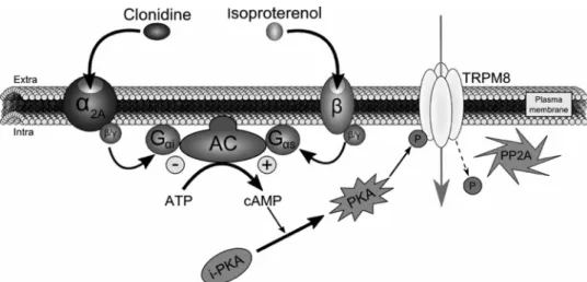
3. Cortright, D. N., Krause, J. E., and Broom, D. C. (2007) Biochim. Biophys. FIGURE 7. Schematic depiction of TRPM8 regulation by␣2A- and -adrenoreceptors. ␣2A-AR stimulation
by clonidine initiates a sequence of intracellular events as follows: G␣iprotein activation following clonidine
fixation on␣2A-AR leads to AC inhibition and decrease of cAMP production. By consequence, PKA transition from an inactive (i-PKA) to an active (PKA) form is reduced. In parallel, the intracellular serine/threonine protein phosphatase 2A (PP2A) dephosphorylates TRPM8 Ser-9 and Thr-17 inhibiting the channel activity. This inhib-itory pathway can be abolished by stimulation of-AR with isoproterenol, because it would lead to a stimula-tion of the G␣sprotein coupled to this receptor and to an up-regulation of the adenylate cyclase activity. The
subsequent increase in cAMP concentration would potentiate PKA phosphorylation, which will fully maintain the functional activity of TRPM8.
by guest on December 21, 2020
http://www.jbc.org/
Acta 1772,978 –988
4. Wang, H., and Woolf, C. J. (2005) Neuron 46, 9 –12
5. McKemy, D. D., Neuhausser, W. M., and Julius, D. (2002) Nature 416, 52–58
6. Voets, T., Owsianik, G., and Nilius, B. (2007) Handb. Exp. Pharmacol.179, 329 –344
7. Brauchi, S., Orio, P., and Latorre, R. (2004) Proc. Natl. Acad. Sci. U.S.A.
101,15494 –15499
8. Voets, T., Droogmans, G., Wissenbach, U., Janssens, A., Flockerzi, V., and Nilius, B. (2004) Nature 430, 748 –754
9. Peier, A. M., Moqrich, A., Hergarden, A. C., Reeve, A. J., Andersson, D. A., Story, G. M., Earley, T. J., Dragoni, I., McIntyre, P., Bevan, S., and Patapou-tian, A. (2002) Cell 108, 705–715
10. Bautista, D. M., Siemens, J., Glazer, J. M., Tsuruda, P. R., Basbaum, A. I., Stucky, C. L., Jordt, S. E., and Julius, D. (2007) Nature 448, 204 –208 11. Colburn, R. W., Lubin, M. L., Stone, D. J., Jr., Wang, Y., Lawrence, D.,
D’Andrea, M. R., Brandt, M. R., Liu, Y., Flores, C. M., and Qin, N. (2007)
Neuron 54,379 –386
12. Dhaka, A., Murray, A. N., Mathur, J., Earley, T. J., Petrus, M. J., and Pata-poutian, A. (2007) Neuron 54, 371–378
13. Chung, M. K., and Caterina, M. J. (2007) Neuron 54, 345–347
14. Xing, H., Chen, M., Ling, J., Tan, W., and Gu, J. G. (2007) J. Neurosci. 27, 13680 –13690
15. Proudfoot, C. J., Garry, E. M., Cottrell, D. F., Rosie, R., Anderson, H., Robertson, D. C., Fleetwood-Walker, S. M., and Mitchell, R. (2006) Curr.
Biol. 16,1591–1605
16. Birder, L. A., and Perl, E. R. (1999) J. Physiol. 515, 533–542
17. Ho¨cker, J., Paris, A., Scholz, J., Tonner, P. H., Nielsen, M., and Bein, B. (2008) Anesthesiology 109, 95–100
18. Duflo, F., Li, X., Bantel, C., Pancaro, C., Vincler, M., and Eisenach, J. C. (2002) Anesthesiology 97, 636 – 641
19. Thebault, S., Lemonnier, L., Bidaux, G., Flourakis, M., Bavencoffe, A.,
Gordienko, D., Roudbaraki, M., Delcourt, P., Panchin, Y., Shuba, Y., Skryma, R., and Prevarskaya, N. (2005) J. Biol. Chem. 280, 39423–39435 20. Beck, B., Bidaux, G., Bavencoffe, A., Lemonnier, L., Thebault, S., Shuba, Y.,
Barrit, G., Skryma, R., and Prevarskaya, N. (2007) Cell Calcium 41, 285–294
21. Pinchenko, V. O., Kostyuk, P. G., and Kostyuk, E. P. (2005) Biochim.
Bio-phys. Acta 1724,1–7
22. Neuberger, G., Schneider, G., and Eisenhaber, F. (2007) Biol. Direct 2, 1 23. Ramos, B. P., Stark, D., Verduzco, L., van Dyck, C. H., and Arnsten, A. F.
(2006) Learn Mem. 13, 770 –776
24. Conklin, B. R., Chabre, O., Wong, Y. H., Federman, A. D., and Bourne, H. R. (1992) J. Biol. Chem. 267, 31–34
25. Liu, B., and Qin, F. (2005) J. Neurosci. 25, 1674 –1681
26. Roha´cs, T., Lopes, C. M., Michailidis, I., and Logothetis, D. E. (2005) Nat.
Neurosci. 8,626 – 634
27. Takezawa, R., Schmitz, C., Demeuse, P., Scharenberg, A. M., Penner, R., and Fleig, A. (2004) Proc. Natl. Acad. Sci. U.S.A. 101, 6009 – 6014 28. Chuang, H. H., Neuhausser, W. M., and Julius, D. (2004) Neuron 43,
859 – 869
29. Karashima, Y., Damann, N., Prenen, J., Talavera, K., Segal, A., Voets, T., and Nilius, B. (2007) J. Neurosci. 27, 9874 –9884
30. Xiao, B., Dubin, A. E., Bursulaya, B., Viswanath, V., Jegla, T. J., and Pata-poutian, A. (2008) J. Neurosci. 28, 9640 –9651
31. Madrid, R., Donovan-Rodríguez, T., Meseguer, V., Acosta, M. C., Bel-monte, C., and Viana, F. (2006) J. Neurosci. 26, 12512–12525
32. Cho, H. J., Kim, D. S., Lee, N. H., Kim, J. K., Lee, K. M., Han, K. S., Kang, Y. N., and Kim, K. J. (1997) Neuroreport 8, 3119 –3122
33. Lefkimmiatis, K., Srikanthan, M., Maiellaro, I., Moyer, M. P., Curci, S., and Hofer, A. M. (2009) Nat. Cell Biol. 11, 433– 442
34. Vanden Abeele, F., Zholos, A., Bidaux, G., Shuba, Y., Thebault, S., Beck, B., Flourakis, M., Panchin, Y., Skryma, R., and Prevarskaya, N. (2006) J. Biol.
Chem. 281,40174 – 40182
by guest on December 21, 2020
http://www.jbc.org/
Roman Skryma and Natalia Prevarskaya
Borowiec, Gabriel Bidaux, Jérôme Busserolles, Alain Eschalier, Yaroslav Shuba,
Alexis Bavencoffe, Dimitra Gkika, Artem Kondratskyi, Benjamin Beck, Anne-Sophie
Adrenoreceptor Signaling Pathway
doi: 10.1074/jbc.M109.069377 originally published online January 28, 2010 2010, 285:9410-9419.
J. Biol. Chem.
10.1074/jbc.M109.069377
Access the most updated version of this article at doi: Alerts:
When a correction for this article is posted
•
When this article is cited
•
to choose from all of JBC's e-mail alerts
Click here
Supplemental material:
http://www.jbc.org/content/suppl/2010/01/28/M109.069377.DC1 http://www.jbc.org/content/285/13/9410.full.html#ref-list-1This article cites 34 references, 11 of which can be accessed free at
by guest on December 21, 2020
http://www.jbc.org/