HAL Id: hal-01792225
https://hal-amu.archives-ouvertes.fr/hal-01792225
Submitted on 18 May 2018HAL is a multi-disciplinary open access archive for the deposit and dissemination of sci-entific research documents, whether they are pub-lished or not. The documents may come from teaching and research institutions in France or abroad, or from public or private research centers.
L’archive ouverte pluridisciplinaire HAL, est destinée au dépôt et à la diffusion de documents scientifiques de niveau recherche, publiés ou non, émanant des établissements d’enseignement et de recherche français ou étrangers, des laboratoires publics ou privés.
in systemic sclerosis patients with anti-centromere or
anti-topoisomerase I antibodies?
Audrey Benyamine, Daniel Bertin, Xavier Heim, Brigitte Granel, Nathalie
Bardin
To cite this version:
Audrey Benyamine, Daniel Bertin, Xavier Heim, Brigitte Granel, Nathalie Bardin. Should we look for RNA polymerase III antibodies in systemic sclerosis patients with centromere or anti-topoisomerase I antibodies?. European Journal of Internal Medicine, Elsevier, 2017, 44, pp.e42-e44. �10.1016/j.ejim.2017.07.033�. �hal-01792225�
European Journal Of Internal Medicine
1
Letter to the Editor
2
Should we look for anti-RNA polymerase III antibodies in Systemic Sclerosis patients
3
with anti-centromere or anti-topoisomerase I antibodies?
4
Running title: anti-RNA polymerase III in Systemic Sclerosis
5 6
Audrey BENYAMINE1,2 M.D, PhD, Daniel BERTIN3 Pharm.D, Xavier HEIM3 Pharm.D,
7
Brigitte GRANEL1,2 M.D, Nathalie BARDIN1,3 Pharm.D, PhD 8
9
1 Aix Marseille University, INSERM, VRCM, UMR_S 1076, Marseille, France
10
2 APHM, Hôpital Nord, Médecine interne, Marseille, France 11
3 APHM, Hôpital La Conception, Laboratoire d’Immunologie, Marseille, France
12 13 Corresponding Author: 14 Audrey BENYAMINE 15
Internal Medicine Department 16
Aix-Marseille Université - Hôpital Nord 17 13915 Marseilles, FRANCE 18 Tél: +33(0)4 91 96 45 13 - Fax : +33(0)4 91 96 80 80 19 Audrey.benyamine@ap-hm.fr 20
Reprint requests should be addressed to Audrey BENYAMINE, Hôpital Nord, Marseilles 21
The authors declare no conflict of interest. 22
Keywords: Systemic Sclerosis; Anti-RNA Polymerase III antibodies; Anti-centromere
23
Antibodies; Anti-topoisomerase I antibodies; Immunofluorescence pattern 24
Systemic sclerosis (SSc) is a chronic autoimmune disease characterised by skin and internal 25
organs fibrosis, vascular damage and positive antinuclear autoantibodies (ANAs). The 26
classical antigenic specificities of ANAs are anti-centromere (ACA), anti-topoisomerase I 27
antibodies (anti-topo I) and the more recently described anti-RNA polymerase III antibodies 28
(anti-RNAPIII). 29
Anti-RNAPIII, firstly described in SSc in 1993[1], are currently admitted as specific
SSc-30
related autoantibodies and have been incorporated into the 2013 ACR/EULAR classification
31
criteria for the disease [2]. The prevalence of anti-RNAPIII partially depends on the 32
geographic origin of patients [3]. In France, the prevalence is low and ranges from 3 to 9% 33
whereas it can reach 14% in North America and 41% in South America [3,4]. The ANA 34
pattern associated with anti-RNAPIII was described as a fine-speckled nuclear stain with 35
additional occasional bright dots, with or without concurrent punctate nucleolar staining but 36
no typical pattern was proposed [5]. Anti-RNAPIII are mainly associated with a diffuse 37
cutaneous subtype, scleroderma renal crisis and a risk of cancer in close temporal relationship 38
to SSc onset [3,4,6,7]. As the specific method for their identification was the
39
radioimmunoprecipitation assay, a cumbersome method not suitable for routine practice, these 40
autoantibodies were not routinely looked for. More recently immunoenzymatic methods have
41
been developed, but, the detection strategy was not clearly established for routine practice [5]. 42
Although the positivity of ACA and anti-topo I is considered to be exclusive, the co-positivity 43
of anti-RNAPIII with these SSc-specific auto-antibodies remains to be clarified. In order to 44
design the best strategy for anti-RNAPIII detection, we aimed to 1/ evaluate the co-positivity 45
of anti-RNAPIII in SSc patients positive for ACA or anti-topo I and to 2/ analyse 46
immunofluorescence patterns and clinical characteristics of anti-RNAPIII positive patients. 47
Firstly, 76 sera from SSc patients (9 men, 67 women) from Marseilles (South of France) 49
positive for either ACA or topo I antibodies were tested for the presence of anti-50
RNAPIII. All patients fulfilled the 2013 ACR/EULAR criteria and were further classified as 51
having diffuse or limited cutaneous SSc [2]. All the sera were collected from 2012 to 2016 52
and were issued from a Biobank (DC 2012-1704) with respect of ethical directives. 53
Antinuclear antibodies (ANAs) were detected by indirect immunofluorescence on HEp-2 cells 54
(Bio-Rad Laboratories, Hercules, CA) at a screening dilution of 1:160. ACA, anti-topo I and 55
anti-RNAPIII were detected by commercially kits (EliA Thermo Fisher). The cutoff value for 56
anti-RNAPIII positivity was 10 Arbitrary Unit/ml (AU). 57
Secondly, the ANAs immunofluorescence patterns and clinical data of 8 SSc patients positive 58
for anti-RNAPIII were collected from 2012 to 2016 and compared to anti-RNAPIII negative 59
SSc patients (<10 AU/ml). Results were expressed as median +/- interquartile range or as 60
frequencies (fq). Medians were compared using Mann Whitney U Test. Frequencies were 61
compared using Chi 2 Test. 62
Among the 76 selected sera of SSc patients, 33 patients (43%) were positive for ACA and 43 63
(57%) for anti-topo I antibodies. Immunofluorescence nuclear patterns were: centromeric 64
(n=32), nucleolar homogeneous (n=41), speckled-centromeric (n=1), speckled-homogeneous 65
(n=1) and mixed speckled-nucleolar-centromeric (n=1). Anti-RNAPIII were investigated in 66
these SSc patients: only one ACA-positive serum (1.3%) was found also positive for anti-67
RNAPIII with a titer of 192 AU/ml. This serum corresponded to the mixed speckled-68
nucleolar-centromeric immunofluorescence nuclear pattern (Figure 1). No other specificities 69
associated with a speckled pattern (anti-Ro/SSA anti-La/SSB, anti-Sm, anti-RNP) were 70
detected. The patient, a 47-year-old female, had a diffuse cutaneous subset, with digital 71
ulcers, joint contractures, a reduced diffuse lung capacity for carbon monoxide and 72
oesophageal reflux disorder. Her medical history was remarkable for an ovarian 73
adenocarcinoma that was diagnosed 6 years before SSc, and considered in remission after 74
surgery and radio-chemotherapy. 75
Then, we retrospectively analysed immunofluorescence pattern and clinical characteristics of 76
8 SSc patients found positive for anti-RNAPIII in our laboratory. The various 77
immunofluorescence aspects were nuclear speckled (n=5), nucleolar (n=1), nucleolar-78
speckled (n=1) (Figure 2) and mixed speckled-nucleolar-centromeric (n=1) (Figure 1). The 79
median titer of anti-RNAPIII was 54 [18-298] AU/ml. 80
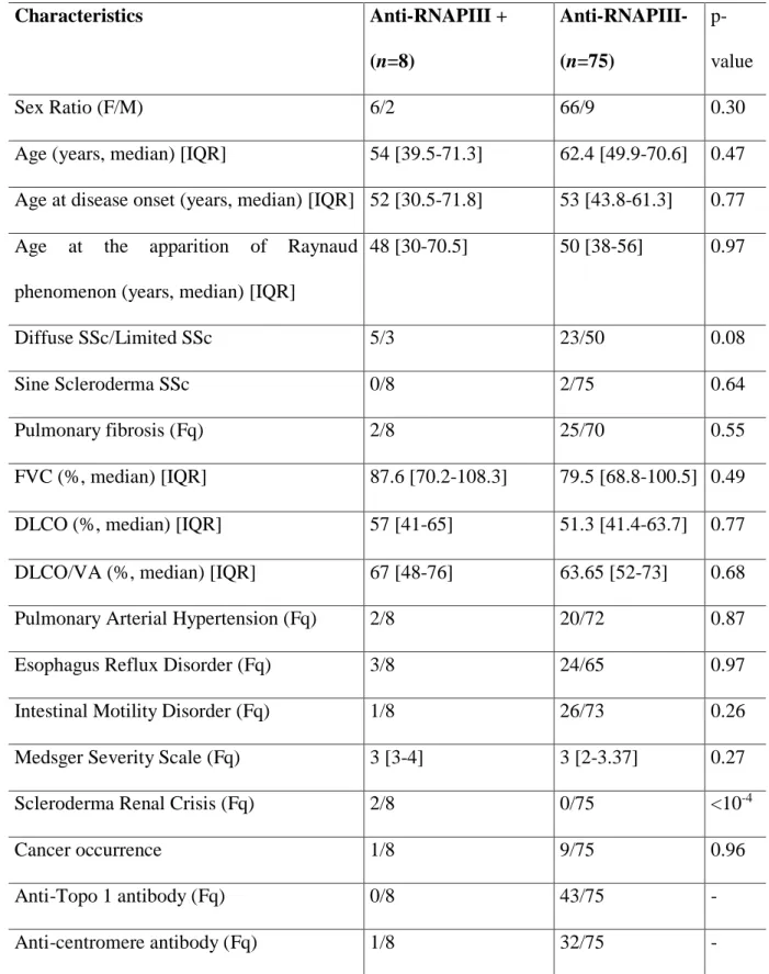
Table 1 illustrates the comparison between anti-RNAPIII positive and negative patients. Sex
81
ratio did not differ between the two groups. A trend to a higher frequency of the diffuse 82
cutaneous form was observed in anti-RNAPIII positive SSc patients. The two patients with 83
sine scleroderma belonged to the group of negative anti-RNAPIII. Scleroderma renal crisis
84
was solely documented in patients with positive anti-RNAPIII. Other variables were not 85
significantly associated with anti-RNAPIII positivity. 86
The present study extends the previously published data performed in South of France [4] 87
about the anti-RNAPIII screening strategy. This study highlights that anti-RNAPIII are rarely 88
encountered in SSc patients already positive for ACA or anti-topo I antibodies. The case of 89
the patient with coexisting ACA and anti-RNAPIII was interesting regarding two aspects. 90
First, the clinical feature was closer to the one described in patients with anti-RNAPIII with a 91
diffuse cutaneous form and a concomitant cancer [8]. Second, the observed ANAs 92
immunofluorescence pattern was highly remarkable due to the peculiar aspect of 93
immunofluorescence. Therefore, in ACA or topo I positive sera, the search for anti-94
RNAPIII can be recommended faced to a mixed fluorescence pattern. Conversely, in case of 95
typical fluorescence aspects related to ACA or anti-topo I positivity, the systematic search for 96
anti-RNAPIII is not mandatory. As observed herein, different immunofluorescence pattern 97
can be associated with anti-RNAPIII [9]. Therefore the search for this auto-antibody should 98
not be restrained to a nucleolar immunofluorescence pattern of ANAs [10]. 99
Regarding the phenotype of anti-RNAPIII positive patients, a higher frequency of 100
scleroderma renal crisis and diffuse cutaneous form was observed. The low number of 101
patients and our geographic location might be a limiting factor to evidence any association 102
with other anti-RNAPIII features such as the frequency of cancer [8]. 103
Although SSc-related autoantibodies are exclusive markers, co-positivity can exist in rare 104
cases. The immunofluorescence reading step appears crucial to focus the screening. In order 105
to improve the benefit cost ratio, anti-RNAPIII should be searched with respect to ANAs 106
fluorescence pattern and clinical characteristics of the patients. 107
108
ACKNOWLEDGEMENT
109
This research did not receive any specific grant from funding agencies in the public, 110
commercial, or not-for-profit sectors. 111
We thank internal medicine physicians for their help in collecting the clinical data (M.Ebbo, 112
N.Schleinitz, G.Kaplanski, C.Gomez). 113
REFERENCES
115 116
[1] Kuwana M, Kaburaki J, Mimori T, Tojo T, Homma M. Autoantibody reactive with three 117
classes of RNA polymerases in sera from patients with systemic sclerosis. J Clin Invest 118
1993;91:1399–404. doi:10.1172/JCI116343. 119
[2] van den Hoogen F, Khanna D, Fransen J, Johnson SR, Baron M, Tyndall A, et al. 2013 120
classification criteria for systemic sclerosis: an American College of 121
Rheumatology/European League against Rheumatism collaborative initiative. Arthritis 122
Rheum 2013;65:2737–47. doi:10.1002/art.38098. 123
[3] Sobanski V, Dauchet L, Lefèvre G, Lambert M, Morell-Dubois S, Sy T, et al. Prevalence 124
of anti-RNA polymerase III antibodies in systemic sclerosis: New data from a French 125
cohort and a systematic review and meta-analysis. Arthritis Rheumatol Hoboken NJ 126
2014;66:407–17. doi:10.1002/art.38219. 127
[4] Faucher B, Stein P, Granel B, Weiller P-J, Disdier P, Serratrice J, et al. Low prevalence 128
of anti-RNA polymerase III antibodies in a French scleroderma population: anti-RNA 129
polymerase III scleroderma. Eur J Intern Med 2010;21:114–7. 130
doi:10.1016/j.ejim.2010.01.004. 131
[5] Parker JC, Burlingame RW, Webb TT, Bunn CC. Anti-RNA polymerase III antibodies 132
in patients with systemic sclerosis detected by indirect immunofluorescence and ELISA. 133
Rheumatol Oxf Engl 2008;47:976–9. doi:10.1093/rheumatology/ken201. 134
[6] Emilie S, Goulvestre C, Bérezné A, Pagnoux C, Guillevin L, Mouthon L. Anti-RNA 135
polymerase III antibodies are associated with scleroderma renal crisis in a French cohort. 136
Scand J Rheumatol 2011;40:404–6. doi:10.3109/03009742.2011.569753. 137
[7] Meyer O, De Chaisemartin L, Nicaise-Roland P, Cabane J, Tubach F, Dieude P, et al. 138
Anti-RNA polymerase III antibody prevalence and associated clinical manifestations in 139
a large series of French patients with systemic sclerosis: a cross-sectional study. J 140
Rheumatol 2010;37:125–30. doi:10.3899/jrheum.090677. 141
[8] Lazzaroni M-G, Cavazzana I, Colombo E, Dobrota R, Hernandez J, Hesselstrand R, et 142
al. Malignancies in Patients with Anti-RNA Polymerase III Antibodies and Systemic 143
Sclerosis: Analysis of the EULAR Scleroderma Trials and Research Cohort and Possible 144
Recommendations for Screening. J Rheumatol 2017. doi:10.3899/jrheum.160817. 145
[9] Codullo V, Morozzi G, Bardoni A, Salvini R, Deleonardi G, De Pità O, et al. Validation 146
of a new immunoenzymatic method to detect antibodies to RNA polymerase III in 147
systemic sclerosis. Clin Exp Rheumatol 2007;25:373–7. 148
[10] Yamasaki Y, Honkanen-Scott M, Hernandez L, Ikeda K, Barker T, Bubb MR, et al. 149
Nucleolar staining cannot be used as a screening test for the scleroderma marker anti-150
RNA polymerase I/III antibodies. Arthritis Rheum 2006;54:3051–6. 151
doi:10.1002/art.22043. 152
153 154
Table 1: Characteristics of SSc patients with respect to positivity of the anti-RNA
155
Polymerase III (anti-RNAPIII) autoantibodies.
156 Characteristics Anti-RNAPIII + (n=8) Anti-RNAPIII- (n=75) p-value Sex Ratio (F/M) 6/2 66/9 0.30
Age (years, median) [IQR] 54 [39.5-71.3] 62.4 [49.9-70.6] 0.47 Age at disease onset (years, median) [IQR] 52 [30.5-71.8] 53 [43.8-61.3] 0.77 Age at the apparition of Raynaud
phenomenon (years, median) [IQR]
48 [30-70.5] 50 [38-56] 0.97
Diffuse SSc/Limited SSc 5/3 23/50 0.08
Sine Scleroderma SSc 0/8 2/75 0.64
Pulmonary fibrosis (Fq) 2/8 25/70 0.55
FVC (%, median) [IQR] 87.6 [70.2-108.3] 79.5 [68.8-100.5] 0.49
DLCO (%, median) [IQR] 57 [41-65] 51.3 [41.4-63.7] 0.77
DLCO/VA (%, median) [IQR] 67 [48-76] 63.65 [52-73] 0.68
Pulmonary Arterial Hypertension (Fq) 2/8 20/72 0.87
Esophagus Reflux Disorder (Fq) 3/8 24/65 0.97
Intestinal Motility Disorder (Fq) 1/8 26/73 0.26
Medsger Severity Scale (Fq) 3 [3-4] 3 [2-3.37] 0.27
Scleroderma Renal Crisis (Fq) 2/8 0/75 <10-4
Cancer occurrence 1/8 9/75 0.96
Anti-Topo 1 antibody (Fq) 0/8 43/75 -
Anti-centromere antibody (Fq) 1/8 32/75 -
Results are expressed as median +/- interquartile range or as frequencies (fq). Medians were 157
compared using Mann Withney U Test. Frequencies were compared using Chi 2 Test. 158
Legends of the figures
159 160
Figure 1: The mixed fluorescence pattern (speckled-nucleolar-centromeric) of the ACA and
161
Anti-RNAPIII positive serum. 162
A: x 400 magnification.
163
B: Typical features of the centromeric pattern are highlighted with characteristic dots at the
164
interphase mitotic stage (left arrow) and a “block” of condensed dots at the metaphase stage 165
(right arrow). 166
167
Figure 2: Various immunofluorescence patterns obtained from sera positive for anti-RNAPIII
168 (x 400 magnification). 169 A: nuclear-speckled pattern 170 B: nucleolar-speckled pattern 171 C: nucleolar pattern 172 173