HAL Id: hal-00088852
https://hal.archives-ouvertes.fr/hal-00088852
Submitted on 14 Apr 2021
HAL is a multi-disciplinary open access
archive for the deposit and dissemination of
sci-entific research documents, whether they are
pub-lished or not. The documents may come from
teaching and research institutions in France or
abroad, or from public or private research centers.
L’archive ouverte pluridisciplinaire HAL, est
destinée au dépôt et à la diffusion de documents
scientifiques de niveau recherche, publiés ou non,
émanant des établissements d’enseignement et de
recherche français ou étrangers, des laboratoires
publics ou privés.
Identification of Morphine-6-glucuronide in Chromaffin
Cell Secretory Granules
Yannick Goumon, A. Müller, E. Glattard, C. Marban, C. Gasnier, J.M. Strub,
S. Chasserot-Golaz, O. Rohr, G.B. Stefano, I.D. Welters, et al.
To cite this version:
Yannick Goumon, A. Müller, E. Glattard, C. Marban, C. Gasnier, et al.. Identification of
Morphine-6-glucuronide in Chromaffin Cell Secretory Granules. Journal of Biological Chemistry, American Society
for Biochemistry and Molecular Biology, 2006, 281 (12), pp.8082-8089. �10.1074/jbc.M502298200�.
�hal-00088852�
Identification of Morphine-6-glucuronide in Chromaffin Cell
Secretory Granules
*
Received for publication, March 1, 2005, and in revised form, January 5, 2006 Published, JBC Papers in Press, January 24, 2006, DOI 10.1074/jbc.M502298200
Yannick Goumon
‡1, Arnaud Muller
‡, Elise Glattard
‡, Ce´line Marban
‡, Claire Gasnier
‡, Jean-Marc Strub
§,
Sylvette Chasserot-Golaz
¶, Olivier Rohr
‡, George B. Stefano
储, Ingeborg D. Welters**, Alain Van Dorsselaer
§,
Franc¸oise Schoentgen
‡‡, Dominique Aunis
‡, and Marie-He´le`ne Metz-Boutigue
‡From the
‡Physiopathologie du Syste`me Nerveux, INSERM U575, 67084 Strasbourg, France, the
§Laboratoire de Spectrome´trie de
Masse BioOrganique, CNRS UMR 7168/LC2, 67087 Strasbourg, France, the
¶Laboratoire de Neurotransmission et Se´cre´tion
Neuroendocrine, CNRS UPR 2356, 67084 Strasbourg, France, the
储Neuroscience Research Institute, State University of New York,
Old Westbury, New York, New York 11568, the **Department of Anesthesiology, Intensive Care, and Pain Medicine,
Justus-Liebig-Universita¨t, 35385 Giessen, Germany, and the
‡‡Centre de Biophysique Mole´culaire, CNRS UPR 4301, 45071 Orle´ans, France
We report for the first time that morphine-6-glucuronide, a highly analgesic morphine-derived molecule, is present in adrenal chromaffin granules and secreted from chromaffin cells upon stimulation. We also demonstrate that phosphatidylethanolamine-binding protein (alternatively named Raf-1 kinase inhibitor protein or RKIP) acts as an endogenous morphine-6-glucuronide-binding protein. An UDP-glucuronosyltransferase 2B-like enzyme, described to transform morphine into morphine-6-glucuronide, has been immunode-tected in the chromaffin granule matrix, and morphine-6-glucuro-nide de novo synthesis has been characterized, demonstrating the possible involvement of intragranular UDP-glucuronosyltrans-ferase 2B-like enzyme in morphine-6-glucuronide metabolism. Once secreted into the circulation, morphine-6-glucuronide may mediate several systemic actions (e.g. on immune cells) based on its affinity for-opioid receptors. These activities could be facilitated by phosphatidylethanolamine-binding protein (PEBP), acting as a molecular shield and preventing morphine-6-glucuronide from rapid clearance. Taken together, our data represent an important observation on the role of morphine-6-glucuronide as a new endo-crine factor.
For 20 years, cerebral endogenous morphine has been isolated and characterized, and its structure has been shown to be identical to mor-phine from poppy (1, 2). In the 1990s, few groups succeeded in charac-terizing endogenous morphine from the organs and fluids of verte-brates, including bovine brain and adrenal gland, rat heart and adrenal gland, and human heart and urine (for review, Refs. 1–3); invertebrates (4 – 6); and parasites (7, 8). In mammals, the endogenous morphine synthetic pathway is associated with the dopamine and catecholamine biosynthetic pathway (for review, see Refs. 1, 3, and 9). Very recently, several crucial steps were reached when Zenk and co-workers (10, 11) demonstrated that morphine can be formed using a multistep biosyn-thetic route, and Zhu et al. (12) showed that human primary polymor-phonuclear cells are able to synthesize morphine. These authors have
shown morphine de novo synthesis in human and animal primary and cancer cell cultures.
Chromaffin cells are neuroendocrine cells originating from the neu-ral crest and are the predominant cell type in the adrenal medulla (for review, see Ref. 13). These cells possess the catecholamine biosynthetic pathway, leading to dopamine and adrenaline/noradrenaline synthesis (13). Chromaffin secretory granules contain a complex mixture of pep-tides and proteins that are co-released with catecholamines into the circulation in response to splanchnic nerve stimulation (13). Based on the morphine biosynthetic pathway and on the presence of morphine in bovine and rat adrenal glands (14, 15), rat pheochromocytoma PC-12 cells (10, 16), and eel chromaffin cells (17), we hypothesized that mam-malian chromaffin cells might have the capacity to synthesize morphine and that their secretory granules could potentially release this alkaloid into the blood.
Several molecular blood transporters of clinically administrated mor-phine have been identified (e.g. serum albumin and␥-globulin (18) and ␣1-acid glycoprotein (19)). In addition, it has been reported that the
phosphatidylethanolamine-binding protein (PEBP)2(20), alternatively named Raf-1 kinase inhibitor protein (RKIP) (21), is also able to bind morphine (22). We have reported recently that PEBP is present in secre-tory granules as well as in the exocytotic medium of stimulated bovine primary cultured chromaffin cells and in bovine serum (23).
Exogenously administered morphine is catabolized in the liver by the UDP-glucuronosyltransferase (UGT) 2B enzyme family (24), leading to the formation of morphine-3-glucuronide (M3G) and morphine-6-glu-curonide (M6G). M3G is totally inactive, whereas M6G appears to dis-play stronger analgesic activity than morphine (50 – 600 times depend-ing on the animal model) (25) and exhibits a much longer half-life (26). Using immunocytochemical, biochemical, and proteomic strategies, we reveal in this study that bovine chromaffin cell secretory granules store the PEBP䡠M6G complex. We show that M6G is secreted from bovine chromaffin cells upon nicotinic stimulation. Co-immunopre-cipitation and affinity chromatography experiments also revealed the interaction between the alkaloid carrier (i.e. PEBP) and a UGT2B-like enzyme necessary for the conversion of morphine into M6G. Finally, M6G de novo synthesis was demonstrated using the UGT2B-like enzyme present in chromaffin granules.
*This work was supported by INSERM, Universite´ Louis Pasteur Strasbourg, Ph.D. grants from the Re´gion Alsace (to C. M.) and the French Ministe`re De´le´gue´ a` la Recherche et a` l’Enseignement Supe´rieur (to E. G. and A. M.), the Fondation pour la Recherche Me´dicale (to M.-H. M.-B.), the Ligue Contre le Cancer (to D. A.), and Hoˆpital Universi-taire de Strasbourg Programme Hospitalier de Recherche Clinique 3150 (to H. M.-B.). The costs of publication of this article were defrayed in part by the payment of page charges. This article must therefore be hereby marked “advertisement” in accordance with 18 U.S.C. Section 1734 solely to indicate this fact.
1To whom correspondence should be addressed: INSERM U575, 5, rue Blaise Pascal, 67084 Strasbourg, France. Tel.: 33-3-8845-6724; Fax: 33-3-8860-0806; E-mail: goumon@neurochem.u-strasbg.fr.
2The abbreviations used are: PEBP, phosphatidylethanolamine-binding protein; UGT, UDP-glucuronosyltransferase; M3G, morphine-3-glucuronide; M6G, morphine-6-glu-curonide; CGA, chromogranin A; RP-HPLC, reverse phase high performance liquid chromatography; MS, mass spectrometry; MS/MS, tandem mass spectrometry; Q-TOF, quadrupole time-of-flight; PEA, proenkephalin A; RKIP, Raf-1 kinase inhibitor protein.
at Rutgers University on August 11, 2015
http://www.jbc.org/
MATERIALS AND METHODS
Immunocytochemistry and Confocal Microscopy Analysis—Bovine
primary chromaffin cells cultured and fixed as described previously (23) were incubated for 2 h at 37 °C with a rabbit anti-bovine chromogranin A (CGA)-(124 –143) polyclonal antibody (1:200 dilution) (25) and a commercial sheep polyclonal antibody against morphine-like com-pounds (i.e. morphine, M3G, and M6G, with minimal cross-reactivity to codeine as declared by the supplying company; 1:1000 dilution; Bio-genesis, Bournemouth, UK) (8) in NaCl/Pibuffer (0.9% (w/v) NaCl and
25 mMsodium phosphate, pH 7.5) containing 5% bovine serum
albu-min. Cells were then washed (6⫻ 5 min) with NaCl/Pibuffer and
incu-bated with secondary antibodies (Cy5-conjugated donkey anti-rabbit IgG at 1:2000 dilution and Alexa 488-conjugated donkey anti-sheep IgG at 1:2000 dilution; Molecular Probes, Eugene, OR) in NaCl/Pibuffer
containing 5% bovine serum albumin for 45 min at 37 °C.
UGT2B was immunodetected using a commercial goat polyclonal antibody against the human UGT2B family (1:500 dilution; Santa Cruz Biotechnology, Inc., Santa Cruz, CA) and Alexa 488-conjugated donkey antibodies (1:2000 dilution). CGA was detected with rabbit anti-bovine CGA-(124 –143) polyclonal antibody (1:200 dilution; see above) and Cy5-conjugated donkey anti-rabbit IgG (1:2000 dilution). All glass cov-erslips were mounted on a glass slide with a drop of Mowiol 4-88.
Various controls were carried out to assess antibody specificity: (i) cells without antibody, (ii) secondary antibody incubated alone; and (iii) primary antibody incubated with the respective antigen prior to immunolabeling. Anti-UGT2B antibody was blocked with the commer-cial peptide (12 h, 4 °C, 1:5 (w/w) ratio; Santa Cruz Biotechnology, Inc.), and anti-morphine antibody with M6G (12 h, 4 °C, 1:50 mol/mol ratio). Immunofluorescence staining was monitored with a Zeiss LSM 510 laser scanning microscope equipped with a Planapo oil immersion objective (⫻63, numerical aperture of 1.4) (23). Co-localization with CGA was quantified using Zeiss CLSM Version 2.8 software.
Isolation of Chromaffin Cell Subcellular Fractions—The chromaffin
cell plasma membrane, cytoplasm, intragranular matrix, and granule membrane were prepared as described by Smith and Winkler (27) and modified by us (23, 28). After protein quantification using the Bradford technique, the purity of subcellular fractions was assayed as described previously using subcellular markers (23). The total microsomal frac-tion was purified according to Levesque et al. (29).
Isolation of Proteins Exocytotically Released from Stimulated
Chro-maffin Cells—Chromaffin cells (2.5⫻ 106) cultured for 3 days were
washed (4⫻ 5 min) with 10 ml of Locke’s solution at 37 °C to get rid of the culture medium and floating dead cells. Cells were subsequently stimulated for 10 min with 10 M nicotine in Locke’s solution as
described previously (28).
The extracellular medium of unstimulated cells and Locke’s solution were treated as controls. Secreted medium and controls were centrifuged at 800⫻ g for 10 min at 4 °C and filtered with a 0.22-m syringe filter to remove cells that might be present in secretions. Samples were acidified up to 0.1% trifluoroacetic acid (TFA) to prevent degradation (23).
Purification of Alkaloids by RP-HPLC—Subcellular fractions (1 ml of
the corresponding sucrose gradient fraction) were deproteinized and prepared as described previously (16). Alkaloids were purified using an A¨ KTA purifier HPLC system (Amersham Biosciences AB, Uppsala, Sweden) and a Nucleosil RP300 –5C18column (4⫻ 250 mm, 5-m
particle size, 300-Å porosity; Macherey-Nagel, Hoerdt, France) as described (23). Gradients are indicated on chromatograms. Buffer A (TFA 0.1% (v/v) in water) and buffer B (acetonitrile 70%, TFA 0.1% in water (v/v)) were used.
Mass Spectral Analysis—Mass spectrometry (MS) and tandem MS
(MS/MS) analyses were performed by electrospray MS on a Q-TOF II (Micromass Ltd., Manchester, UK) in positive mode. Scanning was done from 50 to 700 m/z in 1 s, and calibration was performed using 0.1% phosphoric acid in 50:50 water/acetonitrile. MS and MS/MS anal-ysis were done by nanospray offline using NanoES spray capillaries (Pro-tana, Odense, Denmark). For MS/MS experiments, argon grade 5.5 gas was used for the collision gas, and the collision energy was set to 10 –30 eV. MS/MS spectra were acquired using the selection of the precursor ion by the quadrupole, and fragments were analyzed by time of flight. The absence of M6G prior to sample application was systematically controlled using the MS and MS/MS modes.
Affinity Chromatography—Purified bovine PEBP-(1–11)
anti-body (3 mg prepared as described (30)) or commercial anti-human UGT2B antibody (200g) was coupled to a HiTrap NHS-activated high performance 1-ml column (Amersham Biosciences) according to the manufacturer’s instructions. The antibody-coupled column was first washed with 5 volumes of buffer A (75 mMTris-HCl, pH 8.0). Samples
(i.e. cytoplasm, microsomes, lysosomes, mitochondria, and intragranu-lar matrix) resuspended in buffer A (2 mg in a 1-ml final volume) were loaded onto the column and allowed to stand for 10 min at room tem-perature. The column was then washed with 10 volumes of buffer A prior to elution performed with 1 ml of buffer B (150 mMNaCl and 100 mMglycine, pH 3.0). The eluate was immediately neutralized with 20l of buffer C (1.5M Tris-HCl, pH 8.0) prior to RP-HPLC analysis or desalting.
Co-immunoprecipitation Assays—500g of proteins isolated from
the chromaffin granule matrix was treated as described previously using anti-human UGT2B antibody (31). Immunoprecipitates were analyzed by SDS-PAGE and Western blotting using rabbit anti-bovine PEBP-(1– 11) polyclonal antibody (1:2000 dilution). A semiquantitative approach with different amounts of the granule matrix protein was used to quan-tify the amount of PEBP bound to the UGT2B-like enzyme.
Gel Electrophoresis and Western Blot Analysis—For one-dimensional
electrophoresis analysis, proteins were separated on SDS-polyacryl-amide containing 10 or 12% acrylSDS-polyacryl-amide and electrotransferred as described (23). For two-dimensional gel electrophoresis analysis, iso-electric focusing was performed using the PROTEAN IEFTM cell
isoelectric focusing system (Bio-Rad, Marnes-La-Coquette, France) equipped with dry polyacrylamide gel strips (11-cm immobilized pH gradient strips) using a pH 4 –7 gradient (32). Proteins were immuno-detected with goat polyclonal antibodies raised against the human UGT2B family (60 kDa, 1:500 dilution) and immunorevealed with horseradish peroxidase-conjugated anti-goat antisera. Control experi-ments using the antibodies were performed as described above. For silver staining, the gel was incubated in ethanol/acetic acid/water solu-tion (40:10:50%) and treated using a Dodeca silver stain kit (Bio-Rad) according to the manufacturer’s instructions.
Tryptic Digestion and Mass Spectrometry—Silver-stained bands were
excised, washed with 100l of 25 mMNH4HCO3, dehydrated twice
with 100l of acetonitrile, and dried with a SpeedVac evaporator before reduction (10 mMdithiothreitol in 25 mMNH4HCO3) and alkylation (55
mMiodoacetamide in 25 mMNH4HCO3) (33). For tryptic digestion, gel
pieces were resuspended in 3 gel volumes of trypsin (12.5 ng/l) freshly diluted in 25 mMNH4HCO3and incubated overnight at 35 °C. The
digested peptides were then extracted from the gel in buffer containing 25% H2O, 70% acetonitrile, and 5% formic acid and analyzed by
matrix-assisted laser desorption ionization time-of-flight MS and liquid chro-matography/MS/MS. For nano-HPLC, a CapLC system (Micromass Ltd., Manchester, UK) was used. Samples were concentrated on a
pre-Morphine-6-glucuronide in Chromaffin Secretory Granules
at Rutgers University on August 11, 2015
http://www.jbc.org/
column, and peptides were separated on a column (75m, inner diam-eter⫻ 15 cm) packed with C18 PepMap (3m, 100 Å; LC Packings). The MS and MS/MS analyses were performed with a Q-Tof II hybrid quadrupole time-of-flight (Q-TOF) mass spectrometer (Micromass Ltd.). Data analysis was performed with Global Server software (Micro-mass Ltd.) and Mascote (Matrix Science Ltd., London, UK) against the NCBI Database.
M6G de Novo Synthesis—UGT2B was purified by affinity
chromatog-raphy on an ¨AKTA purifier HPLC System. The eluted material was desalted on a Sep-Pak cartridge and analyzed by Western blotting using anti-UGT2B antibody to confirm the presence of UGT2B-like material. The eluted fraction was also submitted to gel migration, followed by silver staining. Silver-stained bands were excised and treated as described above.
The affinity-purified UGT2B-like enzyme was dried and incubated for 3 h at 37 °C in a reaction volume of 100l (100 MUDP-glucuronic acid, 2 mMsaccharolactone, 0.9Mmorphine, and 50 mMTris-HCl, pH
5.5). The resulting sample was deproteinized and desalted prior to Q-TOF-MS/MS analysis to examine the presence of M6G. Controls experiments were performed with the boiled affinity column eluate, the reaction buffer alone, and morphine used for the assay.
Protein Deglycosylation—The intragranular UGT2B-like enzyme was
deglycosylated using an enzymatic protein deglycosylation kit (Sigma) in presence of Triton X-100. Total deglycosylation was carried on using peptide: N-glycosidase F, O-glycosidase,␣2(3,6,8,9)-neuraminidase, 1,4-galactosidase, and-N-acetylglucosaminidase. The non-deglycosylated control and the resulting deglycosylated products were loaded onto a 12% SDS-polyacrylamide gel prior to electrotransfer. Deglycosylation method efficiency was tested using a control glycosylated protein (fetuin, 10g) provided in the manufacturer’s kit.
RESULTS
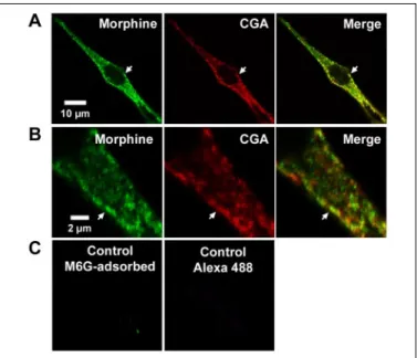
Immunolabeling of Morphine-like Components in Chromaffin Cells—
We extended our previous observations related to the presence of morphine and its derivatives in adrenal medulla and PC-12 cells (15, 16) by investigating the subcellular localization of morphine and its derivatives in chromaffin cells by laser confocal microscopy. Mor-phine localization was compared with CGA immunoreactivity, a specific marker of the intragranular matrix of secretory granules (23). Morphine-like immunolabeling (Fig. 1A, left panel, green) was observed as bright dots in the cytoplasm, similar to that obtained with CGA (middle panel, red). The superimposition of the two labeled materials may suggest a partial intragranular co-localization in chromaffin granules (Fig. 1, A and B, right panels and arrows). Control experiments using anti-morphine antibody blocked with M6G prior to immunocytochemistry experiments or secondary anti-body alone (Fig. 1C) were carried out to determine the specificity of labeling.
Characterization of M6G in the Chromaffin Intragranular Matrix—To
clearly demonstrate the presence of M6G in chromaffin granules, bovine intragranular matrix (33 mg) was isolated and loaded onto an RP-HPLC column to purify and characterize the alkaloids present. The HPLC gradi-ent was specifically designed to separate M3G, morphine, M6G, codeine, and morphine acetate (500 pmol) (Fig. 2A). Comparison of the chromatog-raphy profile of the deproteinized intragranular material with the elution profile of standards indicated the presence of a peak with the same reten-tion time as M6G. Q-TOF-MS/MS analysis of this peak allowed its unam-biguous identification as M6G (462.15 Da) according to its fragmentation profile generating morphine (i.e. 286.13 Da) (Fig. 2B).
FIGURE 1. Immunolabeling of morphine-like components in bovine primary
cul-tured chromaffin cells. A, double immunofluorescence confocal micrographs with
anti-CGA antibody detected with Cy5-conjugated IgG (red) and with antibody against mor-phine-like molecules revealed with Alexa 488-conjugated IgG (green). Co-localized immunolabelings (arrows and merged image) are revealed by yellow staining, showing double-labeled pixels in images of CGA and morphine labeling recorded simultaneously in the same optical section. B, higher resolution of immunolabeling in a chromaffin cell neurite-like extension showing visible individual granules. C, control experiments per-formed with M6G-adsorbed anti-morphine antibody or with secondary antibody alone.
FIGURE 2. HPLC purification and MS analysis of morphine derivatives present in bovine
chromaffin granules. A, RP-HPLC purification of standards (M3G, morphine, M6G, codeine
(COD), and morphine acetate (MA); upper panel) and purification of alkaloid extracted from bovine soluble intragranular material (lower panel). B, Q-TOF-MS/MS analysis of the HPLC fraction corresponding to M6G present in the intragranular material (marked with arrow in A,
lower panel (granule matrix)). mAU, milli-absorbance units; B, buffer B.
at Rutgers University on August 11, 2015
http://www.jbc.org/
Identification of M6G Secreted from Nicotine-stimulated Cultured
Chromaffin Cells—To characterize M6G as a secretory product of
stim-ulated chromaffin cells, alkaloid standards (390 pmol) (Fig. 3A, panel 1) and the extracellular medium were analyzed by RP-HPLC. After stim-ulation with 10Mnicotine, the secreted material was submitted to 1N
HCl and chloroform/isopropyl alcohol extractions. Alkaloids present in the aqueous phase were separated by RP-HPLC on a C18column (panel 2). A major peak corresponding to the M6G standard was observed. In extracellular media corresponding to basal secretions, M6G could not be detected (panel 3), demonstrating the genuine release of M6G upon nicotine stimulation.
Q-TOF-MS/MS analysis of the peak corresponding to M6G unam-biguously demonstrated the presence of M6G (462.15 Da) according to its fragmentation profile whereby morphine is generated (i.e. 286.13 Da) (Fig. 3B). Similar analysis revealed the absence of M6G in secretions from unstimulated cells (Fig. 3C).
Evidence for Interaction between PEBP and M6G—We reported
recently that PEBP is present in chromaffin granules and is able to trans-locate from the cytoplasm to the intragranular space probably via a granule membrane raft-binding mechanism (23). Based on this prop-erty, we then examined the capacity of PEBP to bind intragranular mor-phine-like components by immunoaffinity experiments carried out using the intragranular matrix, cytoplasm, microsomes, mitochondria, and lysosomes. Immunoaffinity experiments using the purified anti-body directed against PEBP-(1–11) were carried out to isolate the puta-tive intragranular PEBP䡠morphine-like complex(es). Subcellular frac-tion extracts (20 mg each) were loaded onto a HiTrap NHS-activated column coupled with anti-(1–11) antibody. The eluted PEBP-binding molecules were concentrated and then purified by RP-HPLC. The chromatograms in Fig. 4A represent the elution profile of a mixture of 500 pmol of standards (M3G, morphine, M6G, codeine, and mor-phine acetate) (panel 1) as well as the purification of immunoaffinity eluate from 20 mg of intragranular matrix (panel 2), cytoplasm and microsomes (panel 3), and mitochondria and lysosomes (panels 4 and
5). Peaks marked with arrows on chromatograms representing the affin-ity elution of the granule matrix, cytoplasm, and microsomes (panels 2 and 3) correspond to the elution time of the M6G standard. Only the intragranular matrix and, at a lower level, the fraction corresponding to the cytoplasm and microsomes contain M6G-like molecules, but not morphine. Q-TOF-MS/MS analysis of the peak corresponding to M6G of the elution from the granule matrix (panel 2) detected M6G (462.15 Da) and its fragmentation-derived fragment (i.e. morphine, 286.13 Da) (Fig. 4B). Control experiments using the MS and MS/MS modes with the elution buffer confirmed that the eluted M6G did not result from contamination (Fig. 4C).
Subcellular Localization of the UGT2B-like Enzyme in Chromaffin
Cells—Given the presence of M6G in secretory granules, the presence
of an enzyme able to convert morphine to M6G was investigated. It has been reported that UGT2B family enzymes, which act in this specific enzymatic reaction, are present in the microsomal fraction of several organs, including the liver (34). Immunocytochemical experiments were performed on bovine primary cultured chromaffin cells using anti-bodies specific to UGT2B enzymes. Confocal laser microscopy revealed UGT2B immunoreactivity as a dispersed dotted pattern in the cyto-plasm (Fig. 5A, green), which was found to co-localize partially with CGA immunoreactivity (red). At a higher magnification, the co-local-ization of UGT2B with the granular marker CGA was further suggested particularly in cell extensions (Fig. 5B). UGT2B labeling specificity was assessed by absorbing antibody with the commercial UGT2B-derived
FIGURE 3. Identification of morphine derivatives secreted from bovine primary
chro-maffin cells. A: panel 1, RP-HPLC purification of alkaloid standards; panel 2, extracted material
present in secreted fluid from primary chromaffin cells in culture stimulated with 10M
nicotine; panel 3, extracted material present in the medium of unstimulated chromaffin cells.
B: Q-TOF-MS/MS analysis of the HPLC fraction corresponding to M6G present in the material
secreted from chromaffin cells (A, panel 2, arrow). C, Q-TOF-MS analysis of the corresponding fraction in the control (basal secretion) (A, panel 3). COD, codeine; MA, morphine acetate;
mAU, milli-absorbance units; B, buffer B.
Morphine-6-glucuronide in Chromaffin Secretory Granules
at Rutgers University on August 11, 2015
http://www.jbc.org/
peptide or by incubating the secondary fluorescent antibody alone (Fig. 5C).
To confirm UGT2B-like enzyme localization, SDS-PAGE followed by Western blot analysis were performed on the intragranular matrix and
microsomal fractions. In this experiment, 10g of each fraction was loaded onto a gel, and immunodetection was carried on with anti-UGT2B antibody. Analysis of the chromaffin granule matrix extract showed strong labeling as a unique band with an apparent molecular mass of 55– 60 kDa (Fig. 6, lane 1), as expected for UGT2B enzymes (34). One band with same molecular mass was also detected in the microso-mal fraction (lane 2). Control experiments using adsorbed anti-UGT2B antibody and a commercial blocking peptide confirmed antibody spec-ificity (lane 5).
Immunoprecipitation experiments using anti-UGT2B antibody were performed to investigate whether a putative complex involving UGT2B
FIGURE 6. Characterization of a UGT2B-like enzyme in the chromaffin granule
matrix by Western blot analysis. Immunodetection was carried out with the antibody
directed against the UGT2B family. The immunoreactivity at 55– 60 kDa corresponds to UGT2B present in the intragranular matrix (10g; lane 1) and microsomal (10 g; lane 2) proteins. Lane 3, UGT2B-like enzyme purified from 3 mg of granule matrix extract; lane 4, total deglycosylation of the purified UGT2B-like enzyme from 3 mg of granule matrix extract; lane 5, immunodetection performed on intragranular matrix extract using anti-UGT2B antibody adsorbed with the commercial corresponding blocking peptide.
FIGURE 4. Characterization of morphine derivatives bound to PEBP. A, RP-HPLC of components eluted by anti-PEBP-(1–11) affinity chromatography: RP-HPLC of M3G, mor-phine, M6G, codeine (COD), and morphine acetate (MA) standards (panel 1) and of the affinity eluate from 20 mg of intragranular matrix (panel 2), cytoplasm and microsomes (panel 3), mitochondria (panel 4), and lysosomes (panel 5). B, Q-TOF-MS/MS analysis of the M6G-like fraction from the affinity-eluted intragranular fraction. C, Q-TOF-MS analysis of the control elution medium. mAU, milli-absorbance units; B, buffer B.
FIGURE 5. Immunolabeling of UGT2B in bovine primary cultured chromaffin cells. A, double immunofluorescence was performed on chromaffin cells with the antibody against the human UGT2B family revealed with Alexa 488-conjugated antibodies (green) and with anti-CGA-(124 –143) antibody detected with Cy5-conjugated IgG (red). Co-lo-calized immunolabelings are revealed by yellow staining (arrows and merged image), showing double-labeled pixels in the images of CGA and UGT2B labeling recorded simul-taneously in the same optical section. B, shown is the immunolabeling of a chromaffin cell extension with individual granules. C, the results from control experiments pre-formed using UGT2B-adsorbed anti-UGT2B antibody and Alexa 488-conjugated second-ary antibody alone are shown.
at Rutgers University on August 11, 2015
http://www.jbc.org/
and PEBP exists in the granule matrix. The data indicated that a com-plex between the UGT2B-like enzyme and PEBP exists in chromaffin granules because PEBP was coprecipitated by the antibody directed against the UGT2B family (data not shown).
Evidence for Glycosylation of the UGT2B-like Enzyme—The UGT2B
family has been described to be N-glycosylated, with glycosylation being involved in their functional properties (35). Deglycosylation experi-ments were therefore designed based on the UGT2B-like enzyme pres-ent in chromaffin granules (Fig. 6, lane 3). Granular extract was treated with peptide: N-glycosidase F, O-glycosidase, ␣2(3,6,8,9)-neuramini-dase,1,4-galactosidase, and -N-acetylglucosaminidase. Total degly-cosylation (lane 4) was accompanied by band shift in comparison with the intragranular material (lanes 1 and 3). A control deglycosylation assay with fetuin showed effective deglycosylation (data not shown).
The physicochemical characteristics of the UGT2B-like enzyme were examined by two-dimensional gel electrophoresis using 300g of
intra-granular protein material. A single broad spot at 55– 60 kDa and pI 5 was observed (data not shown), suggesting the structural heterogeneity of the protein.
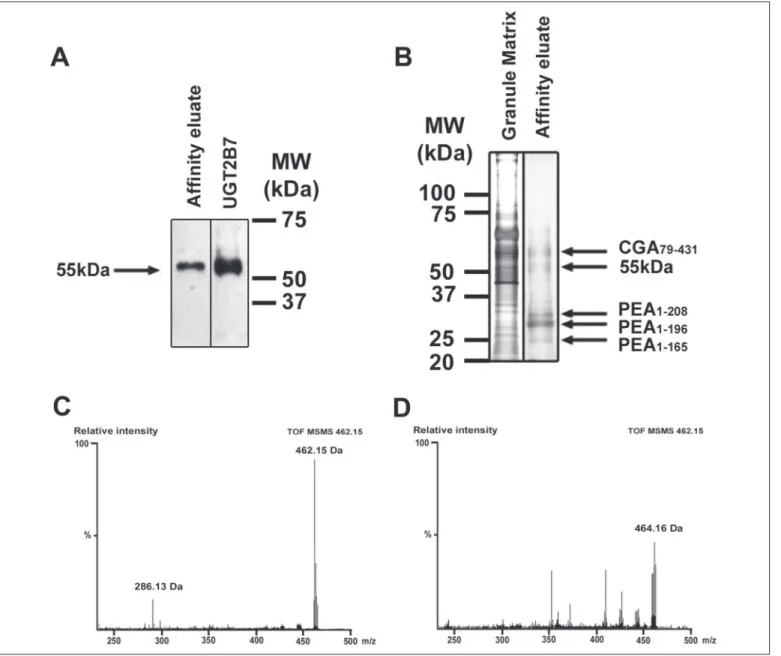
Evidence for M6G de Novo Synthesis by the UGT2B-like Enzyme—A
de novosynthesis experiment was designed to determine whether the
UGT2B-like enzyme present in the intragranular matrix is able to trans-form morphine into M6G. The UGT2B-like enzyme was first purified from the intragranular matrix using an anti-UGT2B affinity column. The resulting eluate was then separated by SDS-PAGE.
Western blot analysis using anti-UGT2B antibody showed an immu-noreactive band at a molecular mass of 55 kDa (Fig. 7A). This immuno-reactive band was identical to the one obtained with 5g of recombi-nant human UGT2B7 (EC 2.4.1.17; Sigma) (Fig. 7A).
To complete this work, a duplicate gel was silver-stained. A band at 55 kDa corresponding to the putative UGT2B-like enzyme as well as other bands were observed (Fig. 7B). Thus, four additional bands were
FIGURE 7. M6G de novo synthesis experiment. A, Western blot analysis of the UGT2B-like enzyme affinity-purified from 4 mg of intragranular matrix and of commercial recombinant human UGT2B7 (5g) using anti-UGT2B antibody recognizing the 55-kDa form. B, silver staining of 5 g of intragranular matrix and of the affinity-purified UGT2B-like enzyme resulting from UGT2B affinity purification performed with 4 mg of intragranular matrix. Silver-stained bands analyzed by MS after digestion corresponded to PEA or CGA fragments.
C, Q-TOF-MS/MS analysis of de novo M6G synthesis performed on the affinity-purified UGT2B-like enzyme collected from an assay with 13 mg of intragranular proteins. D, Q-TOF-MS
analysis of de novo M6G synthesis using the boiled purified UGT2B-like enzyme showing the absence of M6G and morphine.
Morphine-6-glucuronide in Chromaffin Secretory Granules
at Rutgers University on August 11, 2015
http://www.jbc.org/
observed at 24, 26, 28, and 60 kDa. Proteomic analysis indicated the presence in the 24-, 26-, and 28-kDa bands of proenkephalin A (PEA)-(1–165), PEA-(1–196), and PEA-(1–208) fragments, respectively (36) (data not shown). The proteomic analysis of the silver-stained 55-kDa band did not show a similarity to a known protein. The UGT2B enzyme was not identified because of the absence of bovine UGT2B in protein data bases and the poor conservation of UGT2B proteins that was described to be a barrier in the identification of orthologs across species (for review, see Ref. 37).
M6G de novo synthesis experiments were carried out at the intra-granular pH of 5.5 (13) using the affinity eluate. After incubation, the reaction medium was deproteinized and desalted to analyze the pres-ence of newly synthesized M6G. Q-TOF-MS/MS analysis unambigu-ously demonstrated the presence of M6G (462.15 Da) and its character-istic fragmentation profile (286.13 Da) (Fig. 7C). Q-TOF-MS and Q-TOF-MS/MS analyses performed on the reaction buffer (UDP-glu-curonic acid, saccharolactone, and Tris-HCl), the commercial mor-phine, and the affinity eluate prior to incubation indicated the absence of M6G (data not shown). An additional control was performed by incubation with boiled affinity-eluted UGT2B-like enzyme (Fig. 7D). A 464.16-Da component that corresponded to an unidentified molecule was visible; it did not display the specific M6G fragmentation pattern at 286.13Da corresponding to morphine. This last control demonstrated that the formation of M6G was totally abolished upon enzyme heat inactivation.
DISCUSSION
Presence of M6G in Chromaffin Granules—In this work, we have
demonstrated for the first time that M6G (and not morphine) is present in secretory chromaffin granules of bovine adrenal medulla. Because cytochrome P450 enzymes are localized in the endoplasmic reticulum membrane of these cells, we surmise that morphine is synthesized in the endoplasmic reticulum/Golgi apparatus by salutaridine synthase (i.e. cytochrome P450 reductase) (38). Using both immunocytochemical experiments and biochemical techniques, we have identified M6G within chromaffin granules. Some diffuse non-granular labeling can be attributed to morphine-like components present in the endoplasmic reticulum.
Presence of a PEBP䡠M6G Complex in Chromaffin Granules—The
cytoplasmic PEBP, also named Raf-1 kinase inhibitor protein (20), has been identified recently in bovine adrenal gland and chromaffin cells (23, 39). In addition, its presence in the chromaffin intragranular matrix, secretion materials, and serum has been shown by our group (23). PEBP has been described to bind various molecules, including indocyanine, phosphatidylethanolamine, bromosulfophthalein, and hormones such as estradiol-17 and dehydroepiandrosterone (40). It was thus postu-lated that PEBP might represent an organic anion transporter with the same affinity as bovine serum albumin (40). In this study, we have fur-ther demonstrated that, in the chromaffin intragranular matrix, M6G is a novel endogenous ligand for PEBP. These binding data are in agree-ment with a previous report that described PEBP as a morphine-binding protein (22). Under our experimental conditions, it is important to point out that the quantity of M6G could not be determined precisely because part of the M6G bound to PEBP has been lost probably during the deproteinization step (i.e. precipitation of part of the PEBP䡠M6G complex).
Presence of a UGT2B-like Enzyme in Chromaffin Granules—We
sur-mise that, once synthesized, morphine can bind to PEBP in the early granule stage before its presentation to a conversion enzyme (i.e. the UGT2B-like enzyme), producing the final active M6G material. Our
data suggest that such a complex, comprising the UGT2B-like enzyme and PEBP, exists under these experimental conditions.
UGTs represent a superfamily of glycosylated enzymes (35) that are localized in the endoplasmic reticulum (29, 41) and that catalyze the glucuronidation reaction of several drugs and steroid hormones. The glucuronidated products are more polar, less toxic, and easier to excrete through bile or urine. According to their sequence homologies, UGTs fall into two classes: UGT1 and UGT2; the later is subdivided in 2A and 2B components (41). To date, the UGT2B subfamily catabolizes morphine into M3G and M6G (24); UGT2B7 (42, 43) appears to be the only one in this subfamily able to produce M6G from morphine. Using immunochemical techniques, we have shown the presence of UGT2B-like immunoreactivity in chromaffin granules. Our data also demon-strate the presence of glycosylation on the intragranular UGT2B enzyme, a post-translational modification reported to occur on other UGT2B family members (35). The affinity-purified chromaffin granule UGT2B enzyme has been shown to transform morphine into M6G, suggesting for the first time its implication in a metabolic process. Up to now, this property of UGT2B has never been reported.
In our experiments, the proteomic analysis of the silver-stained 55-kDa band (Fig. 7B) did not show similarity to any protein because of the lack of bovine UGT2B sequences in protein data bases or the pres-ence of a new UGT2B variant. Indeed, 12 different human UGT2B genes exist (seven genes and five pseudogenes), and many remnant genes are also present (for review, see Ref. 37), whereas no bovine UGT2B gene or protein has been described. UGT2B proteins show 70% similar sequences, with the higher conservation being present in their C-terminal domains. This low conservation was described to be a bar-rier to the identification of orthologs across species (37).
Relevance of the PEBP䡠M6G Complex in the Stress
Situation—Be-cause PEBP and M6G are bound together within the granule matrix and are co-secreted from bovine chromaffin cells upon stimulation (Fig. 3) (23), it is likely that the PEBP䡠M6G complex exists after secretion and is present in the blood. A large variety of proteolytic enzymes are present in chromaffin secretory granules and act to process precursor proteins such as PEA and chromogranins/secretogranins (36). In contrast, intra-granular PEBP is highly resistant to proteolytic degradation (23). PEBP may also be unaffected by proteolysis in the circulation, thus protecting M6G from bodily clearance (i.e. excretion through urine and/or bile).
Furthermore, our data suggest that the analgesia observed upon acute stress (often related to but never totally explained by enkephalin and corticoid release (44)) might also be due, totally or in part, to M6G secretion, with PEBP acting as a transporter and molecular shield. After secretion, M6G may mediate several systemic actions based on its affin-ity for-opioid receptors present on the cell surface of neurons, neu-roendocrine cells, the endothelium, and immune cells (2, 45– 47).
Interestingly, data from the literature support a separate and select -opioid receptor for M6G. Thus, using an antisense probe targeting G␣i1 in mammals, Rossi et al. (48, 49) found that both heroin- and
M6G-evoked analgesia (but not that induced by morphine) was blocked. These results show that heroin, 6-acetylmorphine, fentanyl, and etonitazine can all produce analgesia through a novel analgesic system that is similar to that activated by M6G (48). Antisense mapping studies on exons 1–3 of MOR-1 (i.e. mu1-opioid receptor-1) in mice
suggested the presence of a novel-receptor subtype responsible for M6G analgesia that may represent a splice variant of MOR-1 (50, 51). In addition, we demonstrated previously that, in three murine macrophage cell lines (J774.2, RAW 264.7, and BAC1.2F5), the-opioid receptor subtype is3because it binds only opiate alkaloids (i.e. M6G), excluding
M3G or any of the opioid peptides tested (52).
at Rutgers University on August 11, 2015
http://www.jbc.org/
Clinical Relevance of M6G in Pain Control—It is known that M6G may have a greater affinity for the1-opioid receptor (responsible for
analgesia) than for the2-opioid receptor (responsible for respiratory
depression), suggesting that M6G can offer a benefit as a systemic anal-gesic (53, 54). M6G is presently under Phase III trial in postoperative pain (CeNeS Pharmaceuticals, Cambridge, UK), highlighting its biolog-ical potential.
Interestingly, for 20 years, cellular therapy using adrenal chromaffin cell transplants has been tested for pain management in both acute and chronic pain models (55, 56). The resulting antinociceptive effect was related to the secretion of opioid peptides and catecholamines from the transplants. Taking into account the present findings and the highly potent analgesic activity of M6G, we suggest that this alkaloid also par-ticipates in the analgesia observed in early experiments. Taken together, the present data may represent important evidence in cell biology and physiology suggesting a new role of M6G in response to stress.
Acknowledgment—We thank N. Aslan for technical help.
REFERENCES
1. Herbert, R. B., Venter, H., and Pos, S. (2000) Nat. Prod. Rep. 17, 317–322 2. Stefano, G. B., Goumon, Y., Casares, F., Cadet, P., Fricchione, G. L., Rialas, C., Peter,
D., Sonetti, D., Guarna, M., Welters, I. D., and Bianchi, E. (2000) Trends Neurosci. 23, 436 – 442
3. Meijerink, W. J., Molina, P. E., and Abumrad, N. N. (1999) Shock 12, 165–173 4. Sonetti, D., Mola, L., Casares, F., Bianchi, E., Guarna, M., and Stefano, G. B. (1999)
Brain Res. 835,137–147
5. Zhu, W., Baggerman, G., Goumon, Y., Casares, F., Brownawell, B., and Stefano, G. B. (2001) Mol. Brain Res. 88, 155–160
6. Zhu, W., Mantione, K. J., Shen, L., Cadet, P., Esch, T., Goumon, Y., Bianchi, E., Sonetti, D., and Stefano, G. B. (2005) Med. Sci. Monit. 11, BR397–BR404
7. Leung, M. K., Dissous, C., Capron, A., Woldegaber, H., Duvaux-Miret, O., Pryor, S., and Stefano, G. B. (1995) Exp. Parasitol. 81, 208 –215
8. Goumon, Y., Casares, F., Pryor, S., Ferguson, L., Brownawell, B., Cadet, P., Rialas, C. M., Welters, I. D., Sonetti, D., and Stefano, G. B. (2000) J. Immunol. 165, 339 –343 9. Stefano, G. B., and Scharrer, B. (1994) Adv. Neuroimmunol. 4, 57– 67
10. Poeaknapo, C., Schmidt, J., Brandsch, M., Drager, B., and Zenk, M. H. (2004) Proc.
Natl. Acad. Sci. U. S. A. 101,14091–14096
11. Boettcher, C., Fellermeier, M., Boettcher, C., Drager, B., and Zenk, M. H. (2005) Proc.
Natl. Acad. Sci. U. S. A. 102,8495– 8500
12. Zhu, W., Cadet, P., Baggerman, G., Mantione, K. J., and Stefano, G. B. (2005) J.
Im-munol. 175,7357–7362
13. Aunis, D. (1998) Int. Rev. Cytol. 181, 213–320
14. Goldstein, A., Barrett, R. W., James, I. F., Lowney, L. I., Weitz, C. J., Knipmeyer, L. L., and Rapoport, H. (1985) Proc. Natl. Acad. Sci. U. S. A. 82, 5203–5207
15. Goumon, Y., and Stefano, G. B. (2000) Mol. Brain Res. 77, 267–269
16. Goumon, Y., Zhu, W., Weeks, B. S., Casares, F., Cadet, P., Bougaeva, M., Brownawell, B., and Stefano, G. B. (2000) Mol. Brain Res. 81, 177–180
17. Epple, A., Nibbio, B., Spector, S., and Brinn, J. E. (1994) Life Sci. 54, 695–702 18. Olsen, G. D. (1975) Clin. Pharmacol. Ther. 17, 31–35
19. Leow, K. P., Wright, A. W., Cramond, T., and Smith, M. T. (1993) Ther. Drug Monit.
15,440 – 447
20. Schoentgen, F., Saccoccio, F., Jolles, J., Bernier, I., and Jolles, P. (1987) Eur. J. Biochem.
166,333–338
21. Yeung, K., Seitz, T., Li, S., Janosch, P., McFerran, B., Kaiser, C., Fee, F., Katsanakis, K. D., Rose, D. W., Mischak, H., Sedivy, J. M., and Kolch, W. (1999) Nature 401, 173–177
22. Grandy, D. K., Hanneman, E., Bunzow, J., Shih, M., Machida, C. A., Bidlack, J. M., and Civelli, O. (1990) Mol. Endocrinol. 4, 1370 –1376
23. Goumon, Y., Angelone, T., Schoentgen, F., Chasserot-Golaz, S., Almas, B., Fukami, M. M., Langley, K., Welters, I. D., Tota, B., Aunis, D., and Metz-Boutigue, M.-H.
(2004) J. Biol. Chem. 279, 13054 –13064
24. Turgeon, D., Carrier, J. S., Levesque, E., Hum, D. W., and Belanger, A. (2001)
Endo-crinology 142,778 –787
25. Lotsch, J., and Geisslinger, G. (2001) Clin. Pharmacokinet. 40, 485– 499
26. Romberg, R., Olofsen, E., Sarton, E., den Hartigh, J., Taschner, P. E., and Dahan, A. (2004) Anesthesiology 100, 120 –133
27. Smith, A. D., and Winkler, H. (1967) Biochem. J. 103, 480 – 482
28. Metz-Boutigue, M.-H., Garcia-Sablone, P., Hogue-Angeletti, R., and Aunis, D. (1993)
Eur. J. Biochem. 217,247–257
29. Levesque, E., Turgeon, D., Carrier, J. S., Montminy, V., Beaulieu, M., and Belanger, A. (2001) Biochemistry 40, 3869 –3881
30. Lugardon, K., Chasserot-Golaz, S., Kieffer, A. E., Maget-Dana, R., Nullans, G., Kieffer, B., Aunis, D., and Metz-Boutigue, M.-H. (2001) J. Biol. Chem. 276, 35875–35882 31. Marban, C., Redel, L., Suzanne, S., Van Lint, C., Lecestre, D., Chasserot-Golaz, S.,
Leid, M., Aunis, D., Schaeffer, E., and Rohr, O. (2005) Nucleic Acids Res. 33, 2318 –2331
32. Gasnier, C., Lugardon, K., Ruh, O., Strub, J.-M., Aunis, D., and Metz-Boutigue, M.-H. (2004) Proteomics 4, 1789 –1801
33. Fraering, P. C., Ye, W., Strub, J.-M., Dolios, G., LaVoie, M. J., Ostaszewski, B. L., Van Dorsselaer, A., Wang, R., Selkoe, D. J., and Wolfe, M. S. (2004) Biochemistry 43, 9774 –9789
34. Armstrong, S. C., and Cozza, K. L. (2003) Psychosomatics 44, 515–520
35. Barbier, O., Girard, C., Breton, R., Belanger, A., and Hum, D. W. (2000) Biochemistry
39,11540 –11552
36. Goumon, Y., Lugardon, K., Gadroy, P., Strub, J.-M., Welters, I. D., Stefano, G. B., Aunis, D., and Metz-Boutigue, M.-H. (2000) J. Biol. Chem. 275, 38355–38362 37. Mackenzie, P. I., Bock, K. W., Burchell, B., Guillemette, C., Ikushiro, S., Iyanagi, T.,
Miners, J. O., Owens, I. S., and Nebert, D. W. (2005) Pharmacogenet. Genomics 15, 677– 685
38. Amann, T., Roos, P. H., Huh, H., and Zenk, M. H. (1995) Heterocycles 40, 425– 440 39. Frayne, J., Ingram, C., Love, S., and Hall, L. (1999) Cell Tissue Res. 298, 415– 423 40. Bernier, I., Tresca, J. P., and Jolles, P. (1986) Biochim. Biophys. Acta 871, 19 –23 41. Mackenzie, P. I., Owens, I. S., Burchell, B., Bock, K. W., Bairoch, A., Belanger, A.,
Fournel-Gigleux, S., Green, M., Hum, D. W., Iyanagi, T., Lancet, D., Louisot, P., Magdalou, J., Chowdhury, J. R., Ritter, J. K., Schachter, H., Tephly, T. R., Tipton, K. F., and Nebert, D. W. (1997) Pharmacogenetics 7, 255–269
42. Stone, A. N., Mackenzie, P. I., Galetin, A., Houston, J. B., and Miners, J. O. (2003) Drug
Metab. Dispos. 31,1086 –1089
43. Yamada, H., Ishii, K., Ishii, Y., Ieiri, I., Nishio, S., Morioka, T., and Oguri, K. (2003) J.
Toxicol. Sci. 28,395– 401
44. Seematter, G., Binnert, C., Martin, J. L., and Tappy, L. (2004) Curr. Opin. Clin. Nutr.
Metab. Care 7,169 –173
45. Massotte, D., and Kieffer, B. L. (1998) Essays Biochem. 33, 65–77
46. Fimiani, C., Arcuri, E., Santoni, A., Rialas, C. M., Bilfinger, T. V., Peter, D., Salzet, B., and Stefano, G. B. (1999) Cancer Lett. 146, 45–51
47. Cadet, P., Rasmussen, M., Zhu, W., Tonnesen, E., Mantione, K. J., and Stefano, G. B. (2004) Front. Biosci. 9, 3176 –3186
48. Rossi, G. C., Brown, G. P., Leventhal, L., Yang, K., and Pasternak, G. W. (1996)
Neurosci. Lett. 216,1– 4
49. Rossi, G. C., Pan, Y. X., Brown, G. P., and Pasternak, G. W. (1995) FEBS Lett. 369, 192–196
50. Rossi, G. C., Leventhal, L., Pan, Y. X., Cole, J., Su, W., Bodnar, R. J., and Pasternak, G. W. (1997) J. Pharmacol. Exp. Ther. 281, 109 –114
51. Schuller, A. G., King, M. A., Zhang, J., Bolan, E., Pan, Y. X., Morgan, D. J., Chang, A., Czick, M. E., Unterwald, E. M., Pasternak, G. W., and Pintar, J. E. (1999) Nat. Neurosci.
2,151–156
52. Makman, M. H., Dvorkin, B., and Stefano, G. B. (1995) Eur. J. Pharmacol. 273, R5–R6 53. Frances, B., Gout, R., Campistron, G., Panconi, E., and Cros, J. (1990) Prog. Clin. Biol.
Res. 328,477– 480
54. Romberg, R., Olofsen, E., Sarton, E., Teppema, L., and Dahan, A. (2003)
Anesthesiol-ogy 99,788 –798
55. Duplan, H., Bes, J. C., Tafani, M., Sallerin, B., Sagen, J., Ohayon, E., Lazorthes, Y., and Tkaczuk, J. (2000) Exp. Neurol. 163, 331–347
56. Sol, J. C., Sallerin, B., Larrue, S., Li, R. Y., Jozan, S., Tortosa, F., Mascott, C., Carraoue, F., Tafani, M., and Lazorthes, Y. (2004) Exp. Neurol. 186, 198 –211
Morphine-6-glucuronide in Chromaffin Secretory Granules
at Rutgers University on August 11, 2015
http://www.jbc.org/
Marie-Hélène Metz-Boutigue
Schoentgen, Dominique Aunis and
Welters, Alain Van Dorsselaer, Françoise
Olivier Rohr, George B. Stefano, Ingeborg D.
Jean-Marc Strub, Sylvette Chasserot-Golaz,
Glattard, Céline Marban, Claire Gasnier,
Yannick Goumon, Arnaud Muller, Elise
in Chromaffin Cell Secretory Granules
Identification of Morphine-6-glucuronide
Bioinformatics:
doi: 10.1074/jbc.M502298200 originally published online January 24, 2006
2006, 281:8082-8089.
J. Biol. Chem.
10.1074/jbc.M502298200
Access the most updated version of this article at doi:
.
JBC Affinity Sites
Find articles, minireviews, Reflections and Classics on similar topics on the
Alerts:
When a correction for this article is posted
•
When this article is cited
•
to choose from all of JBC's e-mail alerts
Click here
http://www.jbc.org/content/281/12/8082.full.html#ref-list-1
This article cites 56 references, 12 of which can be accessed free at
at Rutgers University on August 11, 2015
http://www.jbc.org/