HAL Id: hal-02556202
https://hal.archives-ouvertes.fr/hal-02556202
Submitted on 27 May 2020
HAL is a multi-disciplinary open access archive for the deposit and dissemination of sci-entific research documents, whether they are pub-lished or not. The documents may come from teaching and research institutions in France or abroad, or from public or private research centers.
L’archive ouverte pluridisciplinaire HAL, est destinée au dépôt et à la diffusion de documents scientifiques de niveau recherche, publiés ou non, émanant des établissements d’enseignement et de recherche français ou étrangers, des laboratoires publics ou privés.
Characterization of a black residue in a decorated
Neolithic pot from Dikili Tash (Greece): an unexpected
result
Yannis Maniatis, Zoï Tsirtsoni
To cite this version:
Yannis Maniatis, Zoï Tsirtsoni. Characterization of a black residue in a decorated Neolithic pot from Dikili Tash (Greece): an unexpected result. Archaeometry, Wiley, 2002, 44 (2), pp.229-239. �10.1111/1475-4754.t01-1-00055�. �hal-02556202�
229
CHARACTERIZATION OF A BLACK RESIDUE IN A DECORATED NEOLITHIC POT FROM DIKILI TASH,
GREECE: AN UNEXPECTED RESULT Y. MANIATIS
Laboratory of Archaeometry, NCSR ‘Demokritos’, 153 10 Aghia Paraskevi, Attiki, Greece
and Z. TSIRTSONI
Protohistoire égéenne—UMR 7041 (ArScAn), Maison R. Ginouvès, 21 allée de l’Université, 92023 Nanterre Cedex, France
A black depositional layer on the inner surface of a Neolithic carinated vessel from the archaeological site of Dikili Tash in Eastern Macedonia was examined scientifically. The layer was initially considered as decomposed organic matter and interest was focused on identifying the original organic contents. The scientific investigation, which included FTIR spectroscopy, analytical SEM examination and optical microscopy, revealed that the black substance was not organic but a pure iron oxide layer deposited on the vessel’s inner surface, and reduced in places to black iron oxides, during a destructive fire. The conclusion is that this layer represents the remnants of the vessel’s original content, which was a red hematite pigment. This unexpected find provides, for the first time, a missing link in the evidence of pigments used in Neolithic times, previously attested to only by finished products.
KEYWORDS: DIKILI TASH, GREECE, POTTERY, RESIDUE, CONTENTS, HEMATITE,
PIGMENT, NEOLITHIC, FTIR, SEM, OPTICAL MICROSCOPY, SECONDARY USE, POST-FIRING HEATING
INTRODUCTION
The prehistoric settlement of Dikili Tash is located at the southeastern edge of the Drama plain, in Eastern Macedonia, Greece. Systematic excavations carried out there during the period 1960–70, in a Greek–French collaboration, have allowed the establishment of the settlement’s stratigraphic and chronological sequence (Deshayes 1970; Koukouli and Romiopoulou 1992; Treuil 1992). Four main occupational phases have been distinguished (phases I–IV), each including more than one habitation layer, going from the beginning of the Late Neolithic period (the second half of the sixth millennium bc) to the Late Bronze Age (around 1000 bc). A new research programme, begun in 1986, has undertaken further exploration of habitation levels belonging to the two main phases of the Neolithic period: phase I (Late Neolithic I, c. 5400– 4700 bc) and phase II (Late Neolithic II, c. 4500–3300 bc) (Koukouli et al. 1996).
Excavations in the main French sector V, on the south slope of the tell, were carried out between 1987 and 1995. At the end of the last campaign, in September 1995, an astonishing
Figure 1 The carinated collared pot with painted decoration found on the floor of a Late Neolithic house (c. 5000 bc) at Dikili Tash (Eastern Macedonia, Greece); shown here after restoration (height 14.5 cm, mouth diameter 11.3 cm).
discovery was made: while cleaning up a mass of debris that had fallen on the floor of a house unit in a phase I level, dated both by radiocarbon and thermoluminescence (Guibert and Roque 2000; Maniatis unpublished results) at about 5000 bc, the archaeologists came face to face—almost literally—with a ‘bucranium’, an ox’s skull covered with originally raw clay, that had been baked during the conflagration that destroyed the house (Treuil 1996; Treuil and Darcque 1998). Actually, the ‘bucranium’ had fallen face down, with its mouth inside a clay pot that was lying on the floor beneath the bucranium’s original location. Next to this, another group of three ceramic vases were lying intact on the house floor: one was an open vessel (bowl) and the other two were closed (carinated collared pots). Surprisingly, the bowl and one of the two pots each contained a dozen stone and bone tools (Martinez 1997; Tsirtsoni 1997). The second pot (height 14.5 cm, mouth diameter 11.3 cm; see Fig. 1) seemed to contain nothing but earth, which had obviously fallen into it at the moment of destruction: examination by water sieving revealed no visible trace of any contents. Once this second pot had been emptied, it was noticed that its interior surface was covered in places by a thick layer of black ‘crust’ (Fig. 2).
Clear evidence of destruction by an intense fire had been recorded for all of the finds. The pots showed a certain degree of deformation (Fig. 1), and the bone and stone tools had been heavily damaged by the fire. The most obvious first assumption for the black crust in one of the three pots was that of an organic residue, probably a foodstuff, which had been burnt during the destructive fire.
Before proceeding to a detailed organic residue analysis for the identification of the organic compounds, it was thought useful to run a Fourier transform infra-red (FTIR) analysis of the black residue, that would allow us to confirm, at a preliminary level, the organic nature of the residue; and also to examine the interior surface of the vase with an optical microscope and a scanning electron microscope (SEM), to determine the morphology of the residue and its state of preservation. An additional aim was to examine the ceramic body using the SEM, in order to determine the firing temperature of the vessel and possibly identify the effect of post-firing heating by the destructive fire.
SAMPLES AND ANALYTICAL TECHNIQUES
A number of sherds coming from various parts of the vessel (e.g., the neck and further down below the shoulder) were used for the examination. The interior of the sherds was partially or wholly covered by the black residue or crust, while the exterior had only a red/orange slip.
The sherds as received were examined first in detail and photographed with an optical stereoscopic microscope. Following that, small samples were chipped off from specific places of the inner and outer surface and also fresh-fractured surfaces at a full cross-section of the body were obtained. These samples were not treated any further but just placed on SEM stubs and coated with a thin carbon coating.
The scanning electron microscope used was a Philips 515, equipped with an X-ray energy-dispersive microanalysis system (EDAX PV-9900). The silicon–lithium (Si–Li) X-ray detector has a removable window that also allowed the detection of light elements such as C, O and N, down to boron (atomic number = 5).
Finally, a small sample was obtained from the black crust, by lightly scratching the black surface with a scalpel, and made into a pellet, using dry potassium bromide (KBr) for Fourier transform infra-red (FTIR) spectroscopic analysis with a Bruker instrument.
RESULTS AND DISCUSSION
The first surprising result came from the FTIR spectra. The black residue or crust on the inner surface of the vessel seemed to have no organic content at all. Despite repeated measurements, and having taken care to remove for measurement only the black layer from the surface, the only phase present and dominating the FTIR spectrum was calcite (CaCO3), accompanied by a
much lower Si–O absorption (Fig. 3). If present, organic compounds should produce sharp lines in the spectral region near 3000 cm–1
. This result indicated that the black crust was not of an
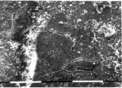
Figure 2 The parts of the pot before restoration: the black ‘crust’ is visible covering one side and the bottom of the inner surface.
Figure 4 A secondary electron SEM image of the interior surface of the vase, showing quite well crystallized and large iron oxide particles. The scale bar represents 10 µm.
Figure 3 The FTIR spectrum of the black residue on the internal surface.
4000 3750 3500 3250 3000 2750 2500 2250 2000 1750 1500 1250 1000 750 500 0.0 0.1 0.2 0.3 0.4 0.5 BLACK CRUST H2O CO2 Si-O CaCO3 CaCO3 CaCO3 Transmittance (%) Wavenumber cm−1
organic nature, but the presence of such a quantity of CaCO3 in a black layer confused the issue
and demanded further investigation.
Scanning electron microscope examination and microanalysis of the black layer on the inner surface showed a large concentration of discrete and well crystallized iron oxide particles on the surface (Fig. 4). Their dimensions varied from place to place on the surface from small particles, less than 1 µm across, to quite large particles, about 5 µm across. In some places, these particles were partially covered or mixed with Ca-rich formations (Fig. 5) of no specific crystal structure and looking mostly amorphous, although these sometimes tended to form structures that resembled the typical rhombic form of freely crystallizing calcite. Detailed spot
analyses on the iron oxide particle layer indicated that the particles were not mixed or covered by clay minerals, as the amounts of silica, alumina and potassia present were negligible and could be considered as traces of soil contamination. This meant that the iron oxide layer was definitely not an iron-rich coating, or slip painted over parts of the inner surface, as it would have to contain sufficient clay material so that it could adhere on to the surface (Middleton 1987). A pure iron-oxide layer was never applied by itself on ceramic surfaces, as it is easily rubbed off, and it is doubtful if it was applied by itself even for specific decoration elements. Calcium was present in small amounts and clearly came from between and around the par-ticles, forming in many instances a kind of bonding material. Clay minerals were practically absent from the Ca-rich formations, but some traces could obviously be detected due to soil contamination.
An examination of the external surface of the vase revealed a heavily damaged surface, exhibiting cracks, detachments and abnormal curving of the surface (Fig. 6). The analysis of this surface layer was different from that of the black inner surface. It exhibited the char-acteristic chemistry of an iron-rich clay slip; in other words, a typical clay concentration with an increased iron content (Noll 1976–7; Aloupi and Maniatis 1990). Some calcium was also present on this surface, and most probably originated from soil depositions.
Examination of the freshly fractured surfaces from the bulk of the ceramic body and microanalysis revealed that the vase is made of a non-calcareous clay. The amounts of lime and potassia are of the order of 4%, and the iron oxide content is about 7–8%. This type of clay can be considered as low refractory according to the nomenclature of Maniatis and Tite (1978–9). The most interesting observation, however, was an extensive network of bloating pores of diameter up to 10 µm, almost everywhere inside the body and more so closer to the external surface (Fig. 7). These bloating pores might be the result of: (1) an original firing of the vase in an intense reducing atmosphere, at a temperature of about 900°C (Maniatis and Tite 1975, 1981); (2) a rapid original firing in an open fire (bonfire) (Maniatis and Tite 1975, 1981); or (3) a rapid or normal original firing in a mixed atmosphere, followed by intense post-firing heating in a destructive fire.
Figure 5 A secondary electron SEM image of the interior surface of the vase. The extended darker regions are Ca-rich formations. The scale bar represents 10 µm.
Figure 6 A secondary electron SEM image of the exterior surface of the vase, showing the iron-rich slip that is heavily damaged due to the destructive fire. The scale bar represents 10 µm.
Figure 7 A secondary electron SEM image of a freshly broken surface of the ceramic body, showing an extensive network of bloating pores. The scale bar represents 10 µm.
Given the extent of vitrification and the non-calcareous nature of the clay, the first option, of an original intense reducing firing, must be excluded, as it would have resulted in an intense grey/ black ceramic body throughout.
The second option by itself can be also excluded, as it usually produces much finer bloating pores—and mostly in the central part of the body, which has no time to oxidize during the rapid firing (Maniatis and Tite 1975). In this case one would also expect a ‘sandwich’ effect, which
is not observed here. Rapid firings are frequently observed in Neolithic times, as a result of a bonfire type of firing, and produce an orange or red overall effect, especially when a decoration or slip is applied (Maniatis et al. 1988). However, a simple original firing without any post-firing effect cannot explain the present state of the vase under study.
The third option is therefore, the most likely one, also taking into account the fact that the bloating is uniform and more pronounced nearer the external surface. This type of post-firing bloating can occur in ceramic bodies that have originally been fired in a mixed atmosphere at relatively low firing temperatures (750–800°C). The atmosphere in these firings is usually reducing at the beginning, while the temperature is still low (up to 600–700°C), but becomes oxidizing during the final stages. The end result is a brown-coloured body (perhaps with a light grey core, especially if the firing was rapid), exhibiting an orange/reddish outer surface colour if an iron-rich slip is applied. No bloating occurs during this original firing, or perhaps some limited fine bloating pores may occur in the central part of the body. However, the refiring of these ceramics in an oxidizing atmosphere at a higher temperature (900°C or higher) activates a kind of ‘incipient’ bloating (Maniatis and Tite 1981), even though no bloating was present in their original state. Judging from the size and extent of the bloating pores in the Dikili Tash vase, the damage to the external iron-rich clay slip covering the surface of the vase and the signs of overall deformation (Fig. 1), we can estimate a post-firing heating during the destruct-ive fire that reached temperatures of the order of 950–1000°C, perhaps for a brief time, in the vicinity of the pot. This intense heating was also responsible for the serious damage to the bone and stone tools found inside the adjacent pots.
In the light of the above evidence for a severely destructive fire, and for heat being applied on the outside of the vessel, one can now assume that the curious black crust on the internal surface, which consists of distinct iron oxides, represents an accumulation or deposition of iron oxides that have been converted to magnetite (Fe3O4), thus producing the black crust in situ.
This might have occurred due to lack of oxygen in these parts of the interior of the vessel during the destructive fire.
In order to clarify this and at the same time investigate in more detail the practically amor-phous calcite material that acts as a bonding between the iron oxide particles, we re-examined the surface with the black crust under an optical microscope. This examination revealed that the black colour is the overall optical effect of a dark purple layer with black patches spread all over it. The purple and black areas had no obvious morphological difference; they both consisted of small iron oxide particles sitting on the surface. This was also verified with a new SEM examination following the optical microscope examination, which showed no difference be-tween the purple areas and the black patches. They were all iron oxide particles of different sizes (Fig. 4). When a strong magnet was brought close to the surface under the optical microscope, it was observed that both the black and the purple particles were strongly attracted, and could easily fly from the surface to the magnet, especially after some tapping or light scratching of the surface. All of this confirmed the evidence that the black crust was made of a layer of iron oxides, deposited on the surface and converted in situ either to magnetite, which formed the black patches, or to maghemite (γ-Fe2O3), which formed the purple areas, obviously
under the action of a partially reducing firing, during the destructive fire. The conversion of iron oxides to maghemite and magnetite under weaker or stronger reducing conditions, respectively, has been documented by Longworth and Tite (1977).
With regard to the calcitic material detected on the surface, the optical microscope examina-tion revealed a transparent material that had covered everything. In some places, where this calcitic cover had flaked off, one could see the purple/black iron oxide layer underneath. The
appearance, extent, morphology and chemistry of this calcite layer closely resemble those of natural depositions of calcite (e.g., in caves, on walls, as limescale in pipes, and so on) from an aqueous saturated solution. It is therefore clear that this is a pure calcitic encrustation layer that was deposited during burial and that covered the inner surface, and the iron oxide layer on it, the latter obviously pre-dating the encrustation event.
Combining all of the above evidence, one is led to the conclusion that a layer of almost pure iron oxides had covered parts of the inner surface of the vessel. This layer was there before the destructive fire, which partially converted the iron oxides to maghemite and magnetite as a result of the partial reducing conditions prevailing in the inner part of the vessel (the mouth of the vessel was most probably covered by something that fell on it during the destruction of the house). Later, and during burial, a pure calcite encrustation was formed on the surface. This covered, trapped and bonded the iron oxide layer on the surface, preserving it in this way until the present day. The pure iron oxide layer deposition on the inner surface can therefore repres-ent nothing other than the remnants of the original substance contained in the vessel; that is, an iron oxide pigment. Given the partial conversion of the iron oxides to maghemite and magnetite, one can assume that the iron oxide pigment contained in this vessel could most probably have been pure hematite (α-Fe2O3), a well-known red pigment. The possibility, initially, of the
existence of iron oxides in the form of hydroxides (brown or yellow ochre) cannot be excluded, although it is not very likely, given the purity and crystallinity of the particles, as well as the overall archaeological evidence (see below). The way in which this iron oxide layer has covered the inside of the vessel and the lack of clay minerals in it suggest that the iron oxide pigment was in a powder form, which—in some places—had stuck to the inner surface. This means that the pigment had been crushed, cleaned and stored in the vessel, ready for use.
THE ARCHAEOLOGICAL IMPLICATIONS
The discovery of pure iron oxide pigment (most probably hematite, a red colouring material), inside a Neolithic pot from Dikili Tash is of great interest from many points of view.
It is, indeed, the first time in Greece, as far as we know, that such a material has been discovered in its raw state, and clearly identified as such, in a Neolithic context. Yellow and red colouring materials are mentioned among the finds from a large pit at the Neolithic site of Elateia, in Central Greece, but no identification of their exact nature has been made (Weinberg 1965, 195). Also, although traces of a red colour have been found on the surface of querns and grinders from several Neolithic settlements, in none of these cases has there been a clear determination of their chemical composition (Runnels 1981, 149; Papathanassopoulos 1996, 215: cat. no. 15–16). Traces of red ochre were recently reported on the surface of a quern and a grinder, and possibly in a jar fragment, from a LN II habitation level at Kryoneri, in Eastern Macedonia; however, identification once again seems to rely on visual criteria and not on analytical results (Malamidou 1997, 514).
The use of hematite as a possible component of Neolithic red pigments is attested to by various decorated objects or wall plasters. Objects include, first, clay vessels, decorated with a purple–red paint applied after firing, usually described in the archaeological literature as a ‘crust’, in order to be distinguished from the red paints made of iron-rich clays that are applied before firing (slip) (Yiouni forthcoming). Such vessels are found predominantly in Northern Greece (Thessaly and Macedonia), as well as in many other parts of the Balkans, by the end of the Late Neolithic (LN II, from the second half of the fifth to the second half of the fourth millennium bc). Clay figurines preserving traces of a similar dark red paint, usually associated
with an incised decoration, are also known from the same horizon (Marangou 1992, 141). In Thessaly, marble figurines with an elaborate red-painted decoration are known from the begin-ning of the Late Neolithic (LN I, from the second half of the sixth to the first half of the fifth millennium bc) (Papathanassopoulos 1996, 303–5: cat. nos. 213–14, 216–17).
The use of hematite in architectural elements is also observed, but more rarely. A very small number of fragments from Neolithic red-painted wall plasters are known in Greece. This would seem to be mostly due to the poor state of preservation of Neolithic architectural remains in general, rather than to the scarcity of the practice. Coloured plasters have, indeed, been in use in other parts of the world (Mesopotamia, Anatolia, the Near East) since at least the ninth millennium bc; that is, almost as early as the first constructions with walls made of earth (see the brief overview in Cameron 1972, 311). In the whole Balkan region, Greece included, we know of no more than a dozen examples, the oldest going back to the beginning of the sixth millennium bc (Treuil 1983, 252, 270–2). At the Dikili Tash site itself, three red-coloured plaster fragments come from the 1987–95 excavations in sector V; analysis using scientific methods (infrared spectroscopy, X-ray diffraction and Raman spectroscopy) has confirmed that the red colour is due to hematite (Dandrau 1997).
The presence of a pure pigment in a 5000 bc vessel from Dikili Tash is not only in accord-ance with the local finds, namely the plaster fragments (since the ‘red-crusted’ vases and figurines are chronologically slightly later here), but also fills a gap in the archaeological record, linking the known finished products with the original raw materials.
With regard to the origin of this pigment, the evidence is still scarce. No ochre mine is known in the immediate region around Dikili Tash, the nearest one known to date being at Tzines, on the island of Thasos, directly opposite to the coast (Koukouli and Weisgerber 1993), but iron-rich deposits do exist in many parts of the Lekani mountains (i.e., the mountains at the foot of which the site of Dikili Tash lies) at a distance of 10–15 km from the settlement (Koukouli 1990, 499). No firm statement can be made about the provenance of the ochre found at Dikili Tash without making comparisons with samples from the above-mentioned sources. Yet, we can convincingly assume that this would not have been an ‘exotic’ material for Dikili Tash’s Neolithic inhabitants.
From a technological point of view, having the pigment in a powder form indicates that the mineral had been prepared for use as a paint component, rather than being in a bulky form for use in a pencil.
The container in which the pigment was stored, a carinated collared pot with painted decora-tion, seems at first sight rather unusual, considering the vessel’s assumed original function. Indeed, vessels of this kind are interpreted as containers for liquids, probably in socially valued contexts (Tsirtsoni 2000, 2001). This does not mean, however, that—in some circumstances— they could not have served for other uses, as long as their morphology was suitable. One does not have to search very far to find further examples of the secondary use of vessels; one need only take a look in the immediate vicinity of the pot in the excavation to find a similar carinated collared pot and a bowl, both having as contents stone and bone tools. It is obvious that neither of these two vessels, which are very different from each other, was originally made with the purpose of containing tools. Yet, this is exactly what happened at that particular moment prior to the destruction of the house.
This concentration of vessels with unexpected, obviously secondary, contents in this particu-lar part of the house could seem suspicious to some, especially if combined with the presence of the ‘bucranium’ in the same room. The idea of a ‘sanctuary’, with offerings for the ‘ox-god’ brought in special containers as part of particular rites, is just too attractive to be rejected at
once. The presence of hematite among the presumed offerings would seem to fit such a picture perfectly, given the much repeated theories about the symbolic, or even magical, qualities of this blood-coloured mineral in prehistoric societies (Schmandt-Besserat 1980).
However, there can be much simpler—or at least less imaginative—explanations. Even if we concede that the ‘bucranium’ had some kind of religious or symbolic value (which is far from certain; see Treuil and Darcque 1998, 25), this does not make the whole room or the whole building a sanctuary. Neolithic people could have just used those empty vessels to store things that they intended to use later: a handful of blades and stone-axes, and a pack of crushed ochre. Was this a ritual niche or a common reserve? As long as we continue to lack clear evidence about both Neolithic peoples’ religious practices and their everyday lives, the question will remain open. Large-scale systematic excavations and multidisciplinary research, such as those carried on at Dikili Tash, could help to elucidate both of these aspects.
CONCLUSIONS
A simple archaeological question concerning the identification of a black substance inside a Neolithic vessel, initially considered to be an organic material which was assumed to have been decomposed by burning, engendered a scientific investigation that turned out to be quite com-plicated, but very interesting and absorbing, and led to unexpected results.
The black substance was not organic, but was a pure iron oxide pigment, deposited on the vessel’s inner surface and reduced in places to black iron oxides, during a destructive fire. The layer was subsequently covered, during burial, by a calcitic encrustation, which preserved it in situ. It is concluded that this layer represents the remnants of the vessel’s original content, which was an iron-oxide ground pigment—most probably hematite.
This result was unexpected and surprising at first, but it was then found to be very interesting and important archaeologically, because it provided a missing link in the evidence for the use of pigments in Neolithic times, which was previously attested to only by finished products, namely wall plasters.
Finally, this work has shown that when the archaeologist’s persistent search for evidence is combined with a detailed scientific examination, this can lead to the extraction of important historical information that would otherwise have been lost.
ACKNOWLEDGEMENTS
This work was performed while Z. Tsirtsoni was a member of the French School at Athens (EFA); we express our gratitude to the EFA director, Professor R. Etienne, for his substantial support. We also thank Professor R. Treuil for his permission to study the materials. Thanks are also due to Dr Y. Facorellis, for some initial analyses with the SEM-EDXA, and to Mrs S. Kalozoumi, for preparing the samples for SEM examination.
REFERENCES
Aloupi, E., and Maniatis, Y., 1990, Investigation of the technology of manufacture of local LBA Theran pottery: body and pigment analysis, in Thera and the Aegean world III (ed. D. A. Hardy), Vol. 1, 459–69, Thera Foundation. Cameron, M. A. S., 1972, Appendix IV. The plasters, in Myrtos: an Early Bronze Age settlement in Crete (ed.
P. M. Warren), The British School of Archaeology at Athens Supplement, 7, 305–14, Thames and Hudson, London.
Dandrau, A., 1997, La construction en terre dans le monde égéen au Néolithique et à l’Age du Bronze: les matériaux
et leurs propriétés, Ph.D. thesis, Université de Paris I.
Deshayes, J., 1970, Les fouilles de Dikili Tash et l’archéologie yougoslave, Zbornik Radova Narodnog Museja u
Beogradu, 6, 21–43.
Guibert, P., and Roque, C., 2000, La datation par thermoluminescence, Dossiers d’Archéologie, 253, 16–23. Koukouli-Chryssanthaki, Ch., 1990, The mines of the Thasians’ coast, Mélanges D. Lazaridis: cité et territoire en
Macédoine et Thrace antiques, Recherches Franco-Helléniques, 1, Greek Ministry of Culture, École française
d’Athènes, Athens (in Greek).
Koukouli-Chryssanthaki, Ch., and Romiopoulou, A., 1992, Excavations in the Greek sector of the prehistoric settle-ment Dikili Tash (1961–1967), in Proceedings of the International Symposium for ancient Thessaly in the memory
of D. R. Theocharis, 226–48, Dimosievmata tou Archaiologikou Deltiou, 48, Athens (in Greek).
Koukouli-Chryssanthaki, Ch., and Weisgerber, G., 1993, Prehistoric ochre mines on Thasos, Archaiologiko Ergo stin
Makedonia kai Thraki, 7, 541–58 (in Greek).
Koukouli-Chryssanthaki, Ch., Treuil, R., and Malamidou, D., 1996, Prehistoric settlement at Philippi ‘Dikili Tash’: ten years of excavations, Archaiologiko Ergo stin Makedonia kai Thraki, 10(2), 681–704 (in Greek).
Longworth, G., and Tite, M. S., 1977, Mössbauer and magnetic susceptibility studies of iron oxides in soils from archaeological sites, Archaeometry, 19(1), 3–14.
Malamidou, D., 1997, The excavation of the prehistoric settlement of Kryoneri, near Nea Kerdyllia, Archaiologiko
Ergo stin Makedonia kai Thraki, 11, 509–22 (in Greek).
Maniatis, Y., and Tite, M. S., 1975, A scanning electron microscope examination of the bloating of fired clays,
Transactions and Journal of the British Ceramic Society, 74, 229–32.
Maniatis, Y., and Tite, M. S., 1978–9, Examination of Roman and medieval pottery using the scanning electron microscope, Acta Praehistorica et Archaeologica, 9/10, 125–30.
Maniatis, Y., and Tite, M. S., 1981, Technological examination of Neolithic—Bronze Age pottery from central and south east Europe and from the Near-East, Journal of Archaeological Science, 8, 59–76.
Maniatis, Y., Perdikatsis, V., and Kotsakis, K., 1988, Assessment of in-site variability of pottery from Sesklo, Thessaly,
Archaeometry, 30, 264–74.
Marangou, Chr., 1992, Eιδ ´ωλια: figurines et miniatures du Néolithique Récent et du Bronze Ancien en Grèce, BAR International Series, 576, Oxford.
Martinez, S., 1997, Dikili Tash à l’époque néolithique: l’homme et ses outils, Dossiers d’Archéologie, 222, 36–9. Middleton, A. P., 1987, Technological investigation of the coatings on some ‘haematite-coated’ pottery from southern
England, Archaeometry, 29(2), 250–61.
Noll, W., 1976–7, Neolithische und chalkolithische bemalte Keramik des Vorderen Orients: Material, Rohstoffe und Herstellungstechnik, Acta Praehistorica et Archaeologica, 7/8, 15–47.
Papathanassopoulos, G. A. (ed.), 1996, Neolithic culture in Greece (catalogue of exposition), N. P. Goulandris Founda-tion, Athens.
Runnels, C. N., 1981, A diachronic study and economic analysis of millstones from the Argolid, Greece, Ph.D. thesis, Indiana University.
Schmandt-Besserat, D., 1980, Ocher in prehistory: 300 000 years of the use of iron ores as pigments, in The coming of
the Age of Iron (eds. T. A. Wertime and J. D. Muhly), 127–50, Yale University Press, New Haven/London.
Treuil, R., 1983, Le Néolithique et le Bronze Ancien égéens: les problèmes stratigraphiques et chronologiques, les
techniques, les hommes, Bibliothèque des Écoles françaises d’Athènes et de Rome, 248, Paris.
Treuil, R., 1992, Dikili Tash, village préhistorique de Macédoine orientale, I. Fouilles de Jean Deshayes (1961–1975),
vol. 1, Bulletin de Correspondance Hellénique Supplément 24, Paris.
Treuil, R., 1996, Dikili Tash, Bulletin de Correspondance Hellénique, 120, 865–74.
Treuil, R., and Darcque, P., 1998, Un ‘bucrane’ néolithique à Dikili Tash (Macédoine orientale): parallèles et perspect-ives d’interprétation, Bulletin de Correspondance Hellénique, 122, 1–25.
Tsirtsoni, Z., 1997, Dikili Tash à l’époque néolithique: morphologie et fonctions des poteries, Dossiers d’Archéologie,
222, 28–35.
Tsirtsoni, Z., 2000, Les poteries du début du Néolithique Récent en Macédoine: 1. Les types des récipients, Bulletin de
Correspondance Hellénique, 124, 1–55.
Tsirtsoni, Z., 2001, Les poteries du début du Néolithique Récent en Macédoine: 2. Les fonctions des récipients, Bulletin
de Correspondance Hellénique, 125, 1–39.
Weinberg, S., 1965, Ceramics and the supernatural: cult and burial evidence in the Aegean world, in Ceramics and man (ed. F. Matson), 187–201, Viking Fund Publications in Anthropology, 41, London.
Yiouni, P., 2001, Surface treatment of Neolithic vessels from Macedonia and Thrace, Annual of the British School at