HAL Id: hal-02613071
https://hal-amu.archives-ouvertes.fr/hal-02613071
Submitted on 19 May 2020
HAL is a multi-disciplinary open access archive for the deposit and dissemination of sci-entific research documents, whether they are pub-lished or not. The documents may come from teaching and research institutions in France or abroad, or from public or private research centers.
L’archive ouverte pluridisciplinaire HAL, est destinée au dépôt et à la diffusion de documents scientifiques de niveau recherche, publiés ou non, émanant des établissements d’enseignement et de recherche français ou étrangers, des laboratoires publics ou privés.
HIV/HCV coinfected subjects: The French nationwide
ANRS CO13 HEPAVIH cohort study
Sarah Shili-Masmoudi, Philippe Sogni, Victor De Ledinghen, Laure Esterle,
Marc-Antoine Valantin, Isabelle Poizot-Martin, Anne Simon, Eric Rosenthal,
Karine Lacombe, Gilles Pialoux, et al.
To cite this version:
Sarah Shili-Masmoudi, Philippe Sogni, Victor De Ledinghen, Laure Esterle, Marc-Antoine Valantin, et al.. Increased liver stiffness is associated with mortality in HIV/HCV coinfected subjects: The French nationwide ANRS CO13 HEPAVIH cohort study. PLoS ONE, Public Library of Science, 2019, 14 (1), pp.e0211286. �10.1371/journal.pone.0211286�. �hal-02613071�
Increased liver stiffness is associated with
mortality in HIV/HCV coinfected subjects: The
French nationwide ANRS CO13 HEPAVIH
cohort study
Sarah Shili-Masmoudi1,2, Philippe Sogni3,4,5, Victor de Ledinghen2,6, Laure EsterleID1, Marc-Antoine Valantin7, Isabelle Poizot-Martin8,9, Anne Simon10, Eric Rosenthal11,12,
Karine Lacombe13,14, Gilles Pialoux15, Olivier Bouchaud16,17, Anne Gervais-Hasenknoff18, Ce´cile Goujard19,20, Lionel Piroth21,22, David Zucman23, Ste´phanie Dominguez24,
Franc¸ois Raffi25, Laurent Alric26,27, Firouze´ Bani-Sadr28,29, Caroline Lascoux-Combe30, Daniel Garipuy31, Patrick Miailhes32, Daniel Vittecoq20,33, Claudine Duvivier34,35,
Hugues Aumaıˆtre36, Didier Neau37, Philippe Morlat1,38, Franc¸ois Dabis1,39, Dominique Salmon5,40, Linda Wittkop
ID1,39*, for the ANRS CO13 HEPAVIH study group¶ 1 Univ Bordeaux, ISPED, Inserm Bordeaux Population Health, team MORPH3EUS, UMR 1219, CIC-EC
1401, Bordeaux, France, 2 Centre Hospitalier Universitaire de Bordeaux, Hoˆpital Haut-Le´ vèque, Service d’He´patologie, Bordeaux, France, 3 Assistance Publique des Hoˆpitaux de Paris, Hoˆpital Cochin, Service d’He´patologie, Paris, France, 4 INSERM U-1223 –Institut Pasteur, Paris, France, 5 Universite´ Paris Descartes, Paris, France, 6 Univ Bordeaux, Inserm, UMR 1053, Bordeaux, France, 7 Assistance Publique des Hoˆpitaux de Paris, Hoˆpital Pitie´-Salpe´ trière, Service Maladies infectieuses et tropicales, Paris, France,
8 Aix Marseille Univ, APHM Sainte-Marguerite, Service d’Immuno-he´matologie clinique, Marseille, France, 9 Inserm U912 (SESSTIM) Marseille, France, 10 Assistance Publique des Hoˆpitaux de Paris, Hoˆpital
Pitie´-Salpe´trière, De´partement de Me´decine Interne et Immunologie Clinique, Paris, France, 11 Centre Hospitalier Universitaire de Nice, Service de Me´decine Interne et Cance´rologie, Hoˆpital l’Archet, Nice, France,
12 Universite´ de Nice-Sophia Antipolis, Nice, France, 13 Assistance Publique des Hoˆpitaux de Paris, Hoˆpital
Saint-Antoine, Service Maladies infectieuses et tropicales, Paris, France, 14 UMPC (Universite´ Pierre et Marie Curie), UMR S1136, Institut Pierre Louis d’Epide´miologie et de Sante´ Publique, Paris, France, 15 Assistance Publique des Hoˆpitaux de Paris, Hoˆpital Tenon, Service Maladies infectieuses et tropicales, Paris, France,
16 Assistance Publique des Hoˆpitaux de Paris, Hoˆpital Avicenne, Service Maladies infectieuses et tropicales,
Bobigny, France, 17 Universite´ Paris 13 Nord, Bobigny, France, 18 Assistance Publique des Hoˆpitaux de Paris, Hoˆpital Bichat Claude Bernard, Service des maladies infectieuses et tropicales, Paris, France,
19 Assistance Publique des Hoˆpitaux de Paris, Hoˆpital Bicêtre, Hoˆpitaux universitaires Paris Sud, Service Me´decine interne et Immunologie clinique, Le Bicêtre, France, 20 Universite´ Paris Sud, Le Kremlin-Bicêtre, France, 21 Centre Hospitalier Universitaire de Dijon, De´partement d’Infectiologie, Dijon, France,
22 Universite´ de Bourgogne, Dijon, France, 23 Hoˆpital Foch, unite´ VIH, Suresnes, France, 24 Assistance
Publique des Hoˆpitaux de Paris, Hoˆpital Henri Mondor, Service Immunologie clinique et maladies infectieuses, Immunologie clinique, Cre´teil, France, 25 Centre Hospitalier Universitaire de Nantes, Service Maladies infectieuses et tropicales, Nantes, France, 26 Centre Hospitalier Universitaire de Toulouse, Hoˆpital Purpan, Me´decine interne, Toulouse, France, 27 Universite´ Toulouse III, Paul Sabatier, Toulouse, France, 28 Centre Hospitalier Universitaire de Reims, Service de me´decine interne, maladies infectieuses et immunologie clinique, Reims, France, 29 Universite´ de Reims, Champagne-Ardenne, Reims, France, 30 Assistance Publique des Hoˆpitaux de Paris, Hoˆpital Saint-Louis, Service Maladies infectieuses et tropicales, Paris, France, 31 Centre Hospitalier Universitaire de Toulouse, Hoˆpital Purpan, Maladies infectieuses et tropicales, Toulouse, France, 32 Service des Maladies Infectieuses et Tropicales, CHU Lyon, Hoˆpital de la Croix Rousse, Lyon, France, 33 Assistance Publique des Hoˆpitaux de Paris, Hoˆpital Bicêtre, Hoˆpitaux universitaires Paris Sud, Service Maladies infectieuses et tropicales, Le Kremlin-Bicêtre, France, 34 APHP-Hoˆpital Necker-Enfants malades, Service de Maladies Infectieuses et Tropicales, Paris, France, 35 Centre d’Infectiologie Necker-Pasteur, Paris, France, 36 Centre Hospitalier de Perpignan, Service Maladies infectieuses et tropicales, Perpignan, France, 37 Centre Hospitalier Universitaire de Bordeaux, Service Maladies infectieuses et tropicales Bordeaux, Hoˆpital Pellegrin, Bordeaux, France, 38 Centre Hospitalier Universitaire de Bordeaux, Service de me´decine interne, hoˆpital Saint-Andre´, Bordeaux, France, 39 Centre Hospitalier Universitaire de Bordeaux, Poˆle de Sante´ Publique, Bordeaux, France, 40 Assistance Publique des Hoˆpitaux de Paris, Hoˆpital Cochin, Service Maladies infectieuses et tropicales, Paris, France
a1111111111 a1111111111 a1111111111 a1111111111 a1111111111 OPEN ACCESS
Citation: Shili-Masmoudi S, Sogni P, de Ledinghen
V, Esterle L, Valantin M-A, Poizot-Martin I, et al. (2019) Increased liver stiffness is associated with mortality in HIV/HCV coinfected subjects: The French nationwide ANRS CO13 HEPAVIH cohort study. PLoS ONE 14(1): e0211286.https://doi.org/ 10.1371/journal.pone.0211286
Editor: Wenyu Lin, Harvard Medical School,
UNITED STATES
Received: October 10, 2018 Accepted: January 10, 2019 Published: January 25, 2019
Copyright:© 2019 Shili-Masmoudi et al. This is an open access article distributed under the terms of theCreative Commons Attribution License, which permits unrestricted use, distribution, and reproduction in any medium, provided the original author and source are credited.
Data Availability Statement: The ANRS CO13
HEPAVIH cohort is a French nationwide cohort sponsored by the ANRS (France REcherche Nord&sud Sida-hiv Hepatites). Data is owned by ANRS and there are also legal restrictions to share data publicly. Nonetheless, data can be accessed upon demand to the scientific committee and the ANRS which can allow a contractual access for collaboration purposes (secretariat-clinique@anrs. fr/laure.esterle@u-bordeaux.fr). Applicants will be asked to complete a Research Application Form
¶ Membership of ANRS CO13 HEPAVIH study group is provided in the Acknowledgments. *Linda.Wittkop@u-bordeaux.fr
Abstract
Background
The association between liver stiffness measurements (LSM) and mortality has not been fully described. In particular the effect of LSM on all-cause mortality taking sustained virolog-ical response (SVR) into account needs further study.
Methods
HIV/HCV participants in the French nation-wide, prospective, multicenter ANRS CO13 HEPAVIH cohort, with�1 LSM by FibroScan (FS) and a detectable HCV RNA when the first valid FS was performed were included. Cox proportional hazards models with delayed entry were performed to determine factors associated with all-cause mortality. LSM and SVR were considered as time dependent covariates.
Results
1,062 patients were included from 2005 to 2015 (69.8% men, median age 45.7 years (IQR 42.4–49.1)). 21.7% had baseline LSM>12.5 kPa. Median follow-up was 4.9 years (IQR 3.2–6.1). 727 (68.5%) were ever treated for HCV: 189 of them (26.0%) achieved SVR. 76 deaths were observed (26 liver-related, 10 HIV-related, 29 non-liver-non-HIV-related, 11 of unknown cause). At the age of 50, the mortality rate was 4.5% for patients with LSM�12.5 kPa and 10.8% for patients with LSM>12.5 kPa. LSM>12.5 kPa (adjusted Hazard Ratio [aHR] = 3.35 [2.06; 5.45], p<0.0001), history of HCV treatment (aHR = 0.53 [0.32; 0.90], p = 0.01) and smoking (past (aHR = 5.69 [1.56; 20.78]) and current (3.22 [0.93; 11.09]) ver-sus never, p = 0.01) were associated with all-cause mortality independently of SVR, age, sex, alcohol use and metabolic disorders.
Conclusion
Any LSM>12.5 kPa was strongly associated with all-cause mortality independently of SVR and other important covariates. Our results suggest that close follow-up of these patients should remain a priority even after achieving SVR.
Background
Hepatitis C Virus (HCV) infection is frequent in people living with Human Immunodefi-ciency Virus (HIV), ranging from 6.2% to 82.4% in injection drug users [1]. HIV/HCV co-infection leads to an increased mortality compared to HIV mono-infected patients [2,3], and a more rapid liver disease progression [4,5]. Liver-related mortality has been ranked first [6,
7] with a decline in more recent calendar years [8,9]. Early administration of combination antiretroviral therapy (cART) and durable suppression of HIV replication have improved
specifying details for their planned study which will then be reviewed by the ANRS CO13 Hepavih Scientific commitee. The ANRS CO13 Hepavih cohort is keen to promote collaboration among researchers and to see our unique co-infected HIV-HCV patients database and biobank used in studies which meet our ethics and consenting process.
Funding: The official sponsor of the study is the
French national agency for HIV and hepatitis research (ANRS) (LW). The funder had no role in study design, data collection and analysis, decision to publish, or preparation of the manuscript.
Competing interests: The authors have declared
that no competing interests exist.
Abbreviations: aHR, adjusted hazard ratio; AIDS,
Acquired immune deficiency syndrome; cART, combination antiretroviral therapy; cHR, crude Hazard ratio; DAA, direct acting antivirals; FS, FibroScan; HBV, Hepatitis B virus; HCV, Hepatitis C Virus; HIV, Human Immunodeficiency Virus; HR, Hazard ratio; LSM, Liver stiffness measurement; PCR, Polymerase Chain Reaction; RNA, ribonucleic acid; SVR, sustained virological response.
overall survival and delayed disease progression [10–12]. Despite cART, liver disease remains currently a leading cause of death [13–15], probably due to hepatic decompensation (14) and/ or hepatocellular carcinoma [15]. In the era of direct acting antivirals (DAAs), sustained viro-logical response (SVR) ranges from 85–100%; these high percentages have been obtained in randomized clinical trials [16,17] as well as in real-life settings [18,19], both HCV mono-infected and HIV/HCV co-mono-infected patients. Nonetheless its impact on long-term disease pro-gression needs further study.
There are several studies linking advanced fibrosis stages or cirrhosis including non-inva-sive markers of fibrosis with a higher risk for all-cause mortality, liver-related mortality or events [13,20–22]. Other studies assessed factors associated including liver fibrosis stages with mortality or liver-related mortality with a focus on the effect of SVR on disease progression [23–26]. Some of these studies may suffer from methodological issues as they did not consider SVR as a time-dependent covariate, implicating a potential immortal time bias or an indica-tion bias (SVR is directly linked to treatment and its indicaindica-tions). Furthermore, liver fibrosis stages may vary over time and can be influenced by SVR [27,28]. In recent studies assessing the association between liver fibrosis stages and hepatocellular carcinoma, liver-related events, or end stage liver disease markers of liver fibrosis were not analyzed as time-dependent vari-ables in their adjusted models. Some recent studies did not consider SVR at all [13,21]. Thus, the association between liver stiffness measurements (LSM, a surrogate marker for liver fibro-sis stages [29]) and its association with all-cause and liver-related mortality has not been fully described. In particular the effect of LSM on all-cause mortality taking SVR and its effect on LSM into account needs further study.
We assessed the effect liver stiffness measurements (and its evolution over time) with all-cause and liver-related mortality in HIV/HCV co-infected patients from the prospective French nationwide ANRS CO13 HEPAVIH cohort.
Patients and methods
Study population
ANRS CO13 HEPAVIH is a French nationwide multicenter, observational cohort study of HIV/HCV co-infected patients with three inclusion phases: 2005–2008, 2011–2015, 2014– 2015 [30]. All patients provided written consent for study participation and the ANRS CO13 HEPAVIH cohort has received approval by an Institutional Review board (CPP Ile de France III, file n˚2234, ref CG/LG/CC 2005–255). Each participant agreed to participate to the ANRS CO13 Hepavih cohort by written consent. Data were prospectively collected using standard-ized case report forms including information on patient demographics, health-related behav-iors (drug, tobacco and alcohol use), clinical diagnoses and laboratory tests on a yearly basis, or every six months if patients had cirrhosis. For this analysis, adult patients above 18 years with at least one LSM by FibroScan (FS) meeting the validity criteria (IQR/LSM�30% and ten measurements, according to current guidelines), a detectable HCV ribonucleic acid (RNA), and at least one follow-up visit before 1stOctober 2015 (administrative censoring) were included. LSM by FS is a non-invasive and repeatable tool that has been validated in HIV/ HCV co-infected patients [29]. Patients entered the study at the date of the first valid FS.
Clinical outcomes
The primary and secondary outcomes were all-cause mortality and liver-related mortality (death from end stage liver disease, liver cancer or complication of liver transplantation), respectively. Patient’s status was ascertained by declaration of participating centers and by research of their vital status in the national death registry for patients lost-to follow-up >24
months. Causes of death were reviewed by a validation committee. Patients were censored at their last follow-up date. For the secondary outcome, patients who died from a non-liver-related event were censored at their death date.
Liver stiffness measurement (LSM)
FS was performed by trained operators in the participating centers. We considered only valid FS with cut-offs for LSM as described in the literature ([2.5–7.1], ]7.1–9.5], ]9.5–12.5] and > 12.5 kPa) [31] or with two categories: �12.5 kPa versus >12.5 kPa. LSM is evolving and therefore we considered it as a time-dependent covariate. LSM was also considered as a contin-uous time-dependent covariate in kPa.
Sustained virological response
SVR was defined as an undetectable HCV RNA assessed by polymerase chain reaction (PCR) six months after the end of treatment as this analysis included mainly patients treated by peg-interferon/ribavirin. We created a three-modalities variable: untreated (patients never receiv-ing any HCV treatment), treated-SVR-negative (patients under HCV treatment and up to six months after the end of treatment), treated-SVR-positive (patients who achieved SVR six months after the end of their HCV treatment). When HCV RNA six months after treatment was detectable, patients were considered in the treated-SVR-negative category until the achievement of SVR with another HCV treatment. SVR was considered as a time-dependent covariate.
Others covariates
Current and past alcohol consumption was assessed according to the patients’ declaration to their physician, and classified as follows: never, past non-excessive, past excessive, current non-excessive and current excessive. “Excessive” was defined as >21 glasses / week of alcohol for men, >14 for women according to the World Health Organization. Tobacco and drug use were categorized as never, past or current. Mode of HIV transmission, presence of cART, Acquire immune deficiency syndrome (AIDS) stage and previous HCV treatment by peg-interferon/ribavirin before inclusion (pretreated versus naive) were also assessed from medical records. Laboratory assessments included HCV RNA, HIV RNA and CD4+ cell count as a time-dependent covariates, HCV genotype, hepatitis B serology, and serum chemistry panel. In order to avoid underestimation of metabolic disorders in HIV/HCV co-infected patients, a broad definition was used: presence of at least one of the following conditions: 1/ diabetes or antidiabetic treatment, 2/ lipodystrophy, 3/ insulin resistance with a HOMA-index > 3.8 (34,35), 4/ metabolic syndrome [32].
Statistical analysis
Survival analysis was conducted to determine factors associated with all-cause mortality and liver-related mortality. The time axis was age allowing for a precise adjustment on age, and being clinically more relevant than time since first valid FS (not corresponding to a particular event for the patient). Survival curves were constructed and compared across initial LSM cate-gories (by Kaplan-Meier method and log-rank test for the primary outcome; Aalen-Johansen method and Gray test for the secondary outcome, to take into account competitive risks).
Cox proportional hazards models with delayed entry were used to study factors associated with mortality. In univariable analysis, factors were selected with a p-value <0.25. In multivari-able analysis, SVR, alcohol use, metabolic disorders, sex and LSM were forced into the model
and factors associated with mortality (p-value <0.05) were determined with a backward step-wise selection procedure among: 1/ fixed variables determined at inclusion: tobacco use, drug use, mode of HIV transmission, AIDS stage, previous HCV treatment, HCV genotype, HBs antigen; 2/ time-dependent variables: HIV RNA, CD4+ level. HCV RNA and cART were not included in the model due to collinearity with SVR and HIV RNA, respectively. Results were based on patients without missing data. For the analysis of liver-related mortality, the same strategy was applied but the model could not be adjusted for SVR as no patient who achieved SVR died from liver-related cause during follow-up.
A more comprehensive analysis using a joint model with shared random effects was per-formed as it allows studying more precisely the association between time-dependent LSM and all-cause mortality [33]. The joint model simultaneously estimated the trajectory of LSM (con-tinuous variable in kPa) for which repeated data were available, and the risk of death, taking into account the link between LSM and occurrence of deaths, and the possible variation of LSM between two measurements. It was adjusted for time-dependent SVR, metabolic disor-ders, sex, CD4+ level, previous HCV treatment and alcohol use determined at inclusion. The effect of LSM on all-cause mortality was estimated through two parameters: the current value of LSM given by a single FS during follow-up, and the trajectory of LSM between two consecutive FS during follow-up. LSM was ln-transformed to satisfy the Gaussian hypothesis.
Statistical analyses were performed using SAS versions 9.3 and 9.4 (SAS Institute Inc., Cary, North Carolina) and JM package in R to estimate the joint model [34].
Results
Study population
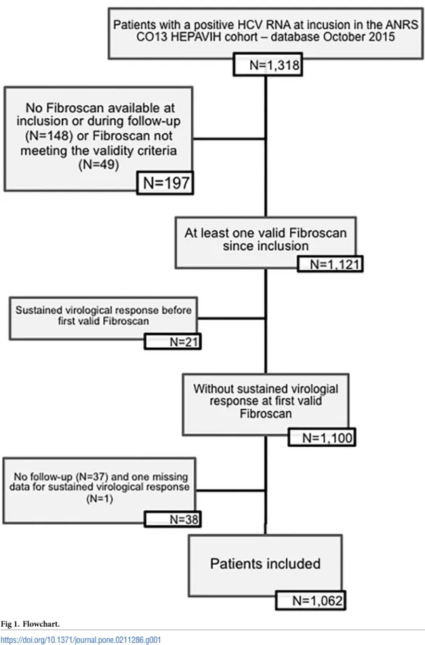
Of the 1,516 patients included from December 2005 to October 2015, 1,170 had at least one FS since their inclusion in the cohort, and 1,121 met the validity criterion. 1,100 patients met the study inclusion criteria but 38 were excluded for missing data on SVR (one) or absence of fol-low-up (n = 37). Finally, 1,062 patients were analyzed (Fig 1).
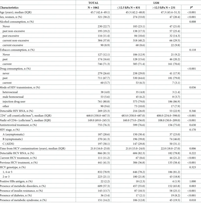
Characteristics of the study population at the time of first valid FS are shown inTable 1. A LSM >12.5 kPa was observed in 21.7% of the patients. In the LSM >12.5 kPa group, patients were older, mostly men, had lower CD4+ counts, had more frequent metabolic disorders, were more often under HCV treatment at inclusion, and were more often pretreated for HCV.
Median follow-up was 4.9 years (IQR, 3.2–6.1 years). At the end of follow-up, 335 (31.5%) patients had never received an HCV treatment and, among the 727 patients treated at least once, 189 (26.0%) had achieved SVR: 141 (74.6%) after peg-interferon/ribavirin, 38 (20.1%) after telaprevir- or boceprevir-based therapy and 10 (5.3%) after DAA. Among those 189 patients, 11 (5.8%) had a LSM > 12.5 kPa at SVR-time.
Death risk during follow-up
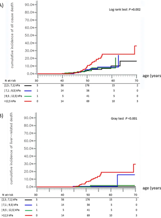
Overall, we observed 76 deaths (Table 2). No liver transplantation occurred during follow-up. Cumulative incidence of all-cause mortality was 5.9% (95% confidence interval (95% CI): [4.4–7.8]) at the age of 50, 13.4% [10.0–17.9] at the age of 60 and 25.5% [14.7–42.0] at the age of 70. It was higher in the LSM >12.5 kPa group than below this threshold (for example, at 50 years, 10.8% versus 4.5%, log rank test,p<0.001) (Fig 2).
Cumulative incidence of liver-related mortality was 1.6% [0.9–2.7] at the age of 50, 5.4% [3.3–8.2] at the age of 60 and 13.6% [4.0–28.9] at the age of 70. It was higher in the LSM >12.5 kPa group than below this threshold (at 50 years, 6.8% versus 0.14%, Gray test,p<0.001)
Fig 1. Flowchart.
Table 1. Characteristics at baseline of HIV/HCV co-infected patients included in this analysis—ANRS CO13 HEPAVIH cohort.
TOTAL LSM
Characteristics N = 1062 �12.5 kPa N = 831 >12.5 kPa N = 231 Pa
Age (years), median (IQR) 45.7 (42.4–49.1) 45.3 (42.2–48.8) 47.3 (43.4–51.3) <0.001
Sex, women, n (%) 321 (30.2) 274 (33.0) 47 (20.4) <0.001 Alcohol consumption, n (%) 0.008 Never 230 (22.7) 183 (23.1) 47 (21.0) past non-excessive 195 (19.2) 138 (17.5) 57 (25.4) past excessive 116 (11.4) 84 (10.6) 32 (14.3) current non-excessive 384 (37.8) 318 (40.2) 66 (29.5) current excessive 90 (8.9) 68 (8.6) 22 (9.8) Tobacco consumption, n (%) 0.118 Never 127 (12.1) 106 (12.9) 21 (9.2) past 174 (16.6) 128 (15.6) 46 (20.2) current 746 (71.3) 585 (71.4) 161 (70.6) Drug consumption, n (%) <0.001 never 279 (26.6) 238 (29.0) 41 (17.9) past 711 (67.7) 530 (64.6) 181 (79.0) current 60 (5.7) 53 (6.5) 7 (3.1)
Mode of HIV transmission, n (%) 0.036
heterosexual 38 (4.0) 35 (4.8) 3 (1.4)
male homosexual 53 (5.6) 45 (6.2) 8 (3.7)
injection drug user 761 (80.8) 575 (79.0) 186 (86.9)
other 90 (9.6) 73 (10.0) 17 (7.9)
Detectable HIV RNA, n (%) 269 (25.3) 216 (26.0) 53 (22.9) 0.346
CD4+cell count(cells/mm3), median (IQR) 468.0 (330.0–667.5) 483.0 (350.0–687.0) 408.0 (254.0–598.0) <0.001
Nadir of CD4+ (cells/mm3), median (IQR) 149.0 (69.0–245.5) 160.0 (75.0–256.0) 108.0 (50.0–209.0) <0.001
Antiretroviral treatment, n (%) 755 (76.3) 599 (76.6) 156 (75.0) 0.630
HIV stage, n (%) 0.170
A (asymptomatic) 187 (28.6) 150 (30.4) 37 (23.0)
B (symptomatic) 270 (41.3) 196 (39.8) 74 (46.0)
C (AIDS) 197 (30.1) 147 (29.8) 50 (31.1)
Time from HCV contamination (years), median (IQR) 21.0 (16.0–25.0) 21.0 (15.0–24.0) 22.0 (18.0–27.0) 0.006
Detectable HCV RNA, n (%) 866 (81.5) 684 (82.3) 182 (78.8) 0.222 Current HCV treatment, n (%) 111 (11.2) 67 (8.6) 44 (21.2) <0.001 Previous HCV treatment, n (%) 441 (41.5) 306 (36.8) 135 (58.4) <0.001 HCV genotype, n (%) 0.323 1, 4 or 5 832 (78.9) 646 (78.2) 186 (81.2) 2 or 3 223 (21.1) 180 (21.8) 43 (18.8) Positive HBs antigen, n (%) 22 (2.2) 18 (2.3) 4 (1.9) 1.000
Presence of metabolic disorders, n (%) 609 (57.3) 457 (55.0) 152 (65.8) 0.003
Presence of insulin resistance, n (%) 145 (13.7) 87 (10.5) 58 (25.1) <0.001
Presence of diabetes, n (%) 36 (3.4) 17 (2.1) 19 (8.2) <0.001
Presence of metabolic syndrome, n (%) 151 (14.2) 106 (12.8) 45 (19.5) 0.010
IQR, Interquartile Range; HIV, Human Immunodeficiency Virus; HCV, Hepatitis C Virus; AIDS, acquired immunodeficiency syndrome; aChi-square test or Fisher test for qualitative covariates, Mann-Whitney-Wilcoxon test for quantitative covariates
There was no difference in all-cause mortality nor in liver-related mortality among the three low LSM categories [2.5–7.1],] 7.1–9.5],] 9.5–12.5] (Fig 3).
LSM and all-cause mortality
In univariable analysis, patients with LSM >12.5 kPa at any point of follow-up had a higher risk of all-cause mortality compared to patients with LSM �12.5 kPa (crude HR [cHR] [95% CI] = 3.11 [1.96–4.92],p<0.001). All-cause mortality was also more frequent in men, tobacco
users and drug users. However, it was lower in ant-HCV pretreated patients and with an increased CD4+ count (Table 3).
In multivariable analysis, LSM >12.5 kPa at any time of follow-up was associated with an increased risk of all-cause mortality (adjusted HR (aHR) = 3.35 [2.06–5.45], p<0.001), inde-pendently of SVR, sex, alcohol use, tobacco use, metabolic disorders and previous HCV treat-ment. In addition, tobacco use (aHR = 5.69 [1.56; 20.78] for past use versus never, 3.22 [0.93; 11.09] for current use versus never, p = 0.015) and previous HCV treatment (aHR = 0.53 [0.32; 0.90], p = 0.017) were also associated with all-cause mortality, independently of SVR and the other covariates (Table 3).
The association of LSM with all-cause mortality was confirmed by the joint model. In the longitudinal sub-model, only SVR was associated with LSM trajectory during follow-up (Table inS1 Table). Furthermore, the model confirmed that an increase in LSM at a given time-point was associated with an increased risk of all-cause mortality, after adjustment for confounders and trajectory of LSM (aHR = 1.64 [1.43–1.88] for a 50% increase of current LSM
Table 2. Causes of death—ANRS CO13 HEPAVIH cohort.
Cause of death n
Overall 76
Liver-related death�: 26 (34.2%)
End stage liver disease�� 17
HCC without end stage liver disease 4
Hepatocholangiocarcinoma 1
Liver transplant complications 3
HIV-related death: 10 (13.1%)
Infectious disease 5
Pulmonary hypertension 1
Progressive multifocal leukoencephalopathy 3
Hodgkin lymphoma 1
Non-HIV and non-liver-related death: 29 (38.2%)
Cancer��� 12 Infectious disease 2 Psychiatric disease 5 Cardiovascular disease 4 Sudden death 1 Toxic cause 4 Pulmonary fibrosis 1 Undetermined 11 (14.5%)
�One patient was classified as a liver-related but the precise cause was not reported. ��Three patients with end stage liver disease were reported to have hepatocellular carcinoma.
���5 broncho-pulmonary, 1 ovarian, 1 esophageal, 1 nasopharyngeal, 1 colon, 2 pancreatic, 1 breast cancers.
Fig 2. Cumulative incidence of all-cause mortality (A) and liver related mortality (B). Cumulative incidence for patients with
LSM � 12.5 kPa (blue line) and patients with LSM >12.5 kPa (red line); 95% confidence intervals are represented by pointed lines. Kaplan-Meier method (A) and Aalen-Johansen method (B).
Fig 3. Cumulative incidence of all-cause, mortality (A) and liver-related mortality (B). Black line: LSM [2.5;7.1] kPa, blue line: LSM ]
7.1;9.5] kPa, green line: LSM ]9.5;12.5] kPa, and red line: LSM >12.5 kPa. Kaplan-Meier method (A) and Aalen-Johansen method (B).
Table 3. Crude (A) and adjusted (B) hazard ratios from a Cox proportional hazards model with delayed entry for all-cause mortality in HIV/HCV patients—ANRS CO13 HEPAVIH cohort.
Univariable analysis (A) cHR [95% CI] P Multivariable analysis (B)� aHR [95% CI] P LSM (t): <0.001 <0.001 �12,5 kPa 1 1 >12,5 kPa 3.11 [1.96; 4.92] 3.35 [2.06; 5.45] SVR (t) 0.002 untreated 1 1 treated-SVR- 3.12 [1.10; 4.02] 3.53 [1.23; 5.47] treated-SVR+ 0.62 [0.13; 2.88] 0.75 [0.17;3.81]
Sex: womenversus men 0.60 [0.34; 1.04] 0.070 0.71 [0.40; 1.26] 0.243
Alcohol consumption 0.063 0.089 Never 1 1 past non-excessive 0.75 [0.36; 1.56] 0.58 [0.27; 1.23] past excessive 0.40 [0.14; 1.21] 0.31 [0.10; 0.94] current non-excessive 0.92 [0.50; 1.68] 0.72 [0.38; 1.38] current excessive 1.80 [0.87; 3.73] 1.22 [0.57; 2.62] Tobacco consumption 0.048 0.015 never 1 1 past 4.43 [1.28; 15.33] 5.69 [1.56; 20.78] current 2.98 [0.91; 9.74] 3.22 [0.93; 11.09] Drug consumption 0.094 0.447 never 1 1 past 2.10 [1.05; 4.20] 1.53 [0.73; 3.17] current 1.30 [0.36; 4.76] 0.99 [0.27; 3.69]
Mode of HIV transmission 0.704
-heterosexual 1
-male homosexual 1.28 [0.20; 8.03]
-injection drug users 1.71 [0.41; 7.17]
-others 1.02 [0.19; 5.47]
-Detectable HIV viral load (t) versus undetectable HIV viral load (t) 0.73 [0.37; 1.43] 0.360 -
-CD4+ level (/50 cells/mm3) (t) 0.95 [0.90; 0.99] 0.008 1.00 [0.95; 1.00] 0.287 Nadir of CD4+ (/50 cells/mm3) 0.96 [0.87; 1.05] 0.341 - -HIV stage: 0.950 -A (asymptomatic) 1 -B (symptomatic) 1.02 [0.54; 1.94] -C (AIDS) 1.11 [0.55; 2.21] -HCV genotype 0.852 -2 or 3versus 1, 4 or 5 0.95 [0.55; 1.64]
-Time from HCV contamination (/year) 1.02 [0.97; 1.07] 0.430 -
-Positive HBs antigen 0.91 [0.13; 6.63] 0.929 -
-Presence of metabolic disorders 1.04 [0.64; 1.67] 0.886 0.92 [0.56; 1.51] 0.737
Presence of previous anti-HCV treatment 0.60 [0.37; 0.99] 0.046 0.53 [0.32; 0.90] 0.017
cHR, crude Hazard ratio; aHR, adjusted Hazard ratio; LSM, liver stiffness measurements; (t) indicates time-dependent covariables;
�52 patients had missing data on alcohol and tobacco and were not included in the final model. There was no difference between analyzed and excluded patients (data not shown).
value,p<0.001). The trajectory of LSM between two measurements was not associated with
all-cause mortality (Table 4).
LSM and liver-related mortality
The risk of liver-related mortality increased when LSM was >12.5 kPa (cHR = 29.65 [8.88– 99.01],p<0.001), in case of metabolic disorders, and in drug users. It decreased for women,
when the CD4+ level increased, for HCV genotype 2 or 3 patients, and for pretreated patients (Table 5).
LSM >12.5 kPa at any time remained associated with an increased risk of liver-related mor-tality (aHR = 20.60 [5.99–70.78],p<0,001) after adjustment for sex, alcohol use, metabolic
dis-orders, CD4+ level and previous HCV treatment. This result was not adjusted for SVR as no liver-related death occurred in the SVR group. Previous HCV treatment (aHR = 0.31 [0.12– 0.85],p = 0.022) and metabolic disorders (aHR = 3.91 [1.12–13.66], p = 0.033) were also
associ-ated with liver-relassoci-ated mortality, after adjustment for the other variables (Table 5).
Discussion
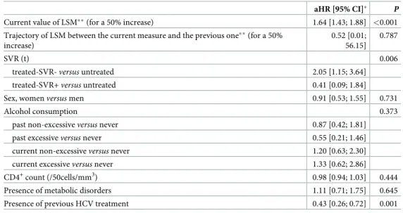
In this large-scale assessment of mortality in a prospective, multicenter, observational cohort study of HIV/HCV co-infected patients, a LSM >12.5 kPa at any time point during follow-up was associated with a 3 fold higher risk of all-cause mortality, independently of SVR and other main confounders. This association has been confirmed by joint modeling of the LSM trajec-tory and all-cause mortality. Furthermore, the latter analysis showed that mortality was related to an increase in the current LSM value at a given time point, and not to the trajectory of LSM.
Table 4. Adjusted hazard ratios for all-cause mortality from the survival sub-model of the joint model with shared random effects in HIV/HCV co-infected patients—ANRS CO13 HEPAVIH cohort.
aHR [95% CI]� P
Current value of LSM��(for a 50% increase) 1.64 [1.43; 1.88] <0.001
Trajectory of LSM between the current measure and the previous one��(for a 50% increase)
0.52 [0.01; 56.15]
0.787
SVR (t) 0.006
treated-SVR-versus untreated 2.05 [1.15; 3.64]
treated-SVR+versus untreated 0.41 [0.09; 1.84]
Sex, womenversus men 0.91 [0.53; 1.55] 0.731
Alcohol consumption 0.373
past non-excessiveversus never 0.87 [0.42; 1.81]
past excessiveversus never 0.55 [0.21; 1.46]
current non-excessiveversus never 1.20 [0.63; 2.30]
current excessiveversus never 1.33 [0.62; 2.86]
CD4+count (/50cells/mm3) 0.98 [0.94; 1.03] 0.444
Presence of metabolic disorders 1.11 [0.71; 1.75] 0.645
Presence of previous HCV treatment 0.43 [0.26; 0.72] 0.001
aHR, adjusted Hazard ratio; LSM, liver stiffness measurements; SVR, sustained virological response; (t) indicates time-dependent covariables;
�Model estimations are based on data from 959 patients without missing data for variables included in the model. In this subgroup, 67 all-cause deaths occurred.
��All LSM available during follow-up were considered to calculate the trajectory of LSM, even those obtained after achieving SVR since the model was adjusted for SVR time-dependent covariate. Current value of LSM represented value at a time point t and the trajectory represented the evolution of LSM.
LSM was also associated with liver-related mortality after adjustment for main confounders. Nevertheless, small number of liver-related deaths hampered the adjustment for SVR and led to large confidence intervals.
Our results on LSM are concordant with previous studies [13,20,21,35]. Limketkai et al found that patients with liver fibrosis stage F4 had a three fold higher risk of death than F0 patients [20]. Sanmartin et al also found a 3.7 fold higher risk of death for patients with
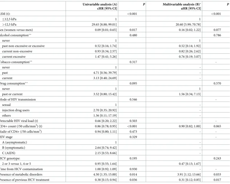
Table 5. Crude (A) and adjusted (B) hazard ratio from a Cox proportional hazards model with delayed entry for occurrence of liver-related death in HIV/HCV co-infected patients from the HEPAVIH cohort.
Univariable analysis (A) cHR [95% CI] P Multivariable analysis (B)� aHR [95% CI] P LSM (t): <0.001 <0.001 �12,5 kPa 1 1 >12,5 kPa 29.65 [8.88; 99.01] 20.60 [5.99; 70.78]
Sex (womenversus men) 0.09 [0.01; 0.65] 0.017 0.16 [0.02; 1.22] 0.077
Alcohol consumption�� 0.480 0.786
never 1 1
past non-excessive or excessive 0.52 [0.16; 1.74] 0.52 [0.14; 1.92]
current non-excessive 0.93 [0.34; 2.57] 0.82 [0.26; 2.62] current excessive 1.47 [0.41; 5.26] 0.76 [0.19; 3.07] Tobacco consumption�� 0.317 -never 1 -past 4.71 [0.56; 39.79] -current 3.13 [0.40; 24.69] -Drug consumption�� 0.095 0.570 never 1 1 past or current 3.52 [0.80; 15.42] 1.56 [0.34; 7.13]
Mode of HIV transmission 0.546
-sexual 1
-injection drug users 2.70 [0.35; 20.92]
-others 1.36 [0.11; 17.19]
-Detectable HIV viral load (t) 0.66 [0.20; 2.22] 0.503 -
-CD4+ count (/50 cells/mm3) (t) 0.86 [0.78; 0.95] <0.001 0.90 [0.82; 1.00] 0.065 Nadir of CD4+ (/50 cells/mm3) 0.94 [0.80; 1.11] 0.473 - -HIV stage 0.329 -A (asymptomatic) 1 -B (symptomatic) 2.64 [0.74; 9.42] -C (AIDS) 2.15 [0.53; 8.66] -HCV genotype: 0.195 0.243 2 or 3versus 1, 4 or 5 0.95 [0.55; 1.64] 0.47 [0.13; 1.67]
Time from HCV contamination 1.00 [0.92; 1.09] 0.930 -
-Presence of metabolic disorders 4.50 [1.35; 15.00] 0.014 3.91 [1.12; 13.66] 0.033
Presence of previous HCV treatment 0.38 [0.15; 0.94] 0.036 0.31 [0.12; 0.85] 0.017
cHR, crude Hazard ratio; aHR, adjusted Hazard ratio; LSM, liver stiffness measurements; HIV, Human immunodefiency virus; HCV, hepatitis C virus; AIDS, acquired immunodeficiency syndrome;
�47 patients had missing data on alcohol and CD4+ count and were not included in the final model. (t) indicates time-dependent covariables. There was no difference between analyzed and excluded patients (data not shown).
��Definition modified for the secondary outcome analysis because no event occurred in some categories of the original variables making the estimation of HRs impossible for these categories.
advanced fibrosis (F3-F4) [35]. This is probably due to a greater incidence of liver-related events (liver decompensation, hepatocellular carcinoma. . .) in case of advanced fibrosis (25). Nonetheless, these studies did not consider SVR or were conducted as natural history studies, i.e. in the absence of any HCV treatment.
Previous HCV treatment was identified as a protective factor whatever the SVR status, which is concordant with results from Butt et al: in patients treated by peg-interferon/ribavi-rin, all-cause mortality was reduced by 30% to 60% according to treatment duration, when compared with untreated patients, whatever the treatment’s outcome (SVR or not) [36]. This beneficial effect could result from liver fibrosis reduction in patients with no cirrhosis.
Treated-SVR negative patients were at higher risk of all-cause mortality, compared with untreated patients, probably linked to an indication bias (treatment was purposed to patients with bad prognosis). Moreover, no significant reduction of all-cause mortality was found in treated-SVR positive patients contrary to what has been described recently [37]. This could be explained in two ways: 1/ a lack of power, 2/ achieving SVR reduced the overmortality of treated patients, without reaching a protective effect.
The level of CD4+ was not associated with all-cause mortality which is in contrast to what has been found in previous studies including HIV/HCV co-infected patients [20,37]. This could be explained in three ways: 1/ CD4+ level was a time-dependent covariate which could have modified its association, 2/ the principal causes of mortality in our population are non-HIV and non- liver-related deaths, 3/ most patients had recovered their immunovirological function before inclusion. Nevertheless, the association between CD4+ level and liver-related mortality almost reached statistical significance with an aHR close to the result reported by Grint et al [8].
No association was found between alcohol use and all-cause or liver-related mortality in our study, probably due to the low proportion of excessive alcohol consumers (90 patients, 8.9%). Furthermore, alcohol use was declared by the patient to his physician which might result in an under-reporting (social desirability bias) considering that HCV-infected patients are often aware of the seriousness of excessive alcohol use.
Our study has some limitations. First, the proportion of patients with SVR was low and only few patients were treated by DAA. Even though the SVR rate in our study did not repre-sent the current context of HIV/ HCV co-infected patients, we do not expect that SVR modi-fies the effect of LSM on all-cause mortality. Indeed, a decrease of LSM values after SVR has been shown, but we would expect that higher LSM markers would still be associated with higher risk of mortality in those with and without SVR. Thus, we do not think that higher SVR rates would have led to different results.
Second, analyses were based on patient records without missing data for variables included in the final models. No imputation procedure was performed because of the small number of patients excluded for missing data and the absence of difference compared to excluded patients. Finally, the main analysis was not adjusted for hepatitis B virus (HBV) status but HBV co-infection was only present in 2.2% of the patients and all were receiving an anti-HBV active cART.
Nonetheless, our study has also several strengths. In the absence of post-SVR cohorts with sufficient follow-up, our approach allows to better understand factors associated with mortality independently of SVR and our primary results were confirmed in several sensitivity analyses. Furthermore, we used an innovative statistical approach to better characterize the association between LSM and all-cause mortality using a joint model. Finally, the interest of our results for clinical practice is important: all HIV/HCV co-infected patients, even those who achieved SVR, have to benefit from a long-term follow-up with LSM to detect patients at high mortality risk.
In conclusion, LSM >12.5 kPa at any point in time was strongly associated with all-cause mortality independently of SVR in HIV/HCV co-infected patients. Close follow-up of these patients should remain a priority even after obtaining SVR. Post-SVR cohorts will be critical in the near future to assess the residual risk especially of liver disease progression and its associ-ated factors. This information is of utmost importance to optimize care especially after the use of DAA.
Supporting information
S1 Table. Factors associated with LSM trajectory in HIV/HCV co-infected patients from the ANRS CO13 HEPAVIH cohort and without missing data (N = 959), longitudinal sub-model of the joint sub-model with shared random effects.
(PDF)
Acknowledgments
Patients of the HEPAVIH cohort
The ANRS CO13 HEPAVIH cohort study group:
Scientific Committee of the ANRS CO13 HEPAVIH Study Group:
L. Wittkop (Principal Investigator and Methodologist) Univ Bordeaux, ISPED, Inserm Bor-deaux Population Health, team MORPH3EUS, UMR 1219, CIC-EC 1401, F-33000 BorBor-deaux, France; Centre Hospitalier Universitaire de Bordeaux, Poˆle de Sante´ Publique, F-33000 Bor-deaux, France; email: linda.wittkopu-bordeaux.fr
D. Salmon (co-Principal investigator): Universite´ Paris Descartes, Paris, France; Assistance Publique des Hoˆpitaux de Paris, HU Paris Centre, Service Maladies infectieuses et tropicales, Paris, France.
P. Sogni (co-Principal Investigator): Assistance Publique des Hoˆpitaux de Paris, Hoˆpital Cochin, Service d’He´patologie, Paris, France; INSERM U-1223 –Institut Pasteur, Paris, France; Universite´ Paris Descartes, Paris, France.
L. Esterle (project manager): Univ Bordeaux, ISPED, Inserm Bordeaux Population Health, team MORPH3EUS, UMR 1219, CIC-EC 1401, F-33000 Bordeaux, France.
P. Trimoulet: CHU de Bordeaux, Hoˆpital Pellegrin, Laboratoire de Virologie, Bordeaux 33000, France.
J. Izopet: CHU de Toulouse, Hoˆpital Purpan, Laboratoire de Virologie, Toulouse, France; INSERM U1043—CNRS UMR5282—Toulouse University Paul Sabatier, CPTP, Toulouse, France.
L. Serfaty: CHU de Strasbourg, Hoˆpital de Hautepierre, Service des maladies du foie, Stras-bourg, France.
V. Paradis: Pathology Department, Beaujon Hospital, Assistance Publique-Hoˆpitaux de Paris, Clichy, France; UMR 1149 INSERM—Paris Diderot University, Inflammation Research Center, Paris, France.
B. Spire, P. Carrieri: Inserm, UMR912, Economics and Social Sciences Applied to Health and Analysis of Medical Information (SESSTIM), 13000 Marseille, France; Aix-Marseille Uni-versity, UMRS912, IRD, 13000 Marseille, France; ORS PACA, Southeastern Health Regional Observatory, 13000 Marseille, France.
M.A. Valantin: Assistance Publique des Hoˆpitaux de Paris, Hoˆpital Pitie´-Salpe´trière, Service Maladies infectieuses et tropicales, Paris, France.
G. Pialoux, J. Chas: Assistance Publique des Hoˆpitaux de Paris, Hoˆpital Tenon, Service Mal-adies infectieuses et tropicales, Paris, France.
I. Poizot-Martin: Aix Marseille Univ, APHM Sainte-Marguerite, Service d’Immuno-he´ma-tologie clinique; Inserm U912 (SESSTIM) Marseille, France.
L. Alric: Centre Hospitalier Universitaire de Toulouse, Hoˆpital Purpan, Me´decine interne, Toulouse, France; Universite´ Toulouse III, Paul Sabatier, Toulouse, France.
K. Barange: CHU Toulouse, Service d’he´patologie et gastoenterologie, Toulouse, France. A. Naqvi: Centre Hospitalier Universitaire de Nice, Service d’Infectiologie, Hoˆpital l’Archet, Nice 06100, France.
E. Rosenthal: Centre Hospitalier Universitaire de Nice, Service de Me´decine Interne et Can-ce´rologie, Hoˆpital l’Archet, Nice, France; Universite´ de Nice-Sophia Antipolis, Nice, France.
A. Bicart-See: E´quipe mobile d’infectiologie, hoˆpital Joseph-Ducuing, Toulouse, France. O. Bouchaud: Assistance Publique des Hoˆpitaux de Paris, Hoˆpital Avicenne, Service Mala-dies infectieuses et tropicales, Bobigny, France; Universite´ Paris 13 Nord, Bobigny, France.
A. Gervais: Assistance Publique des Hoˆpitaux de Paris, Hoˆpital Bichat Claude Bernard, Ser-vice des maladies infectieuses et tropicales, Paris, France.
C. Lascoux-Combe: Assistance Publique des Hoˆpitaux de Paris, Hoˆpital Saint-Louis, Ser-vice Maladies infectieuses et tropicales, Paris, France.
C. Goujard: Assistance Publique des Hoˆpitaux de Paris, Hoˆpital Bicêtre, Hoˆpitaux universi-taires Paris Sud, Service Me´decine interne et Immunologie clinique, Le Kremlin-Bicêtre, France; Universite´ Paris Sud, Le Kremlin-Bicêtre, France.
K. Lacombe: Assistance Publique des Hoˆpitaux de Paris, Hoˆpital Saint-Antoine, Service Maladies infectieuses et tropicales, Paris, France; UMPC (Universite´ Pierre et Marie Curie), UMR S1136, Institut Pierre Louis d’Epide´miologie et de Sante´ Publique, Paris, France.
C. Duvivier: APHP-Hoˆpital Necker-Enfants malades, Service de Maladies Infectieuses et Tropicales, Paris, France; Centre d’Infectiologie Necker-Pasteur, Paris, France.
D. Neau: Centre Hospitalier Universitaire de Bordeaux, Service Maladies infectieuses et tro-picales Bordeaux, Hoˆpital Pellegrin, Bordeaux, France; Universite´ de Bordeaux, Bordeaux, France
P. Morlat: Univ Bordeaux, ISPED, Inserm Bordeaux Population Health, team MOR-PH3EUS, UMR 1219, CIC-EC 1401, F-33000 Bordeaux, France; Centre Hospitalier Universi-taire de Bordeaux, Service de me´decine interne, hoˆpital Saint-Andre´, Bordeaux, France.
F. Bani-Sadr: Centre Hospitalier Universitaire de Reims, Service de me´decine interne, mala-dies infectieuses et immunologie clinique, Reims, France; Universite´ de Reims, Champagne-Ardenne, Reims, France.
L. Meyer: INSERM CESP U1018, Universite´ Paris Sud, Le Kremlin Bicêtre, France. F. Boufassa: Inserm, CESP, U1018, Paris-Sud University, Le Kremlin-Bicêtre, France. B. Autran: Department of Immunology, UPMC, AP-HP, Hospital, Paris, France; U1135, INSERM, CIMI; Sorbonne University, UPMC.
A.M. Roque: Assistance Publique des Hopitaux de Paris, Paris Sud, hoˆpital Paul Brousse, centre he´pato-biliaire, CNR he´patite A, 94800 Villejuif, France; Universite´ Paris-Sud, unite´ mixte de recherche scientifique 785, 92296 Chatenay-Malabry, France; Inserm unite´ 785, 94800 Villejuif, France.
C. Solas: APHM, Hoˆpital La Timone, Laboratoire de Pharmacocine´tique et Toxicologie, Marseille, France.
H. Fontaine: Unite´ d’He´patologie, AP-HP Hoˆpital Cochin, USM20, Institut Pasteur, Uni-versite´ Paris-Descartes, Paris, France.
D. Costagliola: Sorbonne Universite´s UPMC Universite´ Paris 06, INSERM, Institut Pierre Louis d’e´pide´miologie et de Sante´ Publique (UMRS 1136), Paris, France.
L. Piroth: Centre Hospitalier Universitaire de Dijon, De´partement d’Infectiologie, Dijon, France; Universite´ de Bourgogne, Dijon, France.
A. Simon: Assistance Publique des Hoˆpitaux de Paris, Hoˆpital Pitie´-Salpe´trière, De´parte-ment de Me´decine Interne et Immunologie Clinique, Paris, France.
D. Zucman: Hoˆpital Foch, unite´ VIH, Suresnes, France.
F. Boue´: Hoˆpital Antoine-Be´clère, Assistance Publique des Hoˆpitaux de Paris, Universite´ Paris Sud, Service Me´decine interne et immunologie, Clamart 92140, France.
P. Miailhes: Service des Maladies Infectieuses et Tropicales, CHU Lyon, Hoˆpital de la Croix Rousse, Lyon, France.
E. Billaud: Centre Hospitalier Universitaire de Nantes, Service Maladies infectieuses et tro-picales, Nantes, France.
H. Aumaıˆtre: Centre Hospitalier de Perpignan, Service Maladies infectieuses et tropicales, Perpignan, France.
D. Rey: Le Trait d’Union, Centre de soins de l’infection par le VIH, Nouvel Hoˆpital Civil; Strasbourg, France.
G. Peytavin: Assistance Publique des Hoˆpitaux de Paris, Hoˆpital Bichat-Claude Bernard, Laboratoire de Pharmacologie, Paris, France.
V. Petrov-Sanchez, C. Cagnot: .ANRS, 101 rue de Tolbiac, 75013 Paris, France.
Clinical Centres (ward / participating physicians): APHP, Hoˆpitaux Universitaires Paris Centre, Paris (Me´decine Interne et Maladies Infectieuses: D. Salmon, R. Usubillaga; He´pato-gastro-ente´rologie: P. Sogni; Anatomo-pathologie: B. Terris; Virologie: P. Tremeaux); APHP Pitie´-Salpe´trière, Paris (Maladies Infectieuses et Tropicales: C. Katlama, M.A. Valantin, H. Sti-tou; Me´decine Interne: A. Simon, P. Cacoub, S. Nafissa; He´pato-gastro-ente´rologie: Y. Benha-mou; Anatomo-pathologie: F. Charlotte; Virologie: S. Fourati); APHM Sainte-Marguerite, Marseille (Service d’Immuno-He´matologie Clinique: I. Poizot-Martin, O. Zaegel, H. Laroche; Virologie: C. Tamalet); APHP Tenon, Paris (Maladies Infectieuses et Tropicales: G. Pialoux, J. Chas; Anatomo-pathologie: P. Callard, F. Bendjaballah; Virologie: C. Amiel, C. Le Pendeven); CHU Purpan, Toulouse (Maladies Infectieuses et Tropicales: B. Marchou; Me´deicne interne: L. Alric; He´pato-gastro-ente´rologie: K. Barange, S. Metivier; Anatomo-pathologie: J. Selves; Virologie: F. Larroquette); CHU Archet, Nice (Me´decine Interne: E. Rosenthal; Infectiologie: A. Naqvi, V. Rio; Anatomo-pathologie: J. Haudebourg, M.C. Saint-Paul; Virologie: A. De Monte, V. Giordanengo, C. Partouche); APHP Avicenne, Bobigny (Me´decine Interne–Unite´ VIH: O. Bouchaud; Anatomo-pathologie: A. Martin, M. Ziol; Virologie: Y. Baazia, V. Iwaka-Bande, A. Gerber); Hoˆpital Joseph Ducuing, Toulouse (Me´decine Interne: M. Uzan, A. Bicart-See, D. Garipuy, M.J. Ferro-Collados; Anatomo-pathologie: J. Selves; Virologie: F. Nicot); APHP Bichat–Claude-Bernard, Paris (Maladies Infectieuses:, A. Gervais, Y. Yazdanpanah; Anatomo-pathologie: H. Adle-Biassette; Virologie: G. Alexandre, Pharmacologie: G. Peytavin); APHP Saint-Louis, Paris (Maladies infectieuses: C. Lascoux-Combe, J.M. Molina; Anatomo-pathologie: P. Bertheau; Virologie: M.L. Chaix, C. Delaugerre, S. Maylin); APHP Saint-Antoine (Maladies Infectieuses et Tropicales: K. Lacombe, J. Bottero; J. Krause, P.M. Girard, Anatomo-pathologie: D. Wendum, P. Cervera, J. Adam; Virologie: C. Viala); APHP, Hoˆpitaux Paris Sud, Bicêtre, Paris (Maladies Infectieurses et Tropicales: D. Vittecocq; Me´decine Interne: C. Goujard, Y. Quertainmont, E. Teicher; Virologie: C. Pallier); APHP Necker, Paris (Maladies Infectieuses et Tropicales: O. Lortholary, C. Duvivier, C. Rouzaud, J. Lourenco, F. Touam, C. Louisin: Virologie: V. Avettand-Fenoel, E. Gardiennet, A. Me´lard); CHU Bordeaux Hoˆpital Pellegrin, Bordeaux (Maladies Infectieuses et Tropicales: D. Neau, A. Ochoa, E. Blanchard, S. Castet-Lafarie, C. Cazanave, D. Malvy, M. Dupon, H. Dutronc, F. Dauchy, L. Lacaze-Buzy, A. Desclaux; Anatomo-pathologie: P. Bioulac-Sage; Virologie: P. Trimoulet, S. Reigadas); CHU Bordeaux Hoˆpital Saint-Andre´, Bordeaux (Me´decine Interne et Maladies Infectieuses:
Me´decine Interne et Maladies Infectieuses: P. Morlat, D. Lacoste, F. Bonnet, N. Bernard, M. Hessamfar, J, F. Paccalin, C. Martell, M. C. Pertusa, M. Vandenhende, P. Mercie´, D. Malvy, T. Pistone, M.C. Receveur, M. Me´chain, P. Duffau, C Rivoisy, I. Faure, S. Caldato; Anatomo-pathologie: P. Bioulac-Sage; Virologie: P. Trimoulet, S. Reigadas, P. Bellecave, C. Tumiotto); CHU Bordeaux Hoˆpital du Haut-Levêque, Bordeaux (Me´decine Interne: J.L. Pellegrin, J.F. Viallard, E. Lazzaro, C. Greib; Anatomo-pathologie: P. Bioulac-Sage; Virologie: P. Trimoulet, S. Reigadas); Hoˆpital FOCH, Suresnes (Me´decine Interne: D. Zucman, C. Majerholc; Virolo-gie: M. Brollo, E. Farfour); APHP Antoine Be´clère, Clamart (Me´decine Interne: F. Boue´, J. Polo Devoto, I. Kansau, V. Chambrin, C. Pignon, L. Berroukeche, R. Fior, V. Martinez, S. Abgrall, M. Favier; Virologie: C. Deback); CHU Henri Mondor, Cre´teil (Immunologie Clini-que: Y. Le´vy, S. Dominguez, J.D. Lelièvre, A.S. Lascaux, G. Melica); CHU Nantes Hoˆpital Hoˆtel Dieu, Nantes (Maladies Infectieuses et Tropicales: E. Billaud, F. Raffi, C. Allavena, V. Reliquet, D. Boutoille, C. Biron; M. Lefebvre, N. Hall, S. Bouchez; Virologie: A. Rodallec, L. Le Guen, C. Hemon); Hoˆpital de la Croix Rousse, Lyon (Maladies Infectieuses et Tropicales: P. Miailhes, D. Peyramond, C. Chidiac, F. Ader, F. Biron, A. Boibieux, L. Cotte, T. Ferry, T. Per-point, J. Koffi, F. Zoulim, F. Bailly, P. Lack, M. Maynard, S. Radenne, M. Amiri, F Valour; He´pato-gastro-ente´rologie: J. Koffi, F. Zoulim, F. Bailly, P. Lack, M. Maynard, S. Radenne, C. Augustin-Normand; Virologie: C. Scholtes, T.T. Le-Thi); CHU Dijon, Dijon (De´partement d’infectiologie:, L. Piroth, P. Chavanet M. Duong Van Huyen, M. Buisson, A. Waldner-Com-bernoux, S. Mahy, R. Binois, A.L. Simonet-Lann, D. Croisier-Bertin, A. Salmon Rousseau, C. Martins); CH Perpignan, Perpignan (Maladies infectieuses et tropicales: H. Aumaıˆtre, Virolo-gie: S. Galim); CHU Robert Debre´, Reims (Me´decine interne, maladies infectieuses et immu-nologie clinique: F. Bani-Sadr, D. Lambert, Y Nguyen, J.L. Berger, M. Hentzien, Virologie: V. Brodard); CHRU Strasbourg (Le Trait d’Union: D Rey, M Partisani, ML Batard, C Cheneau, M Priester, C Bernard-Henry, E de Mautort, Virologie: P Gantner et S Fafi-Kremer)
Data collection: F. Roustant, P. Platterier, I. Kmiec, L. Traore, S. Lepuil, S. Parlier, V. Sicart-Payssan, E. Bedel, S. Anriamiandrisoa, C. Pomes, F. Touam, C. Louisin, M. Mole, C. Bolliot, P Catalan, M. Mebarki, A. Adda-Lievin, P. Thilbaut, Y. Ousidhoum, F.Z. Makhoukhi, O. Braik, R. Bayoud, C. Gatey, M.P. Pietri, V. Le Baut, R. Ben Rayana, D. Bornarel, C. Chesnel, D. Beni-ken, M. Pauchard, S. Akel, S. Caldato, C. Lions, A. Ivanova, A-S. Ritleg, C. Debreux, L. Chalal, J. Zelie, H. Hue, A. Soria, M. Cavellec, S. Breau, A. Joulie, P. Fisher, S. Gohier, D. Croisier-Ber-tin, S. Ogoudjobi, C. Brochier, V. Thoirain-Galvan, M. Le Cam.
Management, statistical analyses: A. Aurousseau, P. Carrieri, M. Chalouni, V. Conte, L. Dequae-Merchadou, M. Desvallees, N. Douiri, L. Esterle, C. Gilbert, R. Knight, F, T. Lemboub, Marcellin, L. Michel, M. Mora, S. Nordmann, C. Protopopescu, P. Roux, S. Rousseau-Gillet, B. Spire, S. Tezkratt, A. Vilotitch, I. Yaya, L Wittkop.
Presentations
This work has been presented at The Liver Meeting 2016 of the American association for the study of the liver in Boston (11–15 November 2016, Hepatology, october 2016, volume 64, number 1 (suppl) AASLD Abstracts n˚56) as an oral presentation and at the French national conference of the AFEF–socie´te´ francc¸aise d’he´patologie–French society of hepatology (79ième journe´e scientifique) in Bordeaux (28 September– 1 October 2016) as a poster presen-tation (poster #CA-02).
Author Contributions
Formal analysis: Sarah Shili-Masmoudi, Linda Wittkop. Investigation: Firouze´ Bani-Sadr.
Methodology: Sarah Shili-Masmoudi, Linda Wittkop. Project administration: Laure Esterle.
Supervision: Linda Wittkop. Validation: Linda Wittkop.
Writing – original draft: Sarah Shili-Masmoudi, Linda Wittkop.
Writing – review & editing: Philippe Sogni, Victor de Ledinghen, Laure Esterle,
Marc-Antoine Valantin, Isabelle Poizot-Martin, Anne Simon, Eric Rosenthal, Karine Lacombe, Gilles Pialoux, Olivier Bouchaud, Anne Gervais-Hasenknoff, Ce´cile Goujard, Lionel Piroth, David Zucman, Ste´phanie Dominguez, Franc¸ois Raffi, Laurent Alric, Firouze´ Bani-Sadr, Caroline Lascoux-Combe, Daniel Garipuy, Patrick Miailhes, Daniel Vittecoq, Claudine Duvivier, Hugues Aumaıˆtre, Didier Neau, Philippe Morlat, Franc¸ois Dabis, Dominique Salmon.
References
1. Platt L, Easterbrook P, Gower E, McDonald B, Sabin K, McGowan C, et al. Prevalence and burden of HCV co-infection in people living with HIV: a global systematic review and meta-analysis. The Lancet Infectious diseases. 2016; 16(7):797–808. Epub 2016/02/29.https://doi.org/10.1016/S1473-3099(15) 00485-5PMID:26922272.
2. Chen M, Wong WW, Law MG, Kiertiburanakul S, Yunihastuti E, Merati TP, et al. Hepatitis B and C Co-Infection in HIV Patients from the TREAT Asia HIV Observational Database: Analysis of Risk Factors and Survival. PloS one. 2016; 11(3):e0150512. Epub 2016/03/05.https://doi.org/10.1371/journal.pone. 0150512PMID:26933963.
3. Kovari H, Ledergerber B, Cavassini M, Ambrosioni J, Bregenzer A, Stockle M, et al. High hepatic and extrahepatic mortality and low treatment uptake in HCV-coinfected persons in the Swiss HIV cohort study between 2001 and 2013. Journal of hepatology. 2015; 63(3):573–80. Epub 2015/05/06.https:// doi.org/10.1016/j.jhep.2015.04.019PMID:25937433.
4. Macias J, Berenguer J, Japon MA, Giron JA, Rivero A, Lopez-Cortes LF, et al. Fast fibrosis progression between repeated liver biopsies in patients coinfected with human immunodeficiency virus/hepatitis C virus. Hepatology. 2009; 50(4):1056–63. Epub 2009/08/12.https://doi.org/10.1002/hep.23136PMID:
19670415.
5. Sulkowski MS, Mehta SH, Torbenson MS, Higgins Y, Brinkley SC, de Oca RM, et al. Rapid fibrosis pro-gression among HIV/hepatitis C virus-co-infected adults. Aids. 2007; 21(16):2209–16. Epub 2007/12/ 20.https://doi.org/10.1097/QAD.0b013e3282f10de9PMID:18090048.
6. Klein MB, Rollet-Kurhajec KC, Moodie EE, Yaphe S, Tyndall M, Walmsley S, et al. Mortality in HIV-hep-atitis C co-infected patients in Canada compared to the general Canadian population (2003–2013). Aids. 2014; 28(13):1957–65. Epub 2014/09/27.https://doi.org/10.1097/QAD.0000000000000377
PMID:25259703.
7. Morlat P, Roussillon C, Henard S, Salmon D, Bonnet F, Cacoub P, et al. Causes of death among HIV-infected patients in France in 2010 (national survey): trends since 2000. Aids. 2014; 28(8):1181–91. Epub 2014/06/06.https://doi.org/10.1097/QAD.0000000000000222PMID:24901259.
8. Grint D, Peters L, Rockstroh JK, Rakmanova A, Trofimova T, Lacombe K, et al. Liver-related death among HIV/hepatitis C virus-co-infected individuals: implications for the era of directly acting antivirals. Aids. 2015; 29(10):1205–15. Epub 2015/04/15.https://doi.org/10.1097/QAD.0000000000000674
PMID:25870984.
9. Smith CJ, Ryom L, Weber R, Morlat P, Pradier C, Reiss P, et al. Trends in underlying causes of death in people with HIV from 1999 to 2011 (D:A:D): a multicohort collaboration. Lancet. 2014; 384(9939):241– 8. Epub 2014/07/22.https://doi.org/10.1016/S0140-6736(14)60604-8PMID:25042234.
10. Greub G, Ledergerber B, Battegay M, Grob P, Perrin L, Furrer H, et al. Clinical progression, survival, and immune recovery during antiretroviral therapy in patients with HIV-1 and hepatitis C virus coinfec-tion: the Swiss HIV Cohort Study. Lancet. 2000; 356(9244):1800–5. Epub 2000/12/16. PMID:
11. Kitahata MM, Gange SJ, Abraham AG, Merriman B, Saag MS, Justice AC, et al. Effect of early versus deferred antiretroviral therapy for HIV on survival. The New England journal of medicine. 2009; 360 (18):1815–26. Epub 2009/04/03.https://doi.org/10.1056/NEJMoa0807252PMID:19339714.
12. Mocroft A, Ledergerber B, Katlama C, Kirk O, Reiss P, d’Arminio Monforte A, et al. Decline in the AIDS and death rates in the EuroSIDA study: an observational study. Lancet. 2003; 362(9377):22–9. Epub 2003/07/11. PMID:12853195.
13. Gjaerde LI, Shepherd L, Jablonowska E, Lazzarin A, Rougemont M, Darling K, et al. Trends in Inci-dences and Risk Factors for Hepatocellular Carcinoma and Other Liver Events in HIV and Hepatitis C Virus-coinfected Individuals From 2001 to 2014: A Multicohort Study. Clinical infectious diseases: an official publication of the Infectious Diseases Society of America. 2016. Epub 2016/06/17.https://doi. org/10.1093/cid/ciw380PMID:27307505.
14. Graham CS, Baden LR, Yu E, Mrus JM, Carnie J, Heeren T, et al. Influence of human immunodefi-ciency virus infection on the course of hepatitis C virus infection: a meta-analysis. Clinical infectious dis-eases: an official publication of the Infectious Diseases Society of America. 2001; 33(4):562–9. Epub 2001/07/20.https://doi.org/10.1086/321909PMID:11462196.
15. Sanmartin R, Tor J, Sanvisens A, Lopez JJ, Jou A, Muga R, et al. Progression of liver fibrosis in HIV/ hepatitis C virus-coinfected individuals on antiretroviral therapy with early stages of liver fibrosis at base-line. HIV medicine. 2014; 15(4):203–12. Epub 2013/11/20.https://doi.org/10.1111/hiv.12105PMID:
24245909.
16. Molina JM, Orkin C, Iser DM, Zamora FX, Nelson M, Stephan C, et al. Sofosbuvir plus ribavirin for treat-ment of hepatitis C virus in patients co-infected with HIV (PHOTON-2): a multicentre, open-label, non-randomised, phase 3 study. Lancet. 2015; 385(9973):1098–106. Epub 2015/02/11.https://doi.org/10. 1016/S0140-6736(14)62483-1PMID:25659285.
17. Osinusi A, Townsend K, Kohli A, Nelson A, Seamon C, Meissner EG, et al. Virologic response following combined ledipasvir and sofosbuvir administration in patients with HCV genotype 1 and HIV co-infec-tion. Jama. 2015; 313(12):1232–9. Epub 2015/02/24.https://doi.org/10.1001/jama.2015.1373PMID:
25706232.
18. Piroth L, Wittkop L, Lacombe K, Rosenthal E, Gilbert C, Miailhes P, et al. Efficacy and safety of direct-acting antiviral regimens in HIV/HCV-coinfected patients—French ANRS CO13 HEPAVIH cohort. Jour-nal of hepatology. 2017. Epub 2017/02/27.https://doi.org/10.1016/j.jhep.2017.02.012PMID:
28235612.
19. Sogni P, Gilbert C, Lacombe K, Piroth L, Rosenthal E, Miailhes P, et al. All-oral Direct-acting Antiviral Regimens in HIV/Hepatitis C Virus-coinfected Patients With Cirrhosis Are Efficient and Safe: Real-life Results From the Prospective ANRS CO13-HEPAVIH Cohort. Clinical infectious diseases: an official publication of the Infectious Diseases Society of America. 2016; 63(6):763–70. Epub 2016/06/19.
https://doi.org/10.1093/cid/ciw379PMID:27317796.
20. Limketkai BN, Mehta SH, Sutcliffe CG, Higgins YM, Torbenson MS, Brinkley SC, et al. Relationship of liver disease stage and antiviral therapy with liver-related events and death in adults coinfected with HIV/HCV. Jama. 2012; 308(4):370–8. Epub 2012/07/24.https://doi.org/10.1001/jama.2012.7844
PMID:22820790.
21. Lo Re V 3rd, Kallan MJ, Tate JP, Lim JK, Goetz MB, Klein MB, et al. Predicting Risk of End-Stage Liver Disease in Antiretroviral-Treated Human Immunodeficiency Virus/Hepatitis C Virus-Coinfected Patients. Open forum infectious diseases. 2015; 2(3):ofv109. Epub 2015/08/19.https://doi.org/10.1093/ ofid/ofv109PMID:26284259.
22. Peters MG, Bacchetti P, Boylan R, French AL, Tien PC, Plankey MW, et al. Enhanced liver fibrosis marker as a noninvasive predictor of mortality in HIV/hepatitis C virus-coinfected women from a multi-center study of women with or at risk for HIV. Aids. 2016; 30(5):723–9. Epub 2015/11/26.https://doi. org/10.1097/QAD.0000000000000975PMID:26595542.
23. Berenguer J, Alvarez-Pellicer J, Martin PM, Lopez-Aldeguer J, Von-Wichmann MA, Quereda C, et al. Sustained virological response to interferon plus ribavirin reduces liver-related complications and mor-tality in patients coinfected with human immunodeficiency virus and hepatitis C virus. Hepatology. 2009; 50(2):407–13. Epub 2009/07/04.https://doi.org/10.1002/hep.23020PMID:19575364.
24. Berenguer J, Rodriguez E, Miralles P, Von Wichmann MA, Lopez-Aldeguer J, Mallolas J, et al. Sus-tained virological response to interferon plus ribavirin reduces non-liver-related mortality in patients coinfected with HIV and Hepatitis C virus. Clinical infectious diseases: an official publication of the Infec-tious Diseases Society of America. 2012; 55(5):728–36. Epub 2012/05/23.https://doi.org/10.1093/cid/ cis500PMID:22610932.
25. Berenguer J, Zamora FX, Carrero A, Von Wichmann MA, Crespo M, Lopez-Aldeguer J, et al. Effects of sustained viral response in patients with HIV and chronic hepatitis C and nonadvanced liver fibrosis. Journal of acquired immune deficiency syndromes. 2014; 66(3):280–7. Epub 2014/08/27.https://doi. org/10.1097/QAI.0000000000000156PMID:25157646.
26. Mira JA, Rivero-Juarez A, Lopez-Cortes LF, Giron-Gonzalez JA, Tellez F, de los Santos-Gil I, et al. Ben-efits from sustained virologic response to pegylated interferon plus ribavirin in HIV/hepatitis C virus-coinfected patients with compensated cirrhosis. Clinical infectious diseases: an official publication of the Infectious Diseases Society of America. 2013; 56(11):1646–53. Epub 2013/02/23.https://doi.org/ 10.1093/cid/cit103PMID:23429381.
27. Casado JL, Esteban MA, Banon S, Moreno A, Perez-Elias MJ, Mateos ML, et al. Fibrosis Regression Explains Differences in Outcome in HIV-/HCV-Coinfected Patients with Cirrhosis After Sustained Viro-logical Response. Digestive diseases and sciences. 2015; 60(11):3473–81. Epub 2015/06/27.https:// doi.org/10.1007/s10620-015-3773-yPMID:26112991.
28. Cohort ACH. Regression of liver stiffness after sustained hepatitis C virus (HCV) virological responses among HIV/HCV-coinfected patients. Aids. 2015; 29(14):1821–30. Epub 2015/09/16.https://doi.org/10. 1097/QAD.0000000000000787PMID:26372388.
29. de Ledinghen V, Douvin C, Kettaneh A, Ziol M, Roulot D, Marcellin P, et al. Diagnosis of hepatic fibrosis and cirrhosis by transient elastography in HIV/hepatitis C virus-coinfected patients. Journal of acquired immune deficiency syndromes. 2006; 41(2):175–9. Epub 2006/01/06. PMID:16394849.
30. Loko MA, Salmon D, Carrieri P, Winnock M, Mora M, Merchadou L, et al. The French national prospec-tive cohort of patients co-infected with HIV and HCV (ANRS CO13 HEPAVIH): early findings, 2006– 2010. BMC infectious diseases. 2010; 10:303. Epub 2010/10/26. https://doi.org/10.1186/1471-2334-10-303PMID:20969743.
31. Schmid P, Bregenzer A, Huber M, Rauch A, Jochum W, Mullhaupt B, et al. Progression of Liver Fibrosis in HIV/HCV Co-Infection: A Comparison between Non-Invasive Assessment Methods and Liver Biopsy. PloS one. 2015; 10(9):e0138838. Epub 2015/09/30.https://doi.org/10.1371/journal.pone.0138838
PMID:26418061.
32. Alberti KG, Zimmet P, Shaw J, Group IDFETFC. The metabolic syndrome—a new worldwide definition. Lancet. 2005; 366(9491):1059–62. Epub 2005/09/27.https://doi.org/10.1016/S0140-6736(05)67402-8
PMID:16182882.
33. Proust-Lima C, Taylor JM. Development and validation of a dynamic prognostic tool for prostate cancer recurrence using repeated measures of posttreatment PSA: a joint modeling approach. Biostatistics. 2009; 10(3):535–49. Epub 2009/04/17.https://doi.org/10.1093/biostatistics/kxp009PMID:19369642.
34. Rizopoulos D. JM: An R Package for the Joint Modelling of Longitudinal and Time-to-Event Data. Jour-nal of Statistical Software. 2010; 35(9).
35. Sanmartin R, de Felipe E, Tor J, Sanvicens A, Barluenga E, Martinez E, et al. Effect of liver fibrosis on long-term mortality in HIV/hepatitis C virus-coinfected individuals who are evaluated to receive inter-feron therapies in the highly active antiretroviral therapy era. AIDS research and human retroviruses. 2012; 28(10):1235–43. Epub 2012/03/27.https://doi.org/10.1089/AID.2011.0322PMID:22443303.
36. Butt AA, Wang X, Moore CG. Effect of hepatitis C virus and its treatment on survival. Hepatology. 2009; 50(2):387–92. Epub 2009/07/11.https://doi.org/10.1002/hep.23000PMID:19591128.
37. Qurishi N, Kreuzberg C, Luchters G, Effenberger W, Kupfer B, Sauerbruch T, et al. Effect of antiretrovi-ral therapy on liver-related mortality in patients with HIV and hepatitis C virus coinfection. Lancet. 2003; 362(9397):1708–13. Epub 2003/12/04.https://doi.org/10.1016/S0140-6736(03)14844-1PMID:


![Spectroscopic investigations of a semi-synthetic [FeFe] hydrogenase with propane di-selenol as bridging ligand in the binuclear subsite: comparison to the wild type and propane di-thiol variants](data:image/gif;base64,R0lGODlhAQABAIAAAP///wAAACH5BAEAAAAALAAAAAABAAEAAAICRAEAOw==)