HAL Id: hal-01397762
https://hal-univ-rennes1.archives-ouvertes.fr/hal-01397762
Submitted on 4 Jan 2017HAL is a multi-disciplinary open access archive for the deposit and dissemination of sci-entific research documents, whether they are pub-lished or not. The documents may come from teaching and research institutions in France or abroad, or from public or private research centers.
L’archive ouverte pluridisciplinaire HAL, est destinée au dépôt et à la diffusion de documents scientifiques de niveau recherche, publiés ou non, émanant des établissements d’enseignement et de recherche français ou étrangers, des laboratoires publics ou privés.
fibrosis (METAVIR F3/F4) after liver transplantation
results from the CO23 ANRS CUPILT study
Jerome Dumortier, V. Leroy, C. Duvoux, V. Lédinghen, C. Francoz, P.
Houssel-Debry, S. Radenne, L. d’Alteroche, C. Fougerou-Leurent, V. Canva,
et al.
To cite this version:
Jerome Dumortier, V. Leroy, C. Duvoux, V. Lédinghen, C. Francoz, et al.. Sofosbuvir-based treatment of hepatitis C with severe fibrosis (METAVIR F3/F4) after liver transplantation results from the CO23 ANRS CUPILT study. Liver Transplantation, Wiley, 2016, 22 (10), pp.1367–1378. �10.1002/lt.24505�. �hal-01397762�
transplantation: results from the CO23 ANRS CUPILT study
Short Title: Treatment of post-transplant HCV recurrence with severe fibrosis
Jérôme DUMORTIER1, Vincent LEROY,2, Christophe DUVOUX3, Victor de LEDINGHEN4, Claire FRANCOZ5, Pauline HOUSSEL-DEBRY6, Sylvie RADENNE7, Louis d’ALTEROCHE8, Claire FOUGEROU-LEURENT9, Valérie CANVA10, Vincent di MARTINO11, Filomena CONTI12, Nassim KAMAR13, Christophe MORENO14, Pascal LEBRAY12, Albert TRAN15, Camille BESCH16, Alpha DIALLO17, Alexandra ROHEL17, Emilie ROSSIGNOL9, Armand ABERGEL18, Danielle BOTTA-FRIDLUND19, Audrey COILLY20, Didier SAMUEL20, Jean-Charles DUCLOS-VALLEE,20, Georges-Philippe PAGEAUX21
1
Hospices Civils de Lyon, Hôpital Edouard Herriot, and Université Claude Bernard Lyon 1, Lyon, France ;
2
CHU de Grenoble, Pôle Digidune, Clinique Universitaire d’Hépato-Gastroentérologie, and INSERM / Université Grenoble Alpes U823, IAPC Institut Albert Bonniot, Grenoble, France;
3
AP-HP, Hôpital Henri Mondor, Service d’Hépatologie, Créteil, France ; 4
CHU de Bordeaux, Hôpital Haut-Lévêque, Service d’Hépatologie, and INSERM U1053, Université Bordeaux, Bordeaux, France ;
5
AP-HP, Hôpital Beaujon, Service d’Hépatologie, and Université Paris Diderot et INSERM U1149, Centre de Recherche sur l’Inflammation, Clichy, France ;
6
CHU de Rennes, 17Service des Maladies du Foie, Rennes, France; 7
Hospices Civils de Lyon, Hôpital de la Croix Rousse, Service d’Hépatologie, Lyon,, France; 8
CHU Trousseau, Service d’hépato-gastroentérologie, Tours, France; 9
CHU de Rennes, Unité de Pharmacologie clinique, and Centre d’Investigation clinique INSERM 1414, Rennes, France;
10
CHU de Lille, Hôpital Claude Huriez, Services Maladies de l’Appareil Digestif, Lille, France; 11
CHU de Besançon, Hôpital Jean Minjoz, Service d’Hépatologie, and Université de Franche Comté, Besançon, France;
12
AP-HP, Groupe Hospitalier Pitié-Salpétrière, Service d’hépato-gastroentérologie, and Université Pierre et Marie Curie Paris 6, INSERM UMR-S938, Paris, France;
13
CHU Rangueil, Département de néphrologie et de transplantation d’organes, and Université de Toulouse, Toulouse, France;
14
CUB Hôpital Erasme, Université Libre de Bruxelles, Bruxelles, Belgique; 15
CHU de Nice, Hôpital de l'Archet 2, Service d’hépatologie, and INSERM U1065, and Université de Nice-Sophia-Antipolis, Nice, France;
16
CHU de Strasbourg, Service de transplantation, Strasbourg, France ; 17
Unit for Basic and Clinical research on viral hepatitis, ANRS (France REcherche Nord&Sud Sida-hiv Hépatites) Paris, France;
18
CHU Estaing, Service d’hépato-gastroentérologie, and Université d'Auvergne, UMR CNRS 6284, Clermont-Ferrand, France;
19
AP-HM, Hôpital de la Conception, Service d’hépato-gastroentérologie, Marseille, France; 20
AP-HP, Hôpital Paul Brousse, Centre Hépato-Biliaire, and Université Sud, and Université Paris-Saclay, UMR-S 1193, and INSERM Unité 1193, and DHU Hepatinov, Villejuif, France;
21
CHU Saint-Eloi, Département d’hépato-gastroentérologie et de transplantation hépatique, and Université de Montpellier, Montpellier, France;
Abbreviations
ANRS: France REcherche Nord&Sud Sida-hiv Hépatites CUPILT: Compassionate use of protease inhibitors in viral C liver transplantation
BOC: Boceprevir
DAA: Direct-acting antivirals DCV: Daclatasvir
EOT: End-of-treatment
HCC: Hepatocellular carcinoma HCV: Hepatitis C virus
HIV: Human immunodeficiency virus IQR: Interquartile range
LT: Liver Transplantation LDV: Ledipasvir
MDRD: Modified Diet in Renal Disease MELD: Model for end-stage liver disease PEG: Pegylated alpha interferon
RBV: Ribavirin
SAE: Serious adverse event SMV: Simeprevir
SOF: Sofosbuvir
SVR: Sustained virological response TLV: Telaprevir
Correspondence:
Pr Jérôme Dumortier
Service d’Hépato-Gastroentérologie, hôpital Edouard Herriot, CHU de Lyon, France. Tel: +33 472110111
Fax: +33 472110147
E-mail: jerome.dumortier@chu-lyon.fr
Disclosures:
− Jérôme Dumortier has been a clinical investigator, speaker and/or consultant for Astellas, Gilead Sciences, Janssen Pharmaceuticals, Novartis and Roche.
− Vincent Leroy has been a clinical investigator, speaker and/or consultant for Abbvie, Bristol-Myers Squibb, Gilead Sciences, Janssen Pharmaceuticals, Merck Sharp & Dohme, and Roche. − Georges-Philippe Pageaux has been a clinical investigator, speaker and/or consultant for Astellas,
Bristol-Myers Squibb, Gilead Sciences, Janssen Pharmaceuticals, Merck Sharp & Dohme, and Novartis.
− Jean-Charles Duclos-Vallée has been a clinical investigator, speaker and/or consultant for Astellas, Bristol-Myers Squibb, Gilead Sciences, Janssen Pharmaceuticals, Abbvie, Novartis and Roche. − Audrey Coilly has been a clinical investigator, speaker and/or consultant for Abbvie, Astellas,
Bristol-Myers Squibb, Gilead Sciences, Janssen Pharmaceuticals, Merck Sharp & Dohme, and Novartis.
Clinical Trial Identifier: NCT01944527
Grant support: The study was sponsored and funded by The National Agency for Research on AIDS and
Viral Hepatitis (ANRS).
Title character count: 147 Abstract word count: 240
Overall manuscript word count: 3653 Author Contributions:
Jérôme Dumortier, Vincent Leroy, Audrey Coilly, Georges-Philippe Pageaux and Jean-Charles Duclos-Vallée contributed to the conception and design of this study and to the preparation and finalization of the manuscript. Jérôme Dumortier, Vincent Leroy, Christophe Duvoux, Victor de Ledinghen, Claire Francoz, Pauline Houssel-Debry, Sylvie Radenne, Louis d’Alteroche, Valérie Canva, Vincent di Martino, Filomena Conti, Nassim Kamar, Christophe Moreno, Pascal Lebray, Albert Tran, Camille Besch, Armand Abergel, Danielle Botta-Fridlund, Audrey Coilly, Georges-Philippe Pageaux, Jean-Charles Duclos-Vallée and Didier Samuel recruited patients into the study and participated in data collection and data analysis. Claire Fougerou-Leurent, Alpha Diallo, Alexandra Rohel, Emilie Rossignol, contributed to data analysis. All authors critically reviewed the article for important intellectual content and approved the final draft for submission.
ABSTRACT
Recurrence of HCV after liver transplantation (LT) can rapidly lead to liver graft cirrhosis, and therefore graft failure and re-transplantation or death. The aim of the present study was to assess efficacy and tolerance of sofosbuvir (SOF)-based regimens for the treatment of HCV recurrence in patients with severe fibrosis after LT. The CUPILT study is a prospective multicenter cohort including patients with HCV-recurrence following LT treated with second generation direct antivirals. The present study focused on patients included between Oct 2013 and Nov 2014 and diagnosed with HCV recurrence and liver graft extensive fibrosis (METAVIR F3/F4). A SOF-based regimen was administered to 125 patients fulfilling inclusion criteria. The median delay from LT was 95.9 ± 69.6 months. The characteristics of patients were: mean age: 59.4 ± 9.0 years; male: 78.4%, infected by HCV G1: 78.2%, mean HCV RNA: 6.1 ± 1.0 log IU/ml. Eighty patients had failed previous post-LT antiviral therapy (64.0%) including triple therapy with first generation protease inhibitors in 19 (15.2%) cases. The main combination regimen was SOF/daclatasvir (73.6%). Ribavirin was used in 60 patients. Sustained virological response 12 weeks after treatment was 92.8% (on an intent-to-treat basis); seven cases of virological failure were observed. Serious adverse-events occurred in 25.6% of the patients during antiviral treatment. During antiviral treatment and follow-up, 3 patients were re-transplanted and 4 patients died. In conclusion, SOF-based antiviral treatment shows very promising results in patients with HCV recurrence and severe fibrosis after LT.
Introduction
Liver disease related to hepatitis C virus (HCV), including cirrhosis and hepatocellular carcinoma (HCC), is the main indication for liver transplantation (LT) (1, 2). In patients transplanted with detectable HCV RNA, HCV infection of the graft is an almost universal phenomenon responsible for an increased risk of mortality and graft loss (3). Chronic liver disease caused by HCV infection progresses more rapidly in immunosuppressed individuals than in immunocompetent individuals, leading to cirrhosis in up to 20-30% of patients, five years after LT (4, 5). The use of second-generation direct acting antivirals (DAAs) could be a major advance in these difficult-to-treat patients because of their antiviral potency and their usual good tolerance, especially when used as interferon-free combinations. Sofosbuvir (SOF) is a potent inhibitor of the HCV NS5b polymerase that has a pangenotypic activity and a high genetic barrier to resistance, and has been available in France since October 2013, initially in the frame of the French early access program. The ANRS C023 “Compassionate use of Protease Inhibitors in viral C Liver Transplantation” (CUPILT) study is a prospective multicenter cohort study sponsored and funded by ANRS (France REcherche Nord&Sud Sida-hiv Hépatites) that has enrolled liver transplant recipients with HCV-recurrence treated with second generation DAAs. The aim of the present study was to describe the virological, biochemical and clinical outcome of patients included in the CUPILT cohort, presenting with severe recurrence of hepatitis C according to METAVIR fibrosis stage (F3 or F4) and treated with SOF-based therapy.
Patients and methods
Patients
The ANRS CO23-CUPILT study is conducted in 25 French and Belgium LT centers (ClinicalTrials.gov number NCT01944527). To be included in the cohort, patients had to be transplanted for HCV-related liver complication, presented with allograft HCV recurrence, and treated with second generation DAA. The main criteria for exclusion were age less than 18 years and pregnancy. From October 2013 to December 2015, 699 patients with HCV recurrence have been included in the cohort.
Study design
The present study focuses on patients included in the CUPILT study presenting hepatitis C recurrence with severe fibrosis, who were followed at least 12 weeks after the end of their antiviral treatment. The assessment of severe fibrosis (METAVIR F3/F4) was established according to histological analysis of a liver biopsy and/or the result of transient elastography (Fibroscan©). For the latter, patients who had liver stiffness ranging from 9.6 kPa to 14.4 kPa were considered to have F3 METAVIR stage and patients who had values equal or above 14.5 kPa were considered F4 (6).
SOF, daclatasvir (DCV), simeprevir (SMV) and ledipasvir (LDV) (in fixed combination with SOF for the last three) have been successively available throughout the inclusion period. Treatment regimens were at the discretion of investigators. RBV use and dosing were at investigator’s discretion according to weight and renal function. Planned duration of treatment was 12 or 24 weeks. However, investigators were allowed to extend treatment duration according to their clinical judgment in case of sub-optimal response. Modifications in the dose of calcineurin and mTOR inhibitors were performed at investigator’s discretion on the basis of trough levels of tacrolimus, cyclosporine, sirolimus and everolimus.
The patients were not randomized, thus the study did not allow comparisons between the treatment regimens.
The protocol was conducted in accordance with the Declaration of Helsinki and French law for biomedical research and was approved by the "Sud Méditerranée I" Ethics Committee (France).Written informed consent was obtained from each patient before enrolment.
Study assessments
Clinical evaluation including clinical signs of decompensated liver disease (ascites, hepatic encephalopathy) and laboratory tests were performed at baseline and at scheduled visits throughout treatment and follow-up periods (weeks 2, 4, 8, 12, 16, 20, 24, 36). HCV RNA level was measured with a real-time PCR-based assay, either COBAS AmpliPrep® or COBAS TaqMan® (Roche Molecular Systems, Pleasanton, California) with a lower limit of quantification of 15 IU/mL or m2000SP/m2000RT (Abbott Molecular, Des Plaines, Illinois), with a lower limit of quantification of 12 IU/mL.
End-points
Study primary end point was the sustained virological response (SVR) defined as undetectable HCV RNA during and 12 weeks after treatment discontinuation (SVR12). Secondary end points included laboratory liver tests, evaluation of drug-drug interactions with immunosuppressive drugs and evaluation of safety over the full duration of treatment.
Safety
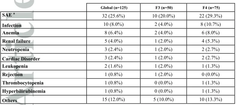
The following adverse events were collected if they occurred after initiation of therapy and up to 48 weeks after the end of treatment (follow-up): serious adverse event (SAE), defined in the Table 6, clinical and laboratory grade 3 or 4 adverse events (assessed with the Inserm-ANRS scale for grading of adverse events seriousness v6 of September 9th 2003 given in Appendix Y) and any grade of adverse events related to neutrophils, platelets, prothrombin, bilirubin, creatinin, haemoglobin or infections. The management of anemia (RBV dose reductions and/or erythropoietin administration, authorized in France when the hemoglobin level is below 10 g/dL, and/or blood transfusion) was at the investigator’s discretion.
Statistical analyses were performed with an intent-to-treat basis using SAS statistical software (SAS Institute, Cary, NC, USA). Continuous variables were expressed by mean and standard deviation and categorical variables were expressed by the number of patients and percentages. Differences in baseline characteristics between groups were evaluated using one-way analysis of variance for continuous data and the chi-square test or Fisher's exact test for categorical data. A paired t-test was used to test for change over time in continuous variable.
Results
Study population
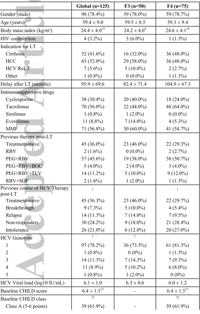
The study population consisted in 125 patients included between Oct 2013 and Nov 2014: 75 patients with METAVIR F4 HCV recurrence and 50 patients with METAVIR F3 HCV recurrence. The main characteristics of patients are presented in Table 1. The majority of patients was male (78.4%), infected by HCV genotype 1 (78.2%), had failed previous post-LT antiviral therapy (63.7%) including triple therapy with first generation protease inhibitors in 19 (15.2%) patients and SOF/RBV combination in 2 patients. Resistance testing before starting antiviral treatment was not systematically performed. This has been added. Four patients (3.2%) were co-infected with HIV. Immunosuppression was based on cyclosporine (30.4%) or tacrolimus (56.0%), and mycophenolate mofetil in 56.8% of cases. The mean time between LT and treatment initiation was 95.9 ± 69.6 months. Among the 73 cirrhotic patients with available data, 14 (19.2%) had ascites.
The following antiviral regimens were used (Table 2): SOF/RBV, SOF/PEG/RBV, SOF/DCV±RBV, SOF/LDV/RBV, SOF/SMV. RBV was used in 60 patients (48.0%). Daily dosages were as follows: 200 mg (n=2 (3.4%)), 400 mg (n=10 (16.9%)), 600 mg (n=18 (30.5%)), 800 mg (n=13 (22.0%)), 1000 mg (n=10 (16.9%)), 1200 mg (n=6 (10.2%)). Planned duration of treatment was 12 (14.4%) or 24 weeks (85.6%). Eventually, 2 patients received 28 weeks of treatment, 4 patients received 32 weeks, 3 patients received 36 weeks and 1 patient received 48 weeks of treatment. This was done because of slow virological response. Treatment regimens were SOF+DCV (n=7), SOF+DCV+RBV (n=1), SOF+RBV (n=1) and SOF+SMV/SOF+DCV (n=1).
Virological response
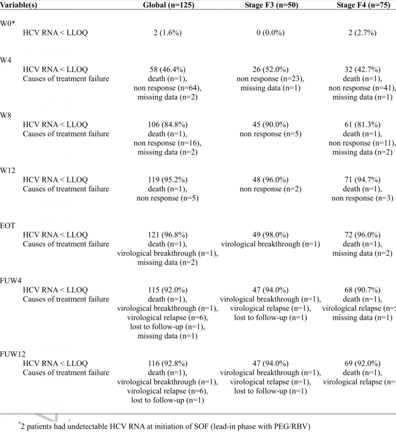
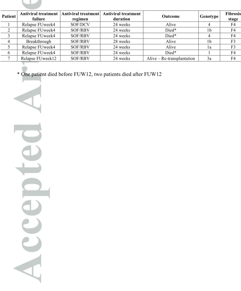
Table 3 summaries the rates of virological response. A rapid HCV RNA decline was observed in all patients after initiation of treatment. As shown in Figure 1, viral kinetics were similar between F3 and F4 groups. All but one patient had undetectable HCV RNA at end of treatment (EOT). In addition, 6 patients experienced relapse: at follow-up week 4 in 5 patients and at follow-up week 12 in one patient in whom
follow-up week 4 time point was missing. Characteristics of the 7 patients who presented virological failure are summarized in Table 4. On an intention-to-treat basis analysis, global SVR12 rate was 92.8%. It was 68.4% in patients who received a SOF/RBV combination (n=19), 100.0% in patients who received the SOF/PEG/RBV (n=5) and 97.0% in patients who received a combination of 2 DAA (n=101). Figure 2 describes the SVR12 rates according to treatment regimen, genotype, duration of treatment and previous HCV therapy post-LT.
Evolution of liver function tests
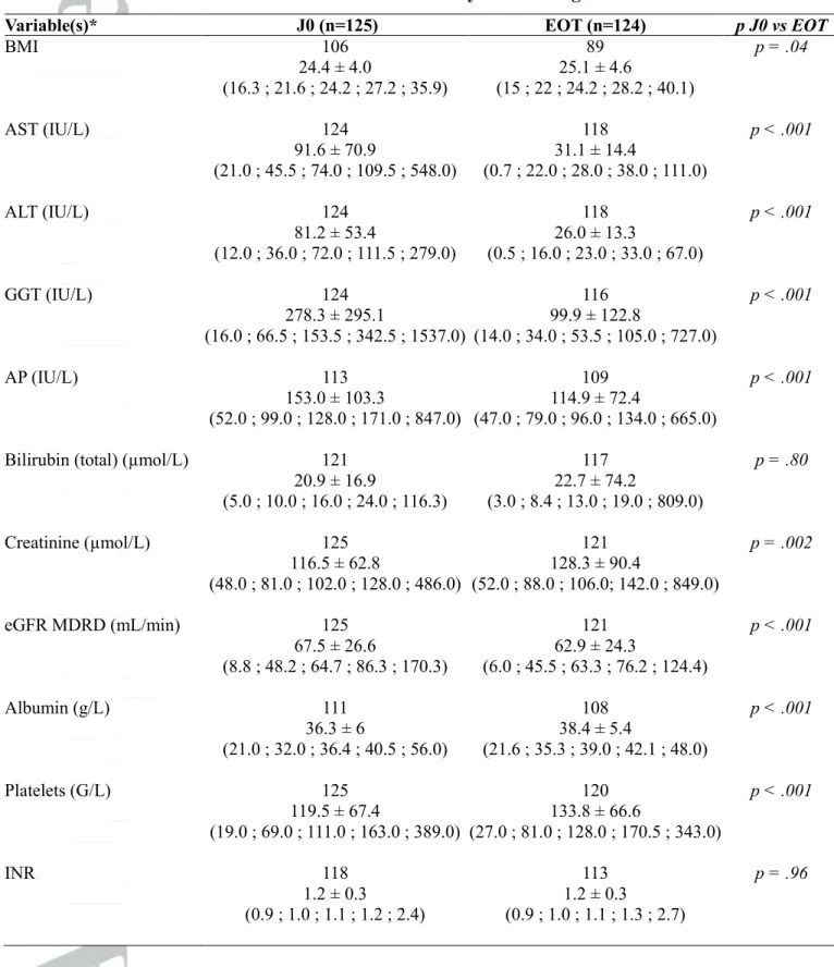
A rapid decrease of ALT, AST, and γGT serum levels was observed after treatment initiation (Table 5). At the end of antiviral treatment and according to the stage of fibrosis, the rate of patients with normal values was 91.8% and 84.1% for ALT, 89.8% and 73.9% for AST, and 63.3% and 41.8% for γGT, for F3 or F4 groups, respectively.
Clinical and liver function outcome
Evolution of biological liver function parameters is described in Table 5. BMI, albumin level and platelet count significantly improved, INR and bilirubin level did not change and creatinine and GFR significantly worsened. In summary, between D0 and EOT, in the group of 75 cirrhotic patients, Child score (when available, n=47) improved in 21 cases (44.7%) and worsened in 5 cases (10.6%) and MELD score (when available, n=64) improved in 25 cases (39.1%) and worsened in 26 cases (40.6%). In addition, between D0 and follow-up week 12, in the group of 75 cirrhotic patients, Child score (when available, n=45) improved in 20 cases (44.4%) and worsened in 5 cases (11.1%) (Figure 3A) and MELD score (when available, n=54) improved in 22 cases (40.7%) and worsened in 19 cases (35.2%) (Figure 3B). Based on Child score, liver disease progression (between D0 and end of treatment) was observed in 5 patients. One patient presented virological relapse. One patient was treated with PEG.
In parallel, mild ascites reversal was observed in 6/7 patients and refractory ascites reversal was observed in 5/7 patients. Two patients developed refractory ascites during treatment and 2 patients developed mild ascites. From the 14 patients with ascites at initiation of antiviral treatment, one had virological failure.
During antiviral treatment and follow-up, 3 patients were re-transplanted (at week 16 (n=1), between week 4 and week 24 post-treatment (n=2)) and 4 patients died (at week 1 (n=1), between week 4 and week 12 post-treatment (n=2), after week 24 post-treatment (n=1)). Causes of death were sepsis (n=2) or liver failure (n=2). All the 4 patients who died were cirrhotic; the 3 patients who died after treatment had virological failure; treatment regimens were SOF+RBV (n=3) or SOF+DCV+RBV (n=1). Deaths were considered unrelated to antiviral treatment in all cases; 3/4 occurred after end of treatment (5, 12 and 25 weeks). Re-transplantation was performed because of liver failure (week 16, n=1), chronic rejection (FU week 16, n=1), or de novo HCC (FU week 7, n=1). One patient presented virological relapse.
Safety
Thirty-two patients (25.6%) experienced at least one serious adverse event (Table 6). Infection was the most common serious adverse event (8.0%). Anemia was more frequent and more severe in patients who received RBV but was not related to fibrosis stage (Table 7).
Dosing of immunosuppressive drugs
Modifications in the dose of calcineurin and mTOR inhibitors, or MMF during antiviral treatment were performed at investigator’s discretion. Dose changes during antiviral treatment were required in 25 patients (34.7%) on tacrolimus, 14 (36.8%) patients on cyclosporine, 6 (50.0%) patients on everolimus, and 7 (9.7%) patients on MMF. Only minimal dose modifications were required, on average of +10.7% for tacrolimus, -3.7% for cyclosporine, -1.7% for everolimus and +0.1% for MMF between baseline and end of treatment. Regarding the main antiviral treatment regimens used, dose changes of tacrolimus or cyclosporine were more frequent in patients treated with SOF+DCV+RBV (51.7%) when compared to patients treated with SOF+DCV (34.0%) or SOF+RBV (18.8%).
No significant over or under-dosage was observed, and one patient had graft rejection during antiviral therapy.
Discussion
Treatment of HCV recurrence after LT is a major goal and results have been impaired for a long time because of poor efficacy and high toxicity of PEG/RBV, even in combination with first generation protease inhibitors (7, 8). To our knowledge, the present study is one of the largest series of patients with recurrent hepatitis C and severe fibrosis (n=125), treated with SOF-based antiviral therapy.
After LT, recurrent hepatitis C begins with a first phase of acute hepatitis and can have thereafter two distinct clinical and histological patterns. The first one is the most frequent and the same than described in non-transplanted patients, characterized by a progression from chronic hepatitis to cirrhosis. The second type of recurrent hepatitis C after LT is specific for immunosuppressed patients and has been described as fibrosing cholestatic hepatitis (FCH). FCH leads to an inexorable deterioration of liver function (9, 10), and is associated with a high probability of death (> 50%) (11). We recently reported, from 23 patients included in the CUPILT cohort and presenting FCH, that SOF-based antiviral therapy could be highly effective, since all patients survived, without retransplantation, and 22 patients (96%) achieved a SVR12 (12). The present study focused on patients with severe fibrosis (METAVIR F3 and F4), outside the field of FCH, and confirms these promising results in a different population with severe hepatitis C recurrence. Indeed, in addition to fibrosis progression, natural history of recurrent HCV cirrhosis is also accelerated after LT. The probability of liver decompensation is > 40% at 1 year and >70% at 3 years in LT recipients
vs. <5% and <10%, respectively, in immuno-competent patients (13, Berenguer, 2000 #1195, 14-16). The
rate of progression from liver decompensation to death is also accelerated, with a 3-year survival of <10% following the first decompensation vs. >60% in immuno-competent patients (13, Berenguer, 2000 #1195, 15, 16). This explains that long-term graft and patient survival is significantly reduced in patients undergoing LT for HCV-related liver disease as compared to other indications (3). Therefore, viral eradication in patients with severe fibrosis is a major goal, in order to avoid death, or re-transplantation. The main result of our study is the high rate of SVR12, observed in a difficult-to-treat population (92.8%). These results look better than that initially reported by Charlton et al. (17) and Forns et al. (18),
from the first series using SOF after LT, in which only 70% and 59% of SVR12 were achieved, respectively. This was probably due to a non-optimal antiviral regimen used (SOF/RBV in all patients), as confirmed in our cohort in the subgroup of patients who received the SOF/RBV combination (68.4% of SVR rate). More recently, Pungpapong et al. reported the results from a multicenter study including 123 patients (all genotype 1, 30% METAVIR F3-F4) treated with a combination of SOF and SMV with or without RBV for 12 weeks ; a SVR12 was achieved in 90% of patients (19). These results were confirmed in the TARGET cohort, from 151 patients (all genotype 1) treated with SOF/SMV ± RBV, for 12 weeks for most patients; a SVR12 was achieved in 88% of patients (20). Similarly, Gutierrez et al. reported a 93.4% rate of SVR12 in a cohort of 61 genotype 1 HCV patients (37.7% METAVIR F3-F4), treated with a combination of SOF and SMV for 12 weeks; interestingly, in METAVIR F3 and F4 patients infected with HCV genotype 1a, SVR12 was only 67% (21). Kwo et al. reported a 97% SVR12 rate in a small cohort of 34 LT recipients treated with the combination of ombitasvir/paritaprevir/dasabuvir ± RBV, but only patients with no fibrosis or mild fibrosis were included (22). Finally, our good virological results are probably related to the frequent use in our series of a combination of SOF and DCV (73.6% of the patients); this is probably due to the pangenotypic effect of this combination and is in accordance with the excellent results of the phase 2 study evaluating this combination in non-transplanted patients (23). An open and extremely relevant question is that of the potential clinical improvement after HCV eradication. We reported a dramatic improvement of liver function in our previous series of LT patients presenting FCH (12). In the present cohort, we observed in a majority of our cirrhotic patients, an improvement of liver function, based on Child score, during antiviral therapy. This was not the same when regarding MELD score but this was due to the worsening of renal function (and not liver function), in a cohort of “old” LT patients (8 years) with previous renal impairment. This observation is consistent with the results from Charlton et al. (24). In a phase 2 study, the efficacy of a combination SOF/LDV/RBV was evaluated from 337 patients infected with HCV genotypes 1 or 4, including a majority of cirrhotic patients, with mild, moderate, or severe hepatic impairment (including LT patients).
The vast majority of patients with Child–Pugh class B and C disease who had or not undergone LT, had improved MELD and Child scores at post-treatment week4 compared with baseline. Similarly, on the cohort of Forns et al., clinical status of compensated and decompensated LT cirrhotic patients receiving SOF/RBV improved in 45% of the cases (18). Nevertheless, evolution of renal function during and after antiviral therapy is of great concern. We observed that worsening of MELD score was less frequent when evaluated at follow-up week 12 (vs. EOT), strongly suggesting that impaired renal function observed during antiviral treatment, partially resolved after the end of treatment. This needs to be more extensively evaluated from larger cohort.
Safety was carefully investigated in our study population with severe fibrosis and multiple co-medications. In our cohort, SAE were not very frequent (25.6% of the patients). The most frequent SAE was bacterial infection, with favorable outcome (no patient’s death during antiviral treatment). Anemia was a frequent adverse event, observed in both patients with or without RBV, leading to RBV dose reduction or early interruption, EPO administration and even blood transfusions. As expected, anemia was more frequent and severe in patients treated with RBV, and this arises with the major issue of the optimal use of RBV (and dosage) in order to provide a significant benefit regarding antiviral efficacy. In the study by Charlton et al. (17) evaluating the SOF/RBV combination the initial dose of ribavirin was 400 mg followed by an escalation dose protocol based on hemoglobin levels. Despite this protocol, still one quarter of patient required RBV dose reduction and 33% had severe anemia. Thus, avoiding RBV from antiviral therapy would undoubtedly increase safety. Interestingly, it has been suggested that RBV could not add any benefit in non-cirrhotic patients treated by SOF/DCV irrespective to genotype and prior treatment exposition (23). Nevertheless, extrapolation of these results to LT patients, especially with severe fibrosis, needs further evaluation. Last but not least, SOF, DCV, SMV and LDV are not supposed to have significant drug-drug interactions with calcineurin inhibitors and mTOR inhibitors (25). Close monitoring of calcineurin inhibitors and mTOR inhibitors concentrations performed in our study confirmed that only mild changes of dosages were required during the antiviral treatment period.
Our study had several limitations. The first one is sample size that was not sufficient to allow precise evaluation of sub-groups according to fibrosis stage, HCV genotype or antiviral treatment regimen. Nevertheless, our results do not support a strong impact of fibrosis stage (F3 vs. F4). We acknowledge that the liver stiffness cut-offs chosen for the diagnosis of F3 and F4 fibrosis stages have been validated in HCV-infection outside the LT setting (6). In their systematic review on the diagnostic performance of transient elastography in HCV-recurrence after LT, Cholangitas et al. found variable cut-offs, ranging from 7.9 to 10.1 kPa and from 10.5 to 26.5 kPa for the diagnosis of F3 and F4, respectively (26). Given these data, we assumed that the use of the well validated cut-offs, falling in the same range than that described in LT, were the most appropriate as a non- invasive estimate of fibrosis in our study population. The second limitation is that it is a cohort study with heterogeneous treatment regimens, which was not designed to determine the optimal antiviral regimen (drugs and duration). Nevertheless, combination of SOF and DCV, with or without RBV, for 24 weeks was the main regimen used in our population and could therefore be considered as the standard option, regarding its pangenotypic activity, when available. In addition, since 6 out of our 7 patients who experienced virological failure were treated with SOF/RBV, this regimen should be used strictly for genotype 2 HCV only. Finally, due to relatively short follow-up we have been unable to provide clinical and histological outcome after HCV eradication; long-term data on survival (graft and patient) and reversion of fibrosis will be of great interest.
In conclusion, our study shows that SOF-based regimens allow achievement of SVR in the vast majority of patients presenting HCV recurrence with severe fibrosis following LT. These promising results are likely to dramatically change the prognosis in this difficult-to-treat population, with expected guarded long-term prognosis.
References
1. Adam R, McMaster P, O'Grady JG, Castaing D, Klempnauer JL, Jamieson N, et al. Evolution of liver
transplantation in Europe: report of the European Liver Transplant Registry. Liver Transpl 2003;9:1231-43. 2. Merion RM. Current status and future of liver transplantation. Semin Liver Dis 2010;30:411-21.
3. Forman LM, Lewis JD, Berlin JA, Feldman HI, Lucey MR. The association between hepatitis C infection and survival after orthotopic liver transplantation. Gastroenterology 2002;122:889-96.
4. Feray C, Caccamo L, Alexander GJ, Ducot B, Gugenheim J, Casanovas T, et al. European collaborative study on factors influencing outcome after liver transplantation for hepatitis C. European Concerted Action on Viral Hepatitis (EUROHEP) Group. Gastroenterology 1999;117:619-25.
5. Gane EJ. The natural history of recurrent hepatitis C and what influences this. Liver Transpl 2008;14 Suppl 2:S36-44.
6. Ziol M, Handra-Luca A, Kettaneh A, Christidis C, Mal F, Kazemi F, et al. Noninvasive assessment of liver fibrosis by measurement of stiffness in patients with chronic hepatitis C. Hepatology 2005;41:48-54.
7. Antonini TM, Furlan V, Teicher E, Haim-Boukobza S, Sebagh M, Coilly A, et al. Therapy with boceprevir or telaprevir in HIV/hepatitis C virus co-infected patients to treat recurrence of hepatitis C virus infection after liver transplantation. AIDS 2015;29:53-8.
8. Coilly A, Roche B, Dumortier J, Leroy V, Botta-Fridlund D, Radenne S, et al. Safety and efficacy of protease inhibitors to treat hepatitis C after liver transplantation: a multicenter experience. J Hepatol 2014;60:78-86.
9. Davies SE, Portmann BC, O'Grady JG, Aldis PM, Chaggar K, Alexander GJ, et al. Hepatic histological findings after transplantation for chronic hepatitis B virus infection, including a unique pattern of fibrosing cholestatic hepatitis. Hepatology 1991;13:150-7.
10. Zylberberg H, Carnot F, Mamzer MF, Blancho G, Legendre C, Pol S. Hepatitis C virus-related fibrosing cholestatic hepatitis after renal transplantation. Transplantation 1997;63:158-60.
11. Narang TK, Ahrens W, Russo MW. Post-liver transplant cholestatic hepatitis C: a systematic review of clinical and pathological findings and application of consensus criteria. Liver Transpl 2010;16:1228-35.
12. Leroy V, Dumortier J, Coilly A, Sebagh M, Fougerou-Leurent C, Radenne S, et al. Efficacy of Sofosbuvir and Daclatasvir in Patients with Fibrosing Cholestatic Hepatitis C After Liver Transplantation. Clin Gastroenterol Hepatol 2015.
13. Fattovich G, Giustina G, Degos F, Tremolada F, Diodati G, Almasio P, et al. Morbidity and mortality in compensated cirrhosis type C: a retrospective follow-up study of 384 patients. Gastroenterology 1997;112:463-72. 14. Pruthi J, Medkiff KA, Esrason KT, Donovan JA, Yoshida EM, Erb SR, et al. Analysis of causes of death in liver transplant recipients who survived more than 3 years. Liver Transpl 2001;7:811-5.
15. Berenguer M, Prieto M, Rayon JM, Mora J, Pastor M, Ortiz V, et al. Natural history of clinically compensated hepatitis C virus-related graft cirrhosis after liver transplantation. Hepatology 2000;32:852-8.
16. Berenguer M, Ferrell L, Watson J, Prieto M, Kim M, Rayon M, et al. HCV-related fibrosis progression following liver transplantation: increase in recent years. J Hepatol 2000;32:673-84.
17. Charlton M, Gane E, Manns MP, Brown RS, Jr., Curry MP, Kwo PY, et al. Sofosbuvir and ribavirin for treatment of compensated recurrent hepatitis C virus infection after liver transplantation. Gastroenterology 2015;148:108-17.
18. Forns X, Charlton M, Denning J, McHutchison JG, Symonds WT, Brainard D, et al. Sofosbuvir compassionate use program for patients with severe recurrent hepatitis C following liver transplantation. Hepatology 2014.
19. Pungpapong S, Aqel B, Leise M, Werner KT, Murphy JL, Henry TM, et al. Multicenter experience using simeprevir and sofosbuvir with or without ribavirin to treat hepatitis C genotype 1 after liver transplant. Hepatology 2015;61:1880-6.
20. Brown RS, Jr., O'Leary JG, Reddy KR, Kuo A, Morelli GJ, Burton JR, Jr., et al. Interferon-free therapy for genotype 1 hepatitis C in liver transplant recipients: Real-world experience from the hepatitis C therapeutic registry and research network. Liver Transpl 2016;22:24-33.
21. Gutierrez JA, Carrion AF, Avalos D, O'Brien C, Martin P, Bhamidimarri KR, et al. Sofosbuvir and simeprevir for treatment of hepatitis C virus infection in liver transplant recipients. Liver Transpl 2015;21:823-30. 22. Kwo PY, Mantry PS, Coakley E, Te HS, Vargas HE, Brown R, Jr., et al. An interferon-free antiviral regimen for HCV after liver transplantation. N Engl J Med 2014;371:2375-82.
23. Sulkowski MS, Gardiner DF, Rodriguez-Torres M, Reddy KR, Hassanein T, Jacobson I, et al. Daclatasvir plus sofosbuvir for previously treated or untreated chronic HCV infection. N Engl J Med 2014;370:211-21.
24. Charlton M, Everson GT, Flamm SL, Kumar P, Landis C, Brown RS, Jr., et al. Ledipasvir and Sofosbuvir Plus Ribavirin for Treatment of HCV Infection in Patients with Advanced Liver Disease. Gastroenterology 2015. 25. Soriano V, Labarga P, Barreiro P, Fernandez-Montero JV, de Mendoza C, Esposito I, et al. Drug interactions with new hepatitis C oral drugs. Expert Opin Drug Metab Toxicol 2015;11:333-41.
26. Cholongitas E, Tsochatzis E, Goulis J, Burroughs AK. Noninvasive tests for evaluation of fibrosis in HCV recurrence after liver transplantation: a systematic review. Transpl Int 2010;23:861-70.
Figure legends
Figure 1:
Viral kinetics according to fibrosis stage (F3/F4). No statistically significant difference was observed between the two groups.
Figure 2:
SVR12 rates according to treatment regimen, genotype, duration of treatment and previous HCV therapy post-LT.
Figure 3:
Evolution of CHILD (A, n=45) and MELD (B, n=54) available scores from initiation of treatment and follow-up week12 (F4 group, patient by patient).
Table 1: Baseline demographics and disease characteristics
Global (n=125) F3 (n=50) F4 (n=75)
Gender (male) 98 (78.4%) 39 (78.0%) 59 (78.7%)
Age (years) 59.4 ± 9.0 59.5 ± 8.5 59.3 ± 9.4
Body mass index (kg/m²) 24.4 ± 4.019 24.2 ± 4.09 24.6 ± 4.110
HIV co-infection 4 (3.2%) 3 (6.0%) 1 (1.3%) Indication for LT Cirrhosis 52 (41.6%) 16 (32.0%) 36 (48.0%) HCC 65 (52.0%) 29 (58.0%) 36 (48.0%) HCV ReLT 7 (5.6%) 5 (10.0%) 2 (2.7%) Other 1 (0.8%) 0 (0.0%) 1 (1.3%)
Delay after LT (months) 95.9 ± 69.6 82.4 ± 71.4 104.9 ± 67.3
Immunosuppressive drugs Cyclosporine 38 (30.4%) 20 (40.0%) 18 (24.0%) Tacrolimus 70 (56.0%) 22 (44.0%) 48 (64.0%) Sirolimus 1 (0.8%) 1 (2.0%) 0 (0.0%) Everolimus 11 (8.8%) 7 (14.0%) 4 (5.3%) MMF 71 (56.8%) 30 (60.0%) 41 (54.7%)
Previous therapy post-LT
Treatment-naive 45 (36.0%) 23 (46.0%) 22 (29.3%) RBV 2 (1.6%) 0 (0.0%) 2 (2.7%) PEG+RBV 57 (45.6%) 19 (38.0%) 38 (50.7%) PEG+RBV+BOC 5 (4.0%) 2 (4.0%) 3 (4.0%) PEG+RBV+TLV 14 (11.2%) 5 (10.0%) 9 (12.0%) RBV+SOF 2 (1.6%) 1 (2.0%) 1 (1.3%)
Previous course of HCV Therapy post-LT 1 1 Treatment-naive 45 (36.3%) 23 (46.0%) 22 (29.7%) Breakthrough 9 (7.3%) 5 (10.0%) 4 (5.4%) Relapse 14 (11.3%) 7 (14.0%) 7 (9.5%) Non-responders 30 (24.2%) 9 (18.0%) 21 (28.4%) Intolerance 26 (21.0%) 6 (12.0%) 20 (27.0%) HCV Genotype 1 1 1 97 (78.2%) 36 (73.5%) 61 (81.3%) 2 1 (0.8%) 0 (0%) 1 (1.3%) 3 14 (11.3%) 7 (14.3%) 7 (9.3%) 4 11 (8.9%) 5 (10.2%) 6 (8.0%) 5 1 (0.8%) 1 (2.0%) 0 (0%)
HCV Viral load (log10 IU/mL) 6.1 ± 1.0 6.3 ± 0.6 6.0 ± 1.2
Baseline CHILD score 6.4 ± 1.312 - 6.4 ± 1.312
Baseline CHILD class 12 12
Class B (7-9 points) 23 (36.5%) - 23 (36.5%)
Class C (10-15 points) 1 (1.6%) - 1 (1.6%)
Baseline MELD 11.8 ± 4.13 - 11.8 ± 4.13
Baseline MELD class 3 3
[6-10[ 26 (36.1%) - 26 (36.1%) [10-15[ 27 (37.5%) - 27 (37.5%) [15-20[ 16 (22.2%) - 16 (22.2%) [20-25[ 3 (4.2%) - 3 (4.2%) Ascites 5 3 2 No ascites 106 (88.3%) 47 (100%) 59 (80.8%) Mild to moderate 7 (5.8%) 0 (0.0%) 7 (9.6%) Refractory 7 (5.8%) 0 (0.0%) 7 (9.6%) Hepatic encephalopathy 4 3 1 No 121 (100%) 47 (100%) 74 (100%) Yes 0 (0.0%) 0 (0.0%) 0 (0.0%) AST (IU/L) 91.6 ± 70.91 82.8 ± 59.01 97.4 ± 77.6 ALT (IU/L) 81.2 ± 53.41 84.7 ± 54.11 79.0 ± 53.2 γGT(IU/L) 278.3 ± 295.11 285.3 ± 300.01 273.7 ± 293.8 ALP (IU/L) 153.0 ± 103.312 147.5 ± 90.26 156.6 ± 111.36
Total bilirubin (µmol/L) 20.9 ± 16.94 16.5 ± 12.52 23.7 ± 18.82 Creatinine (µmol/L) 116.5 ± 62.8 123.2 ± 69.4 112.0 ± 58.0 eGFR MDRD (mL/min) 67.5 ± 26.6 63.6 ± 24.5 70.2 ± 27.9 Albumin (g/L) 36.3 ± 6.014 37.6 ± 6.06 35.5 ± 5.98 Hemoglobin (g/dL) 12.9 ± 1.9 13.1 ± 1.9 12.8 ± 1.9 Platelet count (G/L) 119.5 ± 67.4 128.9 ± 60.8 113.2 ± 71.2 Leukocytes (G/L) 4.3 ± 1.7 4.5 ± 1.5 4.2 ± 1.7 Neutrophil count (G/L) 2.7 ± 1.23 2.8 ± 1.21 2.6 ± 1.32 INR 1.2 ± 0.37 1.2 ± 0.35 1.2 ± 0.32 Prothrombin Ratio (%) 82.4 ± 19.95 88.1 ± 19.62 78.5 ± 19.33
Quantitative results are expressed as mean ± SD. n
Table 2: Antiviral therapy regimens
Expected duration
Regimen Global 12 weeks 24 weeks
125 18 107 SOF+DCV 59 (47.2%) 12 (66.7%) 47 (43.9%) SOF+DCV+RBV 32 (25.6%) 1 (5.6%) 31 (29.0%) SOF+RBV 19 (15.2%) 0 (0.0%) 19 (17.8%) SOF+PEG+RBV 5 (4.0%) 2 (11.1%) 3 (2.8%) SOF+LDV+RBV 4 (3.2%) 1 (5.6%) 3 (2.8%) SOF+SMV 5 (4.0%) 2 (11.1%) 3 (2.8%) SOF/SMV+SOF/DCV* 1 (0.8%) 0 (0.0%) 1 (0.9%)
Table 3: Virological response during and after treatment (intent-to-treat analysis)
Variable(s) Global (n=125) Stage F3 (n=50) Stage F4 (n=75)
W0*
HCV RNA < LLOQ 2 (1.6%) 0 (0.0%) 2 (2.7%)
W4
HCV RNA < LLOQ 58 (46.4%) 26 (52.0%) 32 (42.7%)
Causes of treatment failure death (n=1), non response (n=64), missing data (n=2) non response (n=23), missing data (n=1) death (n=1), non response (n=41), missing data (n=1) W8 HCV RNA < LLOQ 106 (84.8%) 45 (90.0%) 61 (81.3%)
Causes of treatment failure death (n=1), non response (n=16),
missing data (n=2)
non response (n=5) death (n=1), non response (n=11),
missing data (n=2) W12
HCV RNA < LLOQ 119 (95.2%) 48 (96.0%) 71 (94.7%)
Causes of treatment failure death (n=1), non response (n=5)
non response (n=2) death (n=1), non response (n=3)
EOT
HCV RNA < LLOQ 121 (96.8%) 49 (98.0%) 72 (96.0%)
Causes of treatment failure death (n=1),
virological breakthrough (n=1), missing data (n=2)
virological breakthrough (n=1) death (n=1), missing data (n=2)
FUW4
HCV RNA < LLOQ 115 (92.0%) 47 (94.0%) 68 (90.7%)
Causes of treatment failure death (n=1),
virological breakthrough (n=1), virological relapse (n=6), lost to follow-up (n=1), missing data (n=1) virological breakthrough (n=1), virological relapse (n=1), lost to follow-up (n=1) death (n=1), virological relapse (n=5), missing data (n=1) FUW12 HCV RNA < LLOQ 116 (92.8%) 47 (94.0%) 69 (92.0%)
Causes of treatment failure death (n=1),
virological breakthrough (n=1), virological relapse (n=6), lost to follow-up (n=1) virological breakthrough (n=1), virological relapse (n=1), lost to follow-up (n=1) death (n=1), virological relapse (n=5) *
2 patients had undetectable HCV RNA at initiation of SOF (lead-in phase with PEG/RBV)
Table 4: Characteristics of patients with treatment failure
Patient Antiviral treatment failure
Antiviral treatment regimen
Antiviral treatment
duration Outcome Genotype
Fibrosis stage
1 Relapse FUweek4 SOF/DCV 24 weeks Alive 4 F4 2 Relapse FUweek4 SOF/RBV 24 weeks Died* 1b F4 3 Relapse FUweek4 SOF/RBV 24 weeks Died* 4 F4 4 Breakthrough SOF/RBV 28 weeks Alive 1b F3 5 Relapse FUweek4 SOF/RBV 24 weeks Alive 1a F3 6 Relapse FUweek4 SOF/RBV 24 weeks Died* 1 F4 7 Relapse FUweek12 SOF/RBV 24 weeks Alive – Re-transplantation 3a F4
Table 5: Outcome of clinical features and laboratory tests during antiviral treatment
Variable(s)* J0 (n=125) EOT (n=124) p J0 vs EOT
BMI 106 89 p = .04 24.4 ± 4.0 25.1 ± 4.6 (16.3 ; 21.6 ; 24.2 ; 27.2 ; 35.9) (15 ; 22 ; 24.2 ; 28.2 ; 40.1) AST (IU/L) 124 118 p < .001 91.6 ± 70.9 31.1 ± 14.4 (21.0 ; 45.5 ; 74.0 ; 109.5 ; 548.0) (0.7 ; 22.0 ; 28.0 ; 38.0 ; 111.0) ALT (IU/L) 124 118 p < .001 81.2 ± 53.4 26.0 ± 13.3 (12.0 ; 36.0 ; 72.0 ; 111.5 ; 279.0) (0.5 ; 16.0 ; 23.0 ; 33.0 ; 67.0) GGT (IU/L) 124 116 p < .001 278.3 ± 295.1 99.9 ± 122.8 (16.0 ; 66.5 ; 153.5 ; 342.5 ; 1537.0) (14.0 ; 34.0 ; 53.5 ; 105.0 ; 727.0) AP (IU/L) 113 109 p < .001 153.0 ± 103.3 114.9 ± 72.4 (52.0 ; 99.0 ; 128.0 ; 171.0 ; 847.0) (47.0 ; 79.0 ; 96.0 ; 134.0 ; 665.0)
Bilirubin (total) (µmol/L) 121 117 p = .80
20.9 ± 16.9 22.7 ± 74.2 (5.0 ; 10.0 ; 16.0 ; 24.0 ; 116.3) (3.0 ; 8.4 ; 13.0 ; 19.0 ; 809.0) Creatinine (µmol/L) 125 121 p = .002 116.5 ± 62.8 128.3 ± 90.4 (48.0 ; 81.0 ; 102.0 ; 128.0 ; 486.0) (52.0 ; 88.0 ; 106.0; 142.0 ; 849.0) eGFR MDRD (mL/min) 125 121 p < .001 67.5 ± 26.6 62.9 ± 24.3 (8.8 ; 48.2 ; 64.7 ; 86.3 ; 170.3) (6.0 ; 45.5 ; 63.3 ; 76.2 ; 124.4) Albumin (g/L) 111 108 p < .001 36.3 ± 6 38.4 ± 5.4 (21.0 ; 32.0 ; 36.4 ; 40.5 ; 56.0) (21.6 ; 35.3 ; 39.0 ; 42.1 ; 48.0) Platelets (G/L) 125 120 p < .001 119.5 ± 67.4 133.8 ± 66.6 (19.0 ; 69.0 ; 111.0 ; 163.0 ; 389.0) (27.0 ; 81.0 ; 128.0 ; 170.5 ; 343.0) INR 118 113 p = .96 1.2 ± 0.3 1.2 ± 0.3 (0.9 ; 1.0 ; 1.1 ; 1.2 ; 2.4) (0.9 ; 1.0 ; 1.1 ; 1.3 ; 2.7)
Quantitative results are expressed as mean ± SD (range;IQR;median) n
Table 6: Safety profile of antiviral therapy regimens Global (n=125) F3 (n=50) F4 (n=75) SAE a 32 (25.6%) 10 (20.0%) 22 (29.3%) Infection 10 (8.0%) 2 (4.0%) 8 (10.7%) Anemia 8 (6.4%) 2 (4.0%) 6 (8.0%) Renal failure 5 (4.0%) 1 (2.0%) 4 (5.3%) Neutropenia 3 (2.4%) 1 (2.0%) 2 (2.7%) Cardiac Disorder 3 (2.4%) 1 (2.0%) 2 (2.7%) Leukopenia 2 (1.6%) 1 (2.0%) 1 (1.3%) Rejection 1 (0.8%) 1 (2.0%) 0 (0.0%) Thrombocytopenia 1 (0.8%) 0 (0.0%) 1 (1.3%) Hyperbilirubinemia 1 (0.8%) 0 (0.0%) 1 (1.3%) Others 15 (12.0%) 5 (10.0%) 10 (13.3%) a
A serious adverse event refers to any untoward medical occurrence or effect that at any dose: • results in death,
• is life-threatening,
• results in persistent or significant disability or incapacity,
• requires hospitalization or prolongation of existing hospitalization, • is a congenital anomaly or birth defect,
• is a grade 4 clinical adverse event, • is a grade 4 biological adverse event,
• is an "important medical event" (medical events, based upon appropriate medical judgment, which may jeopardize the subject or may require medical or surgical intervention to prevent one of the above characteristics/consequences).
Table 7: Anemia during antiviral therapy (according to RBV use and fibrosis stage) Anemia p Global (n=125) RBV+ (n=60) RBV- (n=65) Grade 0 (>10 g/dl with Trt) 5 (4.0%) 4 (6.7%) 1 (1.5%) p = .03 Grade 1/2 (<10 g/dl) 26 (20.8%) 15 (25.0%) 11 (16.9%) Grade 3/4 (<8 g/dl) 14 (11.2%) 10 (16.7%) 4 (6.2%) Erythropoietin use 34 (27.2%) 22 (36.7%) 12 (18.5%) p = .02 Blood transfusion 10 (8.0%) 8 (13.3%) 2 (3.1%) p = .048
RBV dose reduction for AE 21 (16.8%) 21 (35.0%) 0 (0.0%) NA
Discontinuation of RBV 4 (3.2%) 4 (6.7%) 0 (0.0%) NA
Maximum hemoglobin decrease (g/dL) -1.8 ± 1.8 -2.9 ± 1.7 -0.9 ± 1.3 p < .001
Global (n=125) F3 (n=50) F4 (n=75) Grade 0 (>10 g/dl with Trt) : 5 (4.0%) 2 (4.0%) 3 (4.0%) p = .79 Grade 1/2 (<10 g/dl) 26 (20.8%) 12 (24.0%) 14 (18.7%) Grade 3/4 (<8 g/dl) 14 (11.2%) 4 (8.0%) 10 (13.3%) Erythropoietin use 34 (27.2%) 13 (26.0%) 21 (28.0%) p = .81 Blood transfusion 10 (8.0%) 3 (6.0%) 7 (9.3%) p = .74
RBV dose reduction for AE 21 (16.8%) 8 (16.0%) 13 (17.3%) p = .85
Discontinuation of RBV 4 (3.2%) 0 (0.0%) 4 (5.3%) p = .15
Maximum hemoglobin decrease (g/dL) -1.8 ± 1.8 -1.9 ± 1.7 -1.8 ± 1.8 p = .89
Figure 3A
Figure 3B
