HAL Id: inserm-02375419
https://www.hal.inserm.fr/inserm-02375419
Submitted on 22 Nov 2019
HAL is a multi-disciplinary open access
archive for the deposit and dissemination of
sci-entific research documents, whether they are
pub-lished or not. The documents may come from
teaching and research institutions in France or
abroad, or from public or private research centers.
L’archive ouverte pluridisciplinaire HAL, est
destinée au dépôt et à la diffusion de documents
scientifiques de niveau recherche, publiés ou non,
émanant des établissements d’enseignement et de
recherche français ou étrangers, des laboratoires
publics ou privés.
Viral RNA Degradation Makes Urine a Challenging
Specimen for Detection of Japanese Encephalitis Virus
in Patients With Suspected CNS Infection
Tehmina Bharucha, Onanong Sengvilaipaseuth, Malee Seephonelee, Malavanh
Vongsouvath, Manivanh Vongsouvath, Sayaphet Rattanavong, Géraldine
Piorkowski, Marc Lecuit, Christopher Gorman, Jean-David Pommier, et al.
To cite this version:
Tehmina Bharucha, Onanong Sengvilaipaseuth, Malee Seephonelee, Malavanh Vongsouvath,
Mani-vanh Vongsouvath, et al.. Viral RNA Degradation Makes Urine a Challenging Specimen for Detection
of Japanese Encephalitis Virus in Patients With Suspected CNS Infection: JEV RNA Not Detected
in Urine of Suspected Central Nervous System Infections.. Open Forum Infectious Diseases, Oxford
University Press, 2019, 6 (3), pp.ofz048. �10.1093/ofid/ofz048�. �inserm-02375419�
M A J O R A R T I C L E
Open Forum Infectious Diseases
Received 22 November 2018; editorial decision 30 January 2019; accepted 31 January 2019. Correspondence: Tehmina Bharucha, MBChB BSc(Hons), MRCP MSc DTM&H PGCME, Lao-Oxford-Mahosot Hospital-Wellcome Trust Research Unit (LOMWRU), Microbiology Laboratory, Mahosot Hospital, Vientiane, Lao P.D.R. (t.bharucha@doctors.org.uk).
Open Forum Infectious Diseases®
© The Author(s) 2019. Published by Oxford University Press on behalf of Infectious Diseases Society of America. This is an Open Access article distributed under the terms of the Creative Commons Attribution License (http://creativecommons.org/licenses/by/4.0/), which permits unrestricted reuse, distribution, and reproduction in any medium, provided the original work is properly cited. DOI: 10.1093/ofid/ofz048
Viral RNA Degradation Makes Urine a Challenging
Specimen for Detection of Japanese Encephalitis Virus in
Patients With Suspected CNS Infection
Tehmina Bharucha,1,2 Onanong Sengvilaipaseuth,1 Malee Seephonelee,1 Malavanh Vongsouvath,1 Manivanh Vongsouvath,1 Sayaphet Rattanavong,1 Géraldine Piorkowski,4 Marc Lecuit,5,6 Christopher Gorman,7 Jean-David Pommier,7 Jeremy A. Garson,2,8 Paul N. Newton,1,3 Xavier de Lamballerie,4 and Audrey Dubot-Pérès1,3,4
1Lao-Oxford-Mahosot Hospital-Wellcome Trust Research Unit (LOMWRU), Microbiology Laboratory, Mahosot Hospital, Vientiane, Lao P.D.R.; 2Division of Infection and Immunity, University College
London, London, UK; 3Nuffield Department of Clinical Medicine, Centre for Tropical Medicine and Global Health, University of Oxford, Churchill Hospital, Oxford, UK; 4Unité des Virus Émergents
(UVE: Aix-Marseille Univ – IRD 190 – Inserm 1207 – IHU Méditerranée Infection), Marseille, France; 5Biology of Infection Unit, Inserm U1117, Institut Pasteur, Paris, France; 6Division of Infectious
Diseases and Tropical Medicine, Necker-Enfants Malades University Hospital, Paris Descartes University, Paris, France; 7Institut Pasteur du Cambodge, Institut Pasteur International Network,
Phnom Penh, Cambodia; 8Transfusion Microbiology, NHS Blood and Transplant, London, UK
Background. Japanese encephalitis virus (JEV) is a leading cause of central nervous system (CNS) infections in Asia and results
in significant morbidity and mortality. JEV RNA is rarely detected in serum or cerebrospinal fluid (CSF), and diagnosis of JEV in-fection is usually based on serological tests that are frequently difficult to interpret. Unlike serum or CSF, urine is relatively easy to obtain, but, to date, there has been minimal work on the feasibility of testing urine for JEV RNA.
Methods. We investigated the use of lysis buffer and a Microsep device to optimize urine storage for detection of JEV RNA by
reverse transcription real-time polymerase chain reaction (RT-qPCR). The best of the studied methods was then evaluated in con-secutive patients admitted to the hospital with suspected CNS infections in Laos.
Results. We demonstrated degradation of JEV RNA in urine after even short storage periods at 4°C or –80°C. Although there
was no advantage in using a Microsep concentration device alone, immediate addition of lysis buffer to fresh urine improved the detection of JEV RNA at the limit of detection.
Conclusions. In 2 studies of 41 patients with acute encephalitis syndrome, 11 (27%) were positive for JEV IgM in CSF and/or
serum, and 2 (4.9%) were JEV RT-qPCR positive from throat swabs. JEV RNA was not detected in any of these patients’ urine sam-ples. However, lysis buffer was only used during a prospective study, that is, for only 17/41 (41%) patient urine samsam-ples. Our findings suggest a need for larger studies testing urine for JEV RNA, with urine collected at different times from symptom onset, and using lysis buffer, which stabilizes RNA, for storage.
Keywords. diagnosis; flavivirus; Japanese encephalitis virus; RNA; RT-qPCR. Japanese encephalitis virus (JEV) infection is a mosquito-borne
disease that predominantly affects children in rural parts of Asia with poor access to health care services [1]. It is charac-terized by neurological complications, leading to considerable disability, mortality, and socioeconomic costs. There is no evi-dence for any effective treatment for JEV infection, but there are several licensed vaccines to prevent disease. Nonetheless, vacci-nation programs may only be appropriately implemented and evaluated if there is an accurate diagnostic test demonstrating presence of the infection [2–4].
The mainstay of diagnosis is detection of anti-JEV IgM in cerebrospinal fluid (CSF) on admission to the hospital, as the virus is rarely detected in CSF or serum by the time patients present with neurological complications [5]. Although the gold standard is a neutralization assay, this requires paired acute and convalescent samples, considerable laboratory facilities, and expertise. For this reason, the standard di-agnostic test, and that recommended by the World Health Organization (WHO), is anti-JEV IgM capture enzyme-linked immunosorbent assay (MAC-ELISA) [6, 7]. However, there are increasingly recognized limitations in the use of MAC-ELISA for the diagnosis of JEV infection, with poor ac-curacy or translation to the field. In particular, there is low specificity related to persistence of IgM postinfection or -vac-cination, cross-reactivity with other flaviviruses, and nonspe-cific reactivity [8].
Detection of JEV RNA is specific and informs knowledge of the molecular epidemiology of the virus. Body fluids other than serum and CSF may be suitable for detection of JEV RNA. It
applyparastyle “fig//caption/p[1]” parastyle “FigCapt”
has recently been demonstrated that JEV RNA may be detected in throat swabs of patients with JEV confirmed by JEV MAC-ELISA in CSF [9]. Other mosquito-borne flaviviruses, such as West Nile virus, Zika virus, and dengue virus, have been detected in urine [10–13]. There have been reports of the per-sistence of Zika virus and West Nile virus in patients’ urine after viremia is undetectable, and the possibility of harnessing this sample type to improve the sensitivity of molecular diagnostic techniques has been proposed [10–12]. There are clear advan-tages to the use of urine, as it is an abundant sample that, unlike serum and CSF, does not require an invasive procedure to ob-tain [14, 15].
There has been minimal investigation of the use of urine to detect JEV by reverse transcription polymerase chain reaction (RT-PCR) [16]. A retrospective study of 52 patients with JEV infection suggested that JEV RNA was not detected in urine, al-though there have been 2 recent case reports of detection [17–19]. The aims of this study were (1) to evaluate urine storage with and without a lysis buffer, as well as concentration with a Microsep device, for subsequent detection of JEV RNA by real-time RT-PCR (RT-qPCR) and (2) to evaluate these methods in a study of consecutive patients admitted to the hospital with sus-pected central nervous system (CNS) infections.
METHODS
Evaluation of Urine Preparation and Storage
Preparation of JEV Dilutions in Urine
In Biosafety Level 3, JEV Laos strain JEV_CNS769_Laos_2009 (GenBank KC196115, genotype 1), isolated from the CSF of a patient with a suspected CNS infection, was cultured in Vero cells [20]. The culture supernatant (JEV isolate) was then used to prepare serial 10-fold dilutions in urine at room tempera-ture from a healthy human volunteer for evaluation of storage conditions. Aliquots at each dilution were extracted in triplicate and tested by JEV RT-qPCR (as described below) to identify the limit of detection (LOD; last dilution positive for all 3 repli-cates). JEV isolate dilutions in urine were prepared at 102 LOD
(“high”), 101 LOD (“medium”), and 100 LOD (“low”). Evaluation of Preparation and Storage Conditions One hundred forty microliters
and 5 mL of each JEV urine dilution, “high,” “medium,” and “low,” were stored neat or mixed with an equal volume of RNA-stabilizing lysis buffer, that is, buffer AVL with 1% car-rier RNA (1 μg/μL; Qiagen), each in triplicate. The aliquots were stored at 4°C for 6 hours, and then –80°C for 4 days, 14 days, and 30 days.
After return to room temperature, aliquots at each dilution, with and without buffer AVL carrier RNA, were either extracted neat or concentrated from a 5-mL dilution using a microcentri-fuge device, Microsep Advance centrifugal device 100K MWCO
5 mL (VWR, Thailand). The device allows rapid concentration and purification of up to 5.0 mL of biological samples using an Omega membrane with an ultrafiltration molecular weight cutoff (MWCO) of 100K. Five milliliters was concentrated for 7 minutes at 3220g to a final volume of 140 µL, and if a total of 10 mL (5 mL dilution and 5 mL buffer AVL carrier RNA), then the remaining 5 mL was pipetted into the same Microsep de-vice and concentrated for 7 minutes at 3220g to a final volume of 280 µL.
Nucleic Acid Extraction One hundred forty–microliter
JEV urine aliquots were extracted using a QIAamp Viral RNA Mini Kit (Qiagen) following the manufacturer’s instruction and eluted in 60 μL. A modification was made to the protocol such that, if 140 μL of buffer AVL carrier RNA had been added be-fore freeze-storing as a nucleic acid stabilizer (described above), then 420 μL of buffer AVL carrier RNA was added during the extraction instead of 560 μL of buffer AVL carrier RNA.
RT-qPCR
An in-house JEV RT-qPCR assay targeting the NS2A region, previously developed and validated [5], was used. JEV NS2A RT-qPCR was performed in duplicate using the SuperScript III One-Step RT-PCR System with Platinum Taq DNA Polymerase (Superscript-III kit; Thermo Fisher): 25-μL reaction volume; 600-nM forward (5’-AGCTGGGCCTTCTGGT-3’) and re-verse (5’-CCCAAGCATCAGCACAAG-3’) primers; 300-nM probe (5’ FAM-CTTCGCAAGAGGTGGACGGCCA-TAMRA 3’) and 7.5 μL of RNA. Thermocycling conditions: 50°C for 15 minutes, 95°C for 2 minutes, 45 × [95°C for 15 seconds + 62°C for 45 seconds]. Positive (JEV RNA Genotype 3, UVE/JEV/ UNK/TW/RP9-190 [GenBank KF907505, EVA 001V-02344]) and negative (no template) controls were performed for each RT-qPCR run. An internal control (10-μL MS2 phage) was added to each patient sample before extraction. Subsequently, a separate MS2 RT-qPCR run was performed to control the extraction process and to exclude inhibition, as previously described [21]. All RT-qPCR assays for optimization and sub-sequent experiments were run using a CFX96 qPCR detec-tion system (Biorad Laboratories) with manual baseline and threshold settings. Cq >40 were reported as negative, and ≤40 as positive. The LOD was defined as the last dilution in which all the replicates were positive.
Patient Samples
Ethics
Ethical clearance was granted by the Ethical Review Committee of the Faculty of Medical Sciences, National University of Lao and the Oxford University Tropical Ethics Research Committee, Oxford, UK. The study was performed in accordance with rele-vant guidelines and regulations.
Study Groups
Retrospective Study. We performed a retrospective study of patients
recruited in the South-East Asia Encephalitis (SEAe) study [22] conducted at Mahosot Hospital, Vientiane, Laos, between 2014 and 2017. CSF, serum, urine, and throat swab (using Sigma Virocult, Medicale Wire) samples at patient inclusion, along with follow-up serum at hospital discharge, were collected and stored at –80°C. Patients included in our study met all the following criteria: (1) were admitted to hospital with signs/symptoms ful-filling the SEAe clinical case definition for acute encephalitis syndrome (AES), (2) had no contraindication for lumbar punc-ture, (3) presented within 7 days of onset of symptoms, (4) gave written informed consent, and (5) had urine samples available for testing, collected within 24 hours of admission. The SEAe criteria included altered mental status for over 24 hours and 1 of (1) fever, (2) seizure, or (3) focal neurology.
Prospective Study. A prospective study was performed of SEAe
study participants admitted to Mahosot Hospital from June to December 2017, as described above except that there was no time limit for onset of symptoms. In this study, the urine storage conditions were modified, per the results of optimiza-tion experiments (see below).
Anti-JEV IgM ELISA (JEV MAC-ELISA)
As recommended by the WHO, all CSF and serum samples were tested using the JE Detect Kit (InBios) [23] following the manufacturer’s instructions. A 1/10 dilution was used for CSF samples, as recommended by the WHO [24].
RNA Extraction
Three hundred microliters
of urine was mixed with 100 µL of buffer ATL (Qiagen), and the total 400-µL volume extracted and eluted in 60 µL with the EZ1 Virus Mini Kit v2.0 (Qiagen, Hilden, Germany), following manufacturer’s instruction. Two hundred microliters of serum, CSF, and throat swabs were extracted and eluted in 60 µL with the same kit.
Real-time RT-PCR (RT-qPCR)
The same JEV NS2A RT-qPCR assay was performed, as described above; however, a 50-µL reaction volume was used to improve sensitivity, with a 15-µL sample volume, as reported [5]. An additional in-house RT-qPCR assay, targeting the NS3 re-gion, was also performed [5]. Per extensive optimization and validation experiments previously published [5], the NS2A and NS3 assays are highly sensitive, with LODs ~4 copies per reac-tion. The JEV NS3 RT-qPCR was performed with SuperScript III One-Step RT-PCR System with the Platinum Taq DNA Polymerase Kit (Thermo Fisher): 50-μL reaction volume; 600-nM forward (5’-GCAATGTGYCTCCAAAGAGC-3’) and re-verse (5’-GTCGATGACCCTGCTCGC-3’) primers; 300-nM probe (5’FAM-TCCTATGAYACAGAATAYCCAAA-MGB NFQ 3’); 15 μL of RNA. Thermocycling conditions: 50°C for 15 min-utes; 95°C for 2 minmin-utes; 45 × [95°C for 15 seconds + 56°C for 45 seconds]. An internal control (MS2 phage; details as above) was added to each patient sample. After extraction, MS2 RT-qPCR was performed to control the extraction process and to exclude inhibition, as previously described [21].
RESULTS
Evaluation of Urine Preparation and Storage
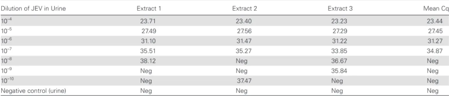
JEV isolate, extracted in duplicate and tested by JEV NS2A RT-qPCR in duplicate, had a mean Cq value of 12.18. JEV isolate was serially diluted 10-fold in urine (10–4 to 10–10); then each
di-lution was extracted in triplicate and submitted to JEV RT-qPCR. The LOD for JEV detection by RT-qPCR in urine was 10–7 (Table 1).
Further experiments were performed utilizing 10–5 dilution
as “high” with a mean Cq of 27.45, 10–6 dilution as “medium”
with a mean Cq of 31.27, and 10–7 dilution as “low” with a mean
Cq of 34.87. The dilutions were compared (1) with or without concentration with the Microsep device, (2) stored neat or with lysis buffer (buffer AVL carrier RNA), and (3) stored at 4°C for 6 hours, or at –80°C for 4 days, 14 days, and 30 days.
Table 2 provides JEV RT-qPCR results for the different urine
preparation and storage conditions. JEV was detected in the
Table 1. JEV RT-qPCR Cq Values of 10-Fold Serial Dilutions of JEV Cell Culture Supernatant in Urine
Dilution of JEV in Urine Extract 1 Extract 2 Extract 3 Mean Cq
10–4 23.71 23.40 23.23 23.44
10–5 27.49 27.56 27.29 27.45
10–6 31.10 31.47 31.22 31.27
10–7 35.51 35.27 33.85 34.87
10–8 38.12 Neg 36.67 Neg
10–9 Neg Neg 35.84 Neg
10–10 Neg 37.47 Neg Neg
Negative control (urine) Neg Neg Neg Neg
Extraction was performed immediately after spiking the urine and preparation of dilutions. Positive (JEV-G3 RNA) and negative (no template) controls were tested in duplicate in the same run; MS2 internal control was added to all samples before extraction, and MS2 PCR from all extracts was positive with expected Cq values. “Neg” = No Cq or Cq > 40. The limit of detection (LOD), defined here as the last dilution positive for all 3 replicates, is shaded in gray.
Abbreviations: JEV, Japanese encephalitis virus; RT-qPCR, reverse transcription real-time polymerase chain reaction.
medium and high neat aliquots, but not in the low aliquot after storage at 4°C for 6 hours, or at –80°C for 4 days, 14 days, and 30 days. Storage in buffer AVL carrier RNA enabled detection of JEV RNA in the “low” dilution for all storage periods. Microsep centrifugation of 5 mL gave similar results without AVL; how-ever, with AVL, the Cq was marginally lower at all dilutions tested.
Patient Samples
Retrospective Study
Twenty-four patients with suspected encephalitis, recruited be-tween 2014 and 2017, were tested. Five (21%) were anti-JEV MAC-ELISA positive in CSF and serum (Table 3). JEV RNA was not detected by RT-qPCR in any (0/24) of the urine samples of the patients, or in the corresponding throat, CSF, or serum
samples. MS2 phage as an internal control was detected by RT-qPCR in all samples with the expected Cq value.
Prospective Study
Seventeen patients with suspected encephalitis, recruited be-tween June and December 2017, were tested. Six (35%) were found anti-JEV MAC-ELISA positive in CSF and serum (Table 3). Results from the evaluation (see “Evaluation of Urine Preparation and Storage”) showed that addition of lysis buffer to fresh urine before freezing improved the preservation of JEV for subsequent detection by RT-qPCR. Therefore, for these patients, 100 µL of buffer ATL carrier RNA was added to 300 µL of urine before storage at –80°C until testing (3–7 days later). The EZ1 extrac-tion kit protocol involves adding 1:4 buffer ATL, so the samples were frozen with this volume of ATL carrier RNA.
Table 2. Results of Evaluation of Urine Preparation and Storage Conditions for the Detection of JEV by RT-qPCR
JEV RT-qPCR Results, Cq Value
140 µL 5 mL, Microcentrifuge Extract 1 Extract 2 Extract 3 Extract 1 Extract 2 Extract 3
1 2 1 2 1 2 Mean 1 2 1 2 1 2 Mean Baseline, t0 Neat High 27.49 ND 27.56 ND 27.29 ND 27.45 Medium 31.10 ND 31.47 ND 31.22 ND 31.27 Low 35.51 ND 35.27 ND 33.85 ND 34.87 Stored for 6 h at 4°C Neat High 30.25 30.7 31.02 30.67 30.29 33.14 31.01 30.64 30.01 29.98 30.11 30.26 30.1 30.18 Medium 34 35.6 35.92 34.95 35.3 34.05 34.97 32.64 34.68 35.09 33.52 33.85 34.83 34.1 Low Neg Neg Neg Neg Neg Neg Neg Neg Neg Neg Neg Neg Neg Neg Stored for 4 d at –80°C
Neat High 27.95 28.03 29.57 28.98 29.08 27.68 28.55 31.73 30.7 31.61 30.31 ND ND 31.08 Medium 32.62 31.85 33.38 32.47 32.1 30.25 32.11 31.71 31.68 33.71 32.18 ND ND 32.32 Low Neg 37.78 36.07 33.75 38.6 39.32 Nega Neg Neg Neg Neg ND ND Neg
With buffer AVL High 27.17 27.46 26.79 26.69 26.36 26.84 26.88 25.95 25.92 26.06 25.75 ND ND 25.92 Medium 30.83 30.6 31.02 30.19 30.32 30.04 30.50 28.86 28.44 28.99 29 ND ND 28.83 Low 36.24 34.11 35.35 36.4 34.18 35.23 35.25 33.33 33.42 33.36 33.78 ND ND 33.47 Stored for 14 d at –80°C
Neat High 32.03 31.78 28.41 29.07 28.46 28.59 29.72 Medium 32.78 32.67 34.63 35.75 33.25 34.11 33.87 Low Neg Neg Neg 38.22 Neg Neg Nega
With buffer AVL High 26.91 26.62 26.56 26.74 26.88 26.86 26.76 Medium 30.93 31.1 30.03 29.74 29.87 29.9 30.26 Low 35.1 34.62 35.58 34.4 33.79 33.9 34.56 Stored for 30 d at –80°C
Neat High 32.13 ND 32.76 ND ND ND 32.44 Medium 37.16 ND 36.03 ND 35.13 ND 36.10 Low Neg ND Neg ND 38.70 ND Nega
With buffer AVL High 30.76 ND 30.38 ND 30.85 ND 30.66 Medium 30.08 ND 34.25 ND 33.65 ND 32.66 Low 39.13 ND 37.66 ND 37.54 ND 38.11
“T0” indicates extracted and tested immediately after virus spiking and preparation of dilutions. Three extraction replicates were performed, on the same run, and 2 RT-qPCR replicates for each extraction. “Controls” indicates PCR-positive controls (x2 JEV G3) and negative controls (no template) in duplicate positive and negative, respectively, MS2 extraction positive control positive for each sample. “ND” indicates not done. This was in an effort to conserve reagents and microcentrifuge kits. Cq >40 were reported as negative and ≤40 as positive. Abbreviations: JEV, Japanese encephalitis virus; RT-qPCR, reverse transcription real-time polymerase chain reaction.
aMean reported as negative if 1 or more extracts were negative.
Table 3. JEV MAC-ELISA and JEV RT-qPCR Results for Patient Samples
JEV MAC-ELISA Result (ISR)a JEV RT-qPCR Result (Cq Value)
ConvaleScent Throat Swab Urine CSF Serum Patients Age, y Sex Acute Serum Serum CSF NS2A NS3 NS2A NS3 NS2A NS3 NS2A NS3 A, Retrospective study
1 42 M Neg (4.9b) - Neg (5.2b) - - Neg Neg Neg - Neg
-2 0.5 M Neg (1.7) - Neg (1.4) Neg Neg Neg Neg Neg - - -3 0.7 F Neg (1.3) - Neg (1.2) Neg Neg Neg Neg - - - -4 20 F Neg (1.3) - Neg (1.3) - - Neg Neg Neg - Neg -5 40 F Neg (1.4) - Neg (1.3) - - Neg Neg Neg - Neg -6 20 M Neg (1.1) Neg (1.3) Neg (1.6) - - Neg Neg Neg - Neg -7 22 F Neg (1.4) - Neg (0.5) - - Neg Neg Neg - Neg -8 17 F Neg (5.7) Pos (13.3) Pos (39.8) - - Neg Neg Neg Neg - Neg 9 2 F Neg (2.2) - Neg (2.4) - - Neg Neg - - - -10 32 M Neg (1.6) Neg (1.6) Neg (2.7) - - Neg Neg Neg - Neg -11 19 M Pos (18.4) Pos (45.1) Pos (29.8) Neg Neg Neg Neg Neg Neg Neg Neg 12 50 M Pos (26.0) Pos (40.8) Pos (34.2) Neg Neg Neg Neg Neg Neg Neg Neg 13 0.3 F Neg (1.6) Neg (2.4) Neg (1.7) Neg Neg Neg Neg Neg Neg - -14 13 M Neg (1.4) Neg (1.8) Neg (2.2) Neg Neg Neg Neg - - - -15 3 F Neg (1.4) Neg (1.1) Neg (1.9) Neg Neg Neg Neg - - - -16 23 M Pos (6.3) Neg (4.8) Pos (6.4) Neg Neg Neg Neg - - - -17 0.6 F Neg (1.1) Neg (1.1) Neg (1.3) Neg Neg Neg Neg - - - -18 4 F Neg (1.1) - Neg (0.7) Neg Neg Neg Neg - - - -19 1.5 M Neg (1.2) Neg (1.2) Neg (1.3) Neg Neg Neg Neg - - - -20 38 M Neg (1.1) Neg (1.3) Neg (1.2) Neg Neg Neg Neg Neg Neg - -21 13 M Pos (11.3) Pos (27.9) Pos (60.3) Neg Neg Neg Neg Neg Neg - -22 14 M Pos (21.7) - Pos (38.2) Neg Neg Neg Neg Neg Neg Neg Neg 23 0.2 F Neg (1.3) Neg (1.3) Neg (1.3) Neg - Neg Neg - - - -24 0.1 M Neg (1.4) - Neg (1.3) - Neg Neg Neg - - - -B, Prospective study
1 13 M Pos (22.3) - Pos (42.8) Pos (36) - Neg - Neg - Neg -2 34 M Neg (1.8) - Neg (2.8) Neg - Neg - Neg - Neg -3 6 F Pos (6.4) - Pos (40.3) Neg - Neg - Neg - - -4 3.8 M Pos (32.6) - Pos (48.5) - - Neg - Neg - Neg -5 24 M Pos (18) - Pos (41.8) Neg - Neg - Neg - Neg -6 18 M Neg (1.2) - Neg (1.3) Neg - Neg - Neg - Neg -7 35 M Pos (17.5) - Pos (46.8) Neg - Neg - Neg - Neg -8 3 M Pos (22.4) - Pos (49.9) Neg - Neg - Neg - Neg -9 27 M Neg (1.3) - Neg (1.3) Neg - Neg - Neg - Neg -10 45 M Neg (1.3) - Neg (1.3) Neg - Neg - Neg - Neg -11 18 M Neg (1.5) - Neg (3.1) Pos (32) - Neg - Neg - Neg -12 25 F Neg (1.5) - Neg (1.4) Neg - Neg - Neg - Neg -13 41 F Neg (1.1) Neg (1.4) Neg (1.4) Neg - Neg - Neg - Neg -14 16 F Neg (1.1) - Neg (1.4) Neg - Neg - - - Neg -15 25 M Neg (0.9) - Neg (0.6) Neg - Neg - - - - -16 55 F Neg (5.3) - Neg (1.3) Neg - Neg - Neg - Neg -17 26 F Neg (1.6) - Neg (1.4) Neg - Neg - Neg - Neg
-Results from testing throat samples, CSF, and serum, but not urine, have previously been published [9]. “Pos” indicates anti-JEV IgM positive. “Neg” indicates anti-JEV IgM negative or equivocal, with ISR cutoffs calculated according to the manufacturer’s instructions. RT-qPCR “Neg” indicates no amplification curve or curve with a Cq >40. RT-qPCR “Pos” indicates ampli-fication curve with Cq ≤40. Hyphen indicates that no sample was available for testing. NS2A and NS3 are the 2 RT-qPCR assays used for testing, targeting the respective segments of the genome [25]. Positive RT-qPCR results were confirmed by sequencing.
Abbreviations: CSF, cerebrospinal fluid; ISR, immune status ratio; JEV, Japanese encephalitis virus; MAC-ELISA, anti-JEV IgM capture enzyme-linked immunosorbent assay; RT-qPCR, re-verse transcription real-time polymerase chain reaction.
aAnti-JEV IgM detection using the JE Detect IgM Antibody Capture ELISA Kit (InBios).
bIndicates that a different kit was used for MAC-ELISA testing, as per the laboratory protocol at that time (Cat. No. E-JED01C; Inverness Medical Innovations [formerly Panbio Ltd.], Brisbane, Australia).
JEV RNA was not detected by RT-qPCR in any (0/17) of the urine samples of the patients, or in the available corresponding CSF or serum samples. MS2 phage as an internal control was detected by RT-qPCR with an expected Cq value in all sam-ples. JEV RNA was detected by RT-qPCR from throat swab in 2 patients, as recently reported [9].
DISCUSSION
A key finding of this study was the degradation of JEV RNA in urine even for short storage periods at 4°C or –80°C. Therefore, to improve the detection of JEV in urine, we investigated the effects of, on the 1 hand, virus concentration using a Microsep device, and, on the other hand, the addition of lysis buffer (AVL or ATL) in fresh urine before storage to play the role of a nucleic acid stabilizer.
Our findings suggest that concentration using a Microsep Advance centrifugal device did not improve detection of JEV RNA in urine without AVL. However, there was possibly a slight synergistic effect with AVL, generating marginally lower Cq values at all dilutions. Microsep devices have been shown to concentrate nonenveloped RNA viruses [25]; however, the lack of improvement at the limit of detection of a flavivirus, dengue, in patients’ urine samples has been reported [26]. This may be due to loss of virus by adsorption onto the membrane or other part of the device. Notably, a recent study highlighted the pres-ence of small viral RNA fragments in urine and the successful application of ultrafiltration devices in conjunction with PCR assays especially designed to detect small amplicons [27].
In contrast to our findings using an ultrafiltration device, we showed that the addition of buffer AVL carrier RNA to fresh urine permitted stabilization of viral RNA for subsequent detec-tion by RT-qPCR even after a freezing step. Optimal detecdetec-tion of low viral titer requires immediate processing of fresh samples, which is often difficult to implement. We showed that this con-straint can be easily overcome with a simple addition of buffer AVL carrier RNA to fresh samples, permitting the deferment of molecular assays without losing analytical sensitivity during storage. Similar findings of the improved detection of Zika virus RNA in urine by storage with a nucleic acid stabilizer have been recently demonstrated [28]. Moreover, the addition of lysis buffer would potentially lead to the release of the cellular JEV RNA present in infected cells, and the subsequent preservation of the JEV RNA at –80°C. It is acknowledged that only AVL and ATL buffers were evaluated in the experimentation, and other stabilizers such as Urine Conditioning Buffer (Zymo Research Corp.) may prove superior to AVL/ATL, as recently reported [29]. However, we used lysis buffer from Qiagen extraction kits, which are widely distributed and utilized worldwide. For this reason, the method has the advantage that it does not require the use of additional reagents.
However, JEV was not detected in the urine of the 41 AES patients that we tested in our pilot study. Those patients were
recruited as part of the South-East Asia Encephalitis study and fulfilled clinical criteria for encephalitis. They had onset of symptoms within 7 days and received comprehensive labo-ratory investigations. Eleven (27%) were positive by the WHO reference standard test for JEV (JEV MAC-ELISA), 5/24 (21%) in the retrospective study and 6/17 (35%) in the prospective study. Two (5%) were positive by JEV RT-qPCR testing, both from throat swab only, as previously reported [9].
There is a paucity of existing literature on this topic; neverthe-less, the finding of no JEV RNA detected in urine is consistent with a previous study of 52 patients in China [17]. However, 2 recent individual case reports have described detection of JEV RNA in the urine of 2 patients with anti-JEV IgM detected by MAC-ELISA in CSF and serum [18, 19]. The first report, by Mai et al., demonstrated JEV RNA detection in urine on day 3 of admission (approximately the fourth day from onset of symp-toms), and not in another urine sample from day 15 of admis-sion. In comparison, the second report, by Huang et al., detected JEV RNA in urine first on day 9 of admission (approximately the 14th day from onset of symptoms), and on repeat testing up to day 20 of admission. All patients in the present study were sampled within 7 days of onset of symptoms. Additionally, the 2 case reports were performed on fresh urine specimens, whereas the 24 retrospective samples in this study were stored at –80°C for up to 3 years before our investigation. Only 17 patients from the prospective study were stored in buffer ATL carrier RNA, permitting similar sensitivity of detection by RT-qPCR to that observed using fresh urine. RNA in urine may be particularly sensitive to degradation, as urine contains RNases with activity as much as 100-fold higher than in serum [28].
Limitations of the study include the limited number of rep-licates and small number of samples stored in optimal condi-tions. It is notable that, although 11 patients were positive by JEV MAC-ELISA, only 2 patients were confirmed as having JEV infection, and for this reason, the inference that urine is a poor sample for the detection of JEV infection may be unreliable, as it is based on only a very small sample of patients [8]. The opti-mization experiments were performed with cell culture super-natant to provide a model of JEV in an infected patient’s urine sample; however, it is recognized that the abundance of RNA as compared with intact virus may not be the same as that in clinical infection. It would have been useful to have tested addi-tional time points between 0 and 6 hours to determine the ra-pidity of RNA degradation in urine more precisely. In addition, patients’ urine samples were only tested at 1 time point, and this was not the same for all patients studied.
The fact that JEV was not detected in urine for the 11 JEV MAC-ELISA–positive patients of this study, in the context of the 3 other publications [17–19], suggests that excretion of JEV RNA may be brief and intermittent, and possibly that human kidney cells are not a site of high levels of replication. Ricklin et al. demonstrated that pigs, the amplifying host for
JEV, do not demonstrate viruria, but instead that the tonsils are a site of high replication [30]. An in vitro study of replica-tion of JEV in various cell types could be used to explore the findings further.
JEV diagnostics, as with many flaviviruses associated with neurological syndromes, are based on serological tests that are notoriously difficult to interpret. Although molecular diag-nosis is invaluable, JEV RNA is only rarely detected in serum or CSF. Urine is relatively easy to obtain and plentiful. There has been minimal investigation into the role of testing urine for JEV RNA to date. Our study suggests that JEV RNA is not easily detected in urine; however, it does demonstrate the value in storage of urine with a nucleic acid stabilizer at the point of bedside collection. This suggests a role for larger rigorous studies testing urine for JEV RNA, with urine collected at dif-ferent times from symptom onset, and using buffer AVL car-rier RNA or a similar nucleic acid stabilizer [29] for immediate storage of fresh urine.
Supplementary Data
Supplementary materials are available at Open Forum Infectious Diseases online. Consisting of data provided by the authors to benefit the reader, the posted materials are not copyedited and are the sole responsibility of the authors, so questions or comments should be addressed to the corre-sponding author.
Acknowledgments
Authors’ contributions. T.B. conceived the concept and design of
the study and performed the laboratory testing, data analysis, and man-uscript preparation. O.S. performed laboratory testing and reviewed the submitted manuscript. M.S. performed laboratory testing and reviewed the submitted manuscript. Mal.V. performed laboratory testing and reviewed the submitted manuscript. Man.V. supervised laboratory testing and reviewed the submitted manuscript. S.R. recruited and managed patients and reviewed the submitted manuscript. G.P. performed the sequencing and reviewed the submitted manuscript. M.L. has jointly had overall re-sponsibility and direction of the SEAe study and reviewed the submitted manuscript. C.G. has jointly had overall responsibility and direction of the SEAe study and reviewed the submitted manuscript. J.D.P. provided the clinical data sets, has jointly had overall responsibility and direction of the SEAe study, and reviewed the submitted manuscript. J.A.G. contributed to the design of the molecular assays, to the interpretation of the data, and reviewed the submitted manuscript. P.N.N. conceived the concept and design of the study, has jointly had overall responsibility and direction of the SEAe study, and supervised drafting and submission of the man-uscript. X.D.L. conceived the concept and design of the study, has jointly had overall responsibility and direction of the SEAe study, and reviewed the submitted manuscript. A.D.P. conceived the concept and design of the study, supervised laboratory testing, has jointly had overall responsibility and direction of the SEAe study, and supervised drafting and submission of the manuscript.
We are very grateful to the patients and to Asst. Prof. Bounthaphany Bounxouei, the Director of Mahosot Hospital, the late Dr. Rattanaphone Phetsouvanh, Director of the Microbiology Laboratory, Dr. Mayfong Mayxay, and the staff of the wards and Microbiology Laboratory of Mahosot Hospital. We also thank Ooyanong Phonemixay for technical help and support, Asst. Prof. Dr. Bounnak Saysanasongkham, Director of the Department of Health and Rehabilitation (DHR), Ministry of Health, and Prof. Bounkong Syhavong, Minister of Health, Lao P.D.R., for their very kind help and support. We thank the stakeholders of the ongoing SEAe pro-ject (www.seaepropro-ject.org).
Financial support. The research has been implemented in
collabo-ration with the ComAcross project (www.onehealthsea.org/ comacross) thanks to the financial support of the European Union (EuropeAid, INNOVATE contract 315-047). It was also supported by Total Foundation, Horizon 2020 research and the innovation program EVAg under grant agreement No. 653316, the Institute of Research for Development, Aix-Marseille University, and the Wellcome Trust of Great Britain.
Data availability. All data generated or analyzed during this study are
included in this published article.
Potential conflicts of interest. All authors: no reported conflicts of
interest. All authors have submitted the ICMJE Form for Disclosure of Potential Conflicts of Interest. Conflicts that the editors consider relevant to the content of the manuscript have been disclosed.
References
1. Campbell GL, Hills SL, Fischer M, et al. Estimated global incidence of Japanese encephalitis: a systematic review. Bull World Health Organ 2011; 89:766–74, 774A–774E.
2. Caldwell JP, Chen LH, Hamer DH. Evolving epidemiology of Japanese encepha-litis: implications for vaccination. Curr Infect Dis Rep 2018; 20:30.
3. Simon-Loriere E, Faye O, Prot M, et al. Autochthonous Japanese encephalitis with yellow fever coinfection in Africa. N Engl J Med 2017; 376:1483–5.
4. Heffelfinger JD, Li X, Batmunkh N, et al. Japanese encephalitis surveillance and immunization - Asia and Western Pacific Regions, 2016. MMWR Morb Mortal Wkly Rep 2017; 66:579–83.
5. Bharucha T, Sengvilaipaseuth O, Vongsouvath M, et al. Development of an improved RT-qPCR assay for detection of Japanese encephalitis virus (JEV) RNA including a systematic review and comprehensive comparison with published methods. PLoS One 2018; 13:e0194412.
6. Robinson JS, Featherstone D, Vasanthapuram R, et al. Evaluation of three com-mercially available Japanese encephalitis virus IgM enzyme-linked immuno-sorbent assays. Am J Trop Med Hyg 2010; 83:1146–55.
7. Khalakdina A, Shrestha SK, Malla S, et al. Field evaluation of commercial im-munoglobulin M antibody capture ELISA diagnostic tests for the detection of Japanese encephalitis virus infection among encephalitis patients in Nepal. Int J Infect Dis 2010; 14(Suppl 3):e79–84.
8. Dubot-Pérès A, Sengvilaipaseuth O, Chanthongthip A, et al. How many patients with anti-JEV IgM in cerebrospinal fluid really have Japanese encephalitis? Lancet Infect Dis 2015; 15:1376–7.
9. Bharucha T, Sengvilaipaseuth O, Seephonelee M, et al. Detection of Japanese en-cephalitis virus RNA in human throat samples in Laos - a pilot study. Sci Rep
2018; 8:8018.
10. Andries AC, Duong V, Ly S, et al. Value of routine dengue diagnostic tests in urine and saliva specimens. PLoS Negl Trop Dis 2015; 9:e0004100.
11. Gourinat AC, O’Connor O, Calvez E, et al. Detection of Zika virus in urine. Emerg Infect Dis 2015; 21:84–6.
12. Nagy A, Bán E, Nagy O, et al. Detection and sequencing of West Nile virus RNA from human urine and serum samples during the 2014 seasonal period. Arch Virol 2016; 161:1797–806.
13. Barzon L, Pacenti M, Franchin E, et al. Excretion of West Nile virus in urine during acute infection. J Infect Dis 2013; 208:1086–92.
14. Mortimer PP. Urine-based viral diagnostics: an innovation in waiting. Rev Med Virol 2015; 25:203–4.
15. Mortimer PP, Parry JV. Non-invasive virological diagnosis: are saliva and urine specimens adequate substitutes for blood? Rev Med Virol 1991; 1:73–8.
16. Turtle L, Solomon T. Japanese encephalitis - the prospects for new treatments. Nat Rev Neurol 2018; 14:298–313.
17. Zhao H, Wang YG, Deng YQ, et al. Japanese encephalitis virus RNA not detected in urine. Clin Infect Dis 2013; 57:157–8.
18. Mai NTH, Phu NH, Nhu LNT, et al. Central nervous system infection diagnosis by next-generation sequencing: a glimpse into the future? Open Forum Infect Dis
2017; 4(X):XXX–XX.
19. Huang GKL, Tio SY, Caly L, et al. Prolonged detection of Japanese encephalitis virus in urine and whole blood in a returned short-term traveler. Open Forum Infect Dis 2017; 4(X):XXX–XX.
20. Aubry F, Vongsouvath M, Nougairede A, et al. Complete genome of a geno-type I Japanese encephalitis virus isolated from a patient with encephalitis in Vientiane, Lao PDR. Genome Announc 2013; 1:e00157–12.
21. Ninove L, Nougairede A, Gazin C, et al. RNA and DNA bacteriophages as molec-ular diagnosis controls in clinical virology: a comprehensive study of more than 45,000 routine PCR tests. PLoS One 2011; 6:e16142.
22. South East Asia Encephalitis Project (SEAe) 2013. 2017. https://research.pasteur.fr/en/ program_project/the-southeast-asia-encephalitis-project/. Accessed June 1, 2018. 23. World Health Organization. Regional Office for the Western Pacific. Western
WHOROft, ed. Training Report Fourth Hands-on Training Workshop on the Laboratory Diagnosis of Japanese Encephalitis. Osong; Manila : WHO Regional Office for the Western Pacific, 2014.
24. Johnson BW, Goodman CH, Jee Y, Featherstone DA. Differential di-agnosis of Japanese encephalitis virus infections with the Inbios JE Detect™ and DEN Detect™ MAC-ELISA kits. Am J Trop Med Hyg 2016; 94:820–8. 25. Brassard J, Seyer K, Houde A, et al. Concentration and detection of hepatitis
A virus and rotavirus in spring water samples by reverse transcription-PCR. J Virol Methods 2005; 123:163–9.
26. Andries AC, Duong V, Ly S, et al. Value of routine dengue diagnostic tests in urine and saliva specimens. PLoS Negl Trop Dis 2015; 9:e0004100.
27. Lu T, Han Y, Zhang R, et al. Quantitative detection of hepatitis C virus RNA in urine of patients with chronic hepatitis C using a novel real-time PCR assay. J Med Virol 2019; 91:115–23.
28. Tan SK, Sahoo MK, Milligan SB, et al. Stability of Zika virus in urine: specimen processing considerations and implications for the detection of RNA targets in urine. J Virol Methods 2017; 248:66–70.
29. Gorchakov R, Berry RM, Patel SM, et al. Optimizing PCR detection of Zika virus from various body fluids. Am J Trop Med Hyg 2019; 100(2):427–33.
30. Ricklin ME, García-Nicolás O, Brechbühl D, et al. Vector-free transmission and persistence of Japanese encephalitis virus in pigs. Nat Commun 2016; 7:10832.