HAL Id: inserm-02967917
https://www.hal.inserm.fr/inserm-02967917
Submitted on 15 Oct 2020
HAL is a multi-disciplinary open access
archive for the deposit and dissemination of
sci-entific research documents, whether they are
pub-lished or not. The documents may come from
teaching and research institutions in France or
abroad, or from public or private research centers.
L’archive ouverte pluridisciplinaire HAL, est
destinée au dépôt et à la diffusion de documents
scientifiques de niveau recherche, publiés ou non,
émanant des établissements d’enseignement et de
recherche français ou étrangers, des laboratoires
publics ou privés.
An unusual Staphylococcus saccharolyticus
spondylodiscitis post kyphoplasty: a case report
Marie-Charlotte Trojani, Brigitte Lamy, Raymond Ruimy, Nicolas Amoretti,
Karine Risso, Christian Roux
To cite this version:
Marie-Charlotte Trojani, Brigitte Lamy, Raymond Ruimy, Nicolas Amoretti, Karine Risso, et al..
An unusual Staphylococcus saccharolyticus spondylodiscitis post kyphoplasty: a case report. BMC
Infectious Diseases, BioMed Central, 2020, 20 (1), pp.539. �10.1186/s12879-020-05263-5�.
�inserm-02967917�
C A S E R E P O R T
Open Access
An unusual
Staphylococcus saccharolyticus
spondylodiscitis post kyphoplasty: a case
report
Marie-Charlotte Trojani
1*, Brigitte Lamy
2,3,4, Raymond Ruimy
2,3,4, Nicolas Amoretti
5, Karine Risso
6and
Christian Roux
7Abstract
Background:Staphylococcus saccharolyticus is a rarely encountered coagulase-negative, which grows slowly and its strictly anaerobic staphylococcus from the skin. It is usually considered a contaminant, but some rare reports have described deep-seated infections. Virulence factors remain poorly known, although, genomic analysis highlights pathogenic potential.
Case presentation: We report a case ofStaphylococcus saccharolyticus spondylodiscitis that followed kyphoplasty, a procedure associated with a low rate but possible severe infectious complication (0.46%), and have reviewed the literature. This case specifically stresses the risk of healthcare-associatedS. saccharolyticus infection in high-risk patients (those with a history of alcoholism and heavy smoking).
Conclusion:S. saccharolyticus infection is difficult to diagnose due to microbiological characteristics of this bacterium; it requires timely treatment, and improved infection control procedure should be encouraged for high-risk patients.
Keywords: Spondylodiscitis, Kyphoplasty, Healthcare-associated infection, Case report,Staphylococcus saccharolyticus
Background
Staphylococcus saccharolyticus (formerly known as Pep-tococcus saccharolyticus) is a rarely encountered coagulase-negative staphylococcus and the only anaer-obic species of the genus Staphylococcus [1]. Although it is usually considered a non-pathogenic microorganism of the human skin flora with no particular known trop-ism to generate specific infections, occasional reports suggest a pathogenic potential through miscellaneous rare deep-seated infections [2–5]. Little is known on its virulence factors, pathogenesis, and determinants of
infection. Recently, genome-sequencing analysis has shown that S. saccharolyticus possesses hyaluronidase activity (similar to that of Staphylococcus aureus), toxins of the phenol-soluble modulin family, and several quorum-sensing systems that may have a tissue-invasive potential [6].
Infectious complications after vertebroplasty/kypho-plasty are rare, but potentially serious life-threatening complications affecting the patient’s functional prognosis can occur (0.46% prevalence rate), which usually result from direct inoculation from skin flora such as Staphylo-coccus aureus, S. epidermidis, and Cutibacterium acnes [7,8].. Here, we report the third case of spondylodiscitis due to S. saccharolyticus and the first to follow a surgical procedure such as kyphoplasty that specifically stresses
© The Author(s). 2020 Open Access This article is licensed under a Creative Commons Attribution 4.0 International License, which permits use, sharing, adaptation, distribution and reproduction in any medium or format, as long as you give appropriate credit to the original author(s) and the source, provide a link to the Creative Commons licence, and indicate if changes were made. The images or other third party material in this article are included in the article's Creative Commons licence, unless indicated otherwise in a credit line to the material. If material is not included in the article's Creative Commons licence and your intended use is not permitted by statutory regulation or exceeds the permitted use, you will need to obtain permission directly from the copyright holder. To view a copy of this licence, visithttp://creativecommons.org/licenses/by/4.0/. The Creative Commons Public Domain Dedication waiver (http://creativecommons.org/publicdomain/zero/1.0/) applies to the data made available in this article, unless otherwise stated in a credit line to the data.
* Correspondence:[email protected]
1Département de Rhumatologie, Université Cote d’Azur, CHU de Nice, Nice,
France
the risk of healthcare-associated S. saccharolyticus infec-tion [2,3].
Case presentation
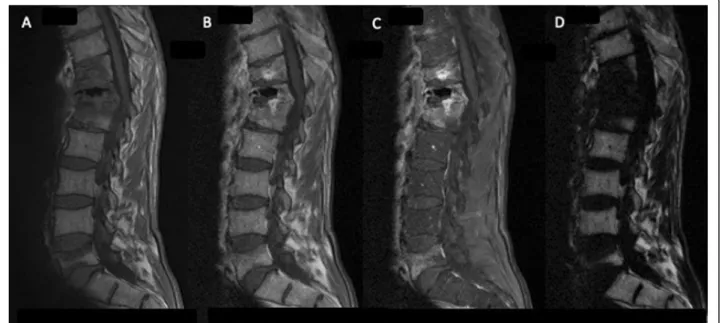
A 57-year-old man with a history of alcoholism and heavy smoking was admitted for disabling back pain. Four months earlier, he had sustained two vertebral frac-tures (T10 and T11) due to falling; these were treated by kyphoplasty under computer tomography (CT) guidance. Because the back pain persisted 2 weeks after the pro-cedure, he received a zygapophyseal joint steroid injec-tion under CT guidance. Three days later, his C-reactive protein level was 12.5 mg/l and hyperleukocytosis was moderate (13 G/L including 10 G/L neutrophils) and the patient had no fever. Magnetic resonance imaging (MRI) findings revealed infectious spondylodiscitis (Fig. 1). A Staphylococcus saccharolyticus isolate was recovered after 90 h of incubation from one single vial of a first series of three blood culture (BC) sets. The same micro-organism was identified from two additional BC series collected 5 and 10 days later after 83 and 100 h of incu-bation, respectively. It was not possible to perform either culturing or molecular diagnostics using the tissue sam-ple as the patient’s condition did not permit disc biopsy. However, the findings from the blood culture tests indi-cated a definite diagnosis of spondylodiscitis, though a catheter or spinal device was not inserted in the patient. Using disk diffusion assay, the isolate was multi-drug susceptible including to penicillin and cefoxitin. The pa-tient was treated with 2 g of amoxicillin three times a day for a total duration of 4 weeks after consultation with the infectious disease team. Pain and inflammatory
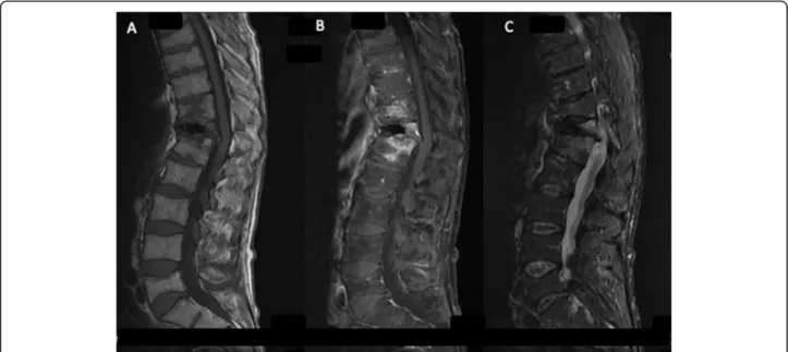
syndrome both gradually regressed, and MRI performed after 12 month showed decrease in hyperintensity (Fig.
2b). No clear source of the bacteria was identified. The infection was presumably from skin and the bacteria was likely introduced in the surgical site during the kypho-plasty procedure. However, we could not identify any defect in the surgical skin preparation and infection con-trol procedures as well as any particular event causing exposure during the kyphoplasty procedure.
Discussion and conclusions
S. saccharolyticus is a lesser known coagulase-negative staphylococcus [1]. Although it is usually considered a skin contaminant, it can cause endocarditis, bone infec-tion, or pyomyositis, which may be associated with poor outcome (Table1) [2–5,9–13]. In addition to the tissue-invasive factors that have been unraveled, the depend-ence on anaerobic conditions is considered to favor the ability to invade human tissue, while biofilm production may contribute to the colonization of medical devices [6]. Further research is needed to understand S. Sacchar-olyticus virulence and the risk of infection better. In this regard, reports should also carefully consider patient co-morbidities as host condition may contribute to the dis-ease development. In this context, a literature review was conducted using the key words “Staphylococcus sac-charolyticus” and “case” on the PubMed database. Eight articles were excluded after reading title and/or abstract because they were not applicable to the topic. A total of 8 articles were selected, which were used to compose the summary table (Table 1). Thus far, only 3 of the 9 reported cases have detailed host risk factors. To date,
Fig. 1 Spinal MRI, sagittal section: Hyperintensity, T11-T12-L1 vertebrae, para-vertebral soft tissue, and T12-L1 disc consistent with infectious spondylodiscitis. a T1-weighted; b, c, d IDEAL sequence
only 3 cases, including our case, of spondylodiscitis have been reported, of which 1 case has been related to a surgi-cal procedure and multiple-level diskographies [3] and 1 (current) case occurred after kyphoplasty; information on surgical procedure was not retrieved for the third case [2].
Although vertebroplasty is a minimally invasive pro-cedure, the possibility of postoperative infection should not be ignored. It requires major salvage surgery and may lead to residual disability and even death in several cases. In addition to standard skin preparation and the administration of prophylactic antibiotics, surgeons should preoperatively consider immune status, urinary tract infection or other infection source within 6 months, and history of pulmonary tuberculosis to prevent infec-tion post vertebroplasty [14].
It is unclear why S. saccharolyticus is specifically asso-ciated with spondylodiscitis. This either reflects a spe-cific bacterial niche that remains to be evidenced or represents a publication bias. To date, there is no means to clarify this point. Bruggeman et al. recently reported 8 strains recovered from hip and shoulder prosthetic infec-tions, which suggests that orthopedic sites other than the spine may be infected by S. saccharolyticus. Unfortu-nately, Brüggeman et al. provided no information about the clinical cases, so it is unclear if this potential con-taminant was actually the causative agent of all the re-ported infections [6].
This case highlights several important considerations in S. saccharolyticus infection and the pitfalls associated with the diagnostic aspects. Symptoms and biological syndrome may be moderate or absent in the early stage of infection [4]. In our case, fever was absent and the in-flammatory biologic syndrome was mild. Possible reasons
could be the proximity of a corticosteroid injection and ef-fective empirical treatment that was timely administered. S. saccharolyticus, in addition to being anaerobic, grows slowly, which may be misinterpreted as a contamination because the bacterium grows only in anaerobic bottles (not in aerobic bottles); thus, very few or only a single bottle may be positive. In addition, the long time to positivity is usually a criterion to suspect BC contamination (together with a single/low number of positive bottle). Thus, the characteristics of the result (long time to positivity, low number of positive bottles) could be misinterpreted for a contamination) [1,13]. This might also lead to under diag-nosis when cultures are not incubated for at least 5 days, which is a regular situation with analyses other than BC. These findings advocate for the following: i) a minimum of 5 days of anaerobic culture study when infection is strongly suspected, and no microorganism is recovered on day 3 of incubation. This implies a preferred cooperation between a rheumatologist and microbiologist to adapt and optimize the diagnostic procedures, including molecular diagnostics, when spinal infection is suspected and ii) a fine interpret-ation of the microbiological findings in order to prevent overlooking an infection etiology when a microorganism that is most frequently a contaminant is recovered.
The favorable evolution after appropriate antibiotics treatment is not a regular option. The rare reported in-fections (9 to our knowledge) have often been fatal (3 of
the 7 available outcomes; Table 1). Timely treatment
may be critical. Comorbidities favoring this opportunis-tic infection are unevenly reported: prostheopportunis-tic heart valves [9,10], poor oral hygiene (2), type II diabetes (5), to which we can importantly add tobacco use, alcohol-ism, and cachexia in this patient.
Table 1 Summary of cases of infections caused by S. saccharolyticus Year (ref) Location Age/ Sex Diagnosis Risk factors Comment on clinical presentation Biology Microbiological diagnosis Antimicrobial susceptibility Final treatment (total duration) Outcome 1990 [4] USA 61/ M Endocarditis No predisposing valvular heart disease Low grade fever at onset; moderate-sized mitral valve vegetation Anemia; thrombocytosis; ESR elevation BC; all bottles positive at day 10 Susceptible to PE, OX, VA, GE, CI, CL; resistant to ME NAF + GE (6 wks) Favorable at day 30 1996 [9] USA 57/ W Prosthetic mitral valve endocarditis NA Fever; large masses at prosthetic valve level Anemia; hyperleukocytosis BC; all anaerobic vials positive at day 1; aerobic vials positive at day 11 Susceptible to VA, CL, CH; resistant to all β -lactamin agents (including OX, CES, ME, TET) Valve change; medical treatment (NA) Died at day 32 of hospitalization 2009 [10 ] USA NA Prosthetic valve endocarditis NA NA NA Mitral valve; anaerobic culture; at day NA NA NA NA 1990 [11 ] China 21/ M Pneumonia NA Blood-stained sputum; multiple spherical focal lesions in the lung (CT scan) Anemia; hyperleukocytosis NA Susceptible to LE, MO; NA for other antibiotics AZ (6 d); TI + PE (3 d); IM + TI (1 d) Died at day 120 of hospitalization 2015 [12 ] China 26/ W Bone marrow infection NA High-grade fever; headache at onset; lymph nodes Anemia; hyperleukocytosis; ESR and CRP strong elevation Lung biopsy; positive anaerobic culture at day 10? Susceptible to VA, LE, PE, CL; resistant to ME; PE + VA (2 d); IM + VA (NA) Died at day 114 2005 [2] France 58/ M Spondylodiscitis No endocarditis; no underlying disease but poor oral hygiene Thoracic posterior pains for 2 months; fever; weight loss; NSAIDs/corticosteroids treatment At admission, hyperleukocytosis; ESR and CRP elevation BC and bone marrow; positive anaerobic cultures at day 3 Susceptible to VA, TEI, RI, ER, PR, TET, OF, CL; Resistant to ME; No β -lactamase production; no mecA gene OF+CL (12 wks) Favorable at year 1 2009 [3] USA 38/ M Spondylodiscitis NA Radicular symptoms treated unsuccessfully by microdiscectomy Elevation of inflammatory parameters Negative aerobic cultures; negative acid-fast bacilli NA NA NA 2017 [5] NZ 48/ M Pyomyositis, spermatic cord infection Type II diabetes; hyperlipidemia Fever Neutrophilia; CRP large increase; CPK normal Multiple muscle biopsies; anaerobic positive culture at 24 h; coinfection S. capitis and S. saccharolyticus Susceptible to FL; Resistant to PE CEFA (1 wk); CEP (2 wks) Favorable at 4 weeks 2017 (our case) France 57/ M Spondylodiscitis Heavy smoking;
alcoholism; unhealthy underweight
Vertebral fractures (treated by kyphoplasty and zygapophyseal joint steroid injection); no fever; unremarkable clinical examination Hyperleukocytosis; CRP moderate increase Aerobic cultures negative at day 7 Susceptible to PE; CEF, MA, RI, TET, FO, OF; no β -lactamase production AM (4 wks) Favorable at 46 months M man, W woman, ESR erythrocyte sedimentation rate, BC blood culture, NA non-available, AM amoxicillin, AZ azithromycin, CEFA cefazolin, CEF cefoxitin, CEP cephalexin, CES cephalosporin, CH chloramphenicol, CI ciprofloxacin, CL clindamycin, ER erythromycin, FL flucloxacillin, FO fosfomycin, GE gentamicin, IM imipenem, LE levofloxacin, MA macrolides, ME metronidazole, MO moxifloxacin, NAF nafcillin, OF ofloxacin, OX oxacillin, PE penicillin, PR pristinamycin, RI rifampicin, TEI teicoplanin, TET tetracycline, TI timidazole, VA vancomycin
Finally yet importantly, infection control procedures de-signed to prevent infection following vertebroplasty proced-ure may require some improvements to achieve infection prevention in patients with poorer condition. This is a chal-lenging goal because, to-date, no suggestions to help reduce the risk of infection are available in the literature when all actions taken have already complied with guidelines. Strat-egies for improvement may arise from further research on antibacterial advanced cement for kyphoplasty [15,16] (e.g., Clarkin et al., 2011; Brauer et al., 2013), as well as from fur-ther research on improved bundle approaches. Improve-ments may also arise from a better understanding of the pathophysiology of surgical site infection. A step in this dir-ection was provided by Romano-Bertrand et al. [17], who showed how disturbances of skin microbiota by antisepsis and prophylactic treatment impacted the dynamics of microbiota in deep tissues during cardiac surgery. Although this model does not exactly fit with kyphoplasty, it does clearly show that diverse bacteria may reach the surgical site during invasive procedures. Further understanding is also needed on how the patient’s condition and innate im-munity may impact the response towards controlling surgi-cal site infection development during the very first steps of invasive procedures. In conclusion, the incidence of S. sac-charolyticus spondylodiscitis is reportedly low, but clini-cians must not fail the diagnosis. We advise that any S. saccharolyticus culture in the context of fever and/or ortho-pedic pain should be cautiously reviewed before being con-sidered a contaminant. Prompt diagnosis and treatment is essential for an improved outcome of this severe infection and overall efforts should be made in infection control dur-ing vertebroplasty.
Abbreviations
CT:Computer tomography; BC: Blood culture; MRI: Magnetic resonance imaging Acknowledgements
None.
Authors’ contributions
MCT was in charge of the case review and preparation of the manuscript. CR, BL, conceived the work, provided clinical expert opinion, and helped to draft the manuscript. KR, NA, and RR provided clinical expert opinion and revised the manuscript. All authors read and approved the manuscript. Funding
None.
Availability of data and materials
Not applicable. Please contact authors for data requests. Ethics approval and consent to participate
Written consent was obtained from the patient. No ethics committee was necessary for this case report.
Consent for publication
Written consent was obtained from the patient for publication of this case report. A copy of the written is available for review by Editor in Chief of this journal.
Competing interests
The authors declare that they have no competing interests. Author details
1Département de Rhumatologie, Université Cote d’Azur, CHU de Nice, Nice,
France.2Laboratoire de Bactériologie, Hôpital L’archet 2, CHU de Nice, Nice,
France.3INSERM U1065, Centre Méditerranéen de Médecine Moléculaire,
Equipe 6, Nice, France.4Faculté de Médecine, Université Côte d’Azur, Nice, France.5Département de Radiologie, Université Cote d’Azur, CHU de Nice,
Nice, France.6Service d’infectiologie, Université Nice Côte d’Azur, CHU de
Nice, Nice, France.7Département de Rhumatologie, Université Cote d’Azur,
LAHMESS EA6309, CNRS, iBV UMR 7277, CHU de Nice, Nice, France.
Received: 17 March 2020 Accepted: 16 July 2020
References
1. Evans CA, Mattern KL, Hallam SL. Isolation and identification of
Peptococcussaccharolyticus from human skin. J Clin Microbiol. 1978;7:261–4. 2. Godreuil S, Jean-Pierre H, Morel J, et al. Unusual case of spondylodiscitis
due toStaphylococcus saccharolyticus. Joint Bone Spine. 2005;72:91–3. 3. Mikhael MM, Bach HG, Huddleston PM, Maus TP, Berbari EF. Multilevel
diskitis and vertebral osteomyelitis after diskography. Orthopedics. 2009;32:60. 4. Westblom TU, Gorse GJ, Milligan TW, Schindzielorz AH. Anaerobic
endocarditis caused by Staphylococcus saccharolyticus. J Clin Microbiol. 1990;28:2818–9.
5. Young N, Bhally H. Bilateral neck Pyomyositis caused byStaphylococcus capitis and Staphylococcus saccharolyticus in a diabetic adult. Case Rep Infect Dis. 2017;2017:3713212.
6. Brüggemann H, Poehlein A, Brzuszkiewicz E, Scavenius C, Enghild JJ, Al-Zeer MA, Brinkmann V, Jensen A, Söderquist B.Staphylococcus saccharolyticus Isolated From Blood Cultures and Prosthetic Joint Infections Exhibits Excessive Genome Decay. Front Microbiol. 2019;10:478.
7. Abdelrahman H, Siam AE, Shawky A, Ezzati A, Boehm H. Infection after vertebroplasty or kyphoplasty. A series of nine cases and review of literature. Spine J. 2013;13:1809–17.
8. Schofer MD, Lakemeier S, Peterlein CD, et al. Primary pyogenic spondylitis following kyphoplasty: a case report. J Med Case Rep. 2011;5:101.
https://doi.org/10.1186/1752-1947-5-101.
9. Krishnan S, Haglund L, Ashfaq A, Leist P, Roat T. Prosthetic valve endocarditis due to Staphylococcus saccharolyticus. Clin Infect Dis. 1996;22: 722–3.
10. Bravo LTC, Oethinger MD. Staphylococcus saccharolyticus: A Rare but Important Cause of Anaerobic Endocarditis. Microbiology No. MB 08–8 (MB-357). Am J Clin Pathol. 2009;131:286–99 (Abst n°46).
11. Wu X, Yu C, Wang X. A case of Staphylococcus saccharolyticus pneumonia. Int J Infect Dis. 2009;13:e43–6.
12. Liu CJ, Sun B, Guo J, et al. A case of bone marrow infection by
Staphylococcus saccharolyticus. Eur Rev Med Pharmacol Sci. 2015;19:1161–3. 13. Lanne S. Staphylococcus saccharolyticus: à propos d’un cas d’infection de
dispositif cardiaque implantable, enjeux de l’identification et pathogénicité d’un germe méconnu. Sci Pharm. 2016; dumas-01323806 .
14. Liao JC, Lai PL, Chen LH, Niu CC. Surgical outcomes of infectious spondylitis after vertebroplasty, and comparisons between pyogenic and tuberculosis. BMC Infect Dis. 2018;18(1):555.https://doi.org/10.1186/s12879-018-3486-x. 15. Clarkin O, Wren A, Thornton R, Cooney J, Towler M. Antibacterial analysis of
a zinc-based glass polyalkenoate cement. J Biomater Appl. 2011;26(3):277–
92.https://doi.org/10.1177/0885328210364430.
16. Brauer DS, Karpukhina N, Kedia G, et al. Bactericidal strontium-releasing injectable bone cements based on bioactive glasses. J R Soc Interface. 2013; 10(78):20120647.https://doi.org/10.1098/rsif.2012.0647.
17. Romano-Bertrand S, Frapier JM, Calvet B, et al. Dynamics of the surgical microbiota along the cardiothoracic surgery pathway. Front Microbiol. 2015; 5:787. Published 2015 Jan 13.https://doi.org/10.3389/fmicb.2014.00787.
Publisher’s Note
Springer Nature remains neutral with regard to jurisdictional claims in published maps and institutional affiliations.