Helena de Puig Guixé
Industrial Engineer (2011)Institut Químic de Sarrià, Universitat Ramon Llull
Submitted to the Department of Mechanical Engineering in partial fulfilment of the requirements for the degree of
Master of Science in Mechanical Engineering at the
MASSACHUSETTS INSTITUTE OF TECHNOLOGY
June 2013
© 2013 Massachusetts Institute of Technology. All rights reserved.
Author………
Department of Mechanical Engineering May 8th, 2013 Certified by.………
Kimberly Hamad-Schifferli Visiting Scientist, Department of Mechanical Engineering & Technical Staff, MIT Lincoln Laboratory Thesis supervisor Accepted by………
Submitted to the Department of Mechanical Engineering, on May 8th, in partial fulfilment of the requirements for the degree of
Master of Science in Mechanical Engineering
Abstract
We have developed a method to externally control blood clotting using gold nanoparticles. Gold nanorods (NRs) have unique size and shape-dependent optical properties that can be used for externally controlled release of biomolecules by laser excitation. Femtosecond pulsed laser irradiation at the NR longitudinal surface plasmon resonance peak (LSPR) can excite the NRs and induce melting, and thus cause release of drug or biomolecular payload on the NR. Because the peak wavelength of the LSPR changes with NR aspect ratio, NRs with different aspect ratios can be independently excited at different wavelengths to release different payloads in a mutually exclusive fashion. This approach can be used to create a biological switch for blood clotting by releasing a single stranded (ssDNA) thrombin binding aptamer (TBA) upon laser irradiation. It is possible to control blood clotting by releasing TBA that binds and inhibits thrombin, and an antidote consisting of a complementary ssDNA sequence that binds to TBA and restores thrombin activity. Both the TBA and the antidote are loaded onto NRs with different aspect ratios. This enables us to use laser excitation at one wavelength to deliver the TBA and inhibit thrombin and consequently blood clotting. We then use a different wavelength to deliver the antidote and reverse the effect of the
current practice is systemically administering drugs though the whole bloodstream and relying on physiological clearance to restore the system.
Thesis supervisor: Kimberly Hamad-Schifferli
Title: Visiting Scientist, Department of Mechanical Engineering & Technical Staff, MIT Lincoln Laboratory
the years I spent in her research group at MIT. Doing research with her has been a great learning experience, due to her kind character and her ability to discuss and share ideas, without those, this research project would have not been feasible. The time she has shared with me has a priceless value, as it has helped me grow both as a researcher and personally. I am as well thankful for the opportunities she has given me to present our work in conferences, both in the US and in Europe. These experiences are very important for my initiation in a research career.
I am grateful for having been able to work also in Prof. Lee Gehrke’s lab. I am thankful for his lab support and also for the help I received from his research group, especially Chunwan, Ann, Irene and José.
I would also like to thank Prof. Mercedes Balcells and Prof. Salvador Borrós, who enabled me to contact Prof. Hamad-Schifferli on the first place. Prof. Mercedes Balcells, as a co-director of the MIT-Spain Program, welcomed me on my arrival in Boston; she has been a helpful guide during my stay at MIT.
for fruitful discussions and help throughout these years. I would also like to thank Dr. Zhichuan Xu, for taking amazing TEM pictures, and for his nice character. Also, the undergraduate students who have helped me in the lab: Helen D’Couto, Dorma C. Flemister, Nate Miska and Ashley Fletcher.
I thank our collaborators at MIT. First, Prof. Andrei Tokmakoff and his students: Sam, Aritra, Luigi, Paul, Carlos, Krupa and Kevin, for the use of their lasers. Prof. Kristala Jones-Prather, Prof. Krystyn Van Vliet, Prof. Moungi Bawendi, Prof. Alexandra A. Techet, for the use of their equipment. Also, the Center for Materials Science and Engineering (CMSE) and the Biophysical Instrumentation Facility (BIF), for the use of their equipment.
I would also like to thank Denise MacPhail and Joan Hutchins for assistance, and Obra Social La Caixa, for my postgraduate fellowship.
I would finally like to thank my parents, family and friends, who have greatly contributed to making this experience even more enjoyable, my roomates: Maria, Sagi, Nuria, Martu, Majo and Gemma, for fruitful philosophical discussions. I thank Bayridge
1.1. Motivation ... 25
1.2. Objectives and strategies ... 28
1.3. Background ... 31
1.3.1. Gold nanoparticles ... 31
1.3.1.1. Surface chemistry ... 33
1.3.1.2. Optical properties ... 35
Nanoparticle assembly ... 38
Photothermal heating of gold nanorods ... 39
Selective photothermal heating of gold nanorods with different SPR ... 42
1.3.2. Thrombin and the coagulation cascade ... 45
1.3.2.1. Thrombin binding aptamer ... 48
1.4. Thesis outline ... 53
1.5. Bibliography ... 54
2. Materials and Methods ... 67
2.1. Synthesis of gold nanoparticles ... 67
2.1.1. Gold nanorods ... 67
2.2.3. DNA conjugation of gold nanoparticles ... 82
2.2.4. Surface modifications on the DNA-conjugated NRs ... 87
2.2.4.1. HSA block ... 87
2.2.4.2. PEG backfill ... 88
2.3. HS Corona conjugation on NP surfaces ... 90
Carbon nanotube solubilization and characterization ... 90
2.4. Laser Irradiation of NR ... 92
2.5. Blood clotting tests ... 93
2.7. Bibliography ... 94
3. Characterization of the covalently bound aptamer-NP conjugates 99
3.1. Introduction ... 993.2. Thermodynamic study of the binding of the aptamer-thrombin on the surface of a NP ... 101
3.2.1. Analysis methods ... 104
3.2.1.1. Langmuir isotherm ... 104
3.2.4. Conclusions ... 133
3.3. Kinetics of biomolecule-induced nanoparticle assembly ... 136
3.3.1. Theoretical ... 141
3.3.2. Results and discussion ... 141
3.3.2.1. Different surface chemistries ... 143
3.3.2.2. Different concentrations of biomolecule added ... 148
3.3.4. Conclusions ... 152
3.4. Reversible nanomachinery at the nanoparticle-biomolecule interface 154
3.4.1. Theoretical ... 155
3.4.2. Results and discussion ... 158
3.4.3. Conclusions ... 160
3.5. Quantifying DNA hybridization at the nanoparticle interface ... 161
3.5.1. Theoretical ... 162
3.5.2. Results and discussion ... 165
3.5.3. Conclusions ... 170
3.6. Conclusions ... 172
4.2.2. Control experiments with single NRs and NBs ... 188
4.2.3. Selective melting of a mixture of NRs and NBs ... 190
4.3. Release of thiolated ssDNA from NRs ... 193
4.3.1. Increasing the loading of thiolated TBA with intramolecular G-quadruplexes ... 193
4.3.2. Aptamers and antidotes effect in blood clotting ... 194
4.3.3. Melting and release of TBA from NRs ... 196
4.3.4. Effect of NPs in blood clotting time ... 199
4.4. Release of dsDNA from NRs ... 201
4.5. Conclusions ... 204
4.6. Bibliography ... 205
5. Increasing loading capacity: NR coronas ... 209
5.1. Introduction ... 209
5.2. Characterization methods for NR-coronas ... 211
5.3.3 Effect of particle material on loading ... 227
5.4. Kinetics of formation of the protein coronas ... 229
5.5. Leakage from the NR-coronas ... 230
5.6. Conclusions ... 238
5.7. Bibliography ... 239
6. Switching on and off blood clotting with light ... 243
6.1. Introduction ... 243
6.2. Effect of TBA and antidote in thrombin clotting tests ... 247
6.3. Preparation and characterization of NR-coronas ... 249
6.4. Single laser irradiations for the release of TBA and antidote ... 253
6.5. Selective release of TBA and antidote for the control of blood clotting ... 257
6.4. Conclusions ... 262
6.5. Bibliography ... 263
7. Conclusions and future directions ... 267
7.2.2. Triggered release for combinational therapy ... 277
7.2.3. Switchable enhancement of in vitro translation with light ... 278
7.2.4. Nanobiomachinery and protein-NR / DNA-NR origamis ... 279
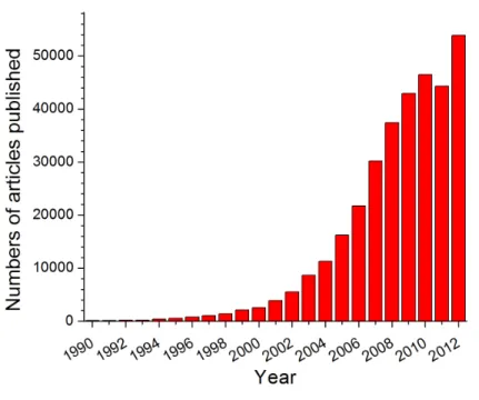
Figure 1-2: Published articles on nanotechnology since 1990. Searched “nanoparticle” in google scholar ... 32
Figure 1-3: Surface Plasmon Absorption of Spherical Nanoparticles[17] ... 36
Figure 1-4: Overview of the selective release[69]. Reprinted with permission from Wijaya A, Schaffer SB, Pallares IG, Hamad-Schifferli K. Selective Release of Multiple DNA Oligonucleotides from Gold Nanorods. ACS Nano 2008;3:80. Copyright 2008 American Chemical Society. ... 42
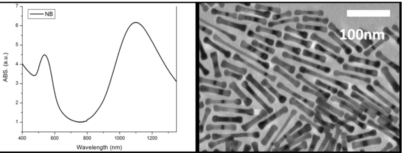
Figure 1-5: Absorbance and TEM image of the NRs and NB after irradiation[69]. Reprinted with permission from Wijaya A, Schaffer SB, Pallares IG, Hamad-Schifferli K. Selective Release of Multiple DNA Oligonucleotides from Gold Nanorods. ACS Nano 2008;3:80. Copyright 2008 American Chemical Society. ... 44
Figure 1-6: Fluorecense of the DNA released after the irradiation[69] Reprinted with permission from Wijaya A, Schaffer SB, Pallares IG, Hamad-Schifferli K. Selective Release of Multiple DNA Oligonucleotides from Gold Nanorods. ACS Nano 2008;3:80. Copyright 2008 American Chemical Society. ... 45
Figure 2-4: TEM Histogram (left), DLS (middle) and extinction (right) of the synthesized
NS ... 72
Figure 2-5: TEM images of the NS ... 73
Figure 2-6: Method for NR ligand exchange[17]. Adapted with permission from Wijaya A, Schaffer SB, Pallares IG, Hamad-Schifferli K. Selective Release of Multiple DNA Oligonucleotides from Gold Nanorods. ACS Nano 2008;3:80. Copyright 2008 American Chemical Society. ... 77
Figure 2-7: Gel electrophoresis of the NRs-CTAB; NRs-MUDA and NRs-MHDA ... 80
Figure 2-8: Gel electrophoresis of PEG-NRs ... 81
Figure 2-9: NR-CTAB; NR-MHA and NR-DNA ... 84
Figure 2-10: Secondary structure of the thrombin binding aptamer (TBA) ... 87
Figure 2-11: Gel of PEG-backfilled NRs ... 88
Figure 3-1: Thermodynamic cycle describing the assembly of NR-TBA in the presence of thrombin. Adapted with permission from de Puig H, Federici S, Baxamusa SH, Bergese P, Hamad-Schifferli K. Quantifying the Nanomachinery of the Nanoparticle-Biomolecule Interface. Small 2011;7. Copyright 2011 WILEY-VCH Verlag GmbH & Co. KGaA,
GmbH & Co. KGaA, Weinheim. ... 116
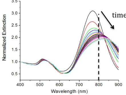
Figure 3-3: Change in absorption spectra of NR-T15-TBA high coverage as a function of time upon incubation with 121 nM of thrombin (arrow indicates increasing time). Adapted with permission from de Puig H, Federici S, Baxamusa SH, Bergese P, Hamad-Schifferli K. Quantifying the Nanomachinery of the Nanoparticle-Biomolecule Interface. Small 2011;7. Copyright 2011 WILEY-VCH Verlag GmbH & Co. KGaA, Weinheim. ... 117
Figure 3-4: A400/A800 as a function of time for NR-T15-TBA high coverage for different thrombin concentrations. Adapted with permission from de Puig H, Federici S, Baxamusa SH, Bergese P, Hamad-Schifferli K. Quantifying the Nanomachinery of the Nanoparticle-Biomolecule Interface. Small 2011;7. Copyright 2011 WILEY-VCH Verlag GmbH & Co. KGaA, Weinheim. ... 118
Figure 3-5: Aggregation isotherm for NR-T15-TBA high coverage. Adapted with permission from de Puig H, Federici S, Baxamusa SH, Bergese P, Hamad-Schifferli K. Quantifying the Nanomachinery of the Nanoparticle-Biomolecule Interface. Small 2011;7. Copyright 2011 WILEY-VCH Verlag GmbH & Co. KGaA, Weinheim. ... 118
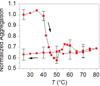
Figure 3-6: A400/A800 as a function of time upon incubation with thrombin (t = 0 min) and complementary DNA sequence (gray area, 60 min). Adapted with permission from de
function of changing the temperature from 25ºC to 80ºC and back down to 25ºC (arrows indicate direction). Adapted with permission from de Puig H, Federici S, Baxamusa SH, Bergese P, Hamad-Schifferli K. Quantifying the Nanomachinery of the Nanoparticle-Biomolecule Interface. Small 2011;7. Copyright 2011 WILEY-VCH Verlag GmbH & Co. KGaA, Weinheim. ... 120
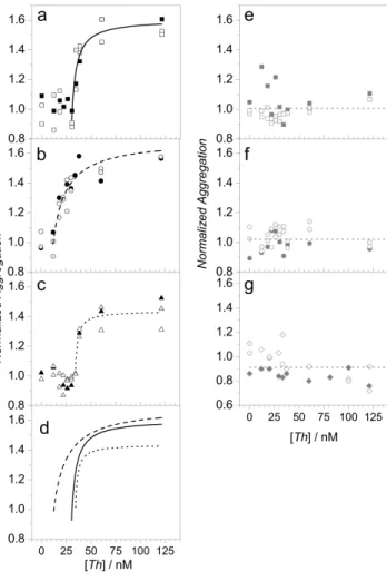
Figure 3-8: Modified Langmuir aggregation isotherms for a) NR-T15-TBA high coverage, b) NR-T15-TBA low coverage, c) NR-T15-TBA mPEG backfill 0.5:1 mPEG:TBA, d) fits for plots a-c , e) NR-TBA without poly T spacer (the dashed line is the average value), f) NR-T15-TBA HSA block (the dashed line is the average value), g) NR- T15-TBA high coverage mPEG backfill 5:1 mPEG:TBA. Reprinted with permission from de Puig H, Federici S, Baxamusa SH, Bergese P, Hamad-Schifferli K. Quantifying the Nanomachinery of the Nanoparticle-Biomolecule Interface. Small 2011;7. Copyright 2011 WILEY-VCH Verlag GmbH & Co. KGaA, Weinheim. ... 124
Figure 3-9: a) θ vs. -Wσ for NR-T15-TBA high coverage (square), NR-T15-TBA low coverage (triangle), and NR-T15-TBA -PEG backfill 0.5:1 (circle), b) ThT vs. - Wσ for NR-T15-TBA high coverage (square), NR-T15-TBA low coverage (triangle), and NR- T15
-Figure 3-11. Nanorod assembly kinetics scheme ... 137
Figure 3-12: Formation of the unit blocks of aggregation scheme ... 138
Figure 3-13: Subsequent monomer addition scheme ... 139
Figure 3-14: Overall kinetic scheme of NR assembly ... 139
Figure 3-15: Evolution of the extinction of the NRs with time ... 142
Figure 3-16: Assembly of the NRs at 121nm thrombin ... 142
Figure 3-17: Kinetic fits with different surface chemistries ... 144
Figure 3-18: Comparison of the fits between different surface chemistries ... 145
Figure 3-19: T15-TBA high coverage kinetics at different thrombin concentrations ... 149
Figure 3-20: T15-TBA low coverage kinetics at different thrombin concentrations ... 150
Figure 3-21: PEG Backfill kinetics at differetn thrombin concentrations ... 151
Figure 3.22: Thrombin-TBA nanomachine ... 155
Figure 3.23: Thermodynamic cycle of NRs disassembly ... 157
Figure 3-24: Reverisble assembly of NRs after the addition of thrombin and anti-TBA 159
Figure 3-25: Disassembly of NRs after antidote addition ... 160
Figure 3-26: DNA hybridization on the NRs surface ... 163
Figure 3-32: DNA hybridization on Low-coverage NRs ... 168
Figure 3-33: Sigmoidal fit for the DNA hybridization (r2=0.998) ... 169
Figure 4-1. Absorbance spectra of NRs (red) and NBs (blue). ... 184
Figure 4-2. Scheme of the selective release of thiolated ssDNA ... 185
Figure 4-3. CTAB-NRs and CTAB-NBs ... 186
Figure 4-4. Melting CTAB-NRs at 800nm ... 187
Figure 4-5. Melting CTAB-NBs at 1100nm ... 188
Figure 4-6. Minimal melting of the CTAB-NRs at 1100nm ... 189
Figure 4-7. No melting of the CTAB-NBs at 800nm ... 189
Figure 4-8. Selective melting of CTAB-NR-NB at 800nm ... 191
Figure 4-9. Selective melting of CTAB-NR-NB at 1100nm ... 192
Figure 4-10. Loading of thiolated TBA with and without PolyT spacer ... 194
Figure 4-11. Effect of TBA in blood and effect of antidote on TBA ... 195
Figure 4-12. ABS spectra of 5nM TBA-NRs at increasing irradiation times ... 196
Figure 4-13. TBA release as a function of irradiation time ... 197
Figure 4-20. Released TBA and thiolated T15-antidote after 800nm CW laser irradiation
... 203
Figure 5-1. Scheme of combined and sequential assembly ... 211
Figure 5-2. Absorbance spectra of the protein coronas ... 213
Figure 5-3. TEM images of NR, NB, CNT and CoNR, CoNB, CoCNT ... 214
Figure 5-4. DLS measurements (top) and Zeta potentials (bottom) of NR, NB, CNT and CoNR, CoNB, CoCNT ... 215
Figure 5-5. Variation of the corona components ... 217
Figure 5-6. Absorbance of NR-coronas ... 217
Figure 5-7. Absorbance of SeNR ... 219
Figure 5-8. Loading from coronas as measured by supernatant loss ... 220
Figure 5-9. Loading of NR-coronas measured by heat displacement ... 221
Figure 5-10. DLS of coronas formed with HS, ES and HSA ... 222
Figure 5-11. Loading of CoNRs, as a function of [PB] ... 224
Figure 5-12. Stability of NR-coronas in different buffer strengths and buffers. ... 224
Figure 5-13. Loading of the NRs as a function of CTAB concentrations ... 226
Figure 5-19. Formation of the protein coronas ... 236
Figure 6-1. Scheme of the selective release approach ... 245
Figure 6-2. Calibration of TBA (top) and antidote:TBA, with [TBA]=500nM(bottom) 248
Figure 6-3. Absorbance spectra of NR, NB, NR-HS-TBA and NB-HS-anti ... 250
Figure 6-4. Loading of NR-HS-TBA and NB-HS-anti ... 251
Figure 6-5. DLS and Zeta potential of NB-CTAB, NB-HS-anti, NRs-CTAB and NR-HS-anti ... 252
Figure 6-6. 1100nm irradiation of NB-HS-anti. Melting and release (inset) ... 253
Figure 6-7. 800nm irradiation of NR-HS-TBA. Melting and release (inset) ... 254
Figure 6-8. Comparison of HS-TBA-Bock release, thiolated TBA-NRs and calibrations 255
Figure 6-9. Comparison of HS-TBA-Bock release and thiolated TBA-NRs ... 256
Figure 6-10. Comparison of HS-TBA-Bock release and thiolated TBA-NRs ... 257
Figure 6-11. Comparison of HS-TBA-Bock release and thiolated TBA-NRs ... 258
Figure 6-12. Comparison of HS-TBA-Bock release and thiolated TBA-NRs ... 259
Figure 6-13. 1100nm irradiation of the NR-HS-TBA and NB-HS-antidote. Left: Absorbance. Right: fluorescence of the released species ... 260
Table 1-2: Kinetic parameters of some aptamers[73] ... 50
Table 2-1: Parameters for the synthesis of NS of different sizes ... 72
Table 3-1: Kinetic binding constants for different surface chemistries ... 143
Table 3-2: Assembly kinetic constants of different surface chemistries ... 146
Table 3-3: Kinetic binding constants for High Coverage NR-T15-TBA ... 148
Table 3-4: Calculated kinetic binding constants for high coverage T15-TBA ... 149
Table 3-5: Kinetic binding constants for Low coverage NR-T15-TBA ... 150
Table 3-6: Calculated binding constants for T15-TBA with Low coverage ... 151
Table 3-7: Kinetic binding constants for PEG backfilled NR-T15-TBA ... 151
Table 3-8: Calculated kinetic binding constants for PEG-backfilled NR-T15-TBA ... 152
Table 3-9: Compared values of DNA hybridization on different surfaces ... 168
1. Introduction: On the control of blood clotting
1.1. Motivation
The coagulation of blood is necessary to avoid bleeding and is achieved by the blood clotting cascade, which is an enzymatic cascade that ensures a rapid and localized response to injury. However, there are a number of thrombophylic disorders, such as deep venous thrombosis (DVT), stokes, heart attacks, peripheral vascular diseases, and many others where precise control of blood clotting by the use of anticoagulants is required. The most widespread anticoagulants are (1) heparin –which acts rapidly by its presence in the blood-, (2) warfarin –which is acts after some delay- and (3) aspirin.
The discovery of heparin by McLean and Howell on 1916[1, 2] is one of the most important advances in medicine. Heparin is a mucopolysaccharide that is extracted from bovine and porcine gut mucosa[3]. Even nowadays, injectable heparin is the most commonly used anticoagulant due to its rapid effectiveness, availability and low cost. It prevents blood coagulation by inhibiting thromboplastin –by depressing factors V, IX, XI and XII- and by enhancing fibrinolysis –and inhibiting the polymerization of fibrinogen-[4].
Warfarin was discovered in the 1920s, when cattle in northern US and Canada started dying of internal bleeding, due to sweet clover disease. Later, in 1948, warfarin was used as rat poison, with rodents dying of internal haemorrhage. In 1950s, warfarin started being used as an anticoagulant, with the name ‘Coumadin’, and is currently the most widespread oral anticoagulant[2]. However, its effect in preventing blood clotting is not immediate, and it requires a controlled dosage and the use of heparin before it can be administered. Warfarin prevents blood clotting by lowering the amount of active vitamin K available for the activation of clotting factors II, VII, IX and X[5].
Aspirin was discovered by Hoffmann in 1898, and was initially used as an antipyretic and anti-inflammatory drug. Aspirin also has antithrombotic effects, and it prevents the coagulation of blood by preventing platelet aggregation, increasing blood clot lysis, and lowering the formation of thrombin by decreasing tissue factor expression[6].
All these anticoagulants have side effects, the most common of them being a high risk of bleeding, if dosage is not correctly monitored[1, 5]. Moreover, anticoagulants are administered to the entire bloodstream, and most of them do not have a specific antidote, so their effect can only be reduced by clearance. Therefore, when surgery is required, it needs to be postponed for several days, until the anticoagulants are cleared from the
bloodstream, with the possibility of eventually causing strokes-. Moreover, in the early 2000’s several deaths were caused by contaminated batches of heparin that were distributed in the US [7-10]. Other adverse reactions of anticoagulants include thrombocytopenia, acute heparin reaction, heparin associated osteoporosis, heparin and warfarin-related skin reactions, abnormal liver functioning, and eosinophilia, among others [4, 11].
Therefore the possibility of manipulating blood clotting in a way that is specific –by using an anticoagulant that can inhibit only one protein in the blood clotting cascade-,
reversible –by using an antidote that can stop the activity of the anticoagulant-, localized
–by enabling the control of blood clotting in a confined area- and externally controlled, would represent a great improvement over the current practice of administering anticoagulants over the whole bloodstream and relying in physiological clearance to restore the system’s activity.
1.2. Objectives and strategies
With the use of gold nanoparticles, it is possible to achieve a localized and external release of biomolecules. The conjugation of gold nanoparticles with biomolecules has enabled important advances in diagnostic and therapeutic nanomedicine. The biocompatibility and unique optical properties of gold nanoparticles has enabled their use in sensing, imaging, drug release and assembly applications.
The interaction of light with noble metal nanoparticles results in collective oscillations of the electrons in the metal known as surface plasmon resonance (SPR). The SPR is dependent on the size and shape of the particles, and can be tuned throughout the visible and IR regions. Gold nanoparticles have a SPR peak, and therefore can be melted by ultrafast laser irradiation at the SPR to release a drug or biomolecule on their surface. Thus, gold nanoparticles of different sizes and shapes have been used for photothermal therapy and delivery.
We have developed a method to externally control blood clotting by exploiting the optical properties of gold nanoparticles. Nanoparticles are attractive for interfacing to biological systems and have been used to manipulate biological processes
inhibit human thrombin, and an antidote (ssDNA complementary sequence) that restores its activity. We use gold nanorods (NRs), as they can be synthesized to absorb light in the infrared region, where biological tissue has minimal absorption. The SPR is tunable by changing the NR aspect ratio (AR); therefore, NRs with different ARs can be independently excited at different wavelengths to release different payloads[12]. By using this approach, we use laser excitation at one wavelength to release the aptamer and trigger the inhibition of thrombin, and then use a different wavelength to deliver the antidote and specifically reverse the effects of the aptamer (Fig. 1-1). Thus, the pair of NRs acts an on/off switch for blood clotting.
We study the binding of the TBA by thiol chemistry and by taking advantage of protein coronas that naturally form around nanoparticles in biological fluids for enhanced loading[13]. We also quantify the interactions of thrombin with gold nanoparticles at the interface, the formation of protein coronas, and the passive release of ssDNA from protein coronas weakly bound to nanoparticles.
1.3. Background
1.3.1. Gold nanoparticles
It was in the Middle Ages when artisans started using gold nanoparticles to give color to stained glasses in the cathedrals. This fact was not recognized until 1857, in M. Faraday’s Bakerian Lecture on the “Experimental Relations of Gold (and other Metals) to Light” [14], when he observed that the red color of colloidal gold comes from the presence of aggregates of gold atoms. Nevertheless, it was not until the twentieth century when colloidal chemistry received a huge growth due to the works of Oswald, Mie [15], Svedberg and Zsigmondy[16].
One fact that was already noticed more than a 100 years ago is how the amount of surface atoms increases with decreasing the particle size. If we imagine a cube of 1cm edge, the percentage of surface atoms would be around 10-5%. Moreover, in a cube of 10nm edge, this percentage would increase to 10%; and in a cube of 1nm edge, each atom would be a surface atom [17]. This is one of the reasons why one can expect very different physical and chemical properties in a nanoparticle than those expected in bulk materials.
Nanotechnology comprises all the methods and techniques to study, design and fabricate devices that work in the nanometer scale (1-100nm). Its interest has increased in the last decade, where more than 35 countries have developed programs in nanotechnology since 2000[18], and the number of published articles regarding nanotechnology has rapidly increased during the last years (Fig. 1-2). The interest on nanotechnology is based on the fact that all biological systems have their first level of organization at the nanoscale [19]; therefore, nanotechnology can lead to important improvements in biology, biotechnology, medicine and healthcare.
The nanoscale interactions of organic and inorganic matter lead to the construction of cells and more complex organisms –the brain and the human body-. Therefore, nanotechnology plays a key role in understanding these processes and in the progress of biological sciences and biotechnology. A significant amount of research has been devoted to studying the interactions of many types of nanoparticles, including quantum dots [20], polymeric nanoparticles[21], carbon nanotubes[22] or gold nanoparticles[23-28], with biological systems.
Gold nanoparticles are especially interesting for interacting with biological systems due to their surface chemistry and optical properties. They can be synthesized with different sizes and shapes, such as spherical[23], stars[29], shells[30-32], bones[33] or rods[34, 35] – among others-, resulting in diverse physical and chemical properties.
Gold nanorods (NRs) are particularly attractive due to the fact that tuning their size tunes their optical properties. They can be synthesized to absorb light in the infrared region, where biological tissue does not absorb. Moreover, their surface properties make them amenable for biological applications.
1.3.1.1. Surface chemistry
in order to be stable in solution, particularly in the case of gold nanorods. This coating on the particles plays a key role in the nanoscale interactions of the nanoparticles with biological materials.
Usually, gold nanoparticles are synthesized in a surfactant solution that keeps them stable [35, 36]. Thiol-gold chemistry has been widely used when working with gold nanoparticles[37-39]. After the synthesis of the nanoparticles, it will usually be necessary to perform a ligand exchange[38, 40] for biofunctionalization and to lower the cytotoxicity, of the synthesizing surfactant in the case of NRs. After the ligand exchange, a DNA conjugation, PEG backfill[41] or other modification to the particles’ surface can be achieved. Therefore, the ligand on the particle surface is the most important parameter when studying its interactions with biological molecules.
Other approaches for nanoparticle functionalization include the use of protein coronas. Its greatest advantage is that it enables for an enhanced loading[13] on the particles, and a potential reduction on biocompatibility, as the proteins that mask the particle surface are already present in blood.
conducted [42-44]. These studies show that the toxicity of nanoparticles both in vivo and
in vitro is low.
1.3.1.2. Optical properties
The optical properties of a material are due to the motion its surface electrons can execute. The electrons in metals are highly delocalized, therefore, when we reduce the size of a metal until it is comparable to the electron mean free path, one can observe an intense absorption in the visible-near-UV, which results from coherent oscillation of the free electrons from one surface of the particle to the other and is called surface plasmon resonance (SPR). Such strong absorption induces strong coupling of the nanoparticles to the electromagnetic radiation of light.
In figure 1-3, we can observe a scheme illustrating the excitation of the dipole surface plasmon oscillation. The electric field of an incoming light wave induces polarization of the free conduction electrons in respect to the core of the spherical gold nanoparticle. There is a net charge difference at the nanoparticle’s boundaries. In this way, the SPR is the dipolar oscillation of the electrons with period T. This condition can be observed in absorption and scattering spectroscopy and depends on the shape, size and dielectric constants of the metal and the surrounding material. The SPR is the physical characteristic most responsible for the optical properties of gold nanoparticles[45]. Mie
was the first to solve Maxwell’s equations of an electromagnetic field interacting with a spherical gold particle under the appropriate boundary conditions.
Figure 1-3: Surface Plasmon Absorption of Spherical Nanoparticles[17]
In the case of gold nanorods, their unique optical properties rely on their lack of symmetry. The aspect ratio (length divided by width) of gold nanorods can be tuned by the synthesis conditions, and it determines the wavelength of the Surface Plasmon Resonance. Therefore, gold nanorods with different aspect ratios will have different SPRs. Gans extended Mie’s theory to account for ellipsoids –which are similar to the shape of gold nanorods[46]-. The extinction coefficient of randomly oriented NRs (εNR) according to Gans is:
2𝜋[𝑁𝑅]𝑉!"𝜀!!! 𝑃𝜀! !!
Where εm is the relative dielectric constant of the medium immediately surrounding the particle, εi is the wavelength-dependent relative complex dielectric constant of gold (Imaginary: ε2; Real: ε1). The wavelength of the interacting light is symbolized by λ, VNR is the volume of one nanorod and Pi is the depolarization factors for the three axes A,B and C for the rod, where A > B =C. They are dependent on the aspect ratio (AR) of the NR, following the equation:
𝑃!= 1 − 𝑒! 𝑒! 1 2𝑒𝑙𝑛 1 + 𝑒 1 − 𝑒 − 1 𝑃! = 𝑃! =1 − 𝑃! 2
Where e is a function of the AR of the NR:
𝑒 = 1 − 1 𝐴𝑅!
The previous equations predict that as the AR of the particles approaches to 1 (spherical particles), there is a peak in the extinction coefficient at around 530nm. As the AR increases, this peak splits in two. The redder peak is due to the plasmon resonance of the longitudinal direction of the NR; while the bluer peak is due to the plasmon resonance of the transverse direction of the NR. The extinction coefficient increases as the AR
Nanoparticle assembly
The assembly of gold nanoparticles to form larger structures leads to a change in their optical properties [47-49], which can be observed as a shift in their SPR peaks. El-Sayed group[50] simulated the changes in the absorption spectra of gold NR after their attachment to form dimers and trimers. They observed that if the NRs attach side by side, their SPR longitudinal absorption peak shifts to the blue region of the spectra, and if the NR attach head to tail, their spectra shifts to the red region of the spectra, resulting from plasmon coupling of neighboring particles interacting in the assembly. Authors agree that the optical properties of gold nanoparticle assemblies depend mainly on the size of the aggregate[49], and that they cannot be explained using small aggregate models[47].
In the last years, the use of nanoparticles as building blocks to form functional devices has gained increasing attention for a number of applications, such as sensing, in vivo targeting and imaging, of single molecule detection by surface enhanced Raman spectroscopy [51, 52]. NRs are especially interesting as they absorb light both in the infrared and in the visible regions. Some authors have reported the design and formation of NR assemblies of controlled size and shape[53-57] in one, two and three dimensions.
DNA hybridization[48, 53], or aptamer-protein[55, 60] recognition has gained much attention in the last years.
Photothermal heating of gold nanorods
Photothermal heating is the phenomenon based on the conversion of the optical energy that certain materials selectively absorb to heat. Gold nanorods can be used as strong absorbers due to their high absorption coefficient in the SPR peak. Moreover, the fact that their absorption peak can be tuned –and therefore the wavelength of the incoming light- by the synthesis conditions, makes them attractive for photothermal therapy[30].
By using photothermal therapy, it is possible selectively kill targeted cells, or enable externally controlled drug delivery. In order to do so, the absorption peak of the gold nanorods has to be inside the “tissue window”, between 600 and 1300nm, where the penetration of light in tissue is maximal. This ensures that the gold nanorods with SPRs inside the tissue window will absorb light preferentially.
Photothermal heating can be accomplished by a variety of sources, such as continuous wave (CW) laser, nanosecond-pulsed laser or femtosecond-pulsed lasers[62]. The local temperature of the NRs will be higher as the laser pulse width decreases, taking into account the same energy per pulse.
During the femtosecond laser irradiation of the gold nanorods at their SPR there is a collective motion of the electrons in the metal nanoparticle that resonantly couples with the oscillating electric field of the incident light. The energy of the photons absorbed is converted to heat via a multi-step process. First, (1) the photons excite the electron cloud during the pulse duration. (2) The electrons transfer the energy to phonons in the lattice; the particle’s temperature increases as a result of the electron-phonon scattering. (3)This heat is dissipated to the surrounding media though phonon-phonon interactions. The dissipation of heat is dependent on the nanoparticle size, ligand and surrounding media.[63]
Photon-Electron
Once the gold nanoparticle is irradiated with light at their SPR, the energy from the photons is transferred to the electron cloud. Exciting the coherent motion of the free electrons by use of femtosecond laser pulses leads to a rapid dephasing of the electronic coherent oscillation due to strong electron-electron repulsion. This leads to the formation of a pulse of hot electrons of tens of thousands of degrees (approaching 1000K) on the femtosecond time scale (~4fs).
Electron-phonon
When the electron cloud is raised to a high temperature, there is an imbalance in the energy level of the electrons and the crystal lattice. This imbalance is relaxed by collisions of the hot electrons with the lattice ions; it results in heating the gold nanocrystal lattice homogenously via electron-phonon interactions, which happens in the picosecond time-scale (≈1.5-4ps)[64, 65]. The energy transfer to the lattice induces vibrations of the lattice ions from their mean position that leads to thermal expansion of the nanoparticle.
Phonon-phonon
As the lattice starts to vibrate, there is an imbalance between the phonons in the nanoparticle lattice and the surrounding fluid. This energy imbalance is relaxed through phonon-phonon interactions, and conduction will be the dominant mode of heat transfer. Therefore, classical diffusion equations characterize nanoparticle cooling. The energy transfer in phonon-phonon interactions happens in the picoseconds time scale (≈100ps)[62, 64].
Ultrafast photothermal heating of the gold nanorods at their SPR induces the melting of the particles. For NRs, this results in a shape transformation to spheres. The timescale of the melting process of gold nanorods has been measured by El-Sayed group to be ~35ps
[66]. Also, the energy needed to melt gold nanorods with fs laser pulses is around 0.01J/cm2 [62, 67]. Moreover, the photothermal heating and melting of the gold nanorods cause the dissociation of the thiol-gold bonds on the surface of the gold nanorods[68].
Selective photothermal heating of gold nanorods with different SPR
Previous work from Hamad-Schifferli group shows that it is possible to selectively release different ssDNA sequences from NRs with different AR (Fig 1-4)[69]. Such experiments were performed by mixing DNA conjugated NRs with SPR maximum at 800nm wavelength and DNA-conjugated gold nanobones (NBs) with SPR maximum at 1100nm, and irradiating them at 1100nm and 800nm with fs lasers at different fluences.
Figure 1-4: Overview of the selective release[69]. Reprinted with permission from Wijaya A, Schaffer SB, Pallares IG, Hamad-Schifferli K. Selective Release of Multiple DNA Oligonucleotides from Gold
The results showed that when the mixture was irradiated at 1100nm, only the ssDNA attached on the NBs was released; and when the mixture was irradiated at 800nm, only the DNA attached on the NRs was released.
They could observe the decrease in the SPR of the nanoparticles as they were irradiated with different fluences (Fig 1-5). In the top image, the particles were irradiated with a 800nm fs laser, and one can observe, both in the absorbance spectrum and in the TEM image, that only the NRs melted. On the bottom image, the mixture was irradiated at 1100nm wavelength, with a femtosecond pulsed (fs) laser. It can be observed both in the TEM image and in the absorbance spectrum that only the NBs melted.
Figure 1-5: Absorbance and TEM image of the NRs and NB after irradiation[69]. Reprinted with permission from Wijaya A, Schaffer SB, Pallares IG, Hamad-Schifferli K. Selective Release of Multiple DNA Oligonucleotides from Gold Nanorods. ACS Nano 2008;3:80. Copyright 2008 American Chemical Society.
Selective DNA release from the gold nanoparticles was also demonstrated (Fig. 1-6). In the image on the left, the mixture was irradiated at 800nm, releasing the DNA attached to the NRs and not the DNA attached on the NB. Moreover, this DNA was still functional as shown in the melting curve of the DNA (Image 10, left on the top). In the
image on the right, one can see the results after the laser irradiation of the mixture at 1100nm.
Figure 1-6: Fluorecense of the DNA released after the irradiation[69] Reprinted with permission from Wijaya A, Schaffer SB, Pallares IG, Hamad-Schifferli K. Selective Release of Multiple DNA Oligonucleotides from Gold Nanorods. ACS Nano 2008;3:80. Copyright 2008 American Chemical Society.
In this thesis we will use this approach and selectively release ssDNA aptamers and antidotes from NRs and NBs, by irradiating them at different wavelengths.
1.3.2. Thrombin and the coagulation cascade
The blood clotting cascade is an enzymatic cascade. Enzymatic cascades are efficient at reaching a rapid response, and can often do so with both temporal and spatial precision. In a cascade, an initial signal starts a series of steps, each of them catalyzed by an enzyme. The signal is amplified at each step[70]. Therefore, small amounts of the initial
Blood clotting is initialized by the intrinsic and the extrinsic pathways (Fig 1-7). The intrinsic pathway starts with the activation of factor XII by contact with abnormal surfaces produced after an injury. The extrinsic pathway is started by trauma, which activates factor VII and releases tissue factor from blood vessels. The extrinsic and intrinsic pathways meet on the common final step where the conversion of fibrinogen into fibrin by thrombin, leads to the formation of a blood clot. The two pathways interact with each other and both are needed for proper clotting. For instance, in patients suffering from Hemophilia A, the factor VIII has a severe loss of activity or is missing. Factor VIII stimulates the formation of factor Xa, by factor IXa. Therefore, the activation of the intrinsic pathway is severely damaged in hemophilia.
Vitamin K is also essential for the synthesis of prothrombin and other clotting factors. Therefore, vitamin K antagonists such as dicoumarol or warfarin are used as anticoagulants in patients prone to clot formation.
The termination of clotting is achieved due to the short life that activated factors have, as they are diluted by blood flow, and degraded by proteases. For example, the factors Va and VIIIa are degraded by protein C, a protease that is switched on by the action of thrombin. Thus, thrombin has a dual function: it catalyzes the formation of fibrin and it initiates the deactivation of the clotting cascade.
Moreover, there are specific inhibitors of clotting factors that are important in the clotting termination. The most important one is antithrombin III, a plasma protein that inactivates thrombin. Antithrombin III also blocks other serine proteases in the clotting cascade: factors XIIa, XIa, IXa, and Xa. The inhibitory action of antithrombin III is enhanced by heparin, which acts as an anticoagulant by increasing the rate of formation of irreversible complexes between antithrombin III and the serine protease clotting factors.
Therefore, in some surgical procedures, such as heart surgery, it is extremely important to control the blood clotting cascade. Moreover, thrombosis is a serious cause of illness
warfarin. These drugs are anticoagulants and prevent the formation of blood clots; therefore, it is extremely important to control the doses, as an overdose could lead to bleeding. Warfarin takes a few days to affect the coagulation cascade, has different reactions in different patients and it is very difficult to dose[71]. In the case of heparin, some patients develop antibodies against it, and there have been cases of adulterated heparin that have causes the death of many people[10]. Common factors of both drugs are that their activity is stopped by clearance of the drug and therefore cannot be effectively controlled. Furthermore, they have an effect in the whole bloodstream and they do not affect specifically one protein in the coagulation cascade.
As thrombin is a key regulatory enzyme in the coagulation cascade[72], a drug that could directly control its activity would be an important step while solving the aforementioned issues. Thrombin is a seriene protease, produced from prothrombin by the action of factor Xa, and converts fibrinogen to fibrin, which is the building block of the fibrin matrix of blood clots. Among other effects, it activates platelets by initiating a change in their shape.
1.3.2.1. Thrombin binding aptamer
even cells-. Nowadays, they can be generated against most target proteins with binding affinities as high as Kd ~ nM, and in most cases they are also able to inhibit the activity of the target protein.
Aptamers are isolated by systematic evolution of ligands by exponential enrichment (SELEX). Initially, a DNA library containing random sequences is –in the case of RNA, transcribed to an RNA pool, and- applied to selection procedures, such as affinity chromatography. The ligand against which the selection is being performed is attached covalently to the column resin. DNA or RNA molecules that do not bind to the ligand are washed off the column, and those that are specifically binding are eluted with the ligand. These sequences are reverse-transcribed -in the case of RNA- and amplified using polymerase chain reaction (PCR) resulting in a selected DNA pool which is submitted to consecutive rounds of selection[73].
Aptamers bind their targets with affinities comparable to those observed for antibodies, with Kd values that range from the low picomolar to the mid-nanomolar range[73]. For instance, thrombin binding aptamer (TBA) binds to thrombin with a Kd of 0.5nmol/l[74]; the aptamer to P-selectin can bind its target with an affinity of 29pmol/l[75] and the aptamer developed to target amyloid beta 4 has an affinity of 29nmol/l[76].
Generally, aptamers are specific binders. In fact, aptamers to heparin binding members of the coagulation cascade are very specific for their targets versus the other members of the coagulation cascade, as illustrated in table 1-1[73]:
Aptamer VEGF Kd (nmol/l) bFGF Kd (nmol/l) KGF Kd (nmol/l) PDGF-AB Kd (nmol/l) Thrombin Kd (nmol/l) VEGF (NX-213) 0.14 286 NA 91 3060 bFGF (m21a) 426 0.35 450 140 >10μmol/l KGF (14F3’T) NA 10 0.0008 50 >10μmol/l
Table 1-1: Specificities of aptamers to heparin-binding molecules[73]
The kinetics of a few aptamer-target pairs have also been studied, usually by calculating the koff by forming the complexes with radiolabeled aptamers, and monitoring the decay of aptamer-protein complexes after the addition of an excess of unlabeled aptamers. kon can be calculated using the approximation kon=koff/Kd :
Aptamer Target koff (s-1) kon
(l/mol.s) Kd (nmol/l) t½ (min) T (ºC) NX-213 NEGF 1.4x10⁻³ 1.0x10⁷ 0.14 8.2 37 m21a bFGF 1.96x10⁻³ 5.6x10⁶ 0.35 5.9 37
One of the first therapeutic aptamers isolated by SELEX was against human thrombin [74, 77, 78]. The 15mer TBA DNA aptamer (TBA): 5’ GGTTGGTGTGGTTGG 3’, prolongs clotting time from 25s to 169s in purified fibrinogen, and from 25s to 43s in human plasma. Moreover, it inhibits platelet aggregation and prolongs thrombin-induced platelet activation [79, 80]. This DNA aptamer has a G-quadruplex secondary structure while binding to thrombin [81, 82], which is stabilized by K+, and other ions in solution [83-87]; and can be even induced in the presence of thrombin[88]. TBA can bind to thrombin at either exosite I or II. As a consequence, two TBAs can bind to one thrombin [55, 81, 89]. As mentioned earlier, thrombin-TBA interaction is highly specific, with a Kd in the picomolar range. Other aptamers that bind and inhibit human thrombin have been discovered[77], however, in this work we will focus on the TBA-thrombin binding.
Thrombin-TBA interaction is reversible. Because TBA needs to be in a G-quadruplex structure in order to bind to thrombin, it is possible to unbind it by unfolding the G-quadruplex structure of TBA. Some authors have reported the use of poryphrins[90]. But the complementary DNA sequence of the aptamer may be used as well to dissociate the TBA-thrombin complexes. Therefore, it is possible to picture a biomachine that can cycle through states of binding and unbinding from thrombin after the addition of ssDNA[91]. On a first stage, TBA would fold in its G-quadruplex structure and bind to thrombin,
and after the addition of the ssDNA complement, TBA would bind to its complement and release thrombin.
Figure 1-8: TBA-thrombin cycle
One can interface such a biomachine to a surface, for example gold nanorods (NRs), by conjugating the surface of the NRs with TBA via a thiol bond. In this case, because TBA can bind to both exosite I and II of thrombin, the binding of thrombin to TBA-NRs results in NRs self-assembly, that can be monitored by optical absorption. Once the complementary ssDNA sequence to TBA is added, the assembly process is reversed. This procedure is useful for obtaining the binding energies of the TBA with thrombin, when the TBA is bound to gold nanoparticles, and will provide insight on the binding energies that are involved in the process.
1.4. Thesis outline
In this thesis we will first explain the Methods used in all of the following projects. Then, we will introduce the first approach we used for the biomolecular control of blood clotting. This involved binding the TBA covalently to gold nanoparticles and testing the release of TBA and its effects on blood clotting. We performed characterization on the interactions of thrombin and antidote with covalently bound TBA on the NPs (Chapter 3). Chapter 4 focuses on the release of the covalently bound TBA from gold nanoparticles. In Chapter 4, we also observe the limitations that binding TBA covalently to the NPs have, such as the loss of activity of the released TBA, and the low binding of TBA to the NPs. In order to increase the loading on the particles and the activity of the released drug, we will test the use of protein coronas (Chapter 5), that enables increased loading on the NPs, and therefore, higher released concentrations of the aptamer. We will characterize the protein coronas, study their formation and the leakage rates that arise from their non-covalent binding nature. In Chapter 6, we will test the selective release idea with protein coronas and observe that blood clotting time can be selectively and externally controlled by taking advantage of the capacities of protein coronas.
1.5. Bibliography
[1] Ehrlich J, Stivala SS. Chemistry and pharmacology of heparin. Journal of Pharmaceutical Sciences 1973;62:517.
[2] Wardrop D, Keeling D. The story of the discovery of heparin and warfarin. British Journal of Haematology 2008;141:757.
[3] Kearon C, Kahn SR, Agnelli G, Goldhaber S, Raskob GE, Comerota AJ. Antithrombotic therapy for venous thromboembolic disease. Chest 2008;133:454S.
[4] Bick RL, Frenkel EP. Clinical aspects of heparin-induced thrombocytopenia and thrombosis and other side effects of heparin therapy. Clinical and Applied Thrombosis-Hemostasis 1999;5:S7.
[5] Holbrook AM, Pereira JA, Labiris R, McDonald H, Douketis JD, Crowther M, Wells PS. Systematic overview of warfarin and its drug and food interactions. Archives of Internal Medicine 2005;165:1095.
[6] Undas A, Brummel-Ziedins KE, Mann KG. Antithrombotic properties of aspirin and resistance to aspirin: beyond strictly antiplatelet actions. Blood 2007;109:2285.
[7] Blossom DB, Kallen AJ, Patel PR, Elward A, Robinson L, Gao G, Langer R, Perkins KM, Jaeger JL, Kurkjian KM, Jones M, Schillie SF, Shehab N, Ketterer D, Venkataraman G, Kishimoto TK, Shriver Z, McMahon AW, Austen KF, Kozlowski S, Srinivasan A, Turabelidze G, Gould CV, Arduino MJ, Sasisekharan R. Outbreak of Adverse Reactions Associated with Contaminated Heparin. New England Journal of
[8] Guerrini M, Beccati D, Shriver Z, Naggi A, Viswanathan K, Bisio A, Capila I, Lansing JC, Guglieri S, Fraser B, Al-Hakim A, Gunay NS, Zhang Z, Robinson L, Buhse L, Nasr M, Woodcock J, Langer R, Venkataraman G, Linhardt RJ, Casu B, Torri G, Sasisekharan R. Oversulfated chondroitin sulfate is a contaminant in heparin associated with adverse clinical events. Nature Biotechnology 2008;26.
[9] Kishimoto TK, Viswanathan K, Ganguly T, Elankumaran S, Smith S, Pelzer K, Lansing JC, Sriranganathan N, Zhao G, Galcheva-Gargova Z, Al-Hakim A, Bailey GS, Fraser B, Roy S, Rogers-Cotrone T, Buhse L, Whary M, Fox J, Nasr M, Dal Pan GJ, Shriver Z, Langer RS, Venkataraman G, Austen KF, Woodcock J, Sasisekharan R. Contaminated heparin associated with adverse clinical events and activation of the contact system. New England Journal of Medicine 2008;358.
[10] Kemsley J. Heparin Undone - A consortium of scientists raced against the clock to identify the cause of adverse reactions. vol. 86: Chemical & Engineering News, 2008. p.38.
[11] Essex DW, Wynn SS, Jin DK. Late-onset warfarin-induced skin necrosis: Case report and review of the literature. American Journal of Hematology 1998;57:233.
[12] Wijaya A, Schaffer SB, Pallares IG, Hamad-Schifferli K. Selective Release of Multiple DNA Oligonucleotides from Gold Nanorods. ACS Nano 2008;3:80.
[13] Kah JCY, Chen J, Zubieta A, Hamad-Schifferli K. Exploiting the protein corona around gold nanorods for loading and triggered release. ACS Nano 2012:Just Accepted. [14] Faraday M. The Bakerian Lecture: Experimental Relations of Gold (and other
[15] Mie G. Articles on the optical characteristics of turbid tubes, especially colloidal metal solutions. Annalen Der Physik 1908;25:377.
[16] Zsigmondy R. Richard Zsigmondy - Nobel Lecture. 1925.
[17] Link S, El-Sayed MA. Shape and size dependence of radiative, non-radiative and photothermal properties of gold nanocrystals. International Reviews in Physical Chemistry 2000;19:409.
[18] Roco MC. Nanotechnology: convergence with modern biology and medicine. Current Opinion in Biotechnology 2003;14:337.
[19] Roco MC. National Nanotechnology Initiative - Past, Present, Future. Handbook on Nanoscience, Engineering and Technology, 2007.
[20] Obonyo O, Fisher E, Edwards M, Douroumis D. Quantum dots synthesis and biological applications as imaging and drug delivery systems. Critical Reviews in Biotechnology 2010;30:283.
[21] Leobandung W, Ichikawa H, Fukumori Y, Peppas NA. Preparation of stable insulin-loaded nanospheres of poly(ethylene glycol) macromers and N-isopropyl acrylamide. Journal of Controlled Release 2002;80:357.
[22] Bianco A, Kostarelos K, Prato M. Applications of carbon nanotubes in drug delivery. Current Opinion in Chemical Biology 2005;9:674.
[24] Jain PK, Huang X, El-Sayed IH, El-Sayad MA. Review of some interesting surface plasmon resonance-enhanced properties of noble metal nanoparticles and their applications to biosystems. Plasmonics 2007;2:107.
[25] Huang XH, Neretina S, El-Sayed MA. Gold Nanorods: From Synthesis and Properties to Biological and Biomedical Applications. Advanced Materials 2009;21:4880. [26] Daniel MC, Astruc D. Gold nanoparticles: Assembly, supramolecular chemistry, quantum-size-related properties, and applications toward biology, catalysis, and nanotechnology. Chemical Reviews 2004;104:293.
[27] Han G, Ghosh P, Rotello VM. Functionalized gold nanoparticles for drug delivery. Nanomedicine 2007;2:113.
[28] Liao HW, Nehl CL, Hafner JH. Biomedical applications of plasmon resonant metal nanoparticles. Nanomedicine 2006;1:201.
[29] Cabrera-Trujillo JM, Montejano-Carrizales JM, Rodriguez-Lopez JL, Zhang W, Velazquez-Salazar JJ, Jose-Yacaman M. Nucleation and Growth of Stellated Gold Clusters: Experimental Synthesis and Theoretical Study. Journal of Physical Chemistry C 2010;114:21051.
[30] Huang XH, Jain PK, El-Sayed IH, El-Sayed MA. Plasmonic photothermal therapy (PPTT) using gold nanoparticles. Lasers in Medical Science 2008;23:217.
[31] Schartl W. Current directions in core-shell nanoparticle design. Nanoscale 2010;2:829.
[32] Lou XW, Archer LA, Yang ZC. Hollow Micro-/Nanostructures: Synthesis and Applications. Advanced Materials 2008;20:3987.
[33] Gou L, Murphy CJ. Fine-Tuning the Shape of Gold Nanorods. Chemistry of Materials 2005:3668
[34] Sau TK, Murphy CJ. Seeded high yield synthesis of short Au nanorods in aqueous solution. Langmuir 2004;20:6414.
[35] Nikoocakht B, El-Sayed MA. Preparation and Growth Mechanism of Gold Nanorods (NRs) Using Seed-Mediated Growth Method. Chemistry of Materials 2003;15:1957
[36] Stakenborg T, Peeters S, Reekmans G, Laureyn W, Jans H, Borghs G, Imberechts H. Increasing the stability of DNA-functionalized gold nanoparticles using mercaptoalkanes. Journal of Nanoparticle Research 2008;10:143.
[37] Hasan M, Bethell D, Brust M. The fate of sulfur-bound hydrogen on formation of self-assembled thiol monolayers on gold: H-1 NMR spectroscopic evidence from solutions of gold clusters. Journal of the American Chemical Society 2002;124:1132.
[38] Wijaya A, Hamad-Schifferli K. Ligand customization and DNA functionalization of gold nanorods via round-trip phase transfer ligand exchange. Langmuir 2008;24:9966.
[39] Larsson JA, Nolan M, Greer JC. Interactions between thiol molecular linkers and the Au-13 nanoparticle. Journal of Physical Chemistry B 2002;106:5931.
[41] Park S, Hamad-Schifferli K. Evaluation of hydrodynamic size and zeta-potential of surface-modified an nanoparticle-DNA conjugates via Ferguson analysis. Journal of Physical Chemistry C 2008;112:7611.
[42] Lewinski N, Colvin V, Drezek R. Cytotoxicity of nanoparticles. Small 2008;4:26.
[43] Fadeel B, Garcia-Bennett AE. Better safe than sorry: Understanding the toxicological properties of inorganic nanoparticles manufactured for biomedical applications. Advanced Drug Delivery Reviews 2010;62:362.
[44] Johnston HJ, Hutchison G, Christensen FM, Peters S, Hankin S, Stone V. A review of the in vivo and in vitro toxicity of silver and gold particulates: Particle attributes and biological mechanisms responsible for the observed toxicity. Critical Reviews in Toxicology 2010;40:328.
[45] Eustis S, El-Sayed MA. Why gold nanoparticles are more precious than pretty gold: Noble metal surface plasmon resonance and its enhancement of the radiative and nonradiative properties of nanocrystals of different shapes. Chemical Society Reviews 2006;35:209.
[46] Lee KS, El-Sayed MA. Dependence of the enhanced optical scattering efficiency relative to that of absorption for gold metal nanorods on aspect ratio, size, end-cap shape, and medium refractive index. Journal of Physical Chemistry B 2005;109:20331.
[47] Lazarides AA, Schatz GC. DNA-linked metal nanosphere materials: Structural basis for the optical properties. Journal of Physical Chemistry B 2000;104:460.
[48] Jin RC, Wu GS, Li Z, Mirkin CA, Schatz GC. What controls the melting properties of DNA-linked gold nanoparticle assemblies? Journal of the American Chemical Society 2003;125:1643.
[49] Ghosh SK, Pal T. Interparticle coupling effect on the surface plasmon resonance of gold nanoparticles: From theory to applications. Chemical Reviews 2007;107:4797. [50] Jain PK, Eustis S, El-Sayed MA. Plasmon coupling in nanorod assemblies: Optical absorption, discrete dipole approximation simulation, and exciton-coupling model. Journal of Physical Chemistry B 2006;110:18243.
[51] Mannelli I, Marco MP. Recent advances in analytical and bioanalysis applications of noble metal nanorods. Anal Bioanal Chem 2010;398:2451.
[52] Mitamura K, Imae T. Functionalization of Gold Nanorods Toward Their Applications. Plasmonics 2009;4:23.
[53] Westerlund F, Bjornholm T. Directed assembly of gold nanoparticles. Current Opinion in Colloid & Interface Science 2009;14:126.
[54] Kim F, Kwan S, Akana J, Yang PD. Langmuir-Blodgett nanorod assembly. Journal of the American Chemical Society 2001;123:4360.
[55] Zhen SJ, Huang CZ, Wang J, Li YF. End-to-End Assembly of Gold Nanorods on the Basis of Aptamer-Protein Recognition. Journal of Physical Chemistry C 2009;113:21543.
[57] Nie ZH, Fava D, Kumacheva E, Ruda HE, Shik A. Plasmon spectra in two-dimensional nanorod arrays. Nanotechnology 2009;20:9.
[58] Chang JY, Wu HM, Chen H, Ling YC, Tan WH. Oriented assembly of Au nanorods using biorecognition system. Chemical Communications 2005:1092.
[59] Connolly S, Cobbe S, Fitzmaurice D. Effects of ligand-receptor geometry and stoichiometry on protein-induced aggregation of biotin-modified colloidal gold. Journal of Physical Chemistry B 2001;105:2222.
[60] Wang WJ, Chen CL, Qian MX, Zhao XS. Aptamer biosensor for protein detection using gold nanoparticles. Analytical Biochemistry 2008;373:213.
[61] Ying-Ying Huang MHaAC-HC. Low-level laser therapy: an emerging clinical paradigm. SPIE Newsroom, 2009.
[62] Link S, Burda C, Nikoobakht B, El-Sayed MA. Laser-induced shape changes of colloidal gold nanorods using femtosecond and nanosecond laser pulses. Journal of Physical Chemistry B 2000;104:6152.
[63] Alpert J, Hamad-Schifferli K. Effect of Ligands on Thermal Dissipation from Gold Nanorods. Langmuir 2010;26:3786.
[64] Link S, Burda C, Mohamed MB, Nikoobakht B, El-Sayed MA. Laser photothermal melting and fragmentation of gold nanorods: Energy and laser pulse-width dependence. Journal of Physical Chemistry A 1999;103:1165.
[65] Link S, El-Sayed MA. Spectral properties and relaxation dynamics of surface plasmon electronic oscillations in gold and silver nanodots and nanorods. Journal of Physical Chemistry B 1999;103:8410.
[66] Link S, Burda C, Nikoobakht B, El-Sayed MA. How long does it take to melt a gold nanorod? A femtosecond pump-probe absorption spectroscopic study. Chemical Physics Letters 1999;315:12.
[67] Link S, El-Sayed MA. Spectroscopic determination of the melting energy of a gold nanorod. Journal of Chemical Physics 2001;114:2362.
[68] Jain PK, Qian W, El-Sayed MA. Ultrafast cooling of photoexcited electrons in gold nanoparticle-thiolated DNA conjugates involves the dissociation of the gold-thiol bond. Journal of the American Chemical Society 2006;128:2426.
[69] Wijaya A, Schaffer SB, Pallares IG, Hamad-Schifferli K. Selective Release of Multiple DNA Oligonucleotides from Gold Nanorods. Acs Nano 2009;3:80.
[70] Stryer L. Biochemistry. New York: W.H. Freeman, 1995.
[71] Drahl C. To Clot, Or Not. vol. 88: Chemical & Engineering News, 2010. p.15.
[72] Coughlin SR. Thrombin signalling and protease-activated receptors. Nature 2000;407:258.
[73] Klussman S. The Aptamer Handbook: Functional Oligonucleotides and Their Applications. Weinheim: Wiley-VCH, 2006.
[75] Jenison RD, Jennings SD, Walker DW, Bargatze RF, Parma D. Oligonucleotide inhibitors of P-selectin-dependent neutrophil-platelet adhesion. Antisense & Nucleic Acid Drug Development 1998;8:265.
[76] Rhie A, Kirby L, Sayer N, Wellesley R, Disterer P, Sylvester I, Gill A, Hope J, James W, Tahiri-Alaoui A. Characterization of 2 '-fluoro-RNA aptamers that bind preferentially to disease-associated conformations of prion protein and inhibit conversion. Journal of Biological Chemistry 2003;278:39697.
[77] Tasset DM, Kubik MF, Steiner W. Oligonucleotide inhibitors of human thrombin that bind distinct epitopes. 1997;272:688.
[78] Pagano B, Martino L, Randazzo A, Giancola C. Stability and binding properties of a modified thrombin binding aptamer. Biophysical Journal 2008;94:562.
[79] Griffin LC, Toole JJ, Leung LLK. The discovery and characterization of a novel nucleotide-based thrombin inhibitor. Gene 1993;137:25.
[80] Griffin LC, Tidmarsh GF, Bock LC, Toole JJ, Leung LLK. Invivo anticoagulant properties of a novel nucleotide-based thrombin inhibitor and demonstration of regional anticoagulation in extracorporeal circuits. Blood 1993;81:3271.
[81] Macaya RF, Schultze P, Smith FW, Roe JA, Feigon J. Thrombin-binding dna aptamer forms a unimolecular quadruplex structure in solution. Proceedings of the National Academy of Sciences of the United States of America 1993;90:3745.
[82] Pagba CV, Lane SM, Cho HS, Wachsmann-Hogiu S. Direct detection of aptamer-thrombin binding via surface-enhanced Raman spectroscopy. Journal of Biomedical
[83] Mao XA, Marky LA, Gmeiner WH. NMR structure of the thrombin-binding DNA aptamer stabilized by Sr2+. Journal of Biomolecular Structure & Dynamics 2004;22:25. [84] Hong ES, Yoon HJ, Kim B, Yim YH, So HY, Shin SK. Mass Spectrometric Studies of Alkali Metal Ion Binding on Thrombin-Binding Aptamer DNA. Journal of the American Society for Mass Spectrometry 2010;21:1245.
[85] Kankia BI, Marky LA. Folding of the thrombin aptamer into a G-quadruplex with Sr2+: Stability, heat, and hydration. Journal of the American Chemical Society 2001;123:10799.
[86] Trajkovski M, Sket P, Plavec J. Cation localization and movement within DNA thrombin binding aptamer in solution. Org Biomol Chem 2009;7:4677.
[87] Hianik T, Ostatna V, Sonlajtnerova M, Grman I. Influence of ionic strength, pH and aptamer configuration for binding affinity to thrombin. Bioelectrochemistry 2007;70:127.
[88] Baldrich E, O'Sullivan CK. Ability of thrombin to act as molecular chaperone, inducing formation of quadruplex structure of thrombin-binding aptamer. Analytical Biochemistry 2005;341:194.
[89] Pavlov V, Xiao Y, Shlyahovsky B, Willner I. Aptamer-functionalized Au nanoparticles for the amplified optical detection of thrombin. Journal of the American Chemical Society 2004;126:11768.
![Figure 1-5: Absorbance and TEM image of the NRs and NB after irradiation[69]. Reprinted with permission from Wijaya A, Schaffer SB, Pallares IG, Hamad-Schifferli K](https://thumb-eu.123doks.com/thumbv2/123doknet/14477743.523522/44.918.284.642.189.629/figure-absorbance-irradiation-reprinted-permission-schaffer-pallares-schifferli.webp)

![Figure 2-6: Method for NR ligand exchange[17]. Adapted with permission from Wijaya A, Schaffer SB, Pallares IG, Hamad-Schifferli K](https://thumb-eu.123doks.com/thumbv2/123doknet/14477743.523522/77.918.186.714.563.850/figure-method-exchange-adapted-permission-schaffer-pallares-schifferli.webp)