HAL Id: hal-01768625
https://hal.umontpellier.fr/hal-01768625
Submitted on 14 Sep 2018
HAL is a multi-disciplinary open access
archive for the deposit and dissemination of
sci-entific research documents, whether they are
pub-lished or not. The documents may come from
teaching and research institutions in France or
abroad, or from public or private research centers.
L’archive ouverte pluridisciplinaire HAL, est
destinée au dépôt et à la diffusion de documents
scientifiques de niveau recherche, publiés ou non,
émanant des établissements d’enseignement et de
recherche français ou étrangers, des laboratoires
publics ou privés.
Value of CT to predict surgically important bowel
and/or mesenteric injury in blunt trauma: performance
of a preliminary scoring system
Claire Faget, Patrice Taourel, Jonathan Charbit, Alban Ruyer, Chakib Alili,
Nicolas Molinari, Ingrid Millet
To cite this version:
Claire Faget, Patrice Taourel, Jonathan Charbit, Alban Ruyer, Chakib Alili, et al.. Value of CT
to predict surgically important bowel and/or mesenteric injury in blunt trauma: performance of a
preliminary scoring system. European Radiology, Springer Verlag, 2015, 25 (12), pp.3620 - 3628.
�10.1007/s00330-015-3771-7�. �hal-01768625�
Value
of CT to predict surgically important bowel
and/or
mesenteric injury in blunt trauma: performance
of
a preliminary scoring system
Claire Faget1 & Patrice Taourel1 & Jonathan Charbit3 & Alban Ruyer1 &
Chakib Alili1 & Nicolas Molinari2 & Ingrid Millet1
Abstract
Objectives To evaluate the performance of a computed to-mography (CT) diagnostic score to predict surgical treatment for blunt bowel and/or mesentery injury (BBMI) in consecu-tive abdominal trauma.
Methods This was a retrospective observational study of 805 consecutive abdominal traumas with 556 patients included and screened by an abdominal radiologist blinded to the pa-tient outcome, to evaluate numerous CT findings and calculate their diagnostic performances. These CT findings were com-pared using univariate and multivariate analysis between pa-tients who had a laparotomy-confirmed BBMI requiring sur-gical repair, and those without BBMI requiring surgery. A CT score was obtained with an internal bootstrap validation. Results Fifty-six patients (10.1 %) had BBMI requiring sur-gery. Nine CT signs were independently associated with BBMI requiring surgery and were used to develop a CT diag-nostic score. The AUC of our model was 0.98 (95 % CI 0.96– 100), with a≥5 cut-off. Its diagnostic performance was deter-mined by internal validation: sensitivity 91.1–100 %, speci-ficity 85.7–97.6 %, positive predictive value 41.4–82.3 % and negative predictive value 98.9–100 %. Bowel wall disconti-nuity and mesenteric pneumoperitoneum had the strongest
association with BBMI requiring surgery (OR =128.9 and 140.5, respectively).
Conclusion We developed a reliable CT scoring system which is easy to implement and highly predictive of BBMI requiring surgery.
Key Points
• Finding of bowel wall discontinuity or mesenteric pneumo-peritoneum indicates BBMI requiring surgery.
• Arterial mesenteric vessel extravasation requires surgery when in association with other CT findings.
• Our CTscoring system has excellent diagnostic performance in predicting BBMI requiring surgery.
Keywords MDCT . Bowel injury . Mesenteric injury . Blunt trauma . Scoring system
Introduction
Blunt bowel and mesenteric injuries (BBMI) occur in about 5 % of blunt abdominal trauma patients [1]. Accurate and timely diagnosis is required because delays in diagnosis as short as 8–12 h after injury may increase the morbidity and mortality [2,3]. Identifying BBMI remains a challenge for trauma care providers as physical examination may be unre-alistic in patients with multiple injuries, and it often takes hours before clinically apparent peritonitis signs and symp-toms appear.
Multidetector computed tomography (MDCT) is the cur-rent accepted standard imaging modality for abdominal trau-ma [4]. It is highly reliable for intra-abdominal solid organ injury diagnosis and is now considered accurate in the diag-nosis of bowel and mesenteric injuries complicating blunt abdominal trauma [5]. Many CT findings of BBMI have been reported, some of which are specific, but most are only
* Ingrid Millet
i-millet@chu-montpellier.fr
1
Department of Medical Imaging, CHU Lapeyronie, 371 avenue Gaston Giraud, 34295 Montpellier, France
2
Department of Medical Information and Statistics, UMR 729 MISTEA, CHU Montpellier, Montpellier, France
3 Department of Intensive Care and Anesthesiology, CHU Lapeyronie,
suggestive, and the exact diagnostic performance of CT re-mains debatable, with experienced readers yielding more ac-curate diagnoses [2,6–12]. Moreover, some BBMI, e.g. su-perficial bowel tear or mesenteric haematoma without bleed-ing, could sometimes be successfully managed with observa-tional therapy. There is still controversy as to how reliably CT alone could help identify those BBMI requiring surgery. Last-ly, some authors have underlined the high negative predictive value of CT for BBMI [9,13,14], but the real predictive value of CT is unknown as, given the low incidence of BBMI, the studies were designed as case–control studies [9], or as surgi-cal cohorts of patients treated by laparotomy for trauma [10,
13–15] but not in consecutive patients with abdominal trauma. Our study was thus designed to assess the predictive value of CT signs for diagnosis of BBMI requiring surgery (Bsurgical BBMI^) in consecutive patients with abdominal blunt trauma with the aim of developing and evaluating the performance of a CT diagnostic score for therapy planning.
Materials and methods
Study population
Institutional research ethics review board approval was ob-tained for this retrospective observational study and informed consent was waived.
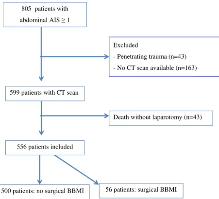
The trauma database of the intensive care unit of our level 1 regional trauma centre was retrospectively reviewed to select all patients with an abbreviated injury score (AIS) for the abdomen-pelvis area of≥1 between April 2004 and December 2011. Among 805 patients who fulfilled this criterion, 249 were excluded for reasons detailed in Fig.1. Finally, 556 consecutive patients were included, 409 male (73.6 %) and 147 female (26.4 %), with a median age of 29 years (inter-quartile range, IQR, 21–46 years). The median injury severity score (ISS) was 14 (IQR 9–22).
CT technique
The CT studies were performed in our institution within 2 h of admission to the trauma centre (n=521, 93.7 %), or in another institution before the patient was transferred to our trauma centre unit (n=35, 6.3 %). All but 35 CTs were performed using a LightSpeed VCT 16- or 64-detector row scanner (GE Healthcare, Milwaukee, Wis), at 120 kVp, and the am-perage setting ranged from 130 to 700 mA, according to the body habitus. The CT images were reconstructed at 3-mm section thickness in the axial, coronal and sagittal plane with native images available for interpretation. Intravenous contrast material (iohexol [Omnipaque 300], GE Healthcare; or iobitridol [Xénétix 350], Guerbet) was administrated at a rate of 3–4 mL/s via a power injector. The CT protocol at our
institution included multi-phase acquisition with an arterial phase initiated using an automatic bolus-tracking program and a venous phase at a 70–80 s delay. A delayed sequence (3–5 min) was performed at the emergency radiologist’s dis-cretion. For the 35 CTs that had been performed outside the institution, the arterial phase was only available in 8 patients (22.9 %), but venous and delayed phases were available in all cases. CT was performed without oral contrast in all cases.
CT analysis
All 556 CT examinations were retrospectively and indepen-dently reviewed by two radiologists (C.F., A.R.) who had respectively 6 and 4 years of experience in abdominal trauma imaging. Reviewers were blinded to the original radiology report, surgical findings and final outcome, but they were aware that the research study involved diagnosis of BBMI at CT.
The CT images were screened for a number of signs, as reported in Table1. Haemoperitoneum was defined by high-attenuation peritoneal fluid (35–60 HU) and was evaluated according to its amount, as proposed by Federle and Jeffrey [16]. Bowel enhancement was assessed subjectively by visual comparison and was classified as abnormal if it was decreased compared to the enhanced adjacent bowel loops. Bowel walls were considered thick if they were greater than 3 mm for the small bowel and 5 mm for the colon [5,9,17]. Bowel wall thickening was considered as focal if it was less than 10 cm in length, and non-focal if it was longer than 10 cm. Diffuse bowel wall thickening with findings of hypovolaemic shock was not considered. Active arterial mesenteric vessel extrava-sation was considered if the extravaextrava-sation appeared in the arterial phase and expanded at the later phases. When CT had not been performed with an arterial phase, it was consid-ered when extravasation increased considerably (at least dou-bled) between the venous and delayed phases with coexis-tence of high and low density areas in the haemorrhage region, suggesting a high rate of active bleeding, as reported by Murakami et al. [18]. Sentinel clot sign was defined by focal high-density collection having an average CT density greater than 50 HU [19]. Pneumoperitoneum was defined as Bmesenteric^ if the extra luminal air was seen only trapped in the mesentery, and asBfree^ if it was extended to the ante-rior part of the abdominal cavity, under the anteante-rior parietal peritoneal layer. Mesenteric stranding was defined by a streaky soft-tissue infiltration of normal mesenteric fat. Beaded appearance of mesenteric vessels was defined as an irregular contour, and abrupt termination indicated by a lack of continuity or tapering of the artery or vein [9].
Injuries to solid visceral organs, bladder, large abdominal vessels, spine and pelvic bones were documented. Anterior abdominal wall injury was noted if there was stranding of
subcutaneous adipose tissue or if there was a rupture in the abdominal muscular wall or an anterior muscular haematoma.
Reference standard
Two of the authors (I.M. and J.C.) reviewed the trauma data-base information, official surgical notes and discharge sum-maries of all patients. Patient demographics, injury severity score and duration of hospitalization were obtained from the trauma registry.
The final diagnosis was established based on the surgical findings and pathology reports, which served as the reference standard for surgical BBMI. As proposed by Atri et al. [9], a surgical bowel injury was defined as a full-thickness
perforation or seromuscular tear or devascularized bowel. A surgical mesenteric injury was defined as involving active mesenteric bleeding or mesenteric injury resulting in an ische-mic bowel loop. Surgical BBMI required therapeutic laparot-omy. Serosal tears of the bowel, bowel wall haematomas with-out tear, and mesenteric haematoma in the absence of active bleeding did not require surgical intervention and were thus considered as negative for surgical BBMI.
All patients referred to our trauma centre were admitted for at least 24 h of observation (more than 3 days for 95 % of the total population). All patients were followed up for 2 months after discharge. Patients treated with non-operative manage-ment and discharged alive were thus considered as true nega-tive for BBMI requiring surgery, as none of them returned for an occult BBMI.
Statistical analysis
Interobserver agreement for CT findings was determined with theκ statistic and classified as follows: κ=0–0.2, slight agree-ment; κ=0.21–0.4, fair agreement; κ=0.41–0.6, moderate agreement;κ=0.61–0.8, substantial agreement; and κ=0.81– 1, almost perfect agreement.
Disagreements between the two reviewers were resolved by consensus. Consensual data were then used for final statistics.
We compared epidemiological data and CT signs between patients with surgical BBMI and those without surgical BBMI. Pearson Chi square or Fisher tests were used for
805 patients with abdominal AIS 1
Excluded
- Penetrating trauma (n=43) - No CT scan available (n=163)
599 patients with CT scan
556 patients included
Death without laparotomy (n=43)
56 patients: surgical BBMI 500 patients: no surgical BBMI
Fig.1 Study flow chart
Table 1 CT signs evaluated
Mesenteric sign Bowel sign
Haemoperitoneum Bowel wall discontinuity
Mesenteric stranding Free intra- or retroperitoneal air
Sentinel clot sign
(=mesenteric haematoma)
Bowel wall thickening (site and length)
Active arterial mesenteric vessel extravasation
Decreased bowel wall enhancement Irregular beading of the mesenteric
vessel
Mesenteric pneumoperitoneum Abrupt termination of the
comparison of categorical variables, and Student t test or Wilcoxon rank-sum test for comparison of continuous vari-ables as appropriate. The diagnostic performance of each CT finding was also calculated.
All CT findings with a univariate p value≤0.1 were entered into a multivariate logistic regression model to gauge their independent association with surgical BBMI. A stepwise pro-cedure was used to select the final model. To establish a score, a rounded up numerical value was assigned to each of the significant variables included in the final prediction model, in relation to theirβ parameter (logistic regression estimates). A receiver operating characteristic (ROC) curve was plotted to estimate the best cut-off of this model and its diagnostic per-formance. We used the bootstrap method to internally validate the score by sampling with replacement for 1000 iterations.
Statistical significance for all tests was set at p<0.05. Com-puter software (SAS, version 9.3, SAS Institute Cary, NC and R, version 3.0.2, R Foundation for Statistical Computing) was used to perform the statistical analyses.
Results
Study population
There was no significant difference for age or sex ratio be-tween patients with or without surgical BBMI [26.5 years (IQR 19–47.5) and 29 years (IQR 21–46), p=0.36; H/F 3.3 and 2.7, p=0.56, respectively]. The hospitalisation duration was significantly longer in the surgical BBMI group (median 21.5 days, IQR 11–32) vs. no surgical BBMI (median 14 days, IQR 8–25) with p=0.005. The ISS was significantly higher in patients with surgical BBMI (18.5 vs. 14, p=0.0036). There were seven deaths, two of which involved surgical BBMI (one devascularized bowel and one seromuscular tear).
Reference standard
A total of 103 patients (18.5 %) underwent laparotomy, in-cluding 56 patients (54.4 %) with surgical BBMI. Eighty-seven patients (82.1 %) were operated on within 24 h from the time of CT, including 50 patients (57.5 %, 50/87) with BBMI requiring surgery, whereas 16 patients were operated on 24 h after CT, including 6 patients (37.5 %, 6/16) with BBMI requiring surgery.
Surgical findings for the 56 patients with BBMI requiring surgery were distributed as follows: 3 had mesenteric lesions which corresponded to active arterial bleeding, 24 bowel in-juries including 17 bowel perforation, 6 seromuscular tears and 1 ischemic bowel wall; 29 had associated bowel and mes-enteric injuries including 14 mesmes-enteric bleeding, 20 bowel ischemia, 9 seromuscular tears and 10 bowel perforation. Among these 56 patients, 19 needed multiple surgical repairs
and 37 single repairs, and treatments were as follows: 33 bow-el resections (21 for ischemic bowbow-el and 12 for multiples perforations), 30 bowel sutures and 17 surgical haemostasis for active bleeding. There was no BBMI requiring surgery in the 47 others patients who underwent laparotomy, and surgical treatments were as follows: 27 splenectomies, 5 nephrecto-mies, 5 hepatic repairs (suture), 8 hepatic packing and 2 dia-phragmatic sutures.
The remaining patients, after excluding the five deaths (n= 448, 80.6 %), were discharged alive without abdominal surgi-cal procedure during the hospitalization or surgery during fol-low-up.
CT univariate analysis
Tables2and3show results of univariate analysis, and diag-nostic performance of each CT finding. All CT findings eval-uated were significantly associated with an increased proba-bility of surgical BBMI, except for length of bowel wall thick-ening (p=0.22).
Bowel wall discontinuity was noted in 20 patients; there was only one false positive discontinuity in the colic bowel in a patient who had undergone laparotomy with no abdominal lesion and who died just after surgery because of non-abdominal haemorrhagic shock.
A free pneumoperitoneum was lacking in 12/27 (44.4 %) patients with a perforated bowel, whereas a mesenteric pneu-moperitoneum was lacking in 19/27 (70.4 %). In 8/27 (29.6 %) patients with small bowel perforation, there was neither free nor mesenteric pneumoperitoneum.
There were six cases of false positives for arterial mesen-teric extravasation. Among them, four underwent laparotomy without active mesenteric bleeding mentioned in the surgical report or without any haemostasis procedure, but two of them died after the surgical procedure due to haemorrhagic shock. The last two had active bleeding noted in the meso-sigmoid and the other in the small omentum, and both were conserva-tively treated favourably with spontaneous regression of bleeding.
Among the associated intra-abdominal lesions, only trauma of the spleen and of the anterior abdominal wall were signif-icantly associated with surgical BBMI (p = 0.0077 and <0.0001, respectively).
Multivariate analysis
Nine CT signs were independently associated with surgical BBMI, as listed in Table4.
The ROC curve derived from our scoring system (Fig. 2) showed an AUC of 0.98 (0.96–1) with a cut-off≥5 for the best discriminative diagnostic performance. Using a cut-off≥5, our CT score had 96.4 % sensitivity, 91.5 % specificity, 56.2 % positive predictive value
(PPV), 99.6 % negative predictive value (NPV), 11.4 positive likelihood ratio (PLR) and 0.04 negative likeli-hood ratio (NLR) in our cohort.
The internal bootstrap validation gave the following 95 % confidence intervals (CI) for the performance of our scoring system: sensitivity, 91.7–100 %; specificity, 85.7–97.6 %; PPV, 41.4–82.3 %; NPV, 98.9–100 %; and AUC, 97.5–99.3 %.
Reproducibility
Interobserver agreement is reported in Table3. Interobserver agreement was substantial to almost perfect for 6/7 direct CT signs of BBMI which belonged to our CT score, and moderate for the bowel wall thickening sign (κ=0.60). It was almost perfect for both detection of anterior abdominal wall injury (κ=0.97, CI 0.94–1) and splenic injury (κ=1).
Discussion
We developed a reliable scoring system which can be easily implemented and could allow surgeons to optimize decisions to operate on multi-trauma patients. To our knowledge, this is the largest reported cohort of BBMI requiring surgery with CT in blunt trauma (n=56), which compares favourably with the findings of the studies by Atri et al. [9], Steenburg et al. [20] and Wu et al. [15], including 38, 15 and 13 patients with surgical BBMI, respectively. Moreover, this is the largest con-secutive cohort of abdominal trauma cases with CT that has been entirely screened to assess the predictive value of CT findings for surgical BBMI.
Risk scoring systems are widely used in trauma centres and ideally have the triple advantage of predicting patient out-come, facilitating decision making and enabling comparisons when benchmarking the performance of clinical units. They
Table 2 Univariate analysis of each CT finding in the general population and according to the presence of surgical BBMI
Overall Surgical BBMI No surgical BBMI p value
N=556 N=56 N=500
Haemoperitoneum None 157 (28.2) 3 (5.4) 154 (30.8) <0.0001
1 (small amount) 233 (41.9) 14 (25) 219 (43.8)
2 (abundant) 166 (29.9) 39 (69.6) 127 (25.4)
Free pneumoperitoneum 21 (3.8) 17 (30.4) 4 (0.8) 4.3E−15b
Mesenteric pneumoperitoneum 12 (2.5) 10 (17.9) 2 (0.4) 2.8E−09b
Bowel wall thickening None 474 (85.2) 18 (32.1) 456 (91.2) <0.0001
Present 82 (14.8) 38 (67.9) 44 (8.8)
Bowel wall thickening length (N=82) Focal 49 (59.8) 20 (52.6) 29 (65.9) 0.22
Non-focal 33 (40.2) 18 (47.4) 15 (34.1)
Arterial mesenteric vessel extravasation a 21 (3.8) 15 (26.8) 6 (1.2) 6E−12b
Mesenteric haematoma 56 (10.1) 24 (42.9) 32 (6.4) <0.0001
Mesenteric stranding 85 (15.3) 33 (58.9) 52 (10.4) <0.0001
Abrupt termination of mesenteric vessel a 18 (3.2) 14 (25) 4 (0.8) 5.3E−12b
Beaded mesenteric vessel a 13 (2.3) 7 (12.5) 6 (1.2) 8E−05b
Decreased bowel wall enhancement a 33 (6) 22 (39.3) 11 (2.2) 1.6E−16b
Bowel wall discontinuity a 20 (3.6) 19 (33.9) 1 (0.2) 9.8E−20b
Associated abdominal injury
Liver 224 (40.4) 18 (32.1) 206 (41.4) 0.1824 Spleen 251 (45.4) 16 (28.6) 235 (47.3) 0.0077 Kidney 102 (18.4) 12 (21.4) 90 (18.1) 0.5437 Adrenal 64 (11.6) 4 (7.1) 60 (12) 0.2762 Aortocaval vessels 14 (2.5) 3 (5.4) 11 (2.2) 0.1605b Pancreas 13 (2.3) 3 (5.4) 10 (2) 0.1349b Bladder 4 (0.7) 2 (3.6) 2 (0.4) 0.0527b
Abdominal anterior wall 132 (23.7) 34 (60.7) 98 (19.6) <0.0001
Lumbar spine 146 (26.3) 11 (19.6) 135 (27) 0.2354
Pelvic ring 151 (27.2) 12 (21.4) 139 (27.8) 0.3094
a
2 missing data because of no abdominal contrast due to heart failure b
must be developed using simple, reliable and reproducible parameters. The CT scoring system we developed fulfils the requirements and goals of trauma scoring systems. It may facilitate the selection of patients requiring abdominal surgical management according to a standardized CT analysis with reliable prediction of surgical BBMI (bootstrap AUC=97.5– 99.3 %). Our scoring system is easy to use, based on a thor-ough CT reading with excellent reproducibility (substantial or almost perfect agreement for 8/9 CT signs), and achieves very high predictive values with an 11-fold increased pretest prob-ability of surgical BBMI if the score is≥5 (PLR 11.4), and a high negative predictive value (99.6 %; 95 % CI bootstrap 98.9–100 %) if the score is <5. This is of paramount impor-tance because the most common missed injuries in the trauma
imaging era are in the bowel region [12], and misdiagnosis of surgical BBMI could delay appropriate management and of-ten results in significant morbidity and mortality. While main-taining this high NPV level, we obtained a PPV ranging from 41.4 to 82.3 % as estimated by the bootstrap calculation, thus limiting the number of non-therapeutic laparotomies.
In fact, two CT signs (mesenteric pneumoperitoneum and bowel wall discontinuity) were sufficient to confirm the pres-ence of surgical BBMI, with a score equal to 5 (Figs.3and4). Mesenteric pneumoperitoneum was highly associated with surgical BBMI (OR 140.5, 95 % CI 9.3–>999.9). Although free pneumoperitoneum also has very good reliability for the diagnosis of traumatic perforation, it does not appear signifi-cant in our multivariate analysis, as there was a statistical
Table 3 Diagnostic performance and interobserver reproducibility of each CT finding
Sensitivity Specificity PPV NPV kappa 95 % CI kappa
Haemoperitoneum 0.79 0.72–0.87
1 (small amount) 25 (14–36) 56.2 (52–61) 6 (3–9) 87 (83–91)
2 (abundant) 69.6 (58–82) 74.6 (70–78) 23.5 (17–31) 95.6 (94–98)
Free pneumoperitoneum 30.4 (18–42) 99.2 (98–100) 81 (64–98) 92.7 (91–95) 0.89 0.77–1
Mesenteric pneumoperitoneum 17.9 (8–28) 99.6 (99–100) 83 (68–100) 91.5 (89–94) 0.72 0.53–0.92
Bowel wall thickness 67.9 (56–80) 91.2 (89–94) 46.3 (36–57) 96.2 (95–98) 0.60 0.48–0.70
Bowel wall thickening length 0.61 0.50–0.72
Focal 35.7 (23–48) 94.2 (92–96) 40.8 (27–55) 92.9 (91–95)
Non-focal 32.1 (20–44) 97 (95–98) 54.5 (36–72) 92.7 (90–95)
Arterial mesenteric vessel extravasation 26.8 (15–38) 98.8 (98–100) 71.4 (53–90) 92.3 (90–95) 0.90 0.78–1
Mesenteric haematoma 42.9 (30–56) 94 (91–96) 42.9 (30–56) 93.6 (91–96) 0.75 0.62–0.88
Mesenteric stranding 58.9 (46–72) 89.6 (87–92) 38.8 (28–49) 95.1 (93–97) 0.64 0.53–0.75
Abrupt termination of mesenteric vessel 25 (14–36) 99.2 (98–100) 77.8 (52–94) 92.1 (90–94) 0.57 0.28–0.86
Beaded mesenteric vessel 12 (4–21) 98.8 (98–100) 53.8 (27–81) 90.9 (89–93) 0.59 0.37–0.81
Decreased bowel wall enhancement 39.3 (26–52) 97.8 (96–99) 66.7 (51–83) 93.5 (91–96) 0.68 0.50–0.85
Bowel wall discontinuity 33.9 (21–46) 99.8 (99–100) 95 (85–100) 93.1 (91–95) 0.75 0.58–0.91
Data are percentages and numbers in brackets are 95 % confidence intervals
Table 4 Significant CT signs in multivariate analysis to predict the risk of surgical BBMI and their assigned numerical CT score according to the
logistic regression estimates (β parameter)
CT signs p value Odds ratio 95 % CI odds ratio β estimate Score
1 Haemoperitoneum 0.0017
Small amount 3.3 0.4–23.8 1.1944 1
Abundant 16.3 2.4–111.4 2.7904 3
2 Mesenteric pneumoperitoneum 0.0003 140.5 9.3–>999.9 4.9456 5
3 Bowel wall thickness 0.0001 9.8 3.1–31.4 2.2878 2
4 Arterial mesenteric vessel extravasation 0.0002 16.8 3.7–75.7 2.8225 3
5 Mesenteric stranding 0.0019 5.2 1.8–14.9 1.6541 2
6 Reduced bowel wall enhancement 0.0306 4.4 1.2–17.0 1.4856 1
7 Bowel wall discontinuity 0.0003 128.9 9.5–>999.9 4.8593 5
8 Splenic injury 0.0467 0.3 0.1–0.9 −1.1123 −1
relationship between free and mesenteric pneumoperitoneum. There are classical causes of free pneumoperitoneum which are not due to bowel wall trauma, such as rupture of bladder with Foley catheter in place or diffusion of a pneumothorax in patients with diaphragmatic rupture [5]. Furthermore, in trau-matic bowel perforation, mesenteric pneumoperitoneum may be useful for localizing the damaged intestinal tract occurring
more commonly in the small bowel [21]. In contrast, mesen-teric air is more commonly missing than free pneumoperito-neum in bowel perforation (these two findings were lacking in 29.6 % of bowel perforations in our study), as already reported in the literature [8,21].
Visualization of a bowel wall discontinuity had the best positive predictive values of surgical BBMI (PPV=95 %)
Fig.2 ROC curve built from our scoring system (a), and ROC curve derived from the bootstrap analysis (b)
Fig.3 Axial abdominal contrast material-enhanced CT image acquired at arterial (a), venous (b, d) and delayed (c) phases, in a 35-year-old man after motor vehicle crash. Note the active mesenteric vessel extravasation appearing in the arterial phase and expanded at the later phases (arrow), and the bowel wall discontinuity in an ileal loop (arrowhead) with free pneumoperitoneum (asterisk). Findings at laparotomy included an ileal perforation requiring suture, and an inferior mesenteric artery rupture requiring haemostasis and left hemi-colectomy for ischemia
among all CT signs studied, as already reported [20]. This is a direct CT sign of a perforated loop.
Arterial mesenteric extravasation had an odds ratio of 16.8 (95 % CI 3.7–75.7). The logistic regression analysis assigned a value of 3, which is inferior to the cut-off of 5 determined by our ROC curve analysis. This could be surprising since it is well established that active mesenteric vessel extravasation is a finding of BBMI requiring surgical exploration both for stopping the bleeding and for investigating the bowel because of the risk of ischemia. However mesenteric vessel extravasa-tion is always associated with at least mesenteric stranding if not an abundant haemoperitoneum. Hence, the combination of both signs results in a score of at least 5, which is predictive of surgical BBMI.
Mesenteric haematoma and mesenteric stranding had very limited reliability for diagnosing surgical BBMI. These two findings had a positive predictive value for the diagnosis of surgical BBMI inferior to 50 %. This confirms that although a mesenteric finding is suggestive and mesenteric haematoma is specific of mesenteric injury, these signs do not always indi-cate a need for surgery [5].
Although we did not consider diffuse thickening of the bowel with other CT findings of shock as a finding of bowel trauma, bowel wall thickness had a positive predictive value for surgical BBMI inferior to 50 % and was assigned a score of 2, insufficient in itself to predict a surgical BBMI. Different criteria have been used in the literature to diagnose small bowel or colon wall thickening [22]. By using the same criteria to define bowel wall thickening as used in our study, Atri et al. [9] have already shown that bowel wall thickening may be present despite the lack of important bowel injury
requiring surgery. Numerous causes may explain bowel thick-ening in patients without surgical BBMI: non-significant bow-el injuries such as a haematoma or a tear limited to the serosa [5], collapsed bowel lumen, or normal variants [22,23].
Areas of decreased or absent bowel wall contrast enhance-ment, which is classically indicative of ischemic bowel wall in abdominal emergencies [24], were significantly associated with surgical BBMI (OR 4.4, 95 % CI 1.2–17). However, there were 11 false positives (33.3 %) in our study. This could be explained by the difficulty of analysing bowel wall en-hancement in multiple trauma patients with heart failure, abundant haemoperitoneum and sometimes hypoperfusion complex [25], because in such cases mucosal bowel enhance-ment is often intense and diffuse, which makes comparisons tricky with the seemingly less enhanced normal adjacent en-hanced bowel.
We note an association between surgical BBMI and abdominal wall injuries. This highlights one of the well-known mechanisms of BBMI due to direct impact [26] and anterior compression of the abdomen classically caused by a fastened seatbelt, whereby the bowel loop may be crushed between the seatbelt and the lumbar spine, leading to bowel perforation. We think that ab-dominal anterior injury visualized in CT examination is helpful for predicting surgical BBMI, when combined with other CT score findings. By contrast, we report an inverse association between spleen lesion and surgical BBMI. Although the weight of this inverse association is weak (assigned score of −1), we think that in patients with an abundant haemoperitoneum (score of 3), the presence of a spleen lesion decreases the risk of surgical BBMI, probably because haemoperitoneum is explained by splenic injury and not by a mesenteric injury.
Our study had some limitations. First, it was a retrospective study with its inherent bias and was conducted in a single centre, but it included consecutive patients with blunt abdom-inal trauma investigated by CT. Secondly, there was a higher prevalence of surgical BBMI in our study (10.1 %) than re-ported in previous studies, i.e. twofold lower [7,17]. This was due to patient selection bias, as patients had more serious injuries in this study (average ISS 14) and were selected on the basis of an abdominal trauma that did not reflect the entire trauma population. The prevalence in our study is actually close to that documented in other cohorts of blunt abdominal trauma patients, with a reported prevalence of surgical BBMI of 12.3 % in the study by Wu et al. [15], and 15.3 % in the study by Tan et al. [14]. Interestingly, in our study, surgical BBMI constituted the majority of blunt abdominal trauma patients explored by laparotomy, and currently represents the main reason for laparotomy [27]. Thirdly, although the inter-nal validation of our score indicated high diagnostic perfor-mance, an external validation set is essential to confirm the diagnostic value before generalization.
Fig.4 Axial abdominal contrast-enhanced CT image acquired at venous phase in a 59-year-old woman after motor vehicle crash. Note the mes-enteric pneumoperitoneum (arrowhead) close to a focal small bowel wall thickening (arrow) and with subcutaneous fat stranding along the course of the fastened seat belt (asterisk). Findings at laparotomy included a 2-mm jejunal perforation and a transverse colic perforation, all requiring suture
In conclusion, we developed an easy to implement and reliable scoring system based on CT findings, which is highly predictive of surgical BBMI. When the CT score is at least 5, then a surgical procedure should be carried out given the high probability of BBMI requiring surgical repair. Conversely, when the CT score is less than 5, then non-operative management should be prescribed along with clinical follow-up and possibly repetition of abdom-inal CT examination within 8–12 h.
Acknowledgements The scientific guarantor of this publication is Prof.
Taourel. The authors of this manuscript declare no relationships with any companies whose products or services may be related to the subject mat-ter of the article. The authors state that this work has not received any funding. One of the authors has significant statistical expertise. Institu-tional review board approval was obtained. Written informed consent was waived by the institutional review board. Methodology: retrospective, diagnostic or prognostic study, performed at one institution.
References
1. Watts DD, Fakhry SM (2003) Incidence of hollow viscus injury in
blunt trauma: an analysis from 275,557 trauma admissions from the East multi-institutional trial. J Trauma 54(2):289–294
2. Killeen KL, Shanmuganathan K, Poletti PA, Cooper C, Mirvis SE
(2001) Helical computed tomography of bowel and mesenteric
in-juries. J Trauma 51(1):26–36
3. Scaglione M, de Lutio di Castelguidone E, Scialpi M, Merola S,
Diettrich AI, Lombardo P et al (2004) Blunt trauma to the gastro-intestinal tract and mesentery: is there a role for helical CT in the
decision-making process? Eur J Radiol 50(1):67–73
4. Frellesen C, Stock W, Kerl JM, Lehnert T, Wichmann JL, Nau C
et al (2014) Topogram-based automated selection of the tube po-tential and current in thoraco-abdominal trauma CT - a comparison
to fixed kV with mAs modulation alone. Eur Radiol 24(7):1725–
1734
5. Brofman N, Atri M, Hanson JM, Grinblat L, Chughtai T,
Brenneman F (2006) Evaluation of bowel and mesenteric blunt
trauma with multidetector CT. Radiographics 26(4):1119–1131
6. Janzen DL, Zwirewich CV, Breen DJ, Nagy A (1998) Diagnostic
accuracy of helical CT for detection of blunt bowel and mesenteric
injuries. Clin Radiol 53(3):193–197
7. Malhotra AK, Fabian TC, Katsis SB, Gavant ML, Croce MA
(2000) Blunt bowel and mesenteric injuries: the role of screening
computed tomography. J Trauma 48(6):991–998, discussion 998–
1000
8. Mirvis SE, Gens DR, Shanmuganathan K (1992) Rupture of the
bowel after blunt abdominal trauma: diagnosis with CT. AJR Am J
Roentgenol 159(6):1217–1221
9. Atri M, Hanson JM, Grinblat L, Brofman N, Chughtai T,
Tomlinson G (2008) Surgically important bowel and/or mesenteric injury in blunt trauma: accuracy of multidetector CT for evaluation.
Radiology 249(2):524–533
10. Ekeh AP, Saxe J, Walusimbi M, Tchorz KM, Woods RJ, Anderson
HL et al (2008) Diagnosis of blunt intestinal and mesenteric injury
in the era of multidetector CT technology–are results better? J
Trauma 65(2):354–359
11. Elton C, Riaz AA, Young N, Schamschula R, Papadopoulos B,
Malka V (2005) Accuracy of computed tomography in the
detec-tion of blunt bowel and mesenteric injuries. Br J Surg 92(8):1024–
1028
12. Matsushima K, Mangel PS, Schaefer EW, Frankel HL (2013) Blunt
hollow viscus and mesenteric injury: still underrecognized. World J
Surg 37(4):759–765
13. Petrosoniak A, Engels PT, Hamilton P, Tien HC (2013) Detection of
significant bowel and mesenteric injuries in blunt abdominal trauma with 64-slice computed tomography. J Trauma Acute Care Surg 74(4):1081–1086
14. Tan K-K, Liu JZ, Go T-S, Vijayan A, Chiu M-T (2010) Computed
tomography has an important role in hollow viscus and mesenteric
injuries after blunt abdominal trauma. Injury 41(5):475–478
15. Wu C-H, Wang L-J, Wong Y-C, Fang J-F, Lin B-C, Chen H-W et al
(2011) Contrast-enhanced multiphasic computed tomography for identifying life-threatening mesenteric hemorrhage and transmural
bowel injuries. J Trauma 71(3):543–548
16. Federle MP, Jeffrey RB (1983) Hemoperitoneum studied by
com-puted tomography. Radiology 148(1):187–192
17. Wisner DH, Chun Y, Blaisdell FW (1990) Blunt intestinal injury.
Keys to diagnosis and management. Arch Surg 125(10):1319–
1322, discussion 1322–1323
18. Murakami AM, Anderson SW, Soto JA, Kertesz JL, Ozonoff A,
Rhea JT (2009) Active extravasation of the abdomen and pelvis in trauma using 64MDCT. Emerg Radiol 16(5):375–382
19. Shanmuganathan K, Mirvis SE, Sover ER (1993) Value of
contrast-enhanced CT in detecting active hemorrhage in patients with blunt
abdominal or pelvic trauma. AJR Am J Roentgenol 161(1):65–69
20. Steenburg SD, Petersen MJ, Shen C, Lin H (2014) Multi-detector
CT of blunt mesenteric injuries: usefulness of imaging findings for predicting surgically significant bowel injuries. Abdom Imaging. doi:10.1007/s00261-014-0262-2
21. Kim HC, Shin HC, Park SJ, Park SI, Kim HH, Bae WK et al (2004)
Traumatic bowel perforation: analysis of CT findings according to the perforation site and the elapsed time since accident. Clin
Imaging 28(5):334–339
22. Macari M, Balthazar EJ (2001) CT of bowel wall thickening:
sig-nificance and pitfalls of interpretation. AJR Am J Roentgenol
176(5):1105–1116
23. Wittenberg J, Harisinghani MG, Jhaveri K, Varghese J, Mueller PR
(2002) Algorithmic approach to CT diagnosis of the abnormal bow-el wall. Radiographics 22(5):1093–1107
24. Geffroy Y, Boulay-Coletta I, Jullès M-C, Nakache S, Taourel P,
Zins M (2014) Increased unenhanced bowel-wall attenuation at multidetector CT is highly specific of ischemia complicating
small-bowel obstruction. Radiology 270(1):159–167
25. Ames JT, Federle MP (2009) CT hypotension complex (shock
bow-el) is not always due to traumatic hypovolemic shock. AJR Am J
Roentgenol 192(5):W230–W235
26. Hughes TMD, Elton C (2002) The pathophysiology and
manage-ment of bowel and mesenteric injuries due to blunt trauma. Injury 33(4):295–302
27. Sorensen VJ, Mikhail JN, Karmy-Jones RC (2002) Is delayed
lap-arotomy for blunt abdominal trauma a valid quality improvement measure in the era of nonoperative management of abdominal