THE BEHAVIORAL AND BRAIN SCIENCES (1979), 2,409-448
Printed in the United States of America
PL Key Dismukes
Neurosciences Research Program, Massachusetts Institute of Technology, Boston, Mass. 02130*
Abstracts Recently a number of complex electrophysiological responses to neurotransmitters have been observed that cannot be described as
simple excitation or inhibition. These responses are often characterized as modulatory, although there is no consensus on what defines
modulation. Morphological studies reveal certain neurotransmitters stored in what might be release sites without synaptic contact. There is no
direct evidence for nonsynaptic release from CNS sites, although such release does occur in the periphery and in invertebrates. Nonsynaptic
release might provide a basis for diffuse one-cell-to-many communication, but it might also simply be a means of sending the transmitter to a
broader area of a single neuron than occurs in typical synapses. Several kinds of macromolecules have been found to be transported in a
retrograde direction - and in some cases transsynaptically. There have been suggestions that some neurons may release more than one type of
transmitter. Particularly intriguing is the possibility of release of substances that modulate actions of a primary transmitter. Taken together this
range of evidence suggests that neurons may use a variety of forms of molecular communication in addition to traditionally described synaptic
transmission.
Several authors have suggested modes of communication distinct from classical synaptic transmission and have classified released substances
using terms such as neurohumor, neurohorrnone, neuroregulator, and modulator. These suggestions have the heuristic value of drawing
together diverse kinds of data, but it remains to be established that the pieces fit together in that fashion - for example, that complex
electrophysiological effects are associated with substances released nonsynaptically. In order to reduce confusion, a flexible, generic approach
to nomenclature for substances released from neurons and for hypothetical modes of communication is recommended. Some behavioral
implications of nonconventional transmission are considered.
Keywords? amines; Dale's Principle; modulators; neurohormones; neurohumors; neurotransmission; neuropeptides; synapses
The notion of synaptic chemical communication is central to our
conceptualization of neuronal function. As early reticular theories of
the nervous system were displaced by the concept of the neuron as a
discrete functional unit, it became evident that there must exist
specialized points of contact for passing information from one cell to
the next (Eccles 1964 and ref. therein). The term "synapse" was
coined by Sherrington (1906), who recognized, almost presciently,
that crucial features of the operation of neuronal circuits could be
explained by the character of synaptic transmission. For example, he
argued that excitatory and inhibitory inputs would be algebraically
summated by a postsynaptic neuron, and that a subthreshold
stimu-lus repeated rapidly would be summated, causing firing.
The concept of synaptic transmission helped bring together the
classical tenets of the neuron doctrine that emerged in the first half of
this century. These tenets suggested that neurons, dynamically
polar-ized into distinct receiving and transmitting zones, summate their
excitatory and inhibitory synaptic inputs and transmit information in
all-or-none impulses along axons, whose terminals release a single
kind of neurotransmitter. These principles were shown to have broad
applicability and provided a conceptual framework for
understand-ing diverse aspects of nervous system function. However, the early
period of apparent clarity and simplicity evoked by successful
theories in biology is often followed by a time of increasing
complex-ity as exceptions and inadequacies are discovered. Thus, for example,
twenty years ago Bullock (1959) pointed out the need for revision of
textbook statements of neuron doctrine, particularly in regard to the
integration of postsynaptic responses and the generation of action
potentials; it was becoming apparent that dendritic potentials
inter-acted in a way more complex than simple summation of excitatory
and inhibitory influences to elicit propagated impulses. His views
have been well borne out by developments in the last two decades.
For example, some interneurons in various regions of the brain have
been found to exhibit such unexpected features as synapses between
dendrites, reciprocal synapses, and bidirectional transmission of
graded potentials rather than impulses (Rakic 1975).
Over the past several years, certain neurotransmitters have been
reported to elicit, in various neuronal systems, complex
electrophys-iological responses that cannot be simply described as either
excita-tory or inhibiexcita-tory. The word "modulation" has often been used to
describe these complex responses, as well as a number of other kinds
of alterations of neuronal activity. Several putative
neurotransmit-ters, particularly norepinephrine, serotonin, and certain
neuropep-tides, have been suggested to be neuromodulators or neuroregulators,
but these terms have generally been used ambiguously and often in
conflicting ways by different authors, and it is not clear how
modulation is to be distinguished from other neural actions.
Recently, certain morphological evidence has been interpreted as
suggesting that some neurotransmitters may be released from some
sites nonsynaptically, for diffusion to targets more distant than those
found in conventional synaptic transmission. Barker (1977; 1978) and
Chan-Palay (1977) have combined the concept of nonsynaptic
trans-mission with evidence for complex electrophysiological responses in
suggestions that there may occur specific modes of molecular
communication among neurons distinct from classical synaptic
trans-mission. These suggestions have raised a certain amount of
controver-sy. There is not yet direct evidence for nonsynaptic release from
central neurons, and even with such evidence many questions will
remain about the targets of such release and the functions served.
Nevertheless, a range of evidence from various neuronal systems
suggests that neurons communicate and process information in ways
that go beyond traditional concepts.
The objectives of this article are:
1. To critically examine evidence for nonclassical modes of molecular
communication among neurons. Are suggestions for distinct modes premature,
Dismukes: Neuronal communication
or do they have heuristic value?
2. To see what common threads can be found among the various ways in
which the concept of modulation has been applied to neuronal function.
3. To examine implications of this research for the ways in which we
formulate principles of neuronal operation.
I will not attempt in this article to review the great wealth of
information now available about synaptic organization and function;
broad summaries of that sort have already been made (e.g., Shepherd
1974). Rather, I will try to draw suggestions together that have been
made about nonconventional transmission and other kinds of
evidence bearing on concepts of neuronal communication. Hopefully
this will provide a better context for evaluating these suggestions and
discussing their implications for neuronal operation in the
Commen-tary.
Can neurotransmitters be released eonsynaptlcally?
The possibility of nonsynaptic release has been suggested for several
amiee neurotransmitters, particularly serotonin and norepinephrine.
These two putative transmitters are contained primarily in pathways
arising from small brainstem nuclei and branching diffusely to
innervate much of the brain (see Moore & Bloom 1978). In both
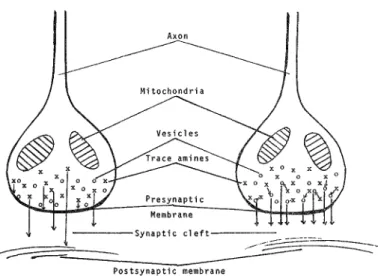
noradrenergic and serotonergic pathways the transmitter is not just
contained in nerve terminals but is also concentrated in varicosities
closely spaced (1-3/*) along tiny unmyelinated axons. Electron
microscopy (EM) has revealed these varicosities to contain aggregates
of small, round, agranular "synaptic" vessels, a few large granular
vesicles, and mitochondria (see ref. in Descarries et al. 1977; and
Descarries et al. 1975). Thus, varicosities of aminergic axons contain
apparatus usually associated with the storage and release of
transmit-ters.
Several ultrastructure studies, using different techniques for
tagging amiee-containing cellular processes, have indicated that
some axonal varicosities make synaptic contact, but the majority do
not appear to do so. Low incidence of synaptic contact by varicosities
has been reported for catecholamines (Ajika & Hokfelt, 1973) and
indolamiees (Calas et al. 1974) in the median eminence, serotonin in
cerebral ventricles (Richards et al. 1973; Chan-Palay 1976),
presumptive dopamine in the neostriatum (Tennyson et al. 1974 and
refs. therein), and serotonin (Descarries et al. 1975) and
norepineph-rine (Descarries et al. 1977) in the neocortex.
Detailed studies of aminergic axonal processes in the neocortex
used the technique of EM-level radioautography. In these
experi-ments 3H-serotonie (Descarries et al. 1975) or 3H-norepinephrine
(Descarries et al. 1977) was topically applied in vivo to the neocortex
to allow cellular processes to become labelled by active uptake. The
relatively high concentrations of 3H-amines used in these procedures
call for careful controls to demonstrate that label has not
accumu-lated in nonaminergic cells through nonspecific uptake. This was
accomplished, for example, in the norepinephrine study by
demon-strating that labelling of axonal varicosities was prevented by
pretreatment of the animals with desmethylimpramine, to
selec-tively block high-affinity uptake of norepinephrine, or with
6-hydroxydopamine, to selectively destory noradrenergic neurons.
Similar controls were performed in experiments with 3H-serotonin.
In these neocortex experiments norepinephrine and
3H-serotonie appeared to accumulate specifically in axonal processes,
particularly varicosities. Label appeared most concentrated over
intracellular organelles, especially the small agranular vesicles.
Extensive topometric ultrastructural analysis was performed on serial
sections, making possible two or even three sequential sections
through a large number of varicosities.
A striking finding of these studies was that less than 5% of either
3H-serotonin- or 3H-norepinephrine-labelled varicosities evincedsynaptic contact. In contrast, synaptic junctions were seen in some
50% of unlabelled boutons in the immediate vicinity. It is not possible
to tell exactly what percentage of either labelled or unlabelled
boutons actually made synapses, since many junctions would be
missed by the sections. The junctions observed with aminergic
varicosities were not of unusual size, and no differences of internal
morphology or constituents were found between aminergic
varicosi-ties displaying synaptic contact and those lacking it. Thus, given the
large number of profiles of axons and boutons studied, it seems likely
that in this region the percentage of noradrenergic and
serotoniner-gic varicosities making synaptic contact is substantially lower than
that which occurs with nonaminergic neurons.
The results of these radioautographic studies in the neocortex
contrast with the findings of a recent investigation of axon terminal
ultrastructure in the dentate gyrus of the hippocampus (Koda et al.
1978). In this study the presence of small granular vesicles (SGV's) in
boutons was used as the criterion for distinguishing noradrenergic
terminals. Various treatments (drugs, lesions) that specifically lower
hippocampal levels of norepinephrine produce correlated reductions
in boutons containing SGV's. The evidence is good that
noradrener-gic boutons contain SGV's and that the preponderance of
SGV-coetaining boutons in the rat dentate gyrus is noradrenergic, but it is
not clear that all such boutons are noradrenergic. Koda and
cowork-ers found that about 20% of boutons in the dentate gyrus, whether
with or without SGV's, displayed synaptic junctions. The actual
incidence of junctions is presumably higher, since random sections
will not reveal all synapses. Thus, this study suggests a higher
incidence of synaptic contact by noradrenergic varicosities than that
found by Descarries and coworkers and does not reveal differences in
incidence between noradrenergic and other boutons.
The difference between the findings of these two groups might be
accounted for by factors such as:
1. Differences in the populations of boutons identified by the criteria of
SGV-presence and 3H-norepinephrine accumulation.
2. Regional differences in the incidence of synaptic contact. These EM
studies, of course, can examine only a miniscule piece of tissue in the few
regions so far examined in detail. Even though noradrenergic fibers examined
in diverse parts of the brain may arise from the same cell bodies (especially
those from the locus coeruleus), it does not necessarily follow that releasing sites
in all branches must bear the same morphological relationship to their target
neurons. Conceivably that relationship, particularly the distance between
releasing sites and targets, depends on the organization and functions of the
circuitry of the target tissue.
3. Occurrence of nonsynaptic boutons along axons that are not
noradrener-gic in some brain regions.
The procedures used for tagging aminergic boutons for EM
identification can affect the apparent incidence of synaptic contact
observed. For example, Tennyson and coworkers (1974) observed
that only about 2% of dopamine boutons in the neostriatum exhibited
synaptic junctions when tagging was accomplished by incubating
tissue slices with 5-hydroxydopamine. In contrast, Arluison et al.
(1978a) observed frequent synaptic contacts by dopamine boutons
when 5-hydroxydopamine was injected in vivo and a different
technique was used for fixation. It appears to be difficult to retain
label in aminergic neuronal elements, and thus the distribution of
label observed is affected by the concentration of label during
incubation and by the technique used for fixation. Nevertheless,
Arluison and coworkers also observed some dopamine boutons not
displaying synaptic contact, and they concede the possibility of
release of dopamine from some boutons synaptically (1978b).
Are serotonin and norepinephrine normally released from
non-synaptic boutons? There is no direct evidence on this point; indeed, it
will be methodologically difficult to settle. However, there is good
reason to ask. As Descarries and coworkers point out (1977),
non-synaptic varicosities appear to have all the apparatus, normally
associated with release, found in varicosities making synapses.
Furthermore, it would be surprising if release from the small fraction
of varicosities demonstrating synaptic junctions could account for the
amount of norepinephrine and serotonin that can be released by
stimulation in vivo (Header et al. 1976; Tanaka et al. 1976) or by
depolarization in vitro (Dismukes & Mulder 1976, and refs. therein).
In the spinal cord, substance P has been immunocytochemically
visualized in extracellular space in patterns suggesting that it can be
released from neurons in packets that diffuse to neighboring cells and
capillaries (Chan-Palay & Palay 1977).
Dismukes: Neuronal communication
Some light may be shed on the question of nonsynaptic release of
CNS transmitters by the much more thoroughly understood
neuro-muscular junctions of the peripheral nervous system (reviewed by
Burnstock & Costa 1975). Junctions between noradrenergic boutons
and effector cells in the periphery vary considerably, depending on
the organ and type of cell. Junctional clefts range from 15-20 nm
(vas deferens and iris) to 1000-2000 nm (large elastic arteries). In
neuroeffector junctions whose membrane separation is greater than
20 nm, no postjunctional specialization is apparent. In closely
apposed junctions, several sorts of postjunctional features have been
described, including increased electron density. This, however, is
rare, and Burnstock & Costa suggest (1975, p. 53) that these areas
might represent mechanical attachment sites between cells rather
than a measure of the region of transmitter action.
These findings raise questions about how synaptic contact is
defined in the CNS. Should we consider close apposition of
membranes with specialized features a sine qua non of chemical
transmission between neurons? The morphology of synaptic
junc-tions in the CNS is in fact diverse, and postsynaptic membrane
specialization is not always apparent (Shepherd 1974, pp. 26-34). It
might turn out that what appear to be nonsynaptic varicosities
release amine neurotransmitters for diffusion to a single adjacent
neuron whose receptors are not confined to a small postjunctional
patch. This would allow the aminergic fiber to bias the largest cell's
responses to its many synaptic inputs more effectively than would be
possible with a single junctional contact. Such an arrangement would
suggest a modulatory role, which is highly consistent with the
character of the electrophysiological responses elicited by amine
neurotransmitters (see discussion below).
The possibility of transmitter release from nonsynaptic boutons has
been suggested by several authors as a basis for modes of
communica-tion in which transmitters released from a single site diffuse to
multiple distant targets. If release into extracellular space did occur,
there would be severe restrictions on information transmission in this
mode. Cellular uptake and enzymatic degradation would sharply
limit how far an amine or peptide transmitter could diffuse. Clearly,
molecules released in such a fashion could not be used to transmit
detailed temporal information about activity in the releasing neuron,
because rapid onset and offset of pulses would be quickly obscured in
diffusion over distance. Thus, transmission of slowly varying or tonic
influence is suggested.
Chan-Palay has examined (1976) the extensive plexuses of
serotin-ergic neurons, originating in raphe nuclei, which form supra- and
subependymal systems in the walls of the cerebral ventricles.
Identi-fied in EM by radioautography, these serotinergic fibers contain
varicosities with dense core "synaptic" vesicles. No evidence was
found for specialized synaptic contact with ependymal cells or axonal
processes. Chan-Palay has suggested (1977) that these fibers release
serotonin into the cerebrospinal fluid (CSF), which would provide a
means of transporting the transmitter to distributed targets. Although
there is no direct evidence for such a function, it is not clear what
purpose these fibers would serve if they did not release serotonin.
Various authors have suggested that the ventricular system may be
more than just a sewer. For example, Dunn (1978) recently proposed
that the cerebrospinal fluid may be used to transport neuropeptides
from release sites to distant target cells. He noted that neuropeptides
are found in significant concentrations in ventricular CSF.
Further-more, proteolytic enzymes are notably lacking in the CSF, and
intraventricular injection has proved an effective means of delivering
exogenous peptides to brain receptors.
Direct evidence for nonsynaptic release of a peptide has been
obtained in Aplysia. Neurosecretory bag cells of the abdominal
ganglion to not appear to make synapses (Coggershall 1970). Their
processes terminate in connective tissue, apparently releasing
prod-ucts directly into the hemolymph for diffusion within the ganglion
and beyond. It appears that bag cells release several kinds of
peptides, affecting the activity of various types of target cell in
different locations (Blankenship 1979). For example, evoking spike
activity in bag cells was found to produce characteristic responses in
a nearby identified neuron, R15 (Branton et al. 1978). In contrast to
conventional postsynaptic actions, these responses were slow in onset
and persisted from minutes to hours. Direct application of bag cell
extract (containing one or more neuropeptides) elicited identical
responses in R15.
Neurotransmitter release is not limited to axonal varicosities and
terminals. Dendrodendritic synaptic contacts have been reported in
several brain regions (Shepherd 1974; Rakic 1975). Neurons
originat-ing in the substantia nigra apparently release dopamine not only
from their distant axonal terminals but also from cell bodies and
dendrites. Release can be stimulated by depolarization either in vivo
(Nieloullon et al. 1977) or in slices (Geffen et al. 1976), and it is
blocked by the removal of Ca++.
Iversen and Cuello (Iversen 1979) found no evidence for synaptic
vesicles or dendrodentritic contacts in nigral cell bodies and
dendrites labelled by either 3H-dopamine or 5-hydroxydopamine.
Their EM studies suggested, rather, that dopamine is stored in
cisterns of smooth endoplasmic reticulum. In contrast, Wilson et al.
(1977), using markedly higher concentrations of
5-hydroxydopa-mine, observed the marker in conjunction with synaptic vesicles and
found the vesicles in dendrites in synaptic contact with other
dopa-minergic dendrites. The different findings of these studies may
reflect the failure of lower concentrations of 5-hydroxydopamine to
effectively label vesicles in outlying dendritic tips. Also arguing for
vesicular storage and release of dopamine is the dependence of
release on Ca++, which is thought to be involved in the interaction of
vesicles with cell membrane in exocytosis.
Indirect evidence suggests that some dopamine released from
nigral dendrites may diffuse to some targets nonsynaptically. GABA
fibers from the striatum terminate in the substantia nigra and cause
inhibition of cell firing therein (see Groves et al. 1975; Iversen 1979).
These GABA terminals may be the site of presynaptic dopamine
receptors coupled to adenylate cyclase that are observed in the
substantia nigra (Spano et al. 1976; Gale et al. 1977). Dopamine at
low concentrations has been found to specifically stimulate the
release of 3H-GABA from nigral terminals (Reubi et al. 1977).
Iversen (1979) has proposed that dopamine released from dendrites
in the substantia nigra may stimulate the release of GABA from
terminals presynaptic to the dopaminergic cell bodies, thus providing
a negative feedback loop. The presynaptic dopamine dendrites
observed by Wilson and coworkers (1977) all appeared to be in
contact with other dendrites, and whenever the postsynaptic element
could be identified, it appeared to be dopaminergic also. No
evidence was found for synaptic contacts between presynaptic
dopaminergic dendrites and axon terminals. Thus, if the Iversen
model is correct, dopamine released from dendrites may diffuse to
GABA terminals that are not in direct synaptic contact. Similarly,
Groves (1979) and coworkers (1975) have proposed a model in which
dopamine released from dendrites produces self-inhibition of firing.
This inhibition might be produced either by dendrodendritic
synapses between adjacent neurons, by nonsynaptic diffusion of
dopamine to autoreceptors, or by some combination of both.
In summary, nonsynaptic release of transmitter has not been
clearly established in any central mammalian neuron other than
those of the neuroendocrine system. However, a range of evidence
from diverse systems suggests that it is an important possibility to
consider and to attempt to test experimentally. Demonstration of
nonsynaptic release would not necessarily be a revolutionary finding;
in fact, it would fit rather nicely with the emerging concepts of the
electrophysiological actions of amines and peptides that will be
described below.
Intercellular transport of eoetransmitter molecules
Neurons exchange molecules in a variety of ways besides chemical
synaptic transmission. Several forms of nontransmitter exchange are
briefly mentioned here for the purpose of illustration. Little is known
about the functions of these processes (Smith & Kreutzberg 1976).
Their nature may be primarily metabolic or trophic, but they may
also provide a means of communication, on a time scale much slower
Dismukes: Neuronal communication
than that of synaptic transmission.
Several exogenous macromolecules (nerve growth factor, tetanus
toxin, cholera toxin, certain lectins, etc.) have been shown to bind
with high affinity to nerve terminals; binding is followed by uptake
and rapid retrograde transport (Schwab & Thoenen 1977). In the
case of tetanus toxin this transport is followed by migration to
dendrites and transsynaptic transport to second-order neurons.
Neurons and glia have been observed to exchange amino acids,
proteins, nucleotides, and nerve growth factor (Smith 1978).
This profuse molecular interchange appears to inform the
peri-karyon of events occurring at the cells' distant projections. Although
metabolic and trophic functions are probably involved, there also
exist mechanisms by which molecules transported into the neuron
could alter its electrical responsiveness and output. For example,
steroid hormones, acting on intracellular receptors that apparently
alter genetic expression, modulate phosphorylation of a cytosol
protein (Liu & Greengard 1976). This protein may be the regulatory
subunit of protein kinase; if so, it might provide a means of
regulating membrane properties. Nerve growth factor taken up by
adrenergic ganglion cell terminals is transported to the perikaryon,
where it triggers a selective increase in tryosine hydroxylase, the
rate-limiting enzyme for synthesizing norepinephrine, the cell's
transmitter (Schwab & Thoenen 1977).
Certain heterologous nonneuronal cells in culture communicate
via gap junctions (Lawrence et al. 1978). Hormones acting on
receptors specific to one type of cell cause the heterologous cell to
respond. This communication may be mediated by cyclic AMP
passed across the gap junctions. Invertebrate neurons also possess gap
junctions, which are thought to provide a means of synchronizing
electrical activity; however, such junctions have been observed only
rarely in mammalian CNS. It would be extremely interesting if
neuronal gap junctions were also found to exchange second
messen-gers.
How valid Is Dale's Principle?
A widely accepted principle of neuronal operation is that the same
transmitter substance is secreted from all branches of a neuron's
terminals (Dale 1985). From time to time this principle has been
questioned, but no clear-cut violation has been established
(Burn-stock 1976; Kandel 1976; Osborne 1979). However, the powerful
techniques recently developed for cytochemical localization reveal a
number of neuronal types that contain more than one substance
thought to act as a transmitter. Eadioenzymatic analysis of several
identified neurons in Aplysia reveals the coexistence of serotonin,
octopamine, and acetylcholine (Brownstein et al. 1974). (However,
the microdissection technique employed has been criticized as
sub-ject to contamination from other cells; Osborne 1977).
Somatostatin-like immunoreactivity has been reported in some noradrenergic
neurons of the sympathetic nervous system (Hokfelt et al. 1977). In
the CNS, cells identified as serotonergic on the basis of
radioautog-raphy were found by immunocytochemical methods to contain
substance P (Chan-Palay et al. 1977).
The fact that a neuron contains some amount of several
transmit-ter substances does not, of course, prove that each substance is being
released as a transmitter. However, the giant cerebral neuron of
Helix does appear to release both acetylcholine and serotonin, each of
which produces postsynaptic responses (Cottrell 1977).
Sympathetic neurons cloned in tissue culture can readily be caused
to form either adrenergic or cholinergic functional synapses by a
choice of appropriate media conditions (Eeichardt & Patterson
1977). Moreover, some microcultures containing only a single neuron
have been found to secrete both norepinephrine and acetylcholine.
Whether this also occurs in mature cells in vivo is not known, but it
clearly indicates that there is no intrinsic biochemical or genetic
reason why a neuron cannot simultaneously manufacture and release
more than one transmitter.
There is an intriguing possibility that some neurons might release
two kinds of molecules from the same terminal - one serving
way to modify the action of the primary transmitter. For example,
ATP is released along with catecholamines from the adrenal gland,
and there is evidence that some ATP is released concurrently with
norepinephrine from sympathetic fibers (Kopin 1967; Burnstock
1976). ATP has been proposed as a primary transmitter in certain
peripheral organs (Burnstock 1975), but there is also a suggestion that
after depletion of norepinephrine a continuing substantial
noncholin-ergic, nonadrenergic response in the cat nictitating membrane may
be due to release of ATP from the adrenergic nerves (Langer & Pinto
1976).
There is evidence that adenosine can be released from central
neurons, can stimulate cyclic AMP formation through specific
recep-tors, and can alter postsynaptic electrical activity (see Fox & Kelley
1978). Schubert and coworkers (1976) have suggested that adenosine
or its derivatives may function as a "secondary" or "additional"
transmitter producing long-lasting alterations in target-cell activity.
However, there is not yet any direct evidence that these nucleotides
are released in conjunction with a primary transmitter.
It has been proposed that minute amounts of amines such as
para-tyramine and phenylethylamine, released along with
norepi-nephrine from adrenergic fibers, act as neuromodulators rather than
as neurotransmitters (Boulton 1976 and refs. therein). However, the
distinction between "neurotransmitter" and "neuromodulator" is
problematic (see discussion below), and it may be better, as
Burn-stock (1976) suggests, to retain in the category of transmitter any
substance produced and physiologically released from nerve
termi-nals to evoke postsynaptic responses through membrane receptors.
Dale's Principle is not invalidated by these findings. Given that
there are very few systems in which we can determine the
transmit-ter released from each branch of a neuron, it remains a useful
operating assumption. However, the principle has at times been
interpreted dogmatically, as an invariant rule, although this was
never intended by Dale himself (1935). The danger of overextending
general working principles is that the exploration of important
exceptions and variations is discouraged.
Complex eiectrophyslological responses to neurotransmitters
Integration of synaptic inputs is a central feature of neuronal
operation. Early concepts portrayed the receptive portion of the
neuron as a linear integrator, adding up excitatory and inhibitory
inputs to evoke an output - firing of impulses - proportional to the
sum of inputs. Later investigations, however, revealed that
postsyn-aptic integration is much more complex, involving a number of
distinct processes (Bullock 1959; Hall 1970; Shepherd 1974).
Synapti-cally evoked dendritic potentials spread passively, for the most part,
declining in magnitude as they spread, because of resistance losses.
(Some dendrites, however, are capable of active conduction;
Shepherd 1974.) Thus their ability to influence
action-potential-generating sites in the cell body depends on geometric relationships,
and in neurons with extensively branching dendritic fields (e.g. the
cerebellar Purkinje cell) those relations can be quite complex.
Further complexity is added by the occurrence in some neurons of
patches of excitable dendritic membrane which by interaction with
nearby simultaneously active dendritic synapses, alter resistance of
the membrane through which passive potentials must pass.
In recent years considerable evidence has accumulated for the
occurrence of postsynaptic potentials that do not fit neatly into either
excitatory or inhibitory categories. It has been suggested that the
function of these potentials is to modulate the effectiveness of
excitatory and inhibitory synpatic inputs rather than directly
iniuencing firing of the postsynaptic neuron.
The excitation or inhibition produced by most synaptic junctions
can be explained in terms of increases in the permeability of the
postsynaptic membrane. The difference in resting potential across
the membrane is caused by differential permeability to ions,
perme-ability for Na+ being much less than that for K+ or Cl~ (Katz 1966).
Decreasing the difference in permeability depolarizes the
membrane; increasing it causes hyperpolarization. In principle,
Dismukes: Neuronal communication
either depolarization and hyperpolarization can be accomplished by
increasing conductance for one set of ions selectively, or by
decreas-ing conductance for another set.
In most cases excitatory postsynaptic potentials (EPSP's) and
inhibitory postsynaptic potentials (IPSP's) are created by selectively
opening membrane channels for one or more ions. However, in
certain neurons, slow postsynaptic potentials have been observed
(Koketsu 1969; Libet 1970) that do not appear to involve opening of
channels, for they are accompanied by increases or no change in
membrane resistance (the reciprocal of conductance). Slow EPSP's
and slow IPSP's in some sympathetic ganglion cells apparently result
from decreased conductance by specific ion channels (Weight 1974),
although other mechanisms may also be involved (Kobayashi & Libet
1974).
Schulman & Weight (1976) have proposed that slow potentials in
which membrane conductance is decreased (and hence the resistance
is increased) may provide a mechanism for modulating
neurotrans-mission by varying the effectiveness of fast PSP's generated by
conventional synaptic inputs. When membrane resistance is
increased, postsynaptic dendritic potentials will be less attentuated,
due to shunting, and thus can spread further. Neurotransmitter
receptors that elicited increases in resistance over wide patches of
postysynaptic membrane could thus modulate the ability of synaptic
inputs to influence postsynaptic firing by altering the spread of fast
PSP's toward the cell-body site that generates action potentials.
Evidence for this sort of mechanism was obtained in bullfrog
sympathetic ganglion cells in which activity in presynaptic fibers
elicits both fast and slow EPSP's (Schulman & Weight 1976).
Pro-duction of slow EPSP's was found to increase the amplitude of fast
EPSP's elicited by presynaptic activity and to enhance greatly the
ability of these fast EPSP's to elicit postsynaptic firing. This
potentia-tion lasted several minutes.
Decreases in membrane conductance during synaptic or
pharma-cologic activation of some slow potentials have been reported for
some central neurons (Krajevic et al. 1971; Siggins et al. 1971). The
effects of norepinephrine on central neurons are generally described
as inhibitory, but recent studies show a complex mode of action
(Freedman et al. 1977). Norepinephrine inhibits the spontaneous
firing of cerebellar Purkinje cells but enhances the fast EPSP's
evoked by climbing fibers and the fast IPSP's induced by basket- and
stellate-cell inputs. The enhancement of convergent inputs persists
for some minutes after spontaneous activity returns to normal. These
results may be explained by the observation that norepinephrine
increases Purkinje cell membrane resistance. Thus, noradrenergic
input (arising from the locus coeruleus) niay alter the Purkinje cell's
mode of operation, switching emphasis from one set of inputs to
another. Such a modulatory role would be consistent with the
anatomical considerations discussed in the early part of this paper.
The suggestion that some transmitters can modulate the efficacy of
transmission of other synapses is intriguing and may have important
implications for the ways in which neurons process information and
even for the mechanisms of learning and memory. However, the
evidence is as yet incomplete, and a number of questions will have to
be dealt with to evaluate this possibility. For example, one might
point out that conventional postsynaptic potentials must also alter
(restrict) the spread of potentials from convergent synapses by
decreasing membrane resistance in the immediate area. In that sense
activity at any synapse could be said to modulate the efficacy of
other nearby synapses. However, this argument would be countered
if it could be shown that the conductance decreases elicited by
certain inputs are spread widely over the postsynaptic membrane.
This mechanism could then influence the efficacy of large numbers
of synaptic inputs, whereas conventional responses would effect only
immediately adjacent synapses. This is in fact quite plausible, since
slow PSP's are generally thought to be mediated by second
messen-gers (Greengard 1976, but see also Phillis 1977) such as cyclic
nucleotides, which could produce membrane changes in almost any
part of the cell.
Another question concerns the possibility that the slow IPSP's
would produce counteracting effects - for example, a decrease in
postsynaptic firing due to membrane hyperpolarization, and an
increase in firing because of enhancement of convergent EPSP's.
Which effect would predominate would of course depend on the
geometry of synapses in the dendritic field, and whether the
conduc-tance increases were in fact widely spread by a second messenger. At
this stage it might be wise to avoid trying to pin any exclusive label
on the function(s) served by these complex electrophysiological
responses.
Neuropeptides may act as conventional neurotransmitters in some
neurons (Iversen et al. 1978). However, a number of reports have
described electrophysiological responses to peptides that are more
complex than conventional excitation or inhibition. For example,
Zieglgansberger & Bayerl (1976), recording from spinal neurons in
vivo, discovered that opiate agonists both depressed spontaneous
activity and blocked excitations produced by glutamate and
acetyl-choline. These effects of opiates were probably mediated by
recep-tors for endogenous peptides, since they were blocked by specific
antagonists. (Similar effects have recently been reported for
rnet-enkephalin by Denavit-Saubie et al. 1978.) The mechanism of this
opiate action is not known, but it does not appear to be due to
hyperpolarization, since the opiates produced no observable change
in membrane potential or resistance. (However, it is hard to
elimi-nate the possibility that in some distal dendritic regions changes in
membrane potential or resistance occurred that would not be
detected in recordings from the cell body.)
Neuropeptides produce diverse effects in invertebrates (Barker
1978). In addition to simple excitation and inhibition, certain
neuro-peptides elicit responses that are unusual in that: (1) they have a
prolonged time course (up to several hours), and (2) they appear to
involve not just the voltage-independent changes in conductance
conventionally associated with postsynaptic responses, but also
volt-age-dependent conductances of the type underlying the generation
of action potentials and pacemaker potentials. For example,
vaso-pressin was found to induce a bursting pattern of firing in an
identified neuron that had not shown bursting in the absence of the
peptide (Barker & Gainer 1974). This was apparently accomplished
by switching the current-voltage relationships of the neuron from a
linear form to a nonlinear form characteristic of bursting cells.
Complex effects of peptides have been examined in detail in
cultured spinal neurons (Barker 1978). Substance P and
leu-enkephalin were found to produce three forms of unusual response in
addition to simple excitation and inhibition: (1) elevation of
thresh-olds for spike generation, (2) abrupt depolarizations not involving
normal activation of membrane conductances, and (3) modulation of
the amplitude of voltage and current responses to glycine and
glutamate, without directly altering membrane polarization or
resis-tance. The physiological significance of these findings is not certain,
since it is not clear what changes may have occurred in the neurons
in culture, or whether they would ever be exposed to peptide
transmitters in vivo. Nevertheless, this is clearly an important model,
particularly since the responses observed resemble to some degree
those occurring in spinal neurons in vivo (where peptide transmission
clearly is involved).
These studies taken together indicate that some neurotransmitters
produce in some neurons rather complicated responses that are not
simply excitation or inhibition, as conventionally defined. The extent
of occurrence and the function of these complex actions remain to be
established. Nevertheless it is worth noting that the effective
conse-quence of these nonconventional actions seems to be a switching of
the mode of operation of the target neuron. In some cases this
appears to involve a change in the way the neuron integrates its
conventional synaptic inputs.
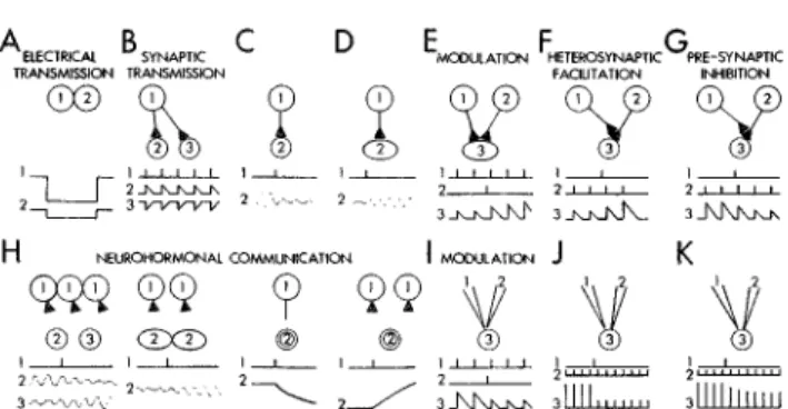
What Is a modulator?
The words "modulation" and "modulator" have appeared in the
titles of several hundred neuroscience articles in the past year
(according to an informal computer survey of the literature).
Modu-lation is being used increasingly to characterize actions of putative
Dismukes: Neuronal communication
transmitters that do not seem to fit within traditional concepts of
neuronal action. By extension, it has been suggested that endogenous
substances that elicit these unconventional responses - particularly
monoamines and neuropeptides - might be considered
neuromodu-lators or neuroreguneuromodu-lators rather than neurotransrnitters.
The concept of modulation has not been applied in any consistent
way, and explicit definitions have seldom been attempted.
Mtodula-tory roles have been ascribed to various neurochemical systems on
the basis of diverse characteristics, such as the nature of the
electro-physiological response, the time course of action, the mode of
transmission to target cells, and the distribution of targets. This
ambiguity is heightened by use of the word in different contexts to
describe actions at different levels of organization. Besides cellular
actions, modulation has been applied at the circuit level to describe
gating of motor outputs, and at the behavioral level to refer to
changes in response strength that might be produced by arousal,
motivation, or learning (Kandel et al. 1979). It is intriguing to
speculate that modulation of behavioral function, arousal, for
exam-ple, might be mediated by neurochemical systems such as the diffuse
ascending aminergic pathways, producing modulation at the cellular
level. Unfortunately there is still little evidence for such conluence,
nor is there likely to be more until our notions of modulation can be
stated more explicitly.
In one of the few explicit definitions that has been made, Florey
(1967) suggested that "modulator substance" be used " . . . for any
compound of cellular and nonsynaptic origin that effects the
excit-ability of nerve cells . . . " and that " . . . can effect the responsiveness
of nerve cells to transsynaptic actions . . . and can alter . . .
sponta-neous activity." Barchas and coworkers (1978) distinguished
neuro-modulators (neuroregulators) from neurotransmitters as compounds
that are important in general communication between nerve cells,
but which operate in a hormone-like fashion, rather than
transsynap-tically. (This definition is similar to Chan-Palay's (1977)
characteri-zation of "neurohumors;" see following section.)
Unfortunately, using nonsynaptic transmission as the primary
defining characteristic fails to draw together all classes of action that
might be considered modulation, and furthermore it creates some
awkward divisions. For example, would the lack of close synaptic
apposition at most noradrenergic boutons en passant in the
periph-eral nervous sytem force us to call norepinephrine a neuromodulator
rather than a neurotransrnitter in these regions? Would modulation
of transmitter release by synapses on nerve terminals not be
consid-ered the work of a modulator, since the modulating compound was
released synaptically? Would the complex electrophysiological
effects described in the previous section not be considered an
example of modulation, unless the monoamines and peptides evoking
them were shown to be released nonsynaptically? The difficulties
raised by these questions suggest that defining the mode of
transmis-sion does not necessarily distinguish the nature of modulatory
actions. The possibility of distinct modes of transmission will be
considered further in the next section.
Given the incomplete state of our knowledge, I suggest that there
is little advantage at this time in attempting to distinguish modulators
from transmitters. On the other hand, there may be some heuristic
value in trying to clarify what actions might be considered to be
modulatory. The various uses of the word modulation seem to have
in common mainly the general English language connotation of
alteration of a primary characteristic over time by a secondary
inluence. At the cellular level most usages imply that a modulator
changes the efficacy of conventional synapses without directly
alter-ing the rate of firalter-ing or release of transmitter. Presynaptic
modula-tion of transmitter release fits this nomodula-tion neatly. The release of
transmitter is clearly caused by axonal impulse activity and is only
modified by presynaptic receptors. Postsynaptically the situation is
more ambiguous. Most central neurons receive many synaptic inputs,
and it is seldom possible to say that any one exerts primary control.
As was previously pointed out, a conventional synaptic excitation or
inhibition can modify the effectiveness of nearby synapses. However,
it is plausible that some transmitters produce responses whose
predominant effect is alteration of other inputs. Such transmitters
might be released either synaptically or nonsynaptically and could
act on nerve terminals, dendrites, or cell bodies.
When it appears that the predominant effect of a transmitter
substance in a particular system is regulation of the efficacy of other
transmitter inputs or of the mode of operation (e.g. bursting vs.
nonbursting) of the target cell, then it may be useful to think of this
action in terms of modulation. I suggest that is is preferable to use the
word modulation as a broad, generic term at present, rather than
trying to pin it down to a particular function, because we are just
beginning to discover the range of responses that transmitters can
evoke in various neurons. It may turn out that an array of functions
beyond simple excitation and inhibition will appear. Furthermore, it
would be well not to label particular transmitters as neuroregulators
or neuromodulators unless the specific system is identified, because a
given agent might evoke various responses in different target cells.
The character of response is, of course, not a function of the
transmitter molecule itself but of the effector mechanisms to which
its receptors are coupled.
Are there distinct modes of chemical communication?
Sharrer pointed out a decade ago (1969) that the borderline between
the two well-established classes of neural mediation - classical
synaptic transmission and neurosecretion of hormones - is less sharp
than had been previously thought, in that some neurons demonstrate
characteristics overlapping both classes. Chan-Palay has suggested
(1977) that the nonsynaptic varicosities of serotonergic and
noradren-ergic axons are the basis of a specific mode of neuronal transmission,
which she terms neurohumoral. This mode is conceived to be
intermediate between classical synaptic transmission and hormonal
secretion. Neurohumors released from diffuse aminergic axonal
projections could alter the responsiveness of vast domains of target
cells, possibly constituting a way of modulating the behavioral state
of the organism. The serotonin fibers lining the walls of the cerebral
ventricles provide a striking illustration of this possibility. Serotonin
released into the cerebrospinal fluid (CSF) might have access to a
wide range of targets, either by re-uptake and transport by certain
ependymal cells lining the ventricles, or by the flow of CSF from the
ventricles over the cortex. By modulating the activity of widespread
target neurons, serotonin might thus have global effects on
behavior-al state.
Barker (1977; 1978) has proposed a similar mode of
communica-tion, termed neurohormonal, on the basis of observations with
neuropeptides. Neurohormonal communication was characterized as
nonsynaptic, acting by regulation of voltage-dependent spike or
pacemaker conductances. In contrast to synaptic
moment-to-moment regulation of single-neuron excitability, it would provide
sustained regulation of neuronal aggregates, perhaps coordinating
their output. Barker (1978) also proposes another mode of
communi-cation - "neuromodulation" - which operates as a form of gain
control over transmission through conventional synapses.
Neuromod-ulation was proposed to operate through alteration of synaptically
activated, voltage-independent conductances and might or might not
be restricted to continguous cells. By defining neuro-modulation
explicitly in terms of electrophysiological phenomena, Barker has
avoided the problem of ambiguity discussed in the previous section.
However there might be some advantage to coining a new term for
this specific electrophysiological action, to avoid confusion with all
the other uses of the word "modulation".
These proposals have heuristic appeal because they draw together
a number of striking features of monoamine and neuropeptide
presumptive transmitters in terms of their possible functional
signifi-cance. Although it is highly speculative, this provides a conceptual
framework for thinking about features as diverse as: the diffuse
anatomy of aminergic pathways, the slow monotonic pattern of
firing in those neurons, the possibility of nonsynaptic release, and
complex electrophysiological responses. At present, evidence is too
fragmentary to evaluate how well these proposed categories of
transmission might accommodate emerging data about the diverse
Bismekes: Neuronal communication
ways in which neurons communicate.
Given this state of affairs, it is important to maintain a flexible
approach to categorizing neuronal communication. Bloom has
proposed (1.979) such an approach, in which transmitters would be
classified in terms of three domains of action: time course, spatial
distribution, and energy. This last domain seems ambiguous but
apparently concerns the character of the response elicited by the
transmitter. This scheme may be useful, for it allows organization of
emerging data without forcing premature labeling. As the growing
number of putative transmitters are plotted on this
three-dimen-sional array, certain groupings that correspond to distinct modes of
transmission may become apparent.
Nomenclature for complex actions of transmitters and for
proposed modes of transmission is becoming chaotic. Substances have
been distinguished as neurotransmitters, neurohumors,
neurohor-mones, neuromodulators, neuroregulators, and neuromediators by
various authors in ways that are sometimes overlapping, sometimes
conflicting. Confusion might be avoided if, at this stage, labels were
avoided and distinctions were spelled out - for example: nonsynaptic
vs. synaptic release, excitation via opening of voltage-independent
ion channels vs. activation of voltage-dependent conductances.
However, in practice this would be tedious, and it seems unavoidable
to use labels. I suggest that the following approach to nomenclature
would be reasonably consistent with conventional definitions found
in technical dictionaries (e.g., Blakiston's 1972) and would allow a
desirable flexibility:
Neurohumor. a generic term for any substance released by a
neuron to alter the activity of other cells, adjacent or distant.
Synaptic (or junctional) neurotransmitter. a neurohumor that is
released for diffusion to an adjacent excitable cell (and to
autoreceptors, when present). The junction may be broader than that
found in Grey's Types I and II synapses, and specialization of the
postsynaptic membrane may not be apparent (Shepherd 1974). The
response to a neurotransmitter, synaptic or nonsynaptic, is mediated
through membrane receptors and may include complex modulatory
actions as well as conventional excitation or inhibition.
Nonsynaptic (or nonfunctional) transmitter, a neurohumor that is
released for diffusion or transport to multiple excitable cells, which
may be distant from the release site.
Neurohormone. a neurohumor capable of regulating mulitple and
distant target cells that do not necessarily have membrane receptors
or electrical responses. Transport is primarily via vascular channels.
Obviously additional terms will be necessary if other classes of
neurohumors or subclasses of nonsynaptic transmitters are
delineated. In that case it would be desirable to coin new words to
avoid confusion with old terminology. Interestingly, the classic
criteria for identifying a substance as a central neurotransmitter, as
formulated by Werman (1966), do not require synaptic action and
thus could be applied to other neurohumors as well. Some other
statements of the criteria, however, do explicitly describe synaptic
transmission. For example, Barchas and coworkers (1978)
distin-guished neuroregulators as operating nonsynaptically and proposed a
modified set of criteria for them.
Some implications and conclusions
It remains to be demonstrated that amine-containing boutons
with-out apparent synaptic contact release their transmitter in vivo. If
such release does occur, the transmitter might diffuse to multiple
target cells, but alternately it might activate only an immediately
adjacent neuron, perhaps through receptors spread more broadly
over the membrane than would occur in a synaptic junction. Thus
the form of release of transmitter from these diffusely-branching
aminergic fibers in the brain may be analogous to that found in
peripheral noradrenergic fibers. The possibility that fibers lining the
walls of the ventricles might release serotonin in the CSF for
transport to distant targets is much more novel and would seem to
represent a mode intermediate between synaptic and hormonal
transmission.
If nonsynaptic transmission does occur, it would not necessarily
require any radical revision of our conceptualization of neuronal
function, but rather it would seem to complement known features of
the diffuse ascending aminergic pathways. For example, the
nor-adrenergic fiber system projecting from the locus coeruleus
through-out many regions of the brain has several striking features:
1. Projections so diffuse that one noradrenergic neuron may influence vast
numbers of target cells.
2. A slow, monotonic pattern of impulse activity that seldom varies.
3. Production of target-cell responses that are long in latency and duration
and that modify the effects of other transmitters.
Such a system seems unlikely to transmit detailed information
about rapidly-changing processes, but rather it is generally conceived
to act in some regulatory fashion, modulating the level or pattern of
responsiveness of its targets.
Both Chan-Palay (1977) and Barker (1977, 1978), in proposing
nonsynaptic modes of transmission, have suggested that these might
serve as a means of coordinating or tuning the activities of large
arrays of target neurons. Modulating the functional state of neuronal
ensembles could be a way of regulating the state of behavioral
responsiveness of the organism (Hobson & Scheibel 1979). This sort
of regulation might underlie functions such as: gating of sensory
information in intermediate processing centers such as the thalamus,
sleep/wakefulness cycles, arousal, reinforcement of behavior,
memory formation, and affective state.
Over the past two decades monoamine transmitters have been
implicated in many aspects of animal behavior and of psychiatric
disorder (Lipton et al. 1978), and recently there have been many
similar suggestions about neuropeptides (Liebeskind & Dismukes
1978). However, only in a very few cases has the actual nature of the
transmitter's involvement been established. Modulation of the
func-tional state of neuronal ensembles is a heuristic concept that may be
very useful in understanding mechanisms underlying behavior. Hints
of behavioral involvement have been observed much more often
with monoamines and peptides than with transmitters such as
acetylcholine or GABA. This might be an indication that
experimen-tal manipulations of transmitters involved in state regulation of
neuronal ensembles produce behavioral manifestations more
concor-dent than would occur with manipulation of an excitatory or
inhibitory transmitter involved in diverse circuits. It should be noted
that the argument for some transmitters acting as regulators of
functional state is based primarily on the nature of their
electrophys-iological action and on behavioral inferences. Nonsynaptic
transmis-sion would not necessarily be required, although it would extend the
range of action.
The studies briefly reviewed in this article suggest several aspects
of neuronal communication not encompassed by conventional
descriptions. This does not mean that traditionally stated principles
of neuronal operation are wrong, but rather that they are incomplete
and perhaps overgeneralized. Current research suggests that an array
of information-processing mechanisms overlie well-known central
themes. Thus, for example, the concept of integration of excitatory
and inhibitory synaptic inputs is not invalidated by evidence that the
summation, rather than being a simple algebraic addition, is instead
a complex and dynamic interaction that includes various regulatory
and adaptive processes. In a similar fashion we still talk of
dopamin-ergic neurons transmitting information from the substantia nigra to
the striatum as a primary theme, even though it is overlaid with
secondary actions such as dendritic release of dopamine in the nigra,
feedback inhibition at autoreceptors on striatel terminals, and
perhaps retrograde transport of macromolecules.
What is invalidated by these various lines of research is any notion
of the neuron as a black box, analogous to an electronic operational
amplifier, which can be described as performing a set transformation
on its inputs to produce a corresponding output. Clearly, neurons are
pretty sophisticated little computers in their own right, with mulitple
Commentary/Dismukes: Neuroeal communication
layers and time scales of information processing. Their modes of
interaction may eventually prove as diverse as their anatomical
forms.
Successful explanations in biology sometimes become
overgeneral-ized as they are extened to new systems and the original
experimen-tal qualifications are forgotten. The traditionally stated principles of
neuron operation have provided a framework for understanding a
wide range of phenomena, but the success of these central themes
should not reduce our sensitivity to additional forms of molecular
exchange and information processing among neurons. It is important
not to let working principles turn into dogma, or to replace old
dogmas with new ones. Ironically, one of the most generalized of
conventional postulates is named after Sir Henry Dale, who
appar-ently intended his principle to be considered only as a guide (Dale
1935; Burnstock 1976).
ACKNOWLEDGMENT
This article was prepared while the author was in residence at the
Neuro-sciences Research Program (NRP). Discussions with NEP staff and participants
in NRP conferences (particularly Hobson & Scheibel 1979 and Kandel et al.
1979) contributed greatly to the development of ideas presented here. NRP is
supported by grants from the National Institute of Mental Health, the National
Institute of Neurological and Communicative Diseases and Stroke, the National
Science Foundation, and private foundations.
NOTE
°R. Key Dismukes's present address is in care of the National Research Council,
Committee on Vision, 2101 Constitution Ave., N.W., Washington, D.C. 20418.
Open Peer Commentary
Commentaries submitted by the qualified professional readership of this journal will be considered for publication in a later issue as Continuing Commentary on this article.
by S. Arch
Biology Department, Reed College, Portland, Ore. 97202
Terminology, modes of communication, and a command
neurohor-mone. The value of the concept of alternative modes of
communica-tion among neurons is apparent only if it leads to a better understand-ing of the normal physiology of the organism. In one of the more thoroughly studied preparations, this understanding seems to be forthcoming. The bag cell neurons in Aplysia californica synthesize and secrete a polypeptide hormone that causes a complex egg-laying response in the animal (reviews: Arch 1976; Blankenship 1979). The egg-laying hormone (ELH) acts on the gonad and other portions of the reproductive tract to cause changes in muscular activity, and on the heart to accelerate and strengthen beating. Moreover, ELH is known to cause a marked change in the behavior of the animal. Thus its effects in the nervous system are of considerable interest. Perhaps the most striking feature of ELH action in the nervous system is the large number of cells it appears to influence. While most of the effects remain to be shown as direct - and many may not be - ELH action results in a significant alteration of central nervous system activity.
The sum of the hormone's effect is to cause the animal to lay its eggs in the species-typical fashion, and each of the known actions of ELH can be interpreted as serving this end. Thus, in a sense, the hormone seizes control of the animal's physiology and directs it to perform this reproductive function. For this reason I believe that it should be considered a command neurohormone. It is both necessary and sufficient (Kupfermann and Weiss 1978) for the initiation and organization of the egg-laying response.
It might seem less than fully responsive to the spirit of Dismukes's article to propose still another term to characterize the functional role of a substance active in the nervous system. I am, in fact, in agreement with Dismukes on the point that we should be explicit about the synaptic, nonsynaptic, or neuorhormonal nature of the influence we are examining. In some respects, however, the development of an "offi-cial" terminology may be either a waste of effort or, worse, a hindrance
to understanding. As we learn more about the physiology of the various systems under active investigation, our terminology may become obsolete. More dangerous is the fact that annointing some terms and proscribing others may impose a dogmatic control over the way we view our results. The problem that should concern us right now is not that of terminology, but that of demonstrating the existence of alterna-tive modes of behaviorally relevant neural intercommunication. For this task we need a set of criteria, whose satisfaction will permit the conclusion that an alternate channel of communication has been identified.
Accumulations of vesicles near cell membranes are not necessarily indications of secretion. Identification of a suspect compound in a cell or brain region tells us nothing about its action (if any). Neural response to an exogenously applied substance is not a demonstration of its significance in normal physiology. Such observations may be sugges-tive, but they permit only speculation. We will not be in a position to conclude that alternative modes of communication exist until we have satisfied essentially the same set of criteria that we recognize for the identification of a synaptic neurotransmitter. Indeed, we probably need to add at least one criterion to the list. Since our concern is with substances that may be released nonsynaptically and may act on extrasynaptic sites, the route taken by the substance becomes impor-tant. Specifically, it should be shown that the putative neuroactive compound can traverse the barriers, if any, between its release site and the structures upon which it is supposed to act.
In the Aplysia system I believe we are close to satisfying these criteria. To call ELH a command neurohormone then, is simply a way of characterizing its role economically. Other substances in other systems have been, and will be, characterized with other words. The only chaos in our literature that need concern us will come not from too many functional names for neuroactive compounds, but from too little empirical support for the functions implied by the names.
by M. A r l u i s o n
Universite de Paris-VI, Laboratoire De Neurocytologie, Paris-5e, France
The problem of nonsynaptic transmission in the neostriatum. This
commentary examines the problem of the occurence of nonsynaptic dopaminergic transmission in the nucleus caudatus-putamen (NCP). In this line of work, our results, dealing with the description of dopaminer-gic contacts in NCP (Arluison et al. 1978a) conflict with those of Tennyson et al. (1974), which strongly supports the existence of nonsynaptic release. I can present only a brief commentary upon the discrepancy between the results:1. In both studies, 5-OH-DA (5-hydroxydopamine) was used in rather high concentrations. In our work (Arluison et al. 1978a), the tagging of synaptic vesicles in nerve endings was studied according to the gradient of diffusion of the marker around the injection site and was controlled by destruction of the substantia nigra by 6-OH-DA. We found that an important nonspecific labelling of nerve endings occurred near the injection site because of the excess concentration. Such nonspecific capture probably also occurs in the work of Tennyson et al., in which 5 x 10~4 M of 5-OH-DA is used.
2. In our own two studies (using, respectively, 5-OH-DA or radioau-tography after administration of 3H-DA), we tried, at first to quantify the presence or absence of labelling in certain nerve endings, studying only those terminals exhibiting differentiated synaptic contacts. In addition, we tried to see whether synaptic contacts were more numerous in the labelled population of nerve endings than in the nonlabelled one. Using 5-OH-DA, we found that the occurence of synapses was approximately the same in both groups. In contrast, with
3H-DA we found that the percentage of labelled nerve endings was higher in the group of nerve endings without synapses.
3. In the studies of Tennyson and coworkers, tissues are incubated in 5-OH-DA, and this procedure alters the tissue such that at least certain synapses are destroyed. Moreover, fixation of tissue by KMnO4
is not well suited for the identification of synapses because postsynap-tic thickening is not visible. Under these conditions, the reported absence of synaptic contacts for dopaminergic fibres is understand-able.