This paper is available online free of all access charges (see http://jxb.oxfordjournals.org/open_access.html for further details)
ReseaRch papeR
Delayed degradation of chlorophylls and photosynthetic
proteins in Arabidopsis autophagy mutants during
stress-induced leaf yellowing
Yasuhito Sakuraba1,*,†, Sang-Hwa Lee1,*, Ye-Sol Kim1, Ohkmae K. Park2, Stefan Hörtensteiner3 and Nam-Chon Paek1,†
1 Department of Plant Science, Plant Genomics and Breeding Institute, and Research Institute for Agriculture and Life Sciences, Seoul National University, Seoul 151-921, Korea
2 Department of Life Sciences, Korea University, Seoul 136-701, Korea 3 Institute of Plant Biology, University of Zurich, CH-8008 Zurich, Switzerland * These authors contributed equally to this work.
† To whom correspondence should be addressed. E-mail: sakuraba0425@gmail.com or ncpaek@snu.ac.kr
Received 1 October 2013; Revised 12 December 2013; Accepted 17 December 2013
Abstract
Plant autophagy, one of the essential proteolysis systems, balances proteome and nutrient levels in cells of the whole plant. Autophagy has been studied by analysing Arabidopsis thaliana autophagy-defective atg mutants, but the rela-tionship between autophagy and chlorophyll (Chl) breakdown during stress-induced leaf yellowing remains unclear. During natural senescence or under abiotic-stress conditions, extensive cell death and early yellowing occurs in the leaves of atg mutants. A new finding is revealed that atg5 and atg7 mutants exhibit a functional stay-green phenotype under mild abiotic-stress conditions, but leaf yellowing proceeds normally in wild-type leaves under these conditions. Under mild salt stress, atg5 leaves retained high levels of Chls and all photosystem proteins and maintained a normal chloroplast structure. Furthermore, a double mutant of atg5 and non-functional stay-green nonyellowing1-1 (atg5
nye1-1) showed a much stronger stay-green phenotype than either single mutant. Taking these results together, it is
proposed that autophagy functions in the non-selective catabolism of Chls and photosynthetic proteins during stress-induced leaf yellowing, in addition to the selective degradation of Chl–apoprotein complexes in the chloroplasts through the senescence-induced STAY-GREEN1/NYE1 and Chl catabolic enzymes.
Key words: Abiotic stress, Arabidopsis thaliana, autophagy, atg5, chlorophyll degradation, leaf senescence, stay-green.
Introduction
Senescence marks the final stage of leaf development in plants. In the early phase of leaf senescence, developmental and envi-ronmental cues signal the plant cells to activate transcription factors (TFs) that modulate the expression of senescence-asso-ciated genes (SAGs) (Guo and Ecker, 2004; Balazadeh et al., 2008). The products of these SAGs conduct the highly ordered breakdown of intracellular organelles, including the degrada-tion of proteins and macromolecules to remobilize leaf nutri-ents into other developing organs such as new leaves or seeds, or into storage organs (Lim et al., 2007; Robinson et al., 2012).
Autophagy, a highly conserved process in eukaryotes, functions as one of the major pathways for the massive deg-radation of intracellular proteins during leaf senescence (Nakatogawa et al., 2009; Reumann et al., 2010) as well as for survival under some biotic/abiotic-stress conditions (Klionsky, 2004). Autophagy occurs by two main mechanisms, micro-autophagy and macromicro-autophagy. In micromicro-autophagy, an invagination of the vacuolar membrane directly engulfs the cytosolic component to be degraded (Klionsky and Ohsumi, 1999). By contrast, in macroautophagy, autophagosomes
This is an Open Access article distributed under the terms of the Creative Commons Attribution License (http://creativecommons.org/licenses/by/3.0/), which permits unrestricted reuse, distribution, and reproduction in any medium, provided the original work is properly cited.
form at the periphery of damaged or overproduced proteins. Autophagosomes enclose organelles or cytosolic compounds, which are transported into the vacuole and broken down by the non-selective degradation pathway (Meijer et al., 2007). To date, more than 30 autophagy-associated (atg) genes have been identified in yeast and Arabidopsis (Arabidopsis
thali-ana) (Bassham et al., 2006).
In pre-senescent leaves during vegetative growth, chloro-plasts contain the majority of plant nutrients. For example, chloroplastic proteins contain 75–80% of total leaf nitrogen in C3 plants (Makino and Osmond, 1991). Thus, the deg-radation of chloroplast proteins in old or inefficient leaves during senescence provides important nutrients for reloca-tion to developing organs. In recent years, the degradareloca-tion mechanisms of chloroplasts and chloroplast proteins during senescence have been widely studied. During leaf senescence, Rubisco, the most abundant stromal protein in the chloro-plasts (Wittenback, 1978), is released from chlorochloro-plasts into the cytoplasm as small double-membrane bodies termed Rubisco-containing bodies (RCBs; Chiba et al., 2003) that are then transported to the central vacuole by autophagy for degradation (Ishida et al., 2008). RCBs were not observed in the leaves of autophagy-defective atg4 (Wada et al., 2009) and
atg5 (Ishida et al., 2008) mutants in Arabidopsis, indicating the direct involvement of macroautophagy in the degrada-tion of Rubisco during leaf senescence. For the degradadegrada-tion of Rubisco and stromal proteins, another extra-chloroplastic degradation system, called senescence-associated vacuoles (SAVs), was also identified. SAVs are clearly smaller than the central vacuole and contain Rubisco and other stromal pro-teins, including glutamine synthetase, but not the photosys-tem proteins (Martinez et al., 2008). This indicates that the SAV-dependent degradation system mainly functions in the degradation of stromal proteins in chloroplasts.
In contrast to the extra-chloroplastic degradation mecha-nisms for Rubisco and other proteins, an intra-chloroplastic degradation system mainly degrades thylakoid proteins. Plastids isolated from senescing leaves can degrade photo-system proteins under light conditions (Feller et al., 2008) indicating that senescing chloroplasts have active systems to degrade photosystem proteins. This system may include the chloroplast protease FtsH, as the Arabidopsis T-DNA inser-tion KO mutants of ftsH6 were unable to degrade Lhcb3 dur-ing dark-induced senescence and were also unable to degrade Lhcb1 and Lhcb3 under high light conditions (Zelisko et al., 2005).
Chlorophyll (Chl) is degraded by several Chl catabolic enzymes (CCEs; Hörtensteiner, 2013). In addition,
STAY-GREEN (SGR), Mendel’s green cotyledon gene encoding a
novel chloroplast protein, functions in the initiation of Chl degradation (Park et al., 2007; Ren et al., 2007). Recently, it was demonstrated that SGR and six CCEs form a complex with light-harvesting complex II (LHCII), which may allow meta-bolic channelling of phototoxic Chl degradation intermedi-ates (Sakuraba et al., 2012b, 2013). Chl degradation ends with the formation of fluorescent chlorophyll catabolite (FCC), a non-toxic Chl degradation intermediate, in chloroplasts. For the final steps of Chl breakdown, FCC is transported into the
vacuole and converted to non-fluorescent Chl catabolite (NCC) (Oberhuber et al., 2003; Hörtensteiner and Krautler., 2011).
Although Chl breakdown generally occurs in the chlo-roplast until the formation of FCC, Wada et al. (2009)
detected Chl fluorescence in the central vacuole of dark-induced senescing leaves in Arabidopsis, strongly indicat-ing that macroautophagy also functions in the transport of Chl–apoprotein complexes from chloroplasts to the vacuole during senescence. Considering this function, the autophagy-dependent degradation system and other intra-chloroplastic degradation systems seem to share target proteins or mac-romolecules, including Chl and Chl-binding photosystem proteins. However, the relationship among these different degradation systems remains enigmatic.
It was found here that atg5 mutants display a stay-green phe-notype only under mild abiotic-stress conditions, but not under strong stress conditions; atg5 leaves showed early leaf yellowing with extensive cell death under strong abiotic-stress conditions. Under mild abiotic-stress conditions, however, atg5 acts as a functional stay-green mutant, maintaining the proper balance of Chls and photosynthetic proteins and retaining the grana thylakoid structure in the chloroplasts. Genetic analysis of atg5 and the non-functional stay-green mutant nye1-1 revealed that autophagy contributes to the non-selective breakdown of Chl-photosynthetic proteins during mild abiotic-stress-induced leaf yellowing, in addition to the selective breakdown of Chl-apoproteins through a dynamic STAY-GREEN1(SGR1)/ NYE1-CCE complex in the senescing chloroplasts (Sakuraba et al., 2012b). The relationship between autophagy-induced and SGR1-dependent degradation of the Chl-apoprotein com-plex in chloroplasts is also discussed.
Materials and methods
Plant materials and growth conditionsThe Arabidopsis thaliana plants were grown on soil at 21–23 °C under long day (LD) conditions (16/8 h light/dark; 90–100 μmol m–2 s–1 cool-fluorescent white light). For dark treatment, detached or attached leaves of 3-week-old plants were placed in complete dark-ness. Wild type, atg5 and nye1-1 are of the Col-0 ecotype. The atg7 mutant (Ws-2 ecotype; CS39995) was obtained from the Arabidopsis Biological Resource Center (ABRC, Ohio, USA).
Trypan blue staining
Leaves were incubated overnight in lactophenol-trypan blue solu-tion (10 ml lactic acid, 10 ml glycerol, 10 g phenol, and 10 mg trypan blue dissolved in 10 ml distilled water) (Koch and Slusarenko, 1990). Stained leaves were then boiled for 1 min and then decolourized in 60% glycerol solution.
Chlorophyll quantification
Chlorophylls (Chls) were extracted from leaf tissues with 80% ice-cold acetone solution at 4 °C. Chl concentration was quantified by a spectrophotometric method (Porra et al., 1989).
Immunoblot analysis
Protein extracts were prepared from rosette leaves of Arabidopsis
and homogenized with 100 μl of sample buffer [50 mM TRIS– HCl, pH 6.8, 2 mM EDTA, 10% (w/v) glycerol, 2% SDS, and 6% 2-mercaptoethanol] was used to suspend the protein extracts. The protein samples were subjected to SDS-PAGE. Gels were stained with Coomassie Brilliant Blue R-250 (Sigma–Aldrich). Antibodies against photosynthetic proteins, including Lhca3, Lhcb1, Lhcb2, Lhcb4, Lhcb5, CP43, D1, and PsaA (Agrisera, Sweden), were used for immunoblot analysis. Each protein was detected using an electro-chemiluminescence (ECL) system (WESTSAVE, AbFRONTIER, Seoul, Korea) according to the manufacturer’s manual.
Chl fluorescence measurement using pulse amplitude modulation
Maximal photochemical efficiency of PSII (Fv/Fm) was measured using the OS-30p+ instrument (OPTI-SCIENCES, USA). Detached leaves before and after salt treatment were adapted in the dark for 5 min and the Fv/Fm ratio was measured at room temperature. This 5 min dark treatment resulted in the complete oxidation of QA. Transmission electron microscopy
Transmission electron microscopy was conducted as previously described by Inada et al. (1998) with some modifications. Leaf tis-sues were fixed with modified Karnovsky’s fixative (2% paraformal-dehyde, 2% glutaralparaformal-dehyde, and 50 mM sodium cacodylate buffer, pH 7.2). Samples were then washed with 0.05 M sodium cacodylate buffer, pH 7.2, three times at 4 °C for 10 min. The samples were post-fixed at 4 ºC for 2 h with 1% osmium tetroxide in 0.05 M sodium cacodylate buffer, pH 7.2, and washed twice with distilled water at room temperature. Samples were stained in 0.5% uranyl acetate at 4 °C overnight and dehydrated in an ethanol gradient and propyl-ene oxide, then finally infiltrated with Spurr’s resin. Polymerization was performed at 70 °C for 24 h and samples were sectioned with an ultramicrotome (MT-X). The sections were mounted on cop-per grids and stained with 2% uranyl acetate for 7 min and with Reynolds’ lead citrate for 7 min. Micrographs were made by using a LIBRA 120 transmission electron microscope (JEOL, Japan).
Ion leakage measurement
To measure ion leakage after treatment, approximately 10 rosette leaves were placed in a tube with 6 ml of 0.4 mM mannitol. The tubes were placed at room temperature for 3 h with shaking. Conductivity of the incubated solution was measured using an electroconductiv-ity meter (CON6 METER, LaMOTTE Co., USA), before and after boiling for 10 min.
Abiotic-stress treatments
Analysis of salt stress was performed as previously described by Wu
et al. (2012) with minor modifications. Detached leaves of 3-week-old plants were floated abaxial side-up, on 3 mM MES buffer (pH 5.8) containing 150, 300, or 450 mM NaCl. For osmotic stress, leaves were floated on buffer containing 50, 200, or 400 mM mannitol. For oxidative stress, leaves were floated on buffer containing 5, 20, or 50 mM H2O2.
Reverse transcription (RT) and quantitative real-time PCR (qPCR) analysis
Total RNA was extracted from the leaf tissues using the Plant RNA Extraction Kit (iNtRON Biotechnology, Seoul, Korea) including the RNase-free DNase I treatment step to remove possible genomic DNA contamination. For RT, the first-strand cDNAs were pre-pared with 5 μg total RNA using M-MLV reverse transcriptase and aqn oligo(dT) primer (Promega). For quantitative real-time PCR (qPCR), 20 μl reactions, including first-strand cDNAs
equivalent to 50 ng total RNA, 10 μl 2× Universal SYBR Green Master Mix (Roche), and gene-specific forward and reverse primers (see Supplementary Table S1 at JXB online), were analysed using a Light Cycler 480 (Roche Diagnostics). Data analysis was conducted using the Roche Optical System software (ver. 1.5). The efficiency of qPCR analysis was calculated by comparing the slope of linear regression of Ct and log10 of gene copies. Relative gene expression levels were normalized against the transcript levels of GAPDH (encoding glyceraldehyde phosphate dehydrogenase; At1g16300) as previously reported by Sakuraba et al. (2010).
Results
atg5 leaves exhibit a stay-green phenotype under mild
abiotic-stress conditions
Plant autophagy affects senescence and stress tolerance; atg mutants exhibit accelerated leaf yellowing during age- and dark-induced senescence (Thompson et al., 2005), and hyper-sensitivity to abiotic stresses such as high salinity, oxidative stress, and drought (Xiong et al., 2007; Liu et al., 2009; Zhou et al., 2013). This indicates that autophagy plays an impor-tant role in maintaining the proper balance of the cellular proteome during abiotic stresses. However, stress-induced chlorophyll (Chl) degradation should be impaired when autophagy does not operate properly, because autophagy is involved in chloroplast degradation, including Chl break-down, during senescence (Ishida et al., 2008; Wada et al., 2009; Ono et al., 2012).
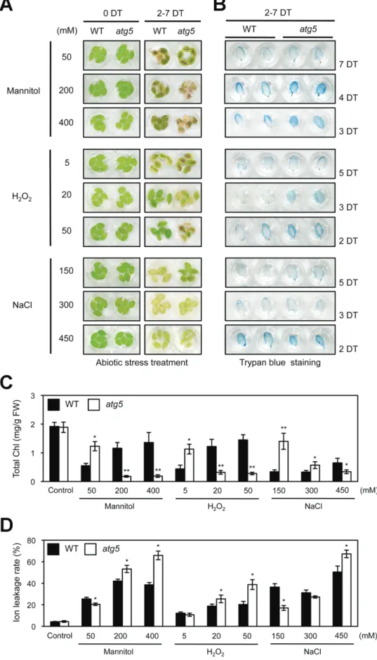
To investigate the relationship between autophagy and Chl degradation in more detail, we examined the visual pheno-types of atg5 leaves under different abiotic-stress conditions (Fig. 1), including salt (NaCl), osmotic pressure (mannitol), and oxidative reagent (H2O2) treatments. To this end, the detached rosette leaves of 3-week-old plants were used to separate the effect of autophagy defects in each plant organ (Thompson et al., 2005). As previously observed in whole plants (Thompson et al., 2005; Zhou et al., 2013), atg5 leaves exhibited an early senescence phenotype under strong abiotic-stress conditions, such as 200 and 400 mM mannitol, 20 and 50 mM H2O2, or 450 mM NaCl (Fig. 1A). Trypan blue stain-ing revealed that early leaf yellowstain-ing of atg5 leaves under these strong abiotic stresses resulted from cell death (Fig. 1B). By contrast, atg5 leaves exhibited a stay-green phenotype under mild abiotic-stress conditions, such as 50 mM man-nitol, 5 mM H2O2, or 150 mM NaCl treatments (Fig. 1A). Notably, under these mild abiotic stresses, atg5 leaves barely showed any cell death phenotype (Fig. 1B), suggesting that
atg5 is defective in Chl degradation, although only under mild
abiotic-stress conditions in which accelerated cell death hardly occurs. To understand the stay-green phenotype of atg5 leaves under mild stress conditions in more detail, total Chl levels and ion leakage rates were measured in each condition as an indicator of membrane disintegration and plant cell death. Consistent with the visible phenotypes, atg5 leaves under mild abiotic-stress conditions had significantly higher total Chl lev-els than wild-type leaves (Fig. 1C). Compared with wild-type leaves, atg5 leaves had significantly lower ion leakage rates, but had significantly higher rates under strong stress conditions
Fig. 1. Phenotypic characterization of atg5 leaves under different abiotic stresses. (A) Visible phenotypes of detached rosette leaves from 3-week-old wild-type (WT) and atg5 mutants under osmotic (50, 200, or 400 mM mannitol), oxidative (5, 20, or 50 mM H2O2), and salt (150, 300, or 450 mM NaCl)
stress conditions. (B) Cell death in WT and atg5 leaves under abiotic stresses, as shown by trypan blue staining. (C, D) The changes of total Chl levels (C) and ion leakage rates (D) in WT and atg5 leaves after abiotic-stress treatments in (A). Black and white bars indicate WT and atg5, respectively. DT, days of treatment. Similar results were obtained from three independent experiments. Student’s t-test (*P<0.05, **P<0.01). (This figure is available in colour at JXB online.)
(Fig. 1D). This confirms that the degree of cell death is closely associated with the phenotype of atg5 leaves under these abi-otic-stress conditions. atg7, another autophagy mutant, was also examined. Similar to atg5 leaves, atg7 leaves also showed a stay-green phenotype under mild abiotic-stress conditions (see Supplementary Fig. S1 at JXB online).
Our results using detached atg5 leaves conflicted with previous results using whole plant bodies under salt-stress conditions (Zhou et al., 2013). Therefore, the whole-plant phenotype of atg5 mutants grown for 2 weeks on phytoagar plates containing a low concentration of NaCl (150 mM) was examined. Consistent with the previous results (Zhou et al., 2013), older leaves (cotyledon and 1st cycle of rosettes) of atg5 plants showed a leaf necrosis phenotype (see Supplementary Fig. S2A and B at JXB online). However, younger leaves (2nd and 3rd cycle of rosettes) stayed green with higher Chl levels compared with those of wild-type leaves, suggesting that both detached and attached leaves of atg5 have a stay-green capac-ity under mild salt-stress conditions, although atg5 mutants exhibit a necrosis phenotype in older leaves.
atg5 exhibits a functional stay-green phenotype under
mild abiotic-stress conditions
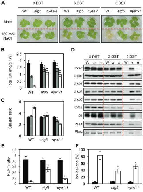
To characterize the stay-green phenotype of atg5 leaves under mild abiotic stresses in more detail, several photosynthetic parameters of atg5 were compared with a non-functional stay-green SGR1 mutant, nye1-1 (Ren et al., 2007), under mild salt-stress conditions (150 mM NaCl). Similar to atg5 mutants, nye1-1 mutants also exhibited a stay-green pheno-type after 3 d and 5 d of salt treatment (Fig. 2A). Consistent with the visible phenotype, atg5 and nye1-1 leaves showed sig-nificantly higher Chl retention than wild-type leaves (Fig. 2B). Stay-green plants can be divided into functional and non-functional types (Thomas and Howarth, 2000; Hörtensteiner, 2009). The Arabidopsis SGR1 mutant, nye1-1, and the Chl catabolism-defective mutants, nyc1-1 and pph-1, belong to the non-functional stay-green type (Kusaba et al., 2007;
Sato et al., 2007; Morita et al., 2009). Several photosynthetic parameters were therefore analysed to examine whether atg5 is a functional or a non-functional stay-green type mutant under mild salt-stress conditions. First, the Chl a/b ratio of leaves was measured. Because SGR1/NYE1 contributes to Chl degradation in the light-harvesting complex of photosystem II (LHCII) with CCEs (Sakuraba et al., 2012b), the Chl a/b ratio of nye1-1 mutants gradually decreased under salt stress (Fig. 2C), and this selective stabilization of LHCII mainly contributes to a non-functional stay-green phenotype. By contrast, the Chl a/b ratio of atg5 leaves did not change under salt stress. Similar to the Chl a/b ratio in nye1-1, LHCII pro-teins (Lhcb1, 2, 4, and 5) were predominantly retained while other photosystem proteins (CP43, D1, Lhca1, Lhca3, and PsaA) gradually decreased in nye1-1 leaves during salt treat-ment (Fig. 2D). By contrast, atg5 leaves retained all photo-system proteins at high levels (Fig. 2D), indicating that ATG5 is involved in the non-selective destabilization of all photo-system proteins under salt stress-induced leaf senescence The Chl fluorescence parameter, Fv/Fm, representing the optimal
yield of PSII, was then compared. After 4 d of salt treatment, the Fv/Fm ratio in atg5 leaves was higher than in wild-type and
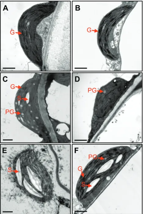
nye1-1 leaves (Fig. 2E). The ion leakage rate, an indicator of membrane disintegration and one of the important factors for determining the stay-green type, was then examined. Ion leakage rates of atg5 and nye1-1 leaves were lower than for wild-type leaves (Fig. 2F). The chloroplast structure of atg5 leaves was also examined. Before salt treatment, atg5 leaves contained normal shapes of chloroplast and grana thylakoid structures, similar to wild-type leaves (Fig. 3A, B). After 4 d of salt treatment, grana thylakoids were hardly found in the chloroplasts of wild-type leaves, and different types of degrading chloroplasts were found (Fig. 3C–E). By contrast,
atg5 leaves retained the grana thylakoids (Fig. 3F).
Taken together, our results indicate that atg5 acts as a func-tional stay-green type mutant under mild abiotic-stress condi-tions, because it retains a proper balance of photosystem proteins, photosynthetic efficiency and grana thylakoid structures.
Expression of SAGs in atg5 leaves under dark- or salt-stress-induced senescence conditions
To reveal the mechanism of conditional delayed senescence in
atg5 leaves under mild abiotic-stress conditions, expression
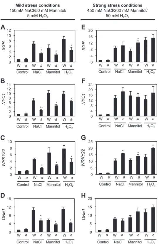
lev-els of senescence-associated genes (SAGs), including two senes-cence-induced TFs, WRKY22 (Zhou et al., 2011) and ORE1 (Kim et al., 2009), and two Chl catabolism-associated proteins,
SGR1/NYE1 (Park et al., 2007; Ren et al., 2007) and NYC1 (Kusaba et al., 2007), were measured under mild stress-induced senescence conditions. Expression levels of all four SAGs in wild-type leaves drastically increased after 3 d of mild stress condi-tions, such as 150 mM NaCl, 50 mM mannitol, and 5 mM H2O2 treatments. However, their expression levels were significantly down-regulated in atg5 leaves (Fig. 4A–D), indicating that the down-regulation of SAGs contributes to the functional stay-green phenotype of atg5 leaves under mild abiotic-stress con-ditions. Although the non-functional stay-green nye1-1 mutant also retained leaf greenness (Fig. 4), it was found that, under mild abiotic stresses, expression levels of WRKY22 and ORE1 in
nye1-1 leaves were almost the same as those of wild-type leaves
(see Supplementary Fig. S3 at JXB online). The expression lev-els of four SAGs were also checked under strong abiotic-stress conditions. Except for the slight up-regulation of WRKY22, the expression levels of the other three SAGs were not significantly different in atg5 leaves and wild-type leaves (Fig. 4E–H).
The stay-green phenotype in atg5 leaves under mild stress conditions could also be caused by defects of other intra-chloroplastic catabolic systems. The expression levels of genes encoding FtsH proteases (FtsH2 and FtsH6), Clp proteases (ClpP4 and ClpC1), and Deg proteases (DegP4 and DegP8) were also examined. Expression levels of these genes signifi-cantly increase under high light, cold, and heat-stress condi-tions (Sinvany-Villalobo et al., 2004), indicating that these chloroplastic proteases have major roles in protein degradation under several abiotic-stress conditions. Under mild salt stress (150 mM NaCl), the gene expression levels in atg5 leaves were almost the same as those in wild-type leaves (see Supplementary Fig. S4 at JXB online), indicating that these three catabolic
systems are not related to the functional stay-green phenotype of atg5 leaves under mild abiotic-stress conditions.
Taking these results together, it is possible that the down-regulation of several SAGs in atg5 leaves during mild abiotic-stress conditions is controlled by retrograde signalling between chloroplasts and the nucleus, which is often observed in many functional stay-green mutants (Sakuraba et al., 2012a).
Early leaf yellowing of atg5 is independent of Chl breakdown in LHCII by the SGR1–CCEs complex in chloroplasts
Chl degradation and autophagy (macroautophagy) affect the degradation of Chls and photosynthetic proteins in the
chloroplasts, a very late step in leaf senescence (Lim et al., 2007). However, atg5 and nye1-1 retain different levels of photosynthetic proteins under mild salt-stress conditions (Fig. 2). Among photosynthetic proteins, nye1-1 selectively retains LHCI and LHCII under salt stress (Fig. 2E), as the
nye1-1 mutant has impaired SGR1 function. SGR1 induces
Chl degradation in LHCII by recruiting Chl catabolic enzymes (CCEs) (Sakuraba et al., 2012b, 2013). However, most photosynthetic proteins are retained in atg5 leaves with a constant Chl a/b ratio (Fig. 2), indicating that the degrada-tion of photosynthetic proteins by the SGR1–CCE complex and macroautophagy may function independently.
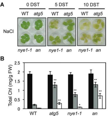
To examine this possibility, the phenotype of atg5 nye1-1 double mutants was investigated under mild salt stress (150 mM Fig. 2. Characterization of atg5 leaves under mild salt stress conditions. (A–F) Visible phenotypes (A), total Chl levels (B), Chl a/b ratios (C),
photosynthetic protein levels (D), Fv/Fm ratios (E), and ion leakage rates (F) of wild-type (WT), atg5, and nye1-1 leaves under the mild salt-stress
conditions. Detached leaves of 3-week-old WT, atg5, and nye1-1 plants were incubated in 3 mM MES buffer (pH 5.8) containing 150 mM NaCl for 3 d and 5 d (3 and 5 DST, days of salt stress). (B, C, E, and F) Black, grey, and white bars indicate 0, 3, and 5 DST, respectively. (D) Antibodies against PSII core (CP43 and D1), PSII antenna (Lhcb1, Lhcb2, Lhcb4, and Lhcb5), PSI antenna (Lhca1 and Lhca3), and PSI core (PsaA) were used. RbcL (Rubisco large subunit) was visualized by Coomassie Brilliant Blue (CBB) staining after immunoblot analysis. These experiments were repeated more than three times with similar results. DST: days of salt treatment. Student’s t-test (*P<0.05, **P<0.01). (This figure is available in colour at JXB online.)
NaCl). Although no differences were observed between atg5 single and atg5 nye1-1 double mutants until 5 d of salt treat-ment, the double mutant exhibited a stronger stay-green phe-notype at 10 d (Fig. 5A) with significantly greater retention of Chls (Fig. 5B), indicating that the two mutations show an addi-tive effect on abiotic-stress-induced leaf senescence. To examine the relationship of the two mutations in more detail, the phe-notype of the double mutants was checked under dark-induced senescence conditions. During dark-induced senescence, the
nye1-1 mutant shows a stay-green phenotype (Ren et al., 2007), but atg5 shows an early leaf yellowing phenotype (Thompson et al., 2005). It was found that, after 4 d of dark incubation, the double mutant exhibited an intermediate phenotype and Chl levels of the two single mutants (see Supplementary Fig. S5 at
JXB online). Collectively, these results indicate that Chl
degra-dation during senescence occurs by two independent processes, SGR1–CCE–LHCII interaction and macroautophagy.
Discussion
atg5 acts as a functional stay-green mutant under mild
abiotic-stress conditions
Defects in chloroplast destruction or senescence-promot-ing mechanisms can cause leaves to retain their green col-our during senescence, a phenomenon called ‘stay-green’ (Hörtensteiner, 2009). For example, the knockout mutants of CCEs exhibit a stay-green phenotype during dark-induced Fig. 3. Transmission electron microscopy of plastids in atg5 leaves under mild salt-stress conditions. (A, B) Chloroplasts in the mesophyll cells of 3-week-old wild-type (WT; A) and atg5 (B) leaves before salt (150 mM) treatment. (C–F) Chloroplasts in the mesophyll cells of WT (C, D, E) and atg5 (F) leaves after 4 DST. DST, days of salt treatment; G, grana thylakoid; PG, plastoglobule; S, starch. Scale bars=1 μm. (This figure is available in colour at JXB online.)
and natural senescence because of the impairment of Chl degradation (Schelbert et al., 2009; Horie et al., 2009). Because the degradation of chloroplast components involves autophagy (Ishida et al., 2008; Wada et al., 2009), it was expected that atg mutants would show a stay-green pheno-type under senescence-inducing conditions. However, under
strong stress conditions, the atg mutants exhibit a phenotype of accelerated yellowing and cell death.
In this study, this apparent inconsistency has been addressed by identifying the conditions under which atg mutants show a stay-green phenotype. Under mild abiotic-stress conditions, such as 150 mM NaCl, 5 mM H2O2, or 50 mM mannitol, atg5 Fig. 4. Altered expression of SAGs in atg5 leaves under mild and strong abiotic-stress conditions. First-strand cDNAs were prepared from total RNA extracted from 3-week-old rosette leaves of WT and atg5 plants before (control) and after 3 d of mild abiotic-stress treatments (A–D) and strong abiotic-stress treatments (E–H). By RT-qPCR analysis, relative expression levels of SGR1/NYE1 (A, E), NYC1 (B, F), WRKY22 (C, G), and ORE1 (D, H) were obtained by normalizing to the mRNA levels of GAPDH. Mean and SD values were obtained from more than three biological replicates. These experiments were replicated at least twice with similar results. Student’s t-test (*P<0.05, **P<0.01).
leaves exhibit a stay-green phenotype (Fig. 1A), and show little cell death (Fig. 1B). Under strong abiotic-stress condi-tions, however, atg5 leaves turn yellow much faster than WT leaves and show extensive cell death (Fig. 1). This rapid leaf yellowing phenotype is consistent with previous studies on the phenotype of atg plants under abiotic-stress conditions (Xiong et al., 2007; Liu et al., 2009; Zhou et al., 2013). It is speculated that the difference in atg phenotype between strong- and weak-stress conditions may reflect the impor-tance of autophagy in adapting to severe-stress conditions. Generally, plants need to change the balance of the proteome drastically under abiotic-stress conditions, for adaptation to these extreme environments. Because atg mutants cannot properly control their proteome balance, they cannot adapt to strong abiotic-stress conditions and thus exhibit extensive cell death. However, adaptation to mild-stress conditions does not require drastic changes in the proteome balance. Thus, atg5 leaves exhibit little cell death, which leads to the stay-green phenotype (Fig. 6).
Autophagy is involved in chloroplast degradation, a downstream step in leaf senescence pathways. It was there-fore expected that the stay-green phenotype of atg mutants would resemble the phenotypes of the cosmetic stay-green mutants of SGR1 and CCEs (Kusaba et al., 2007; Morita et al., 2009). However, it was found that atg5 conditionally acts as a functional, not a cosmetic stay-green type mutant. Under mild salt stress (150 mM NaCl) conditions, atg5 leaves stayed green with the proper balance of photosynthetic
proteins (Fig. 2D), high photosynthetic capacity (Fig. 2E), and well-retained grana thylakoids (Fig. 3). Also, atg5 leaves showed lower expression levels of several SAGs under mild abiotic-stress conditions (Fig. 4), indicating that atg5 leaves during mild abiotic-stress conditions show defects in both autophagy-dependent senescence pathways and other senes-cence pathways. One possibility is that the retained chloro-plast proteins in atg5 leaves may induce retrograde signalling from the chloroplasts to nucleus, leading to the altered expres-sion of the SAGs. Recently, chloroplast homeostasis has been implicated as an important factor in leaf senescence; for example, tobacco plants with reduced NADH dehydro-genase activity exhibited delayed senescence without signifi-cant alteration of their growth rate (Zapata et al., 2005). In addition, Arabidopsis plants over-expressing chlorophyllide a oxygenase (CAO) showed changes in the Chl pigment com-position of the photosynthetic apparatus and also showed a functional stay-green phenotype with wide changes in SAG expression (Sakuraba et al., 2012a). These results indicate that chloroplasts have an important role in regulating nuclear gene expression during leaf senescence.
Together, these data indicate that the multiple effects of two different pathways probably cause the functional stay-green phenotype of atg5 leaves under mild abiotic-stress conditions. Because the macroautophagy pathway does not function in
atg5 leaves, the degradation of chloroplasts and chloroplastic
proteins itself is impaired. Simultaneously, the retention of chloroplast proteins induces chloroplast–nucleus retrograde signalling that affects the regulation of SAGs. Some enig-mas remain for this hypothesis; for instance, the chloroplast component(s) that mediate the chloroplast–nucleus retro-grade signalling during leaf senescence remain to be identi-fied. Further physiological and biochemical analyses of the functional stay-green phenotype of atg mutants are essential for revealing the functions of plant autophagy.
Degradation of Chls and photosynthetic proteins requires both autophagy and intra-chloroplastic catabolic systems during leaf senescence
SGR1/NYE1 and CCEs form a dynamic protein complex for LHCII disassembly and Chl degradation (Sakuraba et al.,
2012a, 2013). However, the SGR1–CCE complex does not interact with other photosynthetic proteins, such as LHCI, the PSII core complex, or the PSI core complex (Sakuraba
Fig. 5. Characterization of atg5 nye1-1 double mutant under mild salt-stress conditions. (A, B) Visible phenotypes (A) and total Chl levels (B) of detached leaves from wild-type (WT), atg5, nye1-1, and atg5 nye1-1 (an) plants during the mild salt stress. Detached leaves from 3-week-old plants were incubated abaxial side-up on 3 mM MES (pH 5.8) buffer containing 150 mM NaCl for 5 d and 10 d. Similar results were obtained from three independent experiments. DST, days of salt treatment. Student’s t-test (*P<0.05, **P<0.01). (This figure is available in colour at JXB online.)
Fig. 6. Leaf phenotypes of atg5 mutants depending on the strength of abiotic stresses. Under strong abiotic-stress conditions, atg5 leaves show an accelerated cell death phenotype; under mild abiotic-stress conditions, atg5 leaves show little or no cell death, which leads to a stay-green phenotype. (This figure is available in colour at JXB online.)
et al., 2012a). Consistent with this LHCII-specific interaction, LHCII proteins were dominantly retained while other photo-system proteins were normally degraded in nye1-1 (Fig. 2D) and a CCE mutant nyc1-1 (Horie et al., 2009). Thus, degrada-tion mechanisms of other photosynthetic proteins and Chls still remain enigmatic.
At least in part, our finding of the functional stay-green phenotype in atg5 leaves under mild salt-stress condition pro-vides an important clue to solve this enigma. The levels of sev-eral photosynthetic proteins retained in atg5 and nye1-1 leaves under mild salt stress were compared. Although both atg5 and nye1-1 leaves exhibited a stay-green phenotype (Fig. 2A) with highly retained Chl levels (Fig. 2B), only LHCII proteins were predominantly retained in nye1-1 leaves, whereas all pho-tosynthetic proteins were substantially retained in atg5 leaves (Fig. 2D). Recent reports showed that one of the autophagy pathways, the so-called chlorophagy pathway, functions in the transportation of Chls and photosystem proteins from the chloroplasts to the vacuole for their degradation (Wada et al., 2009). Considering the low selectivity of proteolysis in autophagy, the chlorophagy pathway non-selectively trans-ported all photosystem proteins from the chloroplasts to the vacuole. Although RCBs also act in chloroplastic autophagy, RCBs do not show Chl fluorescence (Ishida et al., 2008). SAVs, another extra-chloroplastic catabolic system, contain Chl a, but not photosystem proteins. Thus, so far, chlorophagy is the only identified extra-chloroplastic degradation system for photosystem proteins. By contrast with chlorophagy, the SGR–CCE complex seems to concentrate on the destabiliza-tion of LHCII and Chls. LHCII, especially the three major, abundant subunits (Lhcb1, Lhcb2, and Lhcb3), forms aggre-gates because of its abundance (Ruban et al., 2012). In this sense, if specific degradation systems for LHCII exist, it is natural that the SGR1–CCE complex would be one of them.
In this study, it was also found that atg5 nye1-1 double mutants exhibited a very strong stay-green phenotype under mild salt-stress conditions, a seemingly additive phenotype (Fig. 5). This result indicates that the autophagy- and SGR1-dependent degradation pathways function inSGR1-dependently. Reflecting the relationship of these two pathways, the combina-tion of intra- and extra-chloroplastic catabolic pathways acts to degrade photosystem proteins. Another catabolic system may also function in the degradation of photosystem proteins during leaf senescence. For instance, genes encoding several members of the FtsH, Clp, and Deg protease families were sig-nificantly up-regulated in senescing leaves (Gepstein et al., 2003;
Andersson et al., 2004) strongly indicating that these chloro-plastic proteases have important roles in the degradation of chloroplast proteins during leaf senescence as well as abiotic-stress conditions. Indeed, an FtsH protease affects the turno-ver of D1 protein, one of the core subunits of photosystem II, under abiotic-stress conditions (Bailey et al., 2002; Kato et al., 2009). Thus, it is possible that these proteases are involved in the degradation of photosystem proteins during senescence.
Further biochemical analyses of chloroplasts and vacuole fractions during senescence will help us to understand the complete picture of the degradation mechanisms of photo-synthetic proteins and their photophoto-synthetic pigments.
Supplementary data
Supplementary data can be found at JXB online.
Supplementary Fig. S1. Phenotype of wild-type (WT) and
atg7 leaves under mild abiotic-stress conditions.
Supplementary Fig. S2. Phenotype of wild-type (WT) and
atg5 plants grown on phytoagar plates containing NaCl.
Supplementary Fig. S3. Altered expression of SAGs in
nye1-1 leaves under mild abiotic-stress conditions.
Supplementary Fig. S4. Expression analysis of chloro-plastic protease genes in atg5 leaves under mild salt -stress conditions.
Supplementary Fig. S5. Phenotype (A) and total Chl level (B) of atg5 nye1-1 double mutants during dark-induced senescence.
Supplementary Table S1. Primers used for qPCR in this study.
Acknowledgements
We thank Dr Benke Kuai for nye1-1 seeds. This work was supported by the Strategic Korean–Swiss Cooperative Program, the National Research Foundation of Korea (NRF) grant funded by the Ministry of Education, Science, and Technology (MEST) (No. 2013K1A3A1A14055213).
Sequence data from this article can be found in the Arabidopsis Genome Initiative (AGI) or GenBank/EMBL databases under the following acces-sion numbers: ATG5, At5g17290; ATG7, At5g45900; GAPDH, At1g16300; NYC1, At4g13250; ORE1, At5g39610; SGR1/NYE1, At4g22920; WRKY22, At4g01250; FtsH2, At2G20950: FtsH6, At5g15250; ClpP4, At5g45390; ClpC1, At5g50920; DegP4, At1g65640; DegP8, At5g39830.
YS and N-CP designed the research; YS and S-H.L performed the research; Y-SK assisted with the research; YS, OKP, SH, and N-CP analysed the data; YS and N-CP wrote the article. The authors declare that they have no conflict of interest.
References
Andersson A, Keskitalo J, Sjodin A, et al. 2004. A transcriptional timetable of autumn senescence. Genome Biology 5, R24.
Bailey S, Thompson E, Nixon PJ, Horton P, Mullineaux CW,
Robinson C, Mann NH. 2002. A critical role for the Var2 FtsH homologue of Arabidopsis thaliana in the photosystem II repair cycle in vivo. Journal of Biological Chemistry 277, 2006–2011.
Balazadeh S, Riano-Pachon DM, Mueller-Roeber B. 2008. Transcription factors regulating leaf senescence in Arabidopsis thaliana. Plant Biology 10, 63–75.
Bassham DC, Laporte M, Marty F, Moriyasu Y, Ohsumi Y, Olsen LJ, Yoshimoto K. 2006. Autophagy in development and stress responses of plants. Autophagy 2, 2–11.
Chiba A, Ishida H, Nishizawa NK, Makino A, Mae T. 2003. Exclusion of Rubisco from chloroplasts by the vesicle during natural senescence of wheat leaves. Plant and Cell Physiology 44, 914–921.
Feller U, Anders I, Mae T. 2008. Rubiscolytics: fate of Rubisco after its enzymatic function in a cell is terminated. Journal of Experimental Botany 59, 1615–1624.
Gepstein S, Sabehi G, Carp MJ, Hajouj T, Nesher MF, Yariv I, Dor C, Bassani M. 2003. Large-scale identification of leaf senescence-associated genes. The Plant Journal 36, 629–642.
Guo H, Ecker JR. 2004. The ethylene signaling pathway: new insights. Current Opinion in Plant Biology 7, 40–49.
Horie Y, Ito H, Kusaba M, Tanaka R, Tanaka A. 2009. Participation of chlorophyll b reductase in the initial step of the degradation of light-harvesting chlorophyll a/b–protein complexes in Arabidopsis. Journal of Biological Chemistry 284, 17449–17456.
Hörtensteiner S. 2009. Stay-green regulates chlorophyll and chlorophyll-binding protein degradation during senescence. Trends in Plant Science 14, 155–162.
Hörtensteiner S. 2013. Update on the biochemistry of chlorophyll breakdown. Plant Molecular Biology 82, 505–517.
Hörtensteiner S, Krautler B. 2011. Chlorophyll breakdown in higher plants. Biochimica et Biophysica Acta 1807, 977–988.
Inada N, Sakai A, Kuroiwa H, Kuroiwa T. 1998. Three-dimensional analysis of the senescence program in rice (Oryza sativa L.) coleoptile: investigations by fluorescence microscopy and electron microscopy. Planta 206, 585–597.
Ishida H, Yoshimoto K, Izumi M, Reisen D, Yano Y, Makino A, Ohsumi Y, Hanson MR, Mae T. 2008. Mobilization of rubisco and stroma-localized fluorescent proteins of chloroplasts to the vacuole by an ATG gene-dependent autophagic process. Plant Physiology 148, 142–155.
Kato Y, Sakamoto W. 2009. Protein quality control in chloroplasts: a current model of D1 protein degradation in the photosystem II repair cycle. Journal of Biochemstry 146, 463–469.
Kim JH, Woo HR, Kim J, Lim PO, Lee IC, Choi SH, Hwang D, Nam HG. 2009. Trifurcate feed-forward regulation of age-dependent cell death involving miR164 in Arabidopsis. Science 323, 1053–1057.
Klionsky DJ. 2004. Cell biology: regulated self-cannibalism. Nature 431, 31–32.
Klionsky DJ, Ohsumi Y. 1999. Vacuolar import of proteins and organelles from the cytoplasm. Annual Review of Cell and Developmental Biology 15, 1–32.
Koch E, Slusarenko A. 1990. Arabidopsis is susceptible to infection by a downy mildew fungus. The Plant Cell 2, 437–445.
Kusaba M, Ito H, Morita R, et al. 2007. Rice NON-YELLOW COLORING1 is involved in light-harvesting complex II and grana degradation during leaf senescence. The Plant Cell 19, 1362–1375. Lim PO, Kim HJ, Nam HG. 2007. Leaf senescence. Annual Review of Plant Biology 58, 115–136.
Liu YM, Xiong Y, Bassham DC. 2009. Autophagy is required for tolerance of drought and salt stress in plants. Autophagy 5, 954–963. Makino A, Osmond B. 1991. Effects of nitrogen nutrition on nitrogen partitioning between chloroplasts and mitochondria in pea and wheat. Plant Physiology 96, 355–362.
Martinez DE, Costa ML, Guiamet JJ. 2008. Senescence-associated degradation of chloroplast proteins inside and outside the organelle. Plant Biology (Stuttgart) 10, Supplement 1, 15–22.
Meijer WH, van der Klei IJ, Veenhuis M, Kiel JA. 2007. ATG genes involved in non-selective autophagy are conserved from yeast to man, but the selective Cvt and pexophagy pathways also require organism-specific genes. Autophagy 3, 106–116.
Morita R, Sato Y, Masuda Y, Nishimura M, Kusaba M. 2009. Defect in non-yellow coloring 3, an alpha/beta hydrolase-fold family protein, causes a stay-green phenotype during leaf senescence in rice. The Plant Journal 59, 940–952.
Nakatogawa H, Suzuki K, Kamada Y, Ohsumi Y. 2009. Dynamics and diversity in autophagy mechanisms: lessons from yeast. Nature Reviews Molecular Cell Biology 10, 458–467.
Oberhuber M, Berghold J, Breuker K, Hörtensteiner S, Krautler B. 2003. Breakdown of chlorophyll: a non-enzymatic reaction accounts for the formation of the colorless “non-fluorescent” chlorophyll catabolites. Proceedings of the National Academy of Sciences, USA 100, 6910–6915. Ono Y, Wada S, Izumi M, Makino A, Ishida H. 2012. Evidence for contribution of autophagy to Rubisco degradation during leaf senescence in Arabidopsis thaliana. Plant Cell and Environment 36, 1147–1159. Park SY, Yu JW, Park JS, et al. 2007. The senescence-induced staygreen protein regulates chlorophyll degradation. The Plant Cell 19, 1649–1664.
Porra RJ, Thompson WA, Kriedemann PE. 1989. Determination of accurate extinction coefficients and simultaneous-equations for assaying chlorophyll a and chlorophyll b extracted with four different solvents: verification of the concentration of chlorophyll standards by atomic-absorption spectroscopy. Biochimica et Biophysica Acta 975, 384–394. Ren GD, An K, Liao Y, Zhou X, Cao YJ, Zhao HF, Ge XC, Kuai BK. 2007. Identification of a novel chloroplast protein AtNYE1 regulating chlorophyll degradation during leaf senescence in arabidopsis. Plant Physiology 144, 1429–1441.
Reumann S, Voitsekhovskaja O, Lillo C. 2010. From signal
transduction to autophagy of plant cell organelles: lessons from yeast and mammals and plant-specific features. Protoplasma 247, 233–256. Robinson WD, Carson I, Ying S, Ellis K, Plaxton WC. 2012. Eliminating the purple acid phosphatase AtPAP26 in Arabidopsis thaliana delays leaf senescence and impairs phosphorus remobilization. New Phytologist 196, 1024–1029.
Ruban AV, Johnson MP, Duffy CDP. 2012. The photoprotective molecular switch in the photosystem II antenna. Biochimica et Biophysica Acta—Bioenergetics 1817, 167–181.
Sakuraba Y, Balazadeh S, Tanaka R, Mueller-Roeber B, Tanaka A. 2012a. Overproduction of Chl b retards senescence through transcriptional reprogramming in Arabidopsis. Plant and Cell Physiology 53, 505–517.
Sakuraba Y, Kim YS, Yoo SC, Hörtensteiner S, Paek NC. 2013. 7-Hydroxymethyl chlorophyll a reductase functions in metabolic channeling of chlorophyll breakdown intermediates during leaf senescence. Biochemical and Biophysical Research Communications 430, 32–37.
Sakuraba Y, Schelbert S, Park SY, Han SH, Lee BD, Andres CB, Kessler F, Hörtensteiner S, Paek NC. 2012b. STAY-GREEN and chlorophyll catabolic enzymes interact at light-harvesting complex II for chlorophyll detoxification during leaf senescence in Arabidopsis. The Plant Cell 24, 507–518.
Sakuraba Y, Yokono M, Akimoto S, Tanaka R, Tanaka A. 2010. Deregulated chlorophyll b synthesis reduces the energy transfer rate between photosynthetic pigments and induces photodamage in Arabidopsis thaliana. Plant and Cell Physiology 51, 1055–1065. Sato Y, Morita R, Nishimura M, Yamaguchi H, Kusaba M. 2007. Mendel’s green cotyledon gene encodes a positive regulator of the chlorophyll-degrading pathway. Proceedings of the National Academy of Sciences, USA 104, 14169–14174.
Schelbert S, Aubry S, Burla B, Agne B, Kessler F, Krupinska K, Hörtensteiner S. 2009. Pheophytin pheophorbide hydrolase (pheophytinase) is involved in chlorophyll breakdown during leaf senescence in Arabidopsis. The Plant Cell 21, 767–785.
Sinvany-Villalobo G, Davydov O, Ben-Ari G, Zaltsman A, Raskind A, Adam Z. 2004. Expression in multigene families. Analysis of chloroplast and mitochondrial proteases. Plant Physiology 135, 1336–1345. Thomas H, Howarth CJ. 2000. Five ways to stay green. Journal of Experimental Botany 51, 329–337.
Thompson AR, Doelling JH, Suttangkakul A, Vierstra RD. 2005. Autophagic nutrient recycling in Arabidopsis directed by the ATG8 and ATG12 conjugation pathways. Plant Physiology 138, 2097–2110.
Wada S, Ishida H, Izumi M, Yoshimoto K, Ohsumi Y, Mae T, Makino A. 2009. Autophagy plays a role in chloroplast degradation during senescence in individually darkened leaves. Plant Physiology 149, 885–893.
Wittenbach VA. 1978. Breakdown of ribulose bisphosphate carboxylase and change in proteolytic activity during dark-induced senescence of wheat seedlings. Plant Physiology 62, 604–608.
Wu NN, Huang X, Yang XQ, Guo J, Zheng EL, Yin SW, Zhu JH, Qi JR, He XT, Zhang JB. 2012. Stabilization of soybean oil body emulsions using i-carrageenan: effects of salt, thermal treatment and freeze–thaw cycling. Food Hydrocolloids 28, 110–120.
Xiong Y, Contento AL, Nguyen PQ, Bassham DC. 2007. Degradation of oxidized proteins by autophagy during oxidative stress in Arabidopsis. Plant Physiology 143, 291–299.
Zapata JM, Guera A, Esteban-Carrasco A, Martin M, Sabater B. 2005. Chloroplasts regulate leaf senescence: delayed senescence in transgenic ndhF-defective tobacco. Cell Death and Differentiation 12, 1277–1284.
Zelisko A, Garcia-Lorenzo M, Jackowski G, Jansson S, Funk C. 2005. AtFtsH6 is involved in the degradation of the light-harvesting complex II during high-light acclimation and senescence. Proceedings of the National Academy of Sciences, USA 102, 13699–13704.
Zhou J, Wang J, Cheng Y, Chi YJ, Fan BF, Yu JQ, Chen ZX. 2013. NBR1-mediated selective autophagy targets insoluble ubiquitinated protein aggregates in plant stress responses. Plos Genetics 9.
Zhou X, Jiang YJ, Yu DQ. 2011. WRKY22 transcription factor mediates dark-induced leaf senescence in Arabidopsis. Molecules and Cells 31, 303–313.