. . . .
. . . .
Extracellular vesicles from human cardiac
progenitor cells inhibit cardiomyocyte apoptosis and
improve cardiac function after myocardial infarction
Lucio Barile
1*
†, Vincenzo Lionetti
2†, Elisabetta Cervio
1, Marco Matteucci
2,
Mihaela Gherghiceanu
3, Laurentiu M. Popescu
3, Tiziano Torre
1, Francesco Siclari
1,
Tiziano Moccetti
1, and Giuseppe Vassalli
1,4*
1
Laboratory of Molecular and Cellular Cardiology, Departments of Cardiology and Heart Surgery, Fondazione Cardiocentro Ticino, Via Tesserete 48, CH-6900 Lugano, Switzerland;2
Laboratory
of Medical Science, Institute of Life Sciences, Scuola Superiore Sant’Anna, Pisa, Italy;3
‘Victor’ Babes National Institute of Pathology, Bucharest, Romania; and4
Department of Cardiology, Centre Hospitalier Universitaire Vaudois (CHUV), Lausanne, Switzerland
Received 14 December 2013; revised 16 June 2014; accepted 8 July 2014; online publish-ahead-of-print 11 July 2014 Time for primary review: 27 days
Aims Recent evidence suggests that cardiac progenitor cells (CPCs) may improve cardiac function after injury. The underlying mechanisms are indirect, but their mediators remain unidentified. Exosomes and other secreted membrane vesicles, hereafter collectively referred to as extracellular vesicles (EVs), act as paracrine signalling mediators. Here, we report that EVs secreted by human CPCs are crucial cardioprotective agents.
Methods and results
CPCs were derived from atrial appendage explants from patients who underwent heart valve surgery. CPC-conditioned medium (CM) inhibited apoptosis in mouse HL-1 cardiomyocytic cells, while enhancing tube formation in human umbil-ical vein endothelial cells. These effects were abrogated by depleting CM of EVs. They were reproduced by EVs secreted by CPCs, but not by those secreted by human dermal fibroblasts. Transmission electron microscopy and nanoparticle tracking analysis showed most EVs to be 30 – 90 nm in diameter, the size of exosomes, although smaller and larger vesicles were also present. MicroRNAs most highly enriched in EVs secreted by CPCs compared with fibroblasts included miR-210, miR-132, and miR-146a-3p. miR-210 down-regulated its known targets, ephrin A3 and PTP1b, inhibiting apoptosis in cardiomyocytic cells. miR-132 down-regulated its target, RasGAP-p120, enhancing tube formation in endothelial cells. Infarcted hearts injected with EVs from CPCs, but not from fibroblasts, exhibited less cardiomyocyte apoptosis, enhanced angiogenesis, and improved LV ejection fraction (0.8 + 6.8 vs. 221.3 + 4.5%; P , 0.05) compared with those injected with control medium.
Conclusion EVs are the active component of the paracrine secretion by human CPCs. As a cell-free approach, EVs could circumvent many of the limitations of cell transplantation.
-Keywords Extracellular vesicles † Exosomes † Cardiac progenitor cells † MicroRNA † Cardioprotection † Apoptosis † Angiogenesis
1. Introduction
Ischaemic heart disease and ensuing heart failure are leading causes of morbidity and mortality worldwide. An extensively investigated approach for ischaemic heart disease is cell transplantation. Multiple cell sources including bone marrow,1 adipose tissue,2 skeletal
myoblasts,3and cardiac progenitor cells (CPCs)4have been evaluated
in both animal models and humans. Clinical trials of autologous BM cell transplantation in patients after acute myocardial infarction (MI) have provided mixed results, with an overall modest improvement in cardiac function.5 Intramyocardial injection of CPCs isolated from adult hearts improved cardiac function in animal models of MI.6–8
Although both direct cell differentiation and indirect mechanisms, such as secreted growth factors and cytokines, have been implicated
*Corresponding author. Tel:+41 91 805 3359; fax: +41 91 805 3323, Email: lucio.barile@cardiocentro.org (L.B.), giuseppe.vassalli@cardiocentro.org (G.V.)
†L.B. and V.L. contributed equally to this work.
in the therapeutic benefit, accumulating evidence suggests predominant roles of the paracrine secretion by CPCs.8This concept is supported by the observation that CPC-conditioned medium (CM) protects cardiomyocytes against stress-induced apoptosis, while stimulating tube formation in endothelial cells in vitro.8–11The CADUCEUS trial of cardiosphere-derived cells has shown that some degree of cardiac regeneration in injured hearts may be possible. The mechanism of benefit was thought to be indirect.4
A key component of paracrine secretion in many cell types are extra-cellular vesicles (EVs), particularly its exosomal fraction. Exosomes are 40– 90 nm-sized particles stored intracellularly in endosomal compart-ments, which are secreted when these structures fuse with the plasma membrane.12,13Conventional surface markers of exosomes include the tetraspanin family members, CD9, CD63, and CD81, among others. Exo-somes carry proteins, lipids, and nucleic acids including DNA, mRNAs, and microRNAs (miRNAs), which they can shuttle to recipient cells, even at a distance. miRNAs are 18–25 nucleotide, non-coding RNAs that regulate gene expression through post-transcriptional repression. Transfer of mRNAs and miRNAs through exosomes has emerged as a crucial mechanism of genetic exchange between cells.14,15Non-exosomal EVs arise by distinct mechanisms including direct budding from the plasma membrane. While non-exosomal EVs are typically larger than exosomes, exosome purification remains a challenging task. As a result, exosomal preparations used in many studies showed limited purity.16
Exosomes were identified as the active component of the proangio-genic paracrine activity of bone marrow CD34+stem cells.17Moreover, exosomes secreted by mesenchymal stem cells (MSCs) and mouse CPCs mitigated tissue damage and ventricular remodelling in animal models of myocardial ischaemia and reperfusion injury.18,19However, exosomes secreted by human CPCs have not been characterized yet. Using transmission electron microscopy, we recently provided ultra-structural evidence of exosomes and EVs secretion by human CPCs.20 Here, we report that EVs are the active component of the paracrine se-cretion by human CPCs. Transmission electron microscopy and nano-particle tracking analysis showed most EV secretion by CPCs to be 30 – 90 nm in diameter, suggesting they were exosomes, although smaller and larger vesicles were also present. Moreover, the purified particles expressed conventional exosomal markers including CD63. These results suggest that exosomes were the predominant component of the particles purified from CM-CPC. To stress their heterogeneity, however, we hereafter refer to them as EVs. These EVs mitigated apop-tosis triggered by serum deprivation in the neonatal mouse HL-1 cardi-omyocytic cell line, while stimulating tube formation in human umbilical vein endothelial cells (HUVECs), an indicator of angiogenic activity. In vivo, injection of EVs secreted by CPCs into the infarct border zone reduced cardiomyocyte apoptosis and scar, increased both viable mass in the infarct area and blood vessel density, and prevented the early impairment of ventricular function in a rat model of acute MI. EVs secreted by normal human dermal fibroblasts (NHDFs) lacked these beneficial effects, supporting the concept that functional activities of EVs depend on the parent cell.17A comparison of the miRNA tran-scriptional profile of EVs secreted by CPCs with that of EVs secreted by NHDFs identified several miRNAs as being particularly enriched in the former, including miR-210, miR-132, and miR-146a-3p. Because car-dioprotective roles of miR-210 and a role for miR-132 in vascular re-modelling have been reported,21,22 we focused on the functional activities of these miRNAs. Both miR-210 and miR-132 protected HL-1 cardiomyocytic cells against apoptosis. In addition, miR-132 pro-moted the formation of endothelial tube. These results suggest that
EVs secreted by CPCs may impart cardiac protection and the underlying mechanism may involve miRNAs.
2. Methods
2.1 Cell sources
Human right atrial appendage specimens were obtained from patients (n ¼ 22) who underwent surgical repair of heart valve disease and had no concomitant coronary artery disease. The study was approved by the local Ethical Committee and performed according to the Declaration of Helsinki. All patients gave written informed consent to the collection of atrial speci-men. Using the primary ex vivo tissue culture technique,6–8the cellular
out-growth of atrial explants was collected within 14 days and seeded into fibronectin-coated 25 mL flasks. EVs were purified from CPCs after two passages in culture. NHDFs were purchased from Life Technologies.
2.2 EV purification methods
Medium conditioned by CPCs (CM-CPC) or NHDFs was obtained by cul-turing cells in basal medium (BM), a 1 : 1 mix of Iscove’s Modified Dulbecco’s Media and Dulbecco’s Modified Eagle’s Medium (Gibco) supplemented with 1% human serum albumin, for 48 h, followed by centrifugation at 3000 g for 15 min, filtration through a 0.2 mm membrane (BD Biosciences), and pro-cessing using one of the following methods: (i) ExoQuickTMprecipitation solution (System Biosciences; SBI); (ii) ultracentrifugation at 100 000 g for 90 min; and (iii) column precipitation using Exo-spinTMkits (Cell Guidance Systems, Cambridge, UK). The EV pellet was re-suspended in 100 – 500 mL of phosphate-buffered saline (PBS; pH 7.4) and stored at 2808C. Nanoparticle tracking analysis was performed using NanoSight LM10 (Malvern Instruments, Worcestershire, UK). CD63 and CD81 protein levels in EVs were determined by western blotting. Protein lysate was obtained by re-suspending EV pellet in 100 mL of RIPA buffer (Sigma). Antibodies were purchased from SBI. Total proteins were measured using BCA kits (Thermo Fisher Scientific).
2.3 Transmission electron microscopy
The ultrastructure of EVs was analysed using transmission electron micros-copy, as described previously.23Briefly, re-suspended EV pellet (3 mL) was fixed with 2.5% glutaraldehyde, post-fixed in buffered 1% OsO4with 1.5%
K4Fe(CN)6, embedded in 1% agar, and processed according to the standard
Epon812 embedding procedure. EVs were visualized on thin sections (60 nm) using a Morgagni 268 transmission electron microscope (FEI Company, The Netherlands) at 80 kV.
2.4 Flow cytometry
Surface markers expressed on CPCs and EVs were analysed by flow cytome-try. Microbeads coated with anti-CD63 antibody (Life Technologies) were used in EV analyses. Phycoerythrin (PE)-conjugated antibodies against surface markers expressed on CPCs or MSCs (CD90, CD105, CD140, CD146, CD166, CD172, and NG2 proteoglycan; all from Beckman Coulter) or conventional exosomal markers (CD9, CD63, and CD81; all from BD Biosciences) were used. Beads incubated with an antibody in the absence of EVs served as a control. To assess dynamic trafficking of CD63 to exosomes, CPCs were transfected with pCT-CD63-GFP vector (Cyto-Tracer; SBI) expressing a CD63-GFP fusion, using lipofectamine. Analyses were performed on a MACS-Quant flow cytometer (Miltenyi Biotec).
2.5 EV uptake by HL-1 cardiomyocytic cells
To assess EV uptake by HL-1 cardiomyocytic cells, EVs from CPCs were la-belled with DiI. Excess dye was removed by precipitation of EV. Cells cul-tured in complete Claycomb medium24(Sigma), hereafter referred to as growth medium (GM)-FBS, were incubated with DiI-labelled EVs. After 12 h, cells were imaged using a Nikon C2 Plus confocal microscope. RNAextraction, reverse transcription, and real-time PCR protocols were used as described in Supplementary material online, to determine intracellular concentrations of miR-210 and miR-132 in HL-1 cells exposed to EV-CPC. In the time-course experiment, 100 mg of EV-CPC total protein were used (n ¼ 3 experiments). Dose – response study measurements were performed at 48 h (n ¼ 3 experiments).
2.6 In vitro apoptosis assay
To induce apoptosis in the mouse HL-1 cardiomyocytic cell line, GM-FBS was replaced by BM for 12 h. After 12 h, medium was supplemented with CM-CPC, EV-depleted CM-CPC (DCM), or DCM reconstituted with the previously removed EV fraction (DCM+ EV) for 36 h. Cells kept in BM for 48 h or changed to GM-FBS after 12 h served as controls. A pooled sample of purified EVs secreted by CPCs (EV-CPC; n ¼ 4 patients) was com-pared with EVs secreted by NHDF (EV-F). Viable and dead cells were stained using double staining kits (Dojindo EU GmbH, Germany). Activation of apoptotic pathways was detected using activated caspase-3/7 staining kits (Life Technologies).
2.7 In vitro angiogenesis assay
To assess the angiogenic potency of CM-CPC, EV-CPC, and EV-F, HUVECs were seeded at 3× 10⁴cells/cm2density in Matrigel (BD Biosciences). An
endothelial cell medium (Medium 200; Gibco) supplemented with 10% FBS (ECM-FBS) served as a positive control. Images were acquired 5 h later, and tube length was measured using the ImageJ software (National Institutes of Health, USA).
2.8 miRNA analyses
Total RNA was isolated from EV-CPC and EV-F using Quanti-miR kits (SBI). Real-time PCR was performed with Advanced SYBR Green kits (Bio-Rad) using a multistep approach. In a first step, 384 miRNAs were measured in total RNA from a pooled EV-CPC sample (n ¼ 4 patients) using synthetic ‘spike-in’ miRNA sequences, provided with the kits for normalization. Fifty-two miRNAs were found to be enriched more than six-fold compared with ‘spike-in’ miRNA sequences. In the following step, these miRNAs were directly compared with EVs from CPCs and those from NHDFs. Four-teen miRNAs were found to be particularly enriched in EVs from CPCs. These miRNAs were then measured in three pooled EV-CPC samples (n ¼ 4 patients each). Based on the results of this analysis, 7 of 14 miRNAs were then measured in EV-CPC samples from individual patients (n ¼ 6). Data were normalized for the geometric mean of miR-U6 and RNU-6B values and shown as 22DDCt values. Among the most highly enriched miRNAs in EVs secreted by CPCs, miR-210 and miR-132 were chosen for functional studies, owing to their proposed roles in cardioprotection and vascular remodelling.21,22 Cells were transfected with miR-210 and miR-132 mimics, or siRNAs specific for these miRNAs, using RNAiMAXTM
(Life Technologies). Expression of the miR-210 targets, ephrin A3, PTP1b, and FLASH/CASP8AP2,21,25,26 as well as those of miR-132 target, RasGTPase-activating protein (RasGap)-p120,22was measured by western blot using antibodies from Santa Cruz Biotech.
2.9 In vivo myocardial infarction
Experimental protocols were approved by the Animal Care Committee of the Italian Ministry of Health, in accordance with Italian (DL-116, 27 January 1992) and international laws and policies (European Economic Com-munity Council Directive 86/609, OJL 358, 12 December 1987). Animal studies were performed as previously described.27Briefly, acute MI was induced in healthy male Wistar rats (250 – 300 g body weight), anaesthetized with a cocktail of tiletamine and zolazepam (Zoletil 100, Virbac, France; dosage 40 mg/kg IP), intubated, and ventilated. The left anterior descending coronary artery was permanently ligated near its origin with a 6-0 Prolene suture. After 60 min, the viable myocardium bordering the LV infarct zone was injected at three different sites with a total of 150 mL of one of the
following solutions: PBS (n ¼ 9), 30 mg of EV-CPC (n ¼ 9), 300 mg of EV-CPC (n ¼ 10), and 300 mg of Exo-F (n ¼ 4). The sham-operated group included nine animals. At Day 7, rats were anaesthetized with Zoletil 100 (40 mg/kg) and sacrificed with iv KCl (1 – 2 mEq/kg) to induce diastolic arrest of cardiac activity.
2.10 Echocardiography
Echocardiography was performed in rats sedated by IP tiletamine (29 mg/kg) plus xylazine (4.3 mg/kg). Transthoracic echocardiography was performed on Day 2 (baseline) and Day 7 post-MI using the MyLab 30 echocardiography system (Esaote, Italy), equipped with a 12 MHz linear transducer, as described previously.28Under electrocardiographic monitoring of heart
rate, the hearts were imaged in 2D long-axis views at the level of the highest LV diameter. LV end-diastolic and end-systolic diameters (LVEDD/ LVESD), end-diastolic wall thickness (LVEDTHK-BZ) and end-systolic wall thickening in the border zone (LVESTK-BZ), and LV ejection fraction (LVEF) were measured from 2D long-axis views taken through the infarcted area. Changes in LVEF were calculated as LVEF (Day 7) 2 LVEF (baseline)/ LVEF (baseline).
2.11 Histology
Rats were sacrificed at Day 7 after MI. For immunohistochemistry, hearts were cryopreserved in optimal cutting temperature compound (Sigma) at 2808C. For morphometry, they were embedded in paraffin and cut at 8 mm thin sections. Apoptotic cardiomoycytes were detected by TUNEL assay using In Situ Cell Death Detection kits (Roche) and a monoclonal anti-body against a-sarcomeric actinin (a-SA; Sigma).7 From the Masson’s trichrome-stained images (HT15 Trichrome Stain kit; Sigma), viable tissue and scar area within the infarct region were measured on each section by tracing the infarct borders manually using the ImageJ software. Six sections were analysed per heart. Newly formed blood vessels were counted as SMA+/isolectin-IB42(Sigma) structures under the microscope.
2.12 Statistical analysis
Unless otherwise stated, results are presented as means + SEM of n inde-pendent experiments. Statistical analyses were performed using InStat, version 3.0 (GraphPad Software, Inc.). The assumption that data were sampled from populations with identical SDs was tested using the method of Bartlett. One-way analysis of variance (ANOVA) with subsequent post hoc multiple comparison (Tukey– Kramer multiple comparisons test and Kruskal – Wallis test for parametric and non-parametric comparisons, respectively) was performed. Differences with a P-value of ,0.05 were considered statistically significant.
3. Results
3.1 Exosomes are the predominant
component of EVs
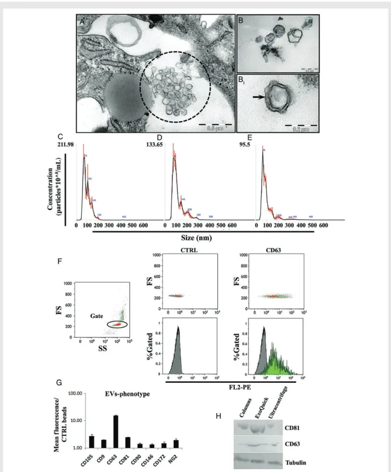
Purified particles from CM-CPC using the ExoQuickTMmethod were
studied by transmission electron microscopy and found to be 72.8 + 20.8 nm in diameter. Many of them exhibited bi-layer membranes (Figure 1B – BI). Nanoparticle tracking analyses of purified EVs from
CPCs using ExoQuickTM, ultracentrifugation, or Exo-spinTMrevealed
modal particle sizes (range; nm) of 73 nm (40 – 250), 85 nm (50 – 400), and 68 nm (40 – 400), respectively (Figure1C – E), and concentrations (×108particles/mL) of 9.8, 7.2, and 4.3, respectively. The ExoQuickTM
method yielded higher CD63 and CD81 protein levels compared with the other methods (Figure1H). Flow cytometric analyses of EVs secreted by CPCs revealed the expression of conventional exosomal markers (CD63, CD9, and CD81; Figure1F and G) along with MSC/stromal markers (CD105, CD90, CD146, CD172, and NG2) also expressed
Figure 1(A) Transmission electron micrographs showing EVs (dotted circle) secreted by human CPCs (EV-CPC) in culture. (B) Purified EVs showing a characteristic lipid bi-layer membrane (BI: higher-magnification view). (C) Nanoparticle tracking analysis of EVs purified using ExoQuickTMprecipitation
solution. (D) EVs purified by ultracentrifugation. (E) EVs purified by Exo-spinTMcolumn precipitation. (F) Flow cytometric analysis of EVs secreted by
CPCs. Left panel: side/forward scatter (SS/FS) plot of CD63-immunobeads in the absence of antibody and EVs; gating on single beads (red dots within the circle; higher FS values are from bead aggregates). Mid panels: control (anti-CD63 antibody; no EVs). Right panels: EVs with PE-conjugated anti-CD63 Ab. (G) Quantitative analysis of surface marker expression on EV-CPC (mean + SEM; n ¼ 3 experiments). (H) CD63 and CD81 western blots for EV-CPC purified using each of the aforementioned methods.
on parent CPCs (see Supplementary material online, Figure S1). Further-more, dynamic trafficking of CD63 to exosomes was visualized in EVs secreted by CPCs transfected with the CD63-GFP vector (see Supple-mentary material online, Figure S2). Taken together, these results indi-cate that exosomes are the predominant component of EVs secreted by CPCs. With respect to the degrees of exosomal purity achieved by the three methods used, ExoQuickTMwas comparable with, or slightly
better than, ultracentrifugation and Exo-spinTM. However, a thorough
comparison of purification methods was beyond the scope of this study.
3.2 EVs are the active component
of CM-CPC
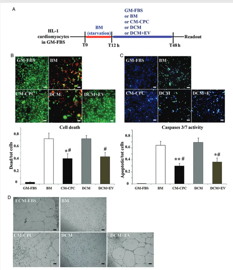
Anti-apoptotic activities of CM-CPC were assessed in the HL-1 cardio-myocytic cell line after 48 h of serum deprivation (Figure2A). CM-CPC added at 12 h reduced dead cells to 40 + 8% compared with 72 + 9% in the control group (P , 0.05; Figure 2B). EV-depleted CM-CPC (DCM) did not reduce the number of dead cells (72 + 6%; NS). Recon-stituting DCM with the previously removed EV fraction (DCM+ EV) restored anti-apoptotic activity, reducing dead cells to 44 + 5% (P , 0.05 vs. BM). Similar changes were found in cells positive for activated caspase-3/7, a marker of programmed cell death activation (Figure2C). Analogously, CM-CPC and DCM+ EV, but not DCM, stimulated tube formation in HUVECs (Figure 2D). We then compared purified EVs secreted by CPCs with those secreted by fibroblasts. Only the former inhibited starvation-induced apoptosis in the HL-1 cardiomyocytic cell line, while stimulating tube formation in HUVECs (Figure 3). These results demonstrate that EVs are the active component of CM-CPC, and that functional activities of EVs depend on the parent cell type.
3.3 EVs secreted by CPCs are taken up by
cardiomyocytes and impact on intracellular
miRNA concentrations
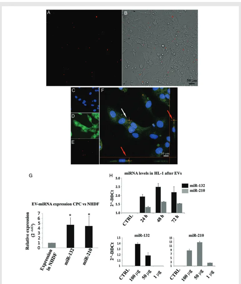
Using laser confocal microscopy, we provided qualitative evidence that HL-1 cardiomyocytic cells take up DiI-labelled EVs secreted by CPCs (Figure 4A – F ). miRNA analyses revealed that miRNA-210 and miRNA-132 were among the most highly enriched miRNAs in EVs secreted by CPCs compared with those secreted by fibroblasts (Figure4G and see Supplementary material online, Figure S3). Incubation of these cells with EVs from CPCs resulted in a time and dose-dependent increase in the intracellular concentrations of miRNA-210 and miRNA-132 (Figure4H). This observation plausibly reflects EV-mediated miRNA transfer to these cells.
3.4 EVs secreted by CPCs are enriched
in miRNAs with anti-apoptotic and
proangiogenic activities
miRNA transcriptional profiling of EVs secreted by CPCs or fibroblasts identified several miRNAs that were enriched in the former, such as miR-210, miR-132, miR-146a-3p, and miR-181 (see Supplementary ma-terial online, Figure S3). Functional roles were studied for two of them: miR-210 and miR-132. Both of these miRNAs inhibited apoptosis in HL-1 cardiomyocytic cells (Figure5A). Conversely, transcriptional silen-cing of miR-210 exacerbated apoptosis (a similar trend was observed for miR-132). The effect of miR-210 was associated with functional down-regulation of its known targets, ephrin A3 and PTP1, but not FLASH/ CASP8AP2 (Figure5B and C ). miR-132 promoted tube formation in HUVECs (Figure5D). This effect was associated with functional down-regulation of its known target, RasGap-p120 (Figure5E and F ). These
results demonstrate that EVs secreted by CPCs are enriched in miRNAs with anti-apoptotic and proangiogenic activities. These miRNAs include miR-210 and miR-132.
3.5 EVs secreted by CPCs improve cardiac
function after MI in vivo
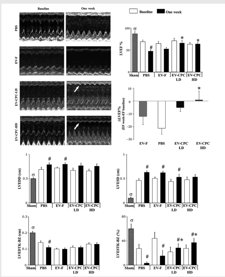
Functional effects of EVs secreted by CPCs were assessed using an in vivo model of acute MI. Echocardiographic data are shown in Figure6. Heart rate was similar with all treatments (not shown). Each group of operated rats showed decreased LVEF, both at baseline and at 7 days, compared with sham-operated animals. LVEF at baseline was comparable in all groups except sham-operated animals. In PBS-injected hearts, LVEF sig-nificantly decreased from baseline to day 7. Infarcted hearts injected with either dosage of EVs secreted by CPCs, but not those injected with EVs secreted by fibroblasts, showed a higher LVEF at 7 days compared with the PBS-injected control group (P , 0.05). LVEF decreased from base-line to Day 7 in hearts injected with PBS, EV-F, or the lower dosage of EV-CPC, whereas it slightly increased in those injected with the higher dosage of EV-CPC (0.8 + 6.8 vs. 221.3 + 4.5% in the PBS group; P , 0.05). The increase in LV diameters observed by Day 7 in the PBS and EV-F groups was prevented by either dosage of EV-CPCs. Systolic LV wall thickening in the infarct border zone was enhanced with either dosage of EV-CPC (P , 0.05), but not with EV-F. These results indicate that EVs secreted by CPCs, but not those secreted by fibroblasts, improve cardiac function after MI.
3.6 EVs secreted by CPCs inhibit
cardiomyocyte apoptosis, reduce scar,
and promote angiogenesis after MI
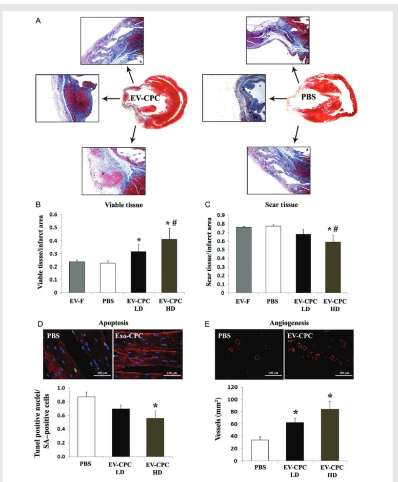
We analysed the morphological consequences of EV injection on scarred areas, cardiomyocyte apoptosis, and blood vessel density in infarcted hearts. Histological analyses showed significantly larger areas of viable tissue in the infarct zone in hearts injected with the higher or lower dosage of EVs secreted by CPCs (41 + 8 and 32 + 5%, respect-ively), compared with those receiving EVs secreted by fibroblasts (24 + 1%; P , 0.05) or PBS control medium (23 + 2%; P , 0.05; Figure7A and B). Reciprocally, scarred areas were decreased in hearts injected with the higher dosage of EVs from CPCs (59 + 8 vs. 77 + 2% in the PBS group; P , 0.05; Figure7A and C ). TUNEL-positive apop-totic cardiomyocytes were significantly decreased (P , 0.05; Figure7D), while blood vessel density being increased in hearts injected with EVs secreted by CPCs (84 + 13 vs. 34 + 5 vessel per mm2 in the PBS group; P , 0.01; Figure7E). These findings demonstrate structural ben-efits of EVs secreted by CPCs and delivered to injured hearts in vivo.
4. Discussion
Recent evidence suggests that CPC transplantation into damaged hearts may be beneficial, and that indirect mechanisms may be responsible for most of the therapeutic benefit. Because EVs are increasingly emerging as the active component of the paracrine secretion by various cell types,17,18we have investigated their potential role as mediators of the cardioprotective effect of human CPCs. Here, we show that EVs fully account for the cardioprotective and proangiogenic paracrine activ-ities of human CPCs. Indeed, CM-CPC depleted of EVs lacked these activities, which were recapitulated by purified EVs. This observation would imply negligible roles for soluble factors secreted by CPCs,
Figure 2 (A) In vitro apoptosis assay protocol: in HL-1 cardiomyocytic cell cultures, growth medium (GM-FBS) was changed to serum-free basal medium (BM) at T0. After 12 h starvation (T12 h), BM was either replaced by GM-FBS (control) or supplemented with one of the followings: CM-CPC, CPC-conditioned medium; DCM, EV-depleted CM; or DCM+ EV, DCM reconstituted with the previously removed EV fraction. (B) Dead (red) and viable cells (green) in the different study groups. (C) Immunostaining for activated caspase 3/7 (green); nuclear counterstaining with DAPI (blue). Quan-titative data are fractions (mean + SEM) of dead and activated caspase 3/7-positive cells (n ¼ 4 experiments; *P , 0.05 vs. BM; **P , 0.01 vs. BM;
#P , 0.05 vs. DCM using the parametric Tukey – Kramer multiple comparison test). (D) In vitro angiogenesis assay. Photomicrographs showing tube
for-mation in HUVECs under the above conditions (ECM-FBS, endothelial cell medium; reference bars, 0.1 mm).
such as growth factors and cytokines, which are not encapsulated in membrane vesicles, possibly due to enzymatic degradation in the extra-cellular compartment. In contrast, membrane vesicles protect their cargoes and deliver them to recipient cells.
Collectively, transmission electron microscopy, nanoparticle tracking analysis, and surface antigen expression data indicated that the purified particles from human CPCs predominantly consists of exosomes. Taking into account the presence of particles .90 nm in diameter in the purified preparations, however, we refer to them as EVs, which encompasses both exosomal and non-exosomal EVs. It should be stressed that many studies of ‘exosomes’ have actually used heteroge-neous EV populations. One notable exception was the study by Lai et al.,18who purified exosomes secreted by MSCs as a homogeneous population of particles with a hydrodynamic radius of 55 – 65 nm by size-exclusion fractionation on a HPLC. These particles reduced infarct size in an ex vivo mouse Langendorff heart model of myocardial ischaemia/reperfusion injury.
When injected into the infarct border zone in vivo, purified EVs secreted by CPCs mitigated cardiomyocyte apoptosis, augmented
blood vessel density, reduced scar while increasing viable areas in the infarct zone, and improved cardiac function compared with hearts injected with control medium. In contrast, purified EVs secreted by dermal fibroblasts lacked therapeutic benefits, illustrating the critical role of the parent cell. These findings are in line with, and expand, the aforementioned data by Lai et al.18on exosomes secreted by MSCs, as well as data by Chen et al.,19showing anti-apoptotic activity of exo-somes secreted by mouse CPCs in a myocardial model of ischaemia/ reperfusion injury.
EVs secreted by CPCs were enriched in several miRNAs including miR-210, miR-132, and miR-146a-3p compared with those secreted by fibroblasts. Both miR-210 and miR-132 inhibited apoptosis in the HL-1 cardiomyocytic cell line in vitro. This effect of miR-210 was asso-ciated with functional down-regulation of its validated targets, ephrin A3 and PTP1, but not of FLASH/CASP8AP2. These results are in line with previous findings.21Moreover, miR-132 stimulated tube formation in HUVECs. This effect was associated with functional down-regulation of the known miR-132 target, RasGap-p120. This observation is in agreement with previous data.22 We also documented dose- and Figure 3 (A) In vitro angiogenesis assay using purified EVs. EVs secreted by CPCs (EV-CPC) promote endothelial tube formation, whereas those secreted by fibroblasts (EV-F) do not (mean + SEM; n ¼ 7 experiments; *P , 0.05 vs. BM;#P , 0.05 vs. EV-F using the parametric Tukey – Kramer multiple
com-parison test; ECM-FBS, endothelial cell medium). (B) In vitro apoptosis assay using serial dilutions of purified EV-CPV and EV-F, showing dose-dependent inhibition of apoptosis by EV-CPV, but not EV-F. Quantitative data are fractions of the HL-1 cardiomyocytic cells staining positive for activated caspase-3/7 (mean + SEM; n ¼ 4 experiments; *P , 0.05; **P , 0.01; ***P , 0.001 vs. EV-F using the parametric Student’s t-test; reference bars, 0.1 mm).
Figure 4 Uptake of EVs from CPCs (EV-CPC) by HL-1 cardiomyocytic cells and changes in the intracellular concentrations of some of the miRNAs enriched in EV-CPC. (A and B) Fluorescence/light photomicrographs of DiI-labelled EV-CPC (red dots) taken up by cardiomyocytic cells. (C) Laser confocal photomicrograph showing DAPI-stained nuclei (blue). (D) Cells visualized with calcein (green). (E) DiI-labelled EV-CPC (red dots). (F) High-magnification merge (white arrow indicates an intracellular EV-CPC aggregate; red arrows indicate the same EV-CPC aggregate depicted in two orthogonal axes, verifying its intracellular location on both axes). (G) Relative expression of miR-210 and miR-132 in EV-CPC compared with EV-F (mean + SEM; n ¼ 6 experiments; *P , 0.05 using the parametric Tukey– Kramer multiple comparison test). (H)Intracellular concentrations of miR-210 and miR-132 in HL-1 cells exposed to EV-CPC (100 mg of total protein) for the indicated periods of time (upper panel; n ¼ 3 experiments). Lower panels: Dose – response studies (measure-ments at 48 h; n ¼ 3 experi(measure-ments).
time-dependent increases in the intracellular concentrations of miR-210 and miR-132 after exposure of the HL-1 cardiomyocytic cell line to EVs secreted by CPCs. This finding shows that purified donor EVs can modify
miRNA concentrations in recipient cardiomyocytes. miRNA transfer therefore appears to be a plausible mechanism for the therapeutic benefit of EVs.
Figure 5 Gain- and loss-of-function studies of miR-210 and miR-132, and regulation of known targets thereof. (A) Apoptotic HL-1 cardiomyocytic cells transfected with a miR-210 and/or a miR-132 mimic, or transcriptional inhibitors thereof (si-miR-210 and si-miR-132; mean + SEM; n ¼ 5 experiments; *P , 0.05 vs. cel-miR-39;#P , 0.05 vs. BM;dP , 0.05 vs. BM and cel-miR-39 using non-parametric Dunn’s multiple comparisons test). (B) Western blots for the miR-210 targets, ephrin A3, PTP1, and FLASH. (C) Densitometric analysis for PTP1 and ephrin A3 (n ¼ 3 experiments; a.u. ¼ arbitrary units). (D) In vitro angiogenesis assay. Photomicrographs showing increased tube formation in HUVECs transfected with a miR-132 mimic, but not with si-miR-132 (mean + SEM; n ¼ 6 experiments; **P , 0.01 vs. BM; #P , 0.05 vs. miR-132;dP , 0.01 vs. miR-132 using the parametric Tukey – Kramer multiple comparison test; dimensional bars, 100 mm). (E) Western blot for RasGap-p120. (F) Densitometric analysis of RasGap-p120 (n ¼ 3 experiments; a.u. ¼ arbitrary units).
Figure 6 Echocardiographic results at baseline (i.e. Day 2; white bars) and at Day 7 after MI (black bars). M-mode images in the different groups at Day 7 (white arrows indicate systolic anterior wall thickening in EV-CPC – injected hearts). Changes in LVEF from baseline to Day 7 were negative in all groups, but in hearts injected with the higher dosage (HD) in EV-CPC (LD, lower dosage; LVEDD/LVESD, LV end-diastolic/end-systolic diameter; LVEDTHK-BZ, LV end-diastolic wall thickness; LVESTK-BZ, end-systolic wall thickening in the border zone; mean + SEM; PBS, n ¼ 9; 30 mg EV-CPC LD, n ¼ 9; 300 mg EV-CPC HD, n ¼ 10; 300 mg Exo-F, n ¼ 4; sham, n ¼ 9; *P , 0.05 vs. PBS;#P , 0.05 at Day 7 vs. baseline;sP , 0.05 sham vs. PBS at Day 7 using the para-metric Tukey – Kramer multiple comparison test).
Figure 7 Morphometric analyses of infarcted rat hearts. (A) Masson’s trichrome-stained sections of EV-CPC- (left) and PBS-injected hearts (right). (B and C) Quantitative analyses of viable and scar areas (%) in the infarct zone. (D) TUNEL-positive (green) apoptotic cardiomyocytes (a-SA -positive cells; red). (E) Smooth muscle actin-positive blood vessels (red). Quantitative analyses of apoptotic cardiomyocytes and vessel density (mean + SEM; EV-F, n ¼ 4; all other groups, n ¼ 5; HD/LD, higher/lower dosage of EV-CPC; *P , 0.05 vs. PBS;#P , 0.05 vs. EV-F using the parametric Tukey– Kramer multiple comparison test).
As cell-free products, EVs including exosomes may have a potential for circumventing many of the limitations of using replicating cells as therapeutic agents.29Indeed, secreted vesicles may be easier to manu-facture and standardize in terms of dosage and biological activity com-pared with their parent cells. Exosomes are stable during storage at 2208C, and their size is unaffected by multiple freezing to 2208C and thawing, whereas it decreases at 4 and 378C, indicating structural changes or degradation.30After all, there may be no interest in trans-planting cells that essentially act through their paracrine secretion. Further studies are needed to address long-term effects of exosome therapy for damaged hearts and whether allogeneic cells could be used as EV donors for clinical use.
In conclusion, we have demonstrated that EVs are the active compo-nent of the paracrine secretion by human CPCs, are enriched in miRNAs with anti-apoptotic and proangiogenic activities, and can improve cardiac function after MI.
Supplementary material
Supplementary material is available at Cardiovascular Research online.
Conflict of interest: none declared.
Funding
The study was supported by the Swiss National Science Foundation (310030_140485), the Swiss Heart Foundation, the Cecilia-Augusta Founda-tion and the Fidinam FoundaFounda-tion, Lugano, Switzerland and, in part, by ‘5× 1000’ funds of Scuola Superiore Sant’Anna, Pisa, Italy (MCARDI13VL), and CNCS – UEFISCDI, IDEI 350/2012 PN-II-ID-PCE-2011-3-0134.
References
1. Scha¨chinger V, Erbs S, Elsa¨sser A, Haberbosch W, Hambrecht R, Ho¨lschermann H, Yu J, Corti R, Mathey DG, Hamm CW, Su¨selbeck T, Assmus B, Tonn T, Dimmeler S, Zeiher AM; REPAIR-AMI Investigators. Intracoronary bone marrow-derived progenitor cells in acute myocardial infarction. N Engl J Med 2006;355:1210 – 1221.
2. Houtgraaf JH, den Dekker WK, van Dalen BM, Springeling T, de Jong R, van Geuns RJ, Geleijnse ML, Fernandez-Aviles F, Zijlsta F, Serruys PW, Duckers HJ. First experience in humans using adipose tissue-derived regenerative cells in the treatment of patients with ST-segment elevation myocardial infarction. J Am Coll Cardiol 2012;59:539 – 540. 3. Menasche´ P, Alfieri O, Janssens S, McKenna W, Reichenspurner H, Trinquart L,
Vilquin JT, Marolleau JP, Seymour B, Larghero J, Lake S, Chatellier G, Solomon S, Desnos M, Hage`ge AA. The Myoblast Autologous Grafting in Ischemic Cardiomyopathy (MAGIC) trial: first randomized placebo-controlled study of myoblast transplantation. Circulation 2008;117:1189 – 1200.
4. Makkar RR, Smith RR, Cheng K, Malliaras K, Thomson LE, Berman D, Czer LS, Marba´n L, Mendizabal A, Johnston PV, Russell SD, Schuleri KH, Lardo AC, Gerstenblith G, Marba´n E. Intracoronary cardiosphere-derived cells for heart regeneration after myo-cardial infarction (CADUCEUS): a prospective, randomised phase 1 trial. Lancet 2012; 379:895 – 904.
5. Marba´n E, Malliaras K. Mixed results for bone marrow– derived cell therapy for ischemic heart disease. JAMA 2012;308:2405 – 2406.
6. Messina E, De Angelis L, Frati G, Morrone S, Chimenti S, Fiordaliso F, Salio M, Battaglia M, Latronico MV, Coletta M, Vivarelli E, Frati L, Cossu G, Giacomello A. Isolation and expan-sion of adult cardiac stem cells from human and murine heart. Circ Res 2004;95:911 – 921. 7. Smith RR, Barile L, Cho HC, Leppo MK, Hare JM, Messina E, Giacomello A, Abraham MR, Marba´n E. Regenerative potential of cardiosphere-derived cells expanded from percu-taneous endomyocardial biopsy specimens. Circulation 2007;115:896 – 908. 8. Chimenti I, Smith RR, Li TS, Gerstenblith G, Messina E, Giacomello A, Marba´n E. Relative
roles of direct regeneration versus paracrine effects of human cardiosphere-derived cells transplanted into infarcted mice. Circ Res 2010;106:971 – 980.
9. Duran JM, Makarewich CA, Sharp TE, Starosta T, Zhu F, Hoffman NE, Chiba Y, Madesh M, Berretta RM, Kubo H, Houser SR. Bone-derived stem cells repair the heart after
myocardial infarction through transdifferentiation and paracrine signaling mechanisms. Circ Res 2013;113:539 – 552.
10. Li TS, Cheng K, Malliaras K, Smith RR, Zhang Y, Sun B, Matsushita N, Blusztajn A, Terrovitis J, Kusuoka H, Marba´n L, Marba´n E. Direct comparison of different stem cell types and subpopulations reveals superior paracrine potency and myocardial repair ef-ficacy with cardiosphere-derived cells. J Am Coll Cardiol 2012;59:942 – 953.
11. Timmers L, Lim SK, Arslan F, Armstrong JS, Hoefer IE, Doevendans PA, Piek JJ, El Oakley RM, Choo A, Lee CN, Pasterkamp G, de Kleijn DP. Reduction of myocardial infarct size by human mesenchymal stem cell conditioned medium. Stem Cell Res 2007; 1:129 – 137.
12. Mathivanan S, Ji H, Simpson RJ. Exosomes: extracellular organelles important in intercel-lular communication. J Proteomics 2010;73:1907 – 1920.
13. Thery C. Exosomes: secreted vesicles and intercellular communications. F1000 Biol Rep 2011;3:15.
14. Valadi H, Ekstrom K, Bossios A, Sjostrand M, Lee JJ, Lotvall JO. Exosome-mediated trans-fer of mRNAs and microRNAs is a novel mechanism of genetic exchange between cells. Nat Cell Biol 2007;9:654 – 659.
15. Stoorvogel W. Functional transfer of microRNA by exosomes. Blood 2012;119: 646 – 648.
16. Bobrie A, Colombo M, Krumeich S, Raposo G, The´ry C. Diverse subpopulations of vesi-cles secreted by different intracellular mechanisms are present in exosome preparations obtained by differential ultracentrifugation. J Extracell Vesicles 2012;1:18397. 17. Sahoo S, Klychko E, Thorne T, Misener S, Schultz KM, Millay M, Ito A, Liu T, Kamide C,
Agrawal H, Perlman H, Qin G, Kishore R, Losordo DW. Exosomes from human CD34(+) stem cells mediate their proangiogenic paracrine activity. Circ Res 2011;109: 724 – 728.
18. Lai RC, Arslan F, Lee MM, Sze NS, Choo A, Chen TS, Salto-Tellez M, Timmers L, Lee CN, El Oakley RM, Pasterkamp G, de Kleijn DP, Lim SK. Exosome secreted by MSC reduces myocardial ischemia/reperfusion injury. Stem Cell Res 2010;4:214 – 222.
19. Chen L, Wang Y, Pan Y, Zhang L, Shen C, Qin G, Ashraf M, Weintraub N, Ma G, Tang Y. Cardiac progenitor-derived exosomes protect ischemic myocardium from acute ische-mia/reperfusion injury. Biochem Biophys Res Commun 2013;431:566 – 571.
20. Barile L, Gherghiceanu M, Popescu LM, Moccetti T, Vassalli G. Ultrastructural evidence of exosome secretion by progenitor cells in adult mouse myocardium and adult human cardiospheres. J Biomed Biotechnol 2012;2012:354605.
21. Hu S, Huang M, Li Z, Jia F, Ghosh Z, Lijkwan MA, Fasanaro P, Sun N, Wang X, Martelli F, Robbins RC, Wu JC. MicroRNA-210 as a novel therapy for treatment of ischemic heart disease. Circulation 2010;122:S124 – S131.
22. Katare R, Riu F, Mitchell K, Gubernator M, Campagnolo P, Cui Y, Fortunato O, Avolio E, Cesselli D, Beltrami AP, Angelini G, Emanueli C, Madeddu P. Transplantation of human pericyte progenitor cells improves the repair of infarcted heart through activation of an angiogenic program involving micro-RNA-132. Novelty and significance. Circ Res 2011; 109:894 – 906.
23. Hinescu ME, Gherghiceanu M, Suciu L, Popescu LM. Telocytes in pleura: two- and three-dimensional imaging by transmission electron microscopy. Cell Tissue Res 2011;343: 389 – 397.
24. Claycomb WC, Lanson NA Jr, Stallworth BS, Egeland DB, Delcarpio JB, Bahinski A, Izzo NJ Jr. HL-1 cells: a cardiac muscle cell line that contracts and retains phenotypic char-acteristics of the adult cardiomyocyte. Proc Natl Acad Sci USA 1998;95:2979 – 2984. 25. Fasanaro P, D’Alessandra Y, Di Stefano V, Melchionna R, Romani S, Pompilio G,
Capogrossi MC, Martelli F. MicroRNA-210 modulates endothelial cell response to hypoxia and inhibits the receptor tyrosine kinase ligand Ephrin-A3. J Biol Chem 2008; 283:15878 – 15883.
26. Kim HW, Haider HK, Jiang S, Ashraf M. Induction of FLASH/CASP8AP2 in miR-210 knocked-down (PC)MSCs resulted in increased cell apoptosis. Taken together, these data demonstrated that cytoprotection afforded by IP was regulated by miR-210 induc-tion via FLASH/Casp8ap2 suppression. J Biol Chem 2009;284:33161 – 33168. 27. Campan M, Lionetti V, Aquaro GD, Forini F, Matteucci M, Vannucci L, Chiuppesi F, Di
Cristofano C, Faggioni M, Maioli M, Barile L, Messina E, Lombardi M, Pucci A, Pistello M, Recchia FA. Ferritin as a reporter gene for in vivo tracking of stem cells by 1.5-T cardiac MRI in a rat model of myocardial infarction. Am J Physiol Heart Circ Physiol 2011;300:H2238 – H2250.
28. Zentilin L, Puligadda U, Lionetti V, Zacchigna S, Collesi C, Pattarini L, Ruozi G, Camporesi S, Sinagra G, Pepe M, Recchia FA, Giacca M. Cardiomyocyte VEGFR-1 acti-vation by VEGF-B induces compensatory hypertrophy and preserves cardiac function after myocardial infarction. FASEB J 2010;24:1467 – 1478.
29. Sahoo S, Losordo DW. Exosomes and cardial repair after myocardial infarction. Circ Res 2014;114:333 – 344.
30. Sokolova V, Ludwig AK, Hornung S, Rotan O, Horn PA, Epple M, Giebel B. Character-isation of exosomes derived from human cells by nanoparticle tracking analysis and scan-ning electron microscopy. Colloids Surf B Biointerfaces 2011;87:146 – 150.