Basic science for the clinician
Biomechanical factors in atherosclerosis:
mechanisms and clinical implications
†
Brenda R. Kwak
1
*
, Magnus Ba¨ck
2
, Marie-Luce Bochaton-Piallat
3
, Giuseppina Caligiuri
4
,
Mat J.A.P. Daemen
5
, Peter F. Davies
6
, Imo E. Hoefer
7
, Paul Holvoet
8
, Hanjoong Jo
9
,
Rob Krams
10
, Stephanie Lehoux
11
, Claudia Monaco
12
, Sabine Steffens
13
, Renu Virmani
14
,
Christian Weber
13
, Jolanda J. Wentzel
15
, and Paul C. Evans
16
*
1
Department of Pathology and Immunology, University of Geneva, CMU, Rue Michel-Servet 1, CH-1211 Geneva, Switzerland;2
Karolinska Institutet, Stockholm, Sweden;3
University of Geneva, Geneva, Switzerland;4
Bichat Hospital, Paris, France;5
Academic Medical Center, Amsterdam, The Netherlands;6
University of Pennsylvania, Philadelphia, PA, USA;7 University Medical Center Urecht, Utrecht, The Netherlands;8
KU Leuven, Leuven, Belgium;9
Emory University, Atlanta, GA, USA;10
Imperial College London, London, UK;11
McGill University, Montreal, QC, Canada;12
University of Oxford, Oxford, UK;13
Ludwig-Maximilians-Universita¨t (LMU), Munich, Germany;14
CVPath Institute, Gaithersburg, MD, USA;15
ErasmusMC, Rotterdam, The Netherlands; and16
Department of Cardiovascular Science, Medical School, University of Sheffield, Beech Hill Road, Sheffield S10 2RX, UK Received 21 May 2014; revised 14 July 2014; accepted 6 August 2014; online publish-ahead-of-print 17 September 2014
Blood vessels are exposed to multiple mechanical forces that are exerted on the vessel wall (radial, circumferential and longitudinal forces) or on
the endothelial surface (shear stress). The stresses and strains experienced by arteries influence the initiation of atherosclerotic lesions, which
develop at regions of arteries that are exposed to complex blood flow. In addition, plaque progression and eventually plaque rupture is influenced
by a complex interaction between biological and mechanical factors—mechanical forces regulate the cellular and molecular composition of
plaques and, conversely, the composition of plaques determines their ability to withstand mechanical load. A deeper understanding of these
inter-actions is essential for designing new therapeutic strategies to prevent lesion development and promote plaque stabilization. Moreover,
integrat-ing clinical imagintegrat-ing techniques with finite element modellintegrat-ing techniques allows for detailed examination of local morphological and biomechanical
characteristics of atherosclerotic lesions that may be of help in prediction of future events. In this ESC Position Paper on biomechanical factors in
atherosclerosis, we summarize the current ‘state of the art’ on the interface between mechanical forces and atherosclerotic plaque biology and
identify potential clinical applications and key questions for future research.
-Keywords
Atherosclerosis † Haemodynamics † Blood flow † Mechanotransduction † Endothelial cell † Plaque rupture
Biomechanical forces
This Position Paper is focused on the influence of biomechanical forces
on the development, function, and pathophysiology of the vasculature.
In each cardiac cycle, blood is transported under pulsatile pressure
through the aorta for distribution to the peripheral organs through
the branching arterial system. The interactions of pulsatile blood
flow with arterial geometries generate complex biomechanical
forces on the vessel wall with spatial and temporal variations.
Thus, arteries are exposed to circumferential and longitudinal
stresses, i.e. perpendicular and longitudinal forces generated by
intra-luminal pressure, and axial stress (shear stress), which acts
longitudinally on the surface of the arterial wall (Figure
1
). Blood
vessels alter their morphology and function in response to changes
in blood flow that are detected by vascular cells through
decentra-lized mechanotransduction mechanisms.
1,2Endothelial cells (ECs)
are exquisitely sensitive to shear stress, the frictional force generated
by blood flow. Average wall shear stress in the healthy human aorta
varies from 10 to 20 dynes/cm
2and circumferential stress varies from
1 to 2
× 10
6dynes/cm
2according to anatomical site. In areas of
ar-terial stenosis (decreased lumen area and thus radius), the same
blood volume is pushed through a lower cross-sectional area and
thus the blood velocity increases and as a consequence the wall
shear stress increases inside the stenotic region. Furthermore, the
†ESC Working Group of Atherosclerosis and Vascular Biology.
*Corresponding author: Tel:+41 223795737 (B.R.K.)/+44 01142712591 (P.C.E.), Fax: +41 223795746 (B.R.K)/+44 1142711863 (P.C.E), Email:brenda.kwakchanson@unige.ch
(B.R.K.)/paul.evans@sheffield.ac.uk(P.C.E.)
endothelium downstream the stenosis is exposed to disturbed flow
and oscillatory shear stress. Flow simulation studies describe the
complex situation near arterial bifurcations and side branches,
regions associated with disturbed blood flow showing repetitive
phases of flow reversal resulting in steep spatial and temporal
gradi-ents in wall shear stress.
3Biomechanical regulation of
arterial homeostasis
Mechanical forces regulate multiple aspects of vascular physiology
and function and play a key role in vascular development and
homeo-static mechanisms as well as during arterial disease. In the short term,
acute increases in shear stress trigger activation of ECs and the
gen-eration of substances such as nitric oxide (NO) and prostacyclin,
which promote vasodilation. On the other hand, long-term
altera-tions in flow can lead to structural adjustments to restore vascular
and mechanical homeostasis. Arterial remodelling processes
includ-ing angiogenesis (growth of new blood vessels from pre-existinclud-ing
vessels) and arteriogenesis (collateral artery growth) are highly
sen-sitive to local mechanical conditions.
4–6Raised levels of shear stress
represent a major stimulus for exercise-induced angiogenesis, a
process that involves NO signalling.
7In addition, increased flow
leads to increases in arterial diameter, which promotes tissue
perfu-sion. For example, animal studies revealed that unilateral carotid
artery occlusion leads to outward remodelling of the contralateral
carotid artery (in response to increased flow) and inward
remodelling of the occluded artery (due to reduced flow).
8,9The
mo-lecular and cellular mechanisms that accompany arterial remodelling
and repair in response to mechanical forces have only been partially
defined. Studies of cultured ECs and animals demonstrated that high
shear stress activates transcriptional programmes that promote
prolif-eration and matrix remodelling, processes that are intimately involved
in structural remodelling of arteries,
10,11as well as survival of ECs by
inhibiting the expression of pro-apoptotic factors.
12–14Flow also
influ-ences EC migration by regulating actin cytoskeleton remodelling, cell
polarity, formation of lamellipodia, and stress fibre contraction;
factors that are essential for cell traction.
15While the effects of shear stress on vascular physiology have been
studied in detail, the effects of mechanical stretch have received little
attention. Thus, although axial and circumferential stretches also play
an important role in regulating EC physiology, vascular cell
prolifer-ation, and matrix remodelling, the mechanisms involved are not
well understood. Mechanical stretch regulates smooth muscle cell
(SMC) functions by inducing deformation of the extracellular
matrix in which SMCs are embedded, a change that is detected by
mechanoreceptors.
16Physiological pulsatile circumferential stress
on the arterial wall maintains medial SMCs in their contractile
differ-entiated state.
17,18In contrast, excessive pressure increase due to
hypertension or compressive forces produced by balloon
angio-plasty and/or stent placement stretches the artery and activates
Figure 1
Biomechanical forces acting on the arterial wall. Blood pressure and blood flow induce forces in the vascular system that deforms the
vessel wall. When forces are to be compared, they need to be normalized to area. Force per area is called stress and is expressed in N/m
2or Pascal
(Pa). Blood pressure produces a force directed perpendicular to the vessel wall. As a consequence, the cylindrical structure will be stretched
cir-cumferentially, resulting in a circumferential stress. Stress in the range of 300 – 500 kPa is associated with plaque rupture. In contrast, the force
induced by a difference in movement of blood and the non-moving vessel wall leads to stress and strain parallel to the surface of endothelial
cells. Due to its shearing deformation, this is called a shear stress. This shear stress is of small amplitude (1 Pa) and exerts its main effects
through the activation of mechanosensitive receptors and signalling pathways.
SMCs, which subsequently undergo phenotypic adaptation to a
ded-ifferentiated synthetic state.
19–21Thus, mechanical circumferential
stress modulates gene expression and SMC functions such as
prolif-eration, survival/apoptosis, migration, and extracellular matrix
remodelling through receptor-tyrosine kinases (e.g. platelet-derived
growth factor receptor), focal adhesions that link the extracellular
matrix and the intracellular cytoskeleton, and ion channels activating
complex intracellular signalling pathways including Ras homologue
gene family, member A (RhoA)/Rho kinase, mitogen-activated
protein kinases (MAPKs), phosphatidylinositol-4,5-bisphosphate
3-kinase (PI3 K)/Akt, forkhead transcription factors of the FoxO
sub-family, and other signalling pathways.
11,19,22–24Of note, some of these
molecular mechanisms have been revealed using in vitro models and
now require validation using ex vivo or in vivo systems.
25,26Biomechanical regulation of focal
atherosclerosis
Shear stress and plaque initiation
Atherosclerosis is characterized by the accumulation of
inflamma-tory cells, lipids, extracellular matrix, and other materials in the
artery wall. Although atherosclerosis is associated with systemic
risk factors (e.g. gender, age, and high serum cholesterol), plaques
form preferentially at branches and bends in arteries that are
exposed to non-uniform, disturbed patterns of blood flow.
27Two
mechanisms have been identified, which could explain the link
between disturbed blood flow and atherosclerosis development,
namely alterations in mass transport and vascular responses to
mech-anical stimuli.
28The ‘mass transport theory’ states that the transport
of certain bioactive substances [e.g. low-density lipoproteins (LDL)]
from the circulation to the vessel wall may be promoted at sites of
dis-turbed flow due to prolonged contact between blood and vascular
ECs. This differs from the ‘shear stress theory’, which emphasizes
the effects of blood flow-induced mechanical forces on vascular
physiology. Of note, these theories are not mutually exclusive.
Both mass transport and shear stress influence plaque formation,
and these factors interact at a functional level, e.g. shear stress
alters vessel permeability that, in turn, regulates molecular
trans-port.
29Several lines of evidence suggest that shear stress regulates
plaque initiation. First, fluid dynamic studies revealed that the
spatial distribution of EC dysfunction, inflammation, and lesion
for-mation in arteries correlates with the magnitude and pattern of
shear stress.
30–32For example, regions exposed to low, oscillatory
shear in the murine aorta are prone to lesion formation. These sites
are also characterized by a highly heterogenous population of ECs
that display enhanced expression of inflammatory molecules, higher
rates of apoptosis and senescence, and a reduced proliferative
reserve, which compromises vascular repair potential.
33–41A
second important evidence for the ‘shear stress theory’ was provided
in studies demonstrating a causal relationship between shear stress and
atherosclerosis by applying a constrictive cuff to generate distinct shear
stress environments (low, low/oscillatory, and high shear fields) in
carotid arteries in rabbits and mice.
42,43Flow-dependent
atheroscler-osis in mice has been confirmed with other models inducing disturbed
flow by partial ligation or tandem ligations of the carotid artery.
44,45There has been considerable debate over the relative importance of
shear stress magnitude, frequency, or direction (e.g. oscillations,
tan-gential shear) in dictating vascular function,
46but it is conceivable
that ECs can detect changes in each of these parameters and
respond accordingly. This question has been addressed using the
shear stress-altering cuff model that demonstrated that low shear
and low, oscillatory shear induced different vascular responses.
42,43Mechanoreceptors
Evidence for the ‘shear stress theory’ has also been obtained through
the identification and characterization of mechanoreceptors.
A large variety of membrane-associated molecules and
microdo-mains have been proposed as potential shear stress sensors including
ion channels [e.g. transient receptor potential (TRP) channels and
P2X4 receptors], receptor-tyrosine kinases [e.g. vascular endothelial
growth factor receptor (VEGFR) and angiopoietin receptor],
adhesion molecules (e.g. PECAM-1/VE-cadherin/VEGFR2), the
gly-cocalyx, membrane microdomains (e.g. primary cilia and caveolae),
the cytoskeleton, and the lipid bilayer plasma membrane
47–49(Figure
2
). Several mechanoreceptors have pleiotropic functions
and, therefore, influence atherosclerosis at multiple levels. For
example, bone marrow cell-derived PECAM-1 has been reported
to be both pro-atherogenic
50and atheroprotective,
51irrespective
of the haemodynamic environment, whereas PECAM-1 in ECs
accel-erates atherogenesis in low shear environments.
50,52While the exact
mechanisms have yet to be elucidated, targeting such receptors
therapeutically will require a cell-type and context-specific strategy.
Despite these insights, the mechanisms that allow cells to respond
specifically to distinct mechanical conditions remain largely unknown.
Thus, further studies involving specialized techniques to apply force
to specific receptors or discrete regions of the cell (e.g. magnetic
tweezers) are required to characterize the mechanisms that
regulate the activity and function of mechanoreceptors.
53Shear stress and inflammatory signalling
The application of flow to cultured ECs has been used to identify
causal relationships between shear stress and EC function and to
define the signalling pathways involved. Shear stress influences EC
in-flammatory responses by modulating the expression of non-coding
RNAs as well as mRNAs. Regions with disturbed flow display a
focal enrichment and luminal redistribution of endothelial junctional
adhesion molecule-A (JAM-A) that promotes mononuclear cell
re-cruitment into the arterial wall. Conversely, atheroprotective
laminar flow mediates repression of JAM-A through microRNA
(miR)-145.
54These data identify endothelial JAM-A as a crucial
effect-or molecule guiding inflammateffect-ory cell entry at predilection sites of
atherosclerosis. Low, oscillatory shear stress influences EC
ex-pression of adhesion proteins and other inflammatory molecules
through multiple mechanisms that target the MAPK pathway and
the nuclear factor-kappa-B (NF-kB) pathway.
36,55In contrast,
atheroprotective shear stress induces several negative regulators of
inflammatory pathways including the transcription factors
Kruppel-like family 2 (KLF2) and 4 (KLF4)
56–58and nuclear factor erythroid
2-related factor (Nrf2).
59–63The mechanism for KLF2 activation
by shear stress involves ERK5-MEF2 signalling, which activates the
KLF2 promoter,
58,64–66and suppression of miR-92a, which is a
nega-tive regulator of KLF2 and KLF4 mRNA expressions.
67,68Conversely,
miR-92a is expressed by ECs in atheroprone low shear stress regions,
increased by hypercholesterolaemia, and in vivo miR-92a blockade
by antagomir treatment protects against the development of
athero-sclerosis.
69High unidirectional shear stress also reduces inflammatory
MAP kinases by inhibiting ASK-1 (an inflammatory MAP kinase kinase
kinase),
70blocking cleavage of protein kinase C epsilon (PKCz),
71in-ducing MAPK phosphatase-1 (MKP-1), a negative regulator of p38
and JNK MAP kinases,
37and via down-regulation of the angiotensin
II type 1 receptor.
72,73In contrast, low shear stress enhances NF-kB expression via
acti-vation of a JNK1-ATF2 transcriptional programme
36and promotes
NF-kB activation via induction of positive regulators [e.g. Toll-like
receptors,
74bone morphogenic proteins,
75–77inhibitor of kB
kinase 2 (IKK2
38), and reactive oxygen species
55,78,79]. In addition
to microRNA control,
68recent epigenetic regulation of pro- and
anti-inflammatory gene expression in disturbed flow regions has
been demonstrated including altered flow-induced DNA
methyla-tion of endothelium mediated by DNA methyltransferases.
80–83Thus, low oscillatory shear stress induces pro-atherogenic epigenetic
and transcriptional programmes in EC, whereas high unidirectional
shear induces multiple anti-inflammatory processes.
Shear stress and endothelial apoptosis,
senescence, and proliferation
Shear stress can also influence EC injury by inducing signalling
pathways that regulate apoptosis or senescence (Figure
3
).
Disturbed flow induces EC apoptosis through multiple
mecha-nisms including activation of PKCz,
40JNK MAP kinase,
84,85and
p53,
86and through up-regulation of an unfolded protein response
signalling pathway.
39In contrast, uniform flow suppresses apoptosis
via the up-regulation and/or activation of protective signalling
path-ways involving superoxide dismutase and NO synthase, for
example.
70,84,87–91Atheroprone sites are associated with higher rates of EC
prolif-eration compared with protected regions,
33,41,85,92a feature that
may enhance vascular permeability to LDL and other atherogenic
molecules. However, a recent study also indicates that low,
oscilla-tory shear stress can induce EC senescence via activation of p53.
93This seemingly paradoxical situation emphasizes the complex
heterogenous nature of EC phenotypes at atheroprone sites. The
molecular mechanisms linking shear stress with EC mitosis are
uncertain, but it has been established that JNK1 positively regulates
proliferation at atheroprone sites, whereas the induction of the
cyclin-dependent kinase regulator GADD45 promotes quiescence
under high shear stress conditions.
94In addition, down-regulation
of miR-126-5p by disturbed flow abrogates EC proliferation at
atherosusceptible sites by up-regulating the Notch1 inhibitor
Dlk1.
41Administration of miR-126-5p rescued EC proliferation at
disease-prone sites and limited atherosclerosis, demonstrating
the importance of an EC proliferative reserve in the prevention
of atherosclerosis and pointing towards a possible therapeutic
approach.
Figure 2
Mechanoreceptors and intracellular signalling in arterial endothelium. Schematic representation of a large variety of
membrane-associated molecules and microdomains that have been proposed as potential shear stress sensors converting a mechanical signal
into a chemical response. Shear stress activates receptor-tyrosine kinase, such as the vascular endothelial growth factor receptor and PECAM-1,
which regulate leukocyte adhesion and endothelial cell – endothelial cell coupling as well as mechanoresponsiveness. In addition to these
mechan-oreceptors, shear stress can also activate ion channels, actin filaments, caveolae, the glycocalyx, primary cilia, and adherence or gap junction proteins.
Shear stress influences activation of endothelial cells through multiple mechanisms that target the mitogen-activated protein kinases, nuclear
factor-kappa-B, and regulators of these pathways including mitogen-activated protein kinase phosphatase-1, Kruppel-like factors-2 and -4,
nuclear factor erythroid 2-related factor, and endothelial nitric oxide synthase.
Gene discovery platforms for endothelial
cell mechanosignalling pathways
The identification of biomarkers for atherosclerosis (e.g. proteins,
lipids, or RNA) is an emerging area of research. While classical
tech-niques focus on individual or few factors, the development of
high-throughput strategies, i.e. -omics approaches, allows comparisons
of protein expression patterns or lipidomic profiles at a broad
level in a single experiment.
95Omics studies have been performed
to characterize mechanosensitive signalling pathways directly in
ECs from arterial sites
33,34and in many cultured cell
experi-ments.
10,96–99In cultured ECs, 900 – 1800 genes were regulated
by varying levels of fluid flow, whereas studies performed in vivo
show a lower number of differentially expressed genes. The gene
profiles associated with the differentially regulated genes show
high variability depending on experimental conditions, and
importantly, the bioinformatics methods used to analyse the data.
Despite this high variability, the current data sets can be
summar-ized with the concept of endothelial priming. In this concept,
unidir-ectional high shear stress confers protection by an up-regulation of
anti-atherogenic, anti-thrombotic, and anti-inflammatory gene
signatures, whereas low oscillatory shear stress induces
pro-thrombotic and pro-inflammatory genes.
99Consequently, regions
exposed to low shear stress are pre-disposed to atherosclerosis
and are more sensitive to high cholesterol levels and inflammatory
mediators, whereas regions exposed to high shear stress are
protected. Recent studies in intact non-atherosclerotic animals
confirmed these in vitro studies and suggest that endothelial
priming occurs in vivo, and this might be one of the reasons for
the presence of predilection sites.
100New studies focused on
obtaining changes in gene networks in ECs during plaque
development are under way and will shed new light on how ECs
react to a combination of mechanical stimuli and an inflamed
sub-endothelium.
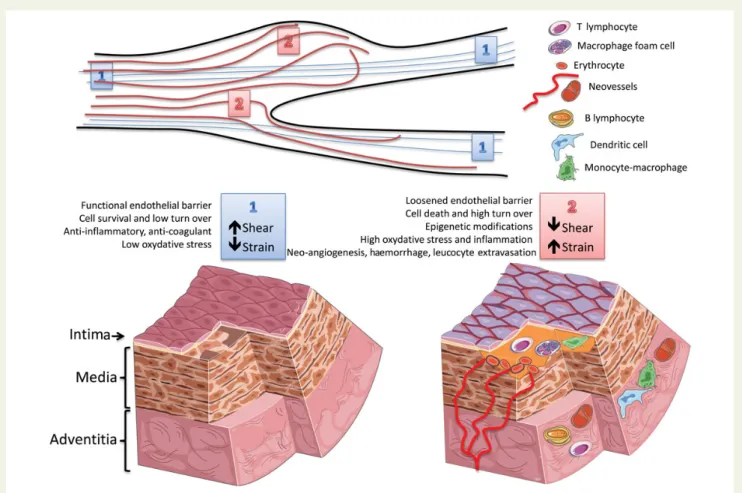
Figure 3
Effects of shear and strain on the arterial wall. (top) Schematic representation of different biomechanical forces along the arterial tree;
1 ¼ laminar flow (blue lines) imposing a high shear stress parallel to the vascular wall and a low circumferential strain; 2 ¼ arterial regions with a
change in the diameter (lack of wall parallelism) and/or proximity to bifurcations (presence of disturbed flow, red line) are subjected to a relatively
lower shear stress and higher strain. (bottom left) High shear stress and low strain (‘1’) contribute to maintenance of the physiological properties of
the endothelial barrier (anti-coagulant, anti-inflammatory, and anti-oxidant properties) and of the vessel wall (homeostatic cell and matrix
turn-over). (bottom right) Low shear and high strain (‘2’) cause endothelial cell death and reduce the physiological endothelial barrier function, thus
favouring the formation of atherosclerotic plaques (yellow matter). Plaque progression can also be affected by biomechanical factors inducing an
accelerated cell and matrix turn-over, modifications of the vascular stromal cells, inflammation, and intraplaque haemorrhage. This can boost
plaque growth and in turn impact on the local flow dynamics, thus generating a vicious circle between biomechanical factors and atherosclerosis.
Plaque progression and remodelling
Arterial sites with developing atherosclerotic plaques undergo
com-pensatory expansive remodelling to maintain their luminal diameter,
a process that presumably normalizes shear stress to a constant
level.
101,102,103Although compensatory remodelling is considered
as the ‘classical’ remodelling response during plaque growth,
con-strictive remodelling (defined as shrinkage of the vessel radius)
104and excessive compensatory remodelling
105(defined as over
com-pensation leading to radius increase) are observed in a small
percent-age of arteries.
106In general, outward remodelling will lead to a
persistence of low shear stress, thereby exaggerating lipid uptake
and inflammation. Since inflammation has been associated with
posi-tive outward remodelling,
107it may contribute to further
develop-ment of vulnerable plaques. As a result, plaques with a large
necrotic core are found at low shear stress locations.
105,108–110Inter-estingly, regional differences have been observed in vascular
remod-elling responses, e.g. there is less compensation for plaque growth in
arteries in the lower extremities.
111Plaque evolution after remodelling
Upon further progression of plaques, positive remodelling can no
longer compensate plaque growth resulting in narrowing of the
vessel lumen. In general, lumen narrowing initiates when plaque
burden exceeds 40%.
101While the precise mechanism underlying
the limitation in outward remodelling is unknown, intraplaque
bleeding,
112,113multiple plaque ruptures,
114and a circumferential
extension of endothelial dysfunction at the plaque site have been
put forward as possible explanations.
101Once atherosclerotic
plaques encroach into the lumen, ECs experience a change in
local shear stress, i.e. high shear stress at the upstream part and
low, oscillatory shear stress at the downstream side of the plaque,
where initially low shear stress was present.
108There is a lack of
detailed information as to whether ECs covering the advanced
ath-erosclerotic lesion remain responsive to changes in local shear
stress. On the one hand, the shear stress-dependent transcription
factor KLF2 seems down-regulated,
57cross-talk between ECs via
connexins is diminished
115and endothelial nitric oxide synthase
ex-pression is decreased at plaques.
116On the other hand, a
preferen-tial occurrence of apoptosis of ECs is present in the downstream
regions of advanced human carotid lesions.
14In addition, studies
of stented arteries suggest that ECs overlaying plaques retain the
ability to respond to flow.
117–119In summary, whereas plaque
initi-ation typically occurs in low shear stress regions, plaque
progres-sion
may
be
accompanied
by
(excessive)
compensatory
remodelling, thereby keeping the lumen open and maintaining the
low shear stress exposure to the plaque. Plaques may also encroach
into the lumen resulting in exposure to high shear stress for ECs in
the upstream region of the plaque.
The influence of biomechanical
forces on plaque destabilization and
rupture
Pathological studies suggest that a large necrotic core, high
macro-phage content, reduced collagen levels, and thin fibrous cap are the
hallmarks of plaque vulnerability
120–123and thus may be the
precur-sors of plaque rupture. However, a recent study indicates that only
5% of the identified vulnerable plaques (thin-capped fibroatheromas,
TCFAs) are associated with plaque rupture and suggests that plaque
morphology is not sufficient to predict plaque rupture.
124,125As
bio-mechanical factors are involved in plaque rupture, they might help to
identify vulnerable plaques.
The role of wall stress in plaque rupture
A plaque ruptures if the local wall stress (i.e. stress within an
athero-sclerotic lesion) exceeds the fracture stress (strength) of the fibrous
cap. Note that the stress in the wall is caused by a variety of factors,
including the blood pressure, local geometry, and local tissue
com-position and is 1
× 10
4to 2
× 10
6times higher than the shear
stress at the endothelium.
126Moreover, maximal predicted plaque
stresses in symptomatic patients are higher than those predicted in
asymptomatic patients, suggesting that plaques with higher stresses
may be more prone to rupture and thus leading to cardiovascular
events.
127Biomechanical stress could therefore potentially act as a
useful tool for risk assessment of plaque rupture. However, the
threshold value for wall stress to be used for risk prediction is
cur-rently under debate.
128Plaque composition influences rupture as it is a key determinant of
cap strength. The highest wall stress is typically found at the thinnest
areas of the fibrous cap,
129,130a region that co-localizes with
increased macrophage density,
131intraplaque haemorrhage,
132and
local microcalcifications.
133The role of low and high shear stress in
plaque destabilization
The causative role of low shear stress in vulnerable plaque formation
was elegantly shown in several animal studies imposing low shear
stress in defined arterial regions.
42,134Although these studies
clearly demonstrate that low shear stress modulates local
inflamma-tion and thereby cap thickness and strength, the majority of such
studies have concentrated on relatively few locations in mature
arter-ies and thus may have introduced an underestimation of the variety of
mechanical factors involved in disease development as was eloquently
pointed out by Peiffer et al.
46The notion that plaque ruptures/ulcerations are most frequently
observed at the upstream side of advanced plaques has strengthened
the idea that high shear stress may be involved in upstream plaque
de-stabilization.
135–137Moreover, plaque composition at the upstream
side of the plaque is markedly different from the downstream side, i.e.
enhanced macrophage accumulation and apoptosis, lipid
accumula-tion, intraplaque haemorrhage, and thinner fibrous caps.
135,137As a
result, upstream plaque regions that are exposed to high shear
stress show an increased strain—a local measure for plaque
weak-ness—implying that those regions are more prone to rupture.
138In
vivo studies on the role of shear stress in plaque destabilization
con-firmed increased vulnerability for the high shear stress plaque
regions at 6 months of follow-up.
110High shear stress is known to
ac-tivate matrix metalloproteinases (MMPs), favouring thinning of the
artery wall and eccentric remodelling in an in vivo arteriovenous
fistula model.
139If a similar process occurs in the advanced
athero-sclerotic lesion, this might account for a thin fibrous cap in high
shear regions of the stenosis. Clearly, more studies are needed to
investigate the potential causative role of high shear stress in plaque
destabilization.
Location of plaque rupture
As plaque rupture depends on both the local wall stress and the local
strength of the tissue, we would like to propose that co-localization of
high wall stress and shear stress-induced plaque weakening will finally
lead to plaque rupture. Figure
4
depicts the relationship between
shear stress, plaque geometry, the plaque strength—the stress
threshold at which a plaque ruptures—and the local wall stress in
the process of plaque rupture. The minor co-incidence that wall
stress exceeds the local plaque strength and the short time frame
may offer an explanation why only 5% of TCFAs rupture. Shear
stress is a biomechanical parameter acting for many years on TCFA
formation and cap weakening through biological processes. On the
other hand, wall stress concentrations are thought to lead to
plaque rupture over much shorter time frames. Further
experimen-tal and clinical imaging studies are needed to investigate the
co-localization of these biomechanical parameters in identifying
‘rupture-prone TCFAs’.
In summary, low shear stress promotes the initiation and
progres-sion of atherosclerotic leprogres-sions. Non-stenotic vulnerable plaques are
typically associated with low shear stress, which can promote
inflam-mation and influence plaque stability. This contrasts with stenotic
high-risk plaques that are typically exposed to high shear stress.
Evi-dence is accumulating for a role of high shear stress in plaque
destabilization. Finally, co-localization of high wall stress and low
plaque strength may be considered as a novel future marker for
iden-tification of vulnerable plaques.
Clinical perspectives
Interactions between drugs and
mechanoresponses
Biomechanical factors may affect the responsiveness of ECs to
pharmacological agents, as demonstrated by the synergy between
statins and laminar shear stress in inducing KLF2-dependent
athero-protective signalling in ECs both in vivo and in vitro.
140,141Consequent-ly, the endothelium lining atherosusceptible sites of low shear stress
may be less responsive to the pleiotropic effects of statins,
140high-lighting the importance of considering biomechanical factors in the
development of atheroprotective therapeutics. In addition, changes
in biomechanical forces can be used for targeted drug delivery to
ath-erosclerotic lesions.
142Mechanosensitive liposomes can be used to
preferentially release preloaded drugs under increased shear
stress
143and could thus potentially selectively target the upstream
segment of the advanced plaque (before the point of maximal
sten-osis) that shows an increased incidence of plaque rupture, as
dis-cussed above. In silico tests with these 1,3-diaminophospholipid
vesicles show promising results,
143but validation in more complex
fluids and large animals are mandatory. Another approach includes
Figure 4
Concept of the influence of shear stress and wall stress on plaque rupture. Co-localization of peak wall stress and shear stress-induced
cap thinning and cap strength will dictate location and timing of plaque rupture. (A) (Excessive) compensatory remodelling induces low shear stress
stimulating local inflammation and thereby fibrous cap thinning and plaque weakening, influencing the cap strength, (B) high shear stress induces cap
thinning and weakening. Wall stress inside the cap is related to blood pressure and the local cap geometry and thickness. If the local wall stress
exceeds the cap strength (the wall stress threshold at which it ruptures), the cap will rupture.
shear-activated nanotherapeutic aggregates.
144This strategy also
uses high shear stress caused by vascular narrowing as a targeting
mechanism to deliver drugs to (partially) obstructed blood vessels.
Microscale aggregates of nanoparticles coated with tissue
plasmino-gen activator (tPA) break into nanoscale components when exposed
to abnormally high fluid shear stress. When administered
intraven-ously in mice, these shear-activated nanotherapeutics induce rapid
clot dissolution in a mesenteric injury model.
Diagnostic and prognostic implications of
biomechanical factors
The implementation of biomechanical factors in the clinical
decision-making for patients with atherosclerosis is today restricted to
mea-surements of flow or pressure alterations. The fractional flow
reserve (FFR) of a coronary atherosclerotic lesion can be measured
as the pressure fall from the proximal aorta to the coronary segment
distal to the lesion, during maximal coronary vasodilatation.
Accord-ing to the latest ESC guidelines, percutaneous coronary intervention
(PCI) is indicated if FFR is
≤0.8.
145FFR-guided PCI has been
asso-ciated with improved clinical outcomes and fewer stents implanted.
Likewise, non-invasively measured coronary flow reserve (CFR)
by transthoracic Doppler echocardiography of the left anterior
des-cending coronary artery is recommended for patients with suspected
coronary microvascular disease.
145However, limiting interventions
to only obstructive coronary disease may be insufficient, since
plaques not causing haemodynamically significant flow restriction
may be prone to rupture. One important clinical application of the
above outlined role of biomechanical factors in atherosclerosis
could be to identify sites exposed to unfavourable biomechanical
forces, associated with high risk of plaque rupture, and to use this
in-formation to guide treatment. The development of novel imaging
tools has rendered the evaluation of wall shear stress possible in
cor-onary patients, by integrating clinical examination techniques
(Doppler ultrasound, CT, IVUS, OCT, MRI, VH) with computational
flow dynamics (CFD). Of note, a recent study revealed that rotational
coronary angiography and CFD could be used to accurately measure
FFR in patients with stable angina, thus informing PCI without the
need for invasive catheter-based measurements.
146,147In addition,
phase-contrast MRI-based shear stress measurement techniques
are currently under development to assess the local wall shear
stress distribution in human carotid arteries, likely facilitating in the
near future the clinical assessment of local wall shear stress
without extensive technical expertise. Finally, intravascular
palpogra-phy can be used for measures of plaque deformation (strain) during
pulsating blood flow.
Flow-mediated dilatation (FMD) of a conduit artery following limb
ischaemia is a measure of endothelial function, and an impaired FMD
is a sign of endothelial dysfunction in for example diabetes and
sub-clinical atherosclerosis.
148In contrast to the transient increase in
shear stress during reactive hyperaemia, exercise-induced elevations
of shear stress may be associated with a more sustained FMD increase
in the supplying conduit artery.
149In addition to these immediate flow
alterations, also long-term effects on FMD have been demonstrated
after repetitive exercise, suggesting that exercise-induced changes in
shear stress induce beneficial effects in terms of flow-mediated
endo-thelial function and vascular remodelling,
150which may be implicated
in the protective value of physical activity in reducing vascular
dys-function and atherosclerosis.
Recently, the PREDICTION study revealed that low shear stress
was an independent predictor for luminal obstruction in patients
with acute coronary syndrome, but was not associated with a
change in plaque area.
108Although clinical events rates were too
low to evaluate the effects of shear stress on outcome in terms of
acute coronary syndromes,
108this study provides an initial indication
that considering biomechanical factors may be clinically relevant for
assessing locations with progressive disease.
Knowledge from basic science is increasingly being translated into
the clinical setting. A deeper understanding of the effects of
mechan-ical forces on vascular biology will further these developments of
novel shear regulated drugs, enhance diagnostic tools and inform
clin-ical decision-making for interventional cardiologists and
cardiovascu-lar surgeons. Simicardiovascu-larly, innovations in the clinic should feedback to
drive new basic science questions in the fields of vascular biology,
engineering, and computational modelling.
Funding
This position paper was funded by the European Society of Cardiology.
Conflict of interest: P.F.D. holds the patent 6399311 B2 issued 4 June
2002. R.V.: Abbott Vascular, Biosensors International, Boston Scientific,
CeloNova, Cordis J&J, Lutonix, Medtronic, Terumo, Merck Speaker’s
Bureau, 480 Biomedical, WL Gore, outside the submitted work. C.W.
has received grants from BMBF, during the conduct of the study; and
grants from DFG and from ERC, outside the submitted work.
References
1. Davies PF. Hemodynamic shear stress and the endothelium in cardiovascular pathophysiology. Nat Clin Pract Cardiovasc Med 2009;6:16 – 26.
2. Shaw A, Xu Q. Biomechanical stress-induced signaling in smooth muscle cells: an update. Curr Vasc Pharmacol 2003;1:41 – 58.
3. Steinman DA. Simulated pathline visualization of computed periodic blood flow patterns. J Biomech 2000;33:623 – 628.
4. Hoefer IE, den Adel B, Daemen MJ. Biomechanical factors as triggers of vascular growth. Cardiovasc Res 2013;99:276 – 283.
5. Chouinard-Pelletier G, Jahnsen ED, Jones EA. Increased shear stress inhibits angiogenesis in veins and not arteries during vascular development. Angiogenesis 2013;16:71 – 83.
6. Egginton S. In vivo shear stress response. Biochem Soc Trans 2011;39:1633 – 1638. 7. Hudlicka O, Brown MD, May S, Zakrzewicz A, Pries AR. Changes in capillary shear
stress in skeletal muscles exposed to long-term activity: role of nitric oxide. Micro-circulation 2006;13:249 – 259.
8. Masuda H, Zhuang YJ, Singh TM, Kawamura K, Murakami M, Zarins CK, Glagov S. Adaptive remodeling of internal elastic lamina and endothelial lining during flow-induced arterial enlargement. Arterioscler Thromb Vasc Biol 1999;19:2298–2307. 9. Langille BL, Odonnell F. Reductions in arterial diameter produced by chronic decreases in blood-flow are endothelium-dependent. Science 1986;231:405 – 407. 10. Chen BPC, Li YS, Zhao YH, Chen KD, Li S, Lao JM, Yuan SL, Shyy JYJ, Chien S. DNA microarray analysis of gene expression in endothelial cells in response to 24-h shear stress. Physiol Genomics 2001;7:55 – 63.
11. Castier Y, Brandes RP, Leseche G, Tedgui A, Lehoux S. P47phox-dependent NADPH oxidase regulates flow-induced vascular remodeling. Circ Res 2005;97: 533 – 540.
12. Urbich C, Walter DH, Zeiher AM, Dimmeler S. Laminar shear stress upregulates integrin expression: role in endothelial cell adhesion and apoptosis. Circ Res 2000;87:683 – 689.
13. Chen BP, Li YS, Zhao Y, Chen KD, Li S, Lao J, Yuan S, Shyy JY, Chien S. DNA micro-array analysis of gene expression in endothelial cells in response to 24-h shear stress. Physiol Genomics 2001;7:55 – 63.
14. Tricot O, Mallat Z, Heymes C, Belmin J, Lese`che G, Tedgui A. Relation between endothelial cell apoptosis and blood flow direction in human atherosclerotic plaques. Circulation 2000;101:2450 – 2453.
15. Li S, Butler P, Wang YX, Hu YL, Han DC, Usami S, Guan JL, Chien S. The role of the dynamics of focal adhesion kinase in the mechanotaxis of endothelial cells. Proc Natl Acad Sci USA 2002;99:3546 – 3551.
16. Lehoux S, Castier Y, Tedgui A. Molecular mechanisms of the vascular responses to haemodynamic forces. J Int Med 2006;259:381 – 392.
17. Birukov KG, Bardy N, Lehoux S, Merval R, Shirinsky VP, Tedgui A. Intraluminal pres-sure is essential for the maintenance of smooth muscle caldesmon and filamin content in aortic organ culture. Arterioscler Thromb Vasc Biol 1998;18:922 – 927. 18. Stegemann JP, Hong H, Nerem RM. Mechanical, biochemical, and extracellular matrix
effects on vascular smooth muscle cell phenotype. J App Physiol 2005;98:2321–2327. 19. Haga J, Li Y-SJ, Chien S. Molecular basis of the effects of mechanical stretch on
vas-cular smooth muscle cells (vol. 40, p. 947, 2007). J Biomech 2008;41:2331. 20. Shyu KG. Cellular and molecular effects of mechanical stretch on vascular cells and
cardiac myocytes. Clin Sci 2009;116:377 – 389.
21. Chaabane C, Otsuka F, Virmani R, Bochaton-Piallat ML. Biological responses in stented arteries. Cardiovasc Res 2013;99:353 – 363.
22. Lemarie CA, Tharaux PL, Esposito B, Tedgui A, Lehoux S. Transforming growth factor-alpha mediates nuclear factor kappa B activation in strained arteries. Circ Res 2006;99:434 – 441.
23. Spassova MA, Hewavitharana T, Xu W, Soboloff J, Gill DL. A common mechanism underlies stretch activation and receptor activation of TRPC6 channels. Proc Natl Acad Sci USA 2006;103:16586 – 16591.
24. Abid MR, Yano K, Guo S, Patel VI, Shrikhande G, Spokes KC, Ferran C, Aird WC. Forkhead transcription factors inhibit vascular smooth muscle cell proliferation and neointimal hyperplasia. J Biol Chem 2005;280:29864 – 29873.
25. Anwar M, Shalhoub J, Lim C, Gohel M, Davies A. The effect of pressure-induced mechanical stretch on vascular wall differential gene expression. J Vasc Res 2012; 49:463 – 478.
26. Qiu J, Zheng Y, Hu J, Liao D, Gregersen H, Deng X, Fan Y, Wang G. Biomechanical regulation of vascular smooth muscle cell functions: from in vitro to in vivo under-standing. J R Soc Interface 2014;11:20130852.
27. Malek AM, Alper SL, Izumo S. Hemodynamic shear stress and its role in atheroscler-osis. JAMA 1999;282:2035 – 2042.
28. Back M, Gasser T, Michel JB, Caligiuri G. Biomechanical factors in the biology of aortic wall and aortic valve diseases. Cardiovasc Res 2013;99:232 – 241. 29. Jo H, Dull RO, Hollis TM, Tarbell JM. Endothelial albumin permeability is shear
de-pendent, time-dede-pendent, and reversible. Am J Physiol 1991;260:H1992 – H1996. 30. Davies PF. Endothelial mechanisms of flow-mediated athero-protection and
sus-ceptibility. Circ Res 2007;101:10 – 12.
31. Suo J, Ferrara DE, Sorescu D, Guldberg RE, Taylor WR, Giddens DP. Hemodynamic shear stresses in mouse aortas—implications for atherogenesis. Arterioscler Thromb Vasc Biol 2007;27:346 – 351.
32. Dai GH, Kaazempur-Mofrad MR, Natarajan S, Zhang YZ, Vaughn S, Blackman BR, Kamm RD, Garcia-Cardena G, Gimbrone MA. Distinct endothelial phenotypes evoked by arterial waveforms derived from atherosclerosis-susceptible and -resistant regions of human vasculature. Proc Natl Acad Sci USA 2004;101:14871–14876. 33. Foteinos G, Hu YH, Xiao QZ, Metzler B, Xu QB. Rapid endothelial turnover in
atherosclerosis-prone areas coincides with stem cell repair in apolipoprotein E-deficient mice. Circulation 2008;117:1856 – 1863.
34. Zeng LF, Zampetaki A, Margariti A, Pepe AE, Alam S, Martin D, Xiao QZ, Wang W, Jin ZG, Cockerill G, Mori K, Li YSJ, Hu YH, Chien S, Xu QB. Sustained activation of XBP1 splicing leads to endothelial apoptosis and atherosclerosis development in response to disturbed flow. Proc Natl Acad Sci USA 2009;106:8326 – 8331. 35. Hajra L, Evans AI, Chen M, Hyduk SJ, Collins T, Cybulsky MI. The NF-kappa B signal
transduction pathway in aortic endothelial cells is primed for activation in regions predisposed to atherosclerotic lesion formation. Proc Natl Acad Sci USA 2000;97: 9052 – 9057.
36. Cuhlmann S, Van der Heiden K, Saliba D, Tremoleda JL, Khalil M, Zakkar M, Chaudhury H, Le AL, Mason JC, Udalova I, Gsell W, Jones H, Haskard DO, Krams R, Evans PC. Disturbed blood flow induces RelA expression via c-Jun N-terminal kinase 1 A novel mode of NF-kappa B regulation that promotes arterial inflammation. Circ Res 2011;108:950 – 959.
37. Zakkar M, Chaudhury H, Sandvik G, Enesa K, Luong LA, Cuhlmann S, Mason JC, Krams R, Clark AR, Haskard DO, Evans PC. Increased endothelial mitogen-activated protein kinase phosphatase-1 expression suppresses proinflammatory activation at sites that are resistant to atherosclerosis. Circ Res 2008;103:726 – 732. 38. Passerini AG, Polacek DC, Shi CZ, Francesco NM, Manduchi E, Grant GR, Pritchard WF, Powell S, Chang GY, Stoeckert CJ, Davies PF. Coexisting proinflam-matory and antioxidative endothelial transcription profiles in a disturbed flow region of the adult porcine aorta. Proc Natl Acad Sci USA 2004;101:2482 – 2487. 39. Civelek M, Manduchi E, Riley RJ, Stoeckert CJ, Davies PF. Chronic endoplasmic
re-ticulum stress activates unfolded protein response in arterial endothelium in regions of susceptibility to atherosclerosis. Circ Res 2009;105:453 – U127.
40. Magid R, Davies PF. Endothelial protein kinase C isoform identity and differential activity of PKC xi in an athero-susceptible region of porcine aorta. Circ Res 2005; 97:443 – 449.
41. Schober A, Nazari-Jahantigh M, Wei Y, Bidzhekov K, Gremse F, Grommes J, Megens RT, Heyll K, Noels H, Hristov M, Wang S, Kiessling F, Olson EN, Weber C. MicroRNA-126-5p promotes endothelial proliferation and limits ath-erosclerosis by suppressing Dlk1. Nat Med 2014;20:368 – 376.
42. Cheng C, Tempel D, van Haperen R, van der Baan A, Grosveld F, Daemen MJAP, Krams R, de Crom R. Atherosclerotic lesion size and vulnerability are determined by patterns of fluid shear stress. Circulation 2006;113:2744 – 2753.
43. Cheng C, van Haperen R, de Waard M, van Damme LCA, Tempel D, Hanemaaijer L, van Cappellen GWA, Bos J, Slager CJ, Duncker DJ, van der Steen AFW, de Crom R, Krams R. Shear stress affects the intracellular distribution of eNOS: direct demon-stration by a novel in vivo technique. Blood 2005;106:3691 – 3698.
44. Nam D, Ni CW, Rezvan A, Suo J, Budzyn K, Llanos A, Harrison D, Giddens D, Jo H. Partial carotid ligation is a model of acutely induced disturbed flow, leading to rapid endothelial dysfunction and atherosclerosis. Am J Physiol Heart Circ Physiol 2009;297: H1535 – H1543.
45. Chen YC, Bui AV, Diesch J, Manasseh R, Hausding C, Rivera J, Haviv I, Agrotis A, Htun NM, Jowett J, Hagemeyer CE, Hannan RD, Bobik A, Peter K. A novel mouse model of atherosclerotic plaque instability for drug testing and mechanis-tic/therapeutic discoveries using gene and microRNA expression profiling. Circ Res 2013;113:252 – 265.
46. Peiffer V, Sherwin SJ, Weinberg PD. Does low and oscillatory wall shear stress cor-relate spatially with early atherosclerosis? A systematic review. Cardiovasc Res 2013; 99:242 – 250.
47. Ando J, Yamamoto K. Flow detection and calcium signalling in vascular endothelial cells. Cardiovasc Res 2013;99:260 – 268.
48. Tzima E, Irani-Tehrani M, Kiosses WB, Dejana E, Schultz DA, Engelhardt B, Cao GY, DeLisser H, Schwartz MA. A mechanosensory complex that mediates the endo-thelial cell response to fluid shear stress. Nature 2005;437:426 – 431.
49. Wang N, Butler JP, Ingber DE. Mechanotransduction across the cell-surface and through the cytoskeleton. Science 1993;260:1124 – 1127.
50. Harrison M, Smith E, Ross E, Krams R, Segers D, Buckley CD, Nash GB, Rainger G. The role of platelet-endothelial cell adhesion molecule-1 in atheroma formation varies depending on the site-specific hemodynamic environment. Arterioscler Thromb Vasc Biol 2013;33:694 – 701.
51. Goel R, Schrank BR, Arora S, Boylan B, Fleming B, Miura H, Newman PJ, Molthen RC, Newman DK. Site-specific effects of PECAM-1 on atherosclerosis in LDL receptor-deficient mice. Arterioscler Thromb Vasc Biol 2008;28:1996 – 2002. 52. Harry BL, Sanders JM, Feaver RE, Lansey M, Deem TL, Zarbock A, Bruce AC, Pryor AW, Gelfand BD, Blackman BR, Schwartz MA, Ley K. Endothelial cell PECAM-1 promotes atherosclerotic lesions in areas of disturbed flow in ApoE-deficient mice. Arterioscler Thromb Vasc Biology 2008;28:2003 – 2008.
53. Collins C, Guilluy C, Welch C, O’Brien ET, Hahn K, Superfine R, Burridge K, Tzima E. Localized tensional forces on PECAM-1 elicit a global mechanotransduc-tion response via the integrin-RhoA pathway. Curr Biol 2012;22:2087 – 2094. 54. Schmitt MMN, Megens RTA, Zernecke A, Bidzhekov K, van den Akker NM,
Rademakers T, van Zandvoort MA, Hackeng TM, Koenen RR, Weber C. Endothe-lial junctional adhesion molecule-A guides monocytes into flow-dependent predi-lection sites of atherosclerosis. Circulation 2014;129:66 – 76.
55. Xiao H, Lu M, Lin TY, Chen Z, Chen G, Wang WC, Marin T, Shentu Tp, Wen L, Gongol B, Sun W, Liang X, Chen J, Huang HD, Pedra JH, Johnson DA, Shyy JYJ. Sterol regulatory element binding protein 2 activation of NLRP3 inflammasome in endothelium mediates hemodynamic-induced atherosclerosis susceptibility. Cir-culation 2013;128:632 – 642.
56. Dekker RJ, van Soest S, Fontijn RD, Salamanca S, de Groot PG, VanBavel E, Pannekoek H, Horrevoets AJG. Prolonged fluid shear stress induces a distinct set of endothelial cell genes, most specifically lung Kruppel-like factor (KLF2). Blood 2002;100:1689 – 1698.
57. Dekker RJ, van Thienen JV, Rohlena J, de Jager SC, Elderkamp YW, Seppen J, de Vries CJ, Biessen EA, van Berkel TJ, Pannekoek H, Horrevoets AJ. Endothelial KLF2 links local arterial shear stress levels to the expression of vascular tone-regulating genes. Am J Pathol 2005;167:609 – 618.
58. Parmar KM, Larman HB, Dai GH, Zhang YH, Wang ET, Moorthy SN, Kratz JR, Lin ZY, Jain MK, Gimbrone MA, Garcia-Cardena G. Integration of flow-dependent endothelial phenotypes by Kruppel-like factor 2. J Clin Invest 2006;116:49 – 58. 59. Fledderus JO, Boon RA, Volger OL, Hurttila H, Yla-Herttuala S, Pannekoek H,
Levonen AL, Horrevoets AJ. KLF2 Primes the antioxidant transcription factor Nrf2 for activation in endothelial cells. Arterioscler Thromb Vasc Biol 2008;28:1339–1346. 60. Dai G, Vaughn S, Zhang Y, Wang ET, Garcia-Cardena G, Gimbrone MA.
Biomech-anical forces in atherosclerosis-resistant vascular regions regulate endothelial redox balance via phosphoinositol 3-kinase/akt-dependent activation of Nrf2. Circ Res 2007;101:723 – 733.
61. Hosoya T, Maruyama A, Kang MI, Kawatani Y, Shibata T, Uchida K, Itoh K, Yamamoto M. Differential responses of the Nrf2-Keap1 system to laminar and os-cillatory shear stresses in endothelial cells. J Biol Chem 2005;280:27244 – 27250. 62. Chen XL, Varner SE, Rao AS, Grey JY, Thomas S, Cook CK, Wasserman MA,
Medford RM, Jaiswal AK, Kunsch C. Laminar flow induction of antioxidant response element-mediated genes in endothelial cells—a novel anti-inflammatory mechan-ism. J Biol Chem 2003;278:703 – 711.
63. Zakkar M, Van der Heiden K, Luong LA, Chaudhury H, Cuhlmann S, Hamdulay SS, Krams R, Edirisinghe I, Rahman I, Carlsen H, Haskard DO, Mason JC, Evans PC. Ac-tivation of nrf2 in endothelial cells protects arteries from exhibiting a proinflamma-tory state. Arterioscler Thromb Vasc Biol 2009;29:1851 – 1857.
64. Yan C, Takahashi M, Okuda M, Lee JD, Berk BC. Fluid shear stress stimulates big mitogen-activated protein kinase 1 (BMK1) activity in endothelial cells—depend-ence on tyrosine kinases and intracellular calcium. J Biol Chem 1999;274:143 – 150. 65. Yan C, Luo HL, Lee JD, Abe JI, Berk BC. Molecular cloning of mouse ERK5/BMK1 splice variants and characterization of ERK5 functional domains. J Biol Chem 2001;276:10870 – 10878.
66. Young A, Wu W, Sun W, Larman HB, Wang N, Li YS, Shyy JY, Chien S, Garcia-Cardena G. Flow activation of AMP-activated protein kinase in vascular endothelium leads to Kruppel-like factor 2 expression. Arterioscler Thromb Vasc Biol 2009;29:1902 – 1908.
67. Wu W, Xiao H, Laguna-Fernandez A, Villarreal G Jr, Wang KC, Geary GG, Zhang Y, Wang WC, Huang HD, Zhou J, Li YS, Chien S, Garcia-Cardena G, Shyy JYJ. Flow-dependent regulation of Kruppel-like factor 2 is mediated by microRNA-92a. Circu-lation 2011;124:633 – 641.
68. Fang Y, Davies PF. Site-specific microRNA-92a regulation of Kruppel-like factors 4 and 2 in atherosusceptible endothelium. Arterioscler Thromb Vasc Biol 2012;32:979–987. 69. Loyer X, Potteaux S, Vion AC, Gue´rin CL, Boulkroun S, Rautou PE,
Ramkhelawon B, Esposito B, Dalloz M, Paul JL, Julia P, Maccario J, Boulanger CM, Mallat Z, Tedgui A. Inhibition of microRNA-92a prevents endothelial dysfunction and atherosclerosis in mice. Circ Res 2014;114:434 – 443.
70. Liu YM, Yin GY, Surapisitchat J, Berk BC, Min W. Laminar flow inhibits TNF-induced ASK1 activation by preventing dissociation of ASK1 from its inhibitor 14-3-3. J Clin Invest 2001;107:917 – 923.
71. Traub O, Monia BP, Dean NM, Berk BC. PKC-epsilon is required for mechano-sensitive activation of ERK1/2 in endothelial cells. J Biol Chem 1997;272:31251–31257. 72. Ramkhelawon B, Vilar J, Rivas D, Mees B, de Crom R, Tedgui A, Lehoux S. Shear stress regulates angiotensin type 1 receptor expression in endothelial cells. Circ Res 2009;105:869 – 875.
73. Ramkhelawon B, Rivas D, Lehoux S. Shear stress activates extracellular signal-regulated kinase 1/2 via the angiotensin II type 1 receptor. FASEB J 2013;27:3008–3016. 74. Mullick AE, Soldau K, Kiosses WB, Bell TA, Tobias PS, Curtiss LK. Increased
endo-thelial expression of Toll-like receptor 2 at sites of disturbed blood flow exacer-bates early atherogenic events. J Exp Med 2008;205:373 – 383.
75. Sorescu GP, Sykes M, Weiss D, Platt MO, Saha A, Hwang J, Boyd N, Boo YC, Vega JD, Taylor WR, Jo H. Bone morphogenic protein 4 produced in endothelial cells by oscillatory shear stress stimulates an inflammatory response. J Biol Chem 2003;278:31128 – 31135.
76. Chang K, Weiss D, Suo J, Vega JD, Giddens D, Taylor WR, Jo H. Bone morphogenic protein antagonists are coexpressed with bone morphogenic protein 4 in endothe-lial cells exposed to unstable flow in vitro in mouse aortas and in human coronary arteries—role of bone morphogenic protein antagonists in inflammation and ath-erosclerosis. Circulation 2007;116:1258 – 1266.
77. Ni CW, Qiu H, Rezvan A, Kwon K, Nam D, Son DJ, Visvader JE, Jo H. Discovery of novel mechanosensitive genes in vivo using mouse carotid artery endothelium exposed to disturbed flow. Blood 2010;116:E66 – E73.
78. Mohan S, Koyoma K, Thangasamy A, Nakano H, Glickman RD, Mohan N. Low shear stress preferentially enhances IKK activity through selective sources of ROS for persistent activation of NF-kappa B in endothelial cells. Am J Physiol Cell Physiol 2007;292:C362 – C371.
79. Wang C, Baker BM, Chen CS, Schwartz MA. Endothelial cell sensing of flow direc-tion. Arterioscler Thromb Vasc Biol 2013;33:2130 – 2136.
80. Jiang Y-Z, Jime´nez JM, Ou K, McCormick ME, Davies PF. Differential DNA methy-lation of endothelial Kruppel-like factor 4 (KLF4) promoter in response to hemo-dynamic disturbed flow in vitro and in vivo. Circ Res 2014;115:32 – 43.
81. Dunn J, Qiu H, Kim S, Jjingo D, Hoffman R, Kim CW, Jang I, Son DJ, Kim D, Pan C, Fan Y, Jordan IK, Jo H. Flow alters genome-wide methylation, regulating gene ex-pression and atherosclerosis. J Clin Invest 2014;124:3187 – 3199.
82. Zhou J, Li Y-S, Wang K-C, Chien S. Epigenetic mechanism in regulation of endothe-lial function by disturbed flow: induction of DNA hypermethylation by DNMT1. Cell Mol Bioeng 2014;7:218 – 224.
83. Davies PF, Manduchi E, Stoeckert CJ, Jime´nez JM, Jiang Y-Z. Emerging topic: flow-related epigenetic regulation of endothelial phenotype through DNA methylation. Vascul Pharmacol 2014;62:88 – 93.
84. Garin G, Abe JI, Mohan A, Lu W, Yan C, Newby AC, Rhaman A, Berk BC. Flow antagonizes TNF-alpha signaling in endothelial cells by inhibiting caspase-dependent PKC zeta processing. Circ Res 2007;101:97 – 105.
85. Chaudhury H, Zakkar M, Boyle J, Cuhlmann S, van der Heiden K, Luong LA, Davis J, Platt A, Mason JC, Krams R, Haskard DO, Clark AR, Evans PC. c-Jun N-terminal kinase primes endothelial cells at atheroprone sites for apoptosis. Arterioscler Thromb Vasc Biol 2010;30:546 – 553.
86. Heo KS, Lee H, Nigro P, Thomas T, Le NT, Chang E, McClain C, Reinhart-King CA, King MR, Berk BC, Fujiwara K, Woo CH, Abe JI. PKC zeta mediates disturbed flow-induced endothelial apoptosis via p53 SUMOylation. J Cell Biol 2011;193:867–884. 87. dela Paz NG, Walshe TE, Leach LL, Saint-Geniez M, D’Amore PA. Role of shear-stress-induced VEGF expression in endothelial cell survival. J Cell Sci 2012; 125:831–843.
88. Dimmeler S, Fleming I, Fisslthaler B, Hermann C, Busse R, Zeiher AM. Activation of nitric oxide synthase in endothelial cells by Akt-dependent phosphorylation. Nature 1999;399:601 – 605.
89. Dimmeler S, Hermann C, Galle J, Zeiher AM. Upregulation of superoxide dismu-tase and nitric oxide synthase mediates the apoptosis-suppressive effects of shear stress on endothelial cells. Arterioscler Thromb Vasc Biol 1999;19:656 – 664. 90. Jin X, Mitsumata M, Yamane T, Yoshida Y. Induction of human inhibitor of apoptosis
protein-2 by shear stress in endothelial cells. FEBS Lett 2002;529:286 – 292. 91. Taba Y, Miyagi M, Miwa Y, Inoue H, Takahashi-Yanaga F, Morimoto S, Sasaguri T.
15-Deoxy-Delta(12,14)-prostaglandin J(2) and laminar fluid shear stress stabilize c-IAP1 in vascular endothelial cells. Am J Physiol Heart Circ Physiol 2003;285:H38–H46. 92. Hansson GK, Chao S, Schwartz SM, Reidy MA. Aortic endothelial-cell death and rep-lication in normal and lipopolysaccharide-treated rats. Am J Path 1985;121:123– 127. 93. Warboys CM, de Luca A, Amini N, Luong L, Duckles H, Hsiao S, White A, Biswas S, Khamis R, Chong CK, Cheung WM, Sherwin SJ, Bennett MR, Gil J, Mason JC, Haskard DO, Evans PC. Disturbed flow promotes endothelial senescence via a p53-dependent Pathway. Arterioscler Thromb Vasc Biol 2014;34:985 – 995. 94. Lin K, Hsu PP, Chen BP, Yuan S, Usami S, Shyy JYJ, Li YS, Chien S. Molecular
mech-anism of endothelial growth arrest by laminar shear stress. Proc Natl Acad Sci USA 2000;97:9385 – 9389.
95. Didangelos A, Stegemann C, Mayr M. The -omics era: proteomics and lipidomics in vascular research. Atherosclerosis 2012;221:12 – 17.
96. White SJ, Hayes EM, Lehoux S, Jeremy JY, Horrovoets AJ, Newby AC. Character-ization of the differential response of endothelial cells exposed to normal and ele-vated laminar shear stress. J Cell Physiol 2011;226:2841 – 2848.
97. Butcher JT, Tressel S, Johnson T, Turner D, Sorescu G, Jo H, Nerem RM. Transcrip-tional profiles of valvular and vascular endothelial cells reveal phenotypic differ-ences—influence of shear stress. Arterioscler Thromb Vasc Biol 2006;26:69 – 77. 98. Conway DE, Williams MR, Eskin SG, McIntire LV. Endothelial cell responses to
ather-oprone flow are driven by two separate flow components: low time-average shear stress and fluid flow reversal. Am J Physiol Heart Circ Physiol 2010;298:H367 –H374. 99. Frueh J, Maimari N, Homma T, Bovens SM, Pedrigi RM, Towhidi L, Krams R. Systems
biology of the functional and dysfunctional endothelium. Cardiovasc Res 2013;99: 334 – 341.
100. Davies PF, Civelek M, Fang Y, Fleming I. The atherosusceptible endothelium: endo-thelial phenotypes in complex haemodynamic shear stress regions in vivo. Cardiovasc Res 2013;99:315 – 327.
101. Glagov S, Weisenberg E, Zarins CK, Stankunavicius R, Kolettis GJ. Compensatory enlargement of human atherosclerotic coronary-arteries. N Engl J Med 1987;316: 1371 – 1375.
102. Zarins CK, Zatina MA, Giddens DP, Ku DN, Glagov S. Shear-stress regulation of artery lumen diameter in experimental atherogenesis. J Vasc Surg 1987;5:413 – 420. 103. Inaba S, Mintz GS, Shimizu T, Weisz G, Mehran R, Marso SP, Xu K, de Bruyne B, Serruys PW, Stone GW, Maehara A. Compensatory enlargement of the left main coronary artery: insights from the PROSPECT study. Coronary Artery Dis 2014;25: 98 – 103.
104. Pasterkamp G, Wensing PJW, Post MJ, Hillen B, Mali WPTM, Borst C. Paradoxical arterial-wall shrinkage may contribute to luminal narrowing of human atheroscler-otic femoral arteries. Circulation 1995;91:1444 – 1449.
105. Chatzizisis YS, Jonas M, Coskun AU, Beigel R, Stone BV, Maynard C, Gerrity RG, Daley W, Rogers C, Edelman ER, Feldman CL, Stone PH. Prediction of the localiza-tion of high-risk coronary atherosclerotic plaques on the basis of low endothelial shear stress—an intravascular ultrasound and histopathology natural history study. Circulation 2008;117:993 – 1002.
106. Koskinas KC, Feldman CL, Chatzizisis YS, Coskun AU, Jonas M, Maynard C, Baker AB, Papafaklis MI, Edelman ER, Stone PH. Natural history of experimental coronary atherosclerosis and vascular remodeling in relation to endothelial shear stress. Circulation 2010;121:2092 – 2101.
107. Varnava AM, Mills PG, Davies MJ. Relationship between coronary artery remodel-ing and plaque vulnerability. Circulation 2002;105:939 – 943.
108. Stone PH, Saito S, Takahashi S, Makita Y, Nakamura S, Kawasaki T, Takahashi A, Katsuki T, Nakamura S, Namiki A, Hirohata A, Matsumura T, Yamazaki S,