325 © The Author 2014. Published by Oxford University Press on behalf of the European Orthodontic Society. All rights reserved.
For permissions, please email: journals.permissions@oup.com
Original article
Microradiographic and histological evaluation
of the bone-screw and bone-plate interface of
orthodontic miniplates in patients
Simon Vandergugten
*
, Marie A. Cornelis
**
, Pierre Mahy
***
and
Catherine Nyssen-Behets
****
*Pole of Morphology, Institut de recherche expérimentale et clinique, Université catholique de Louvain, Brussels, Belgium, **Department of Orthodontics, School of Dentistry, Université de Genève, Geneva, Switzerland, ***Department of Oral & Maxillofacial Surgery, Cliniques Universitaires St-Luc, Institut de recherche expérimentale et clinique, Université catholique de Louvain, Brussels, Belgium, ****Pole of Morphology, Institut de recherche expérimentale et clinique, Université catholique de Louvain, Brussels, Belgium
Correspondence to: Marie A. Cornelis, Division d’orthodontie, Médecine Dentaire, Université de Genève, Rue Barthélemy-Menn 19, 1205 Geneva, Switzerland. E-mail: macornel@hotmail.com
Summary
Objectives: To describe the tissue reactions at the bone-titanium interface of orthodontic miniplates
in humans.
Materials and methods: Forty-two samples, consisting of tissue fragments attached or not to
miniplates or their fixation screws, were collected from 24 orthodontic patients treated with miniplate anchorage, at the time of removal of their miniplates. The samples were embedded in methylmethacrylate and cut into undecalcified sections which were submitted to microradiographic analysis. The sections were also stained and examined under ordinary light.
Results: Three types of reactions were observed both on the histological sections and on the
microradiographs. 1. The majority of the stable miniplates were easy to remove (34/42). The tissue samples collected consisted mainly in mature lamellar bone with some medullary spaces containing blood vessels, 2. two screws were highly osseointegrated and required the surgeon to remove them by trephining (2/42). They were surrounded by bone tissue which extended to the miniplate. The histological features were similar to the previous group, though the bone-screw contact was higher, and 3. in six samples obtained after unstable miniplate removal during the treatment, we observed either some woven bone trabeculae or loose connective tissue, without any histological sign of inflammation.
Limitations and Conclusion: For evident ethical reasons, our data were limited by the size of the
tissue fragments and the limited number of patients and variety of clinical presentations. The healing reactions consisted mainly in mature lamellar bone tissue sparsely in contact with the screw or the miniplate, with signs of a moderate remodelling activity.
Introduction
Skeletal anchorage is now part of contemporary orthodontics because of its advantages over traditional anchorage systems (1, 2). Conventional orthodontics relies on the use of several teeth as an
anchorage unit to move other teeth. Additional compliance-depend-ent devices such as intermaxillary elastics or headgear are often necessary to reach therapeutic success. Furthermore, traditional anchorage tools reach their limitation when dental anchorage is
doi:10.1093/ejo/cju051 Advance Access publication 1 September 2014
lacking, for example in periodontally compromised patients. In these cases, the use of skeletal anchorage can be considered. Prosthetic (3), retromolar (4) and palatal (5) implants, still useful in limited indi-cations, have been progressively substituted by miniscrews (6) and miniplates (7) which are smaller, less expensive, less traumatic, and can provide a direct anchorage. We use the term ‘miniplate system’ to refer to the miniplate or the fixation screws.
The tissue reactions of the bone-screw interface of orthodontic miniplates have been exhaustively described in a dog model at 7 and 29 weeks (8). Although these miniplates were shown to be efficient and innocuous in patients, no microscopic data are available about their tissue interface in humans. Moreover, the duration of use of these miniplates in clinical conditions is usually longer than what has been reported in animal models (9).
Therefore, the purpose of the present study is to evaluate the tissue reactions at the bone-screw and bone-plate interface of an orthodontic miniplate system in humans. Our hypothesis is that the healing reactions in human samples would be typical of mature lamellar bone with some bone contact with the screws or miniplate.
Materials and methods
Samples, consisting of tissue adjacent to miniplates or their fixation screws, were collected from patients treated with orthodontic mini-plates, at the time of miniplates’ removal. The inclusion criteria for the samples were: macroscopic bone fragments, free or sticking to the miniplate system or macroscopic soft tissue fragments on the miniplate or on the fixation screws. Forty-two samples coming from 24 patients were included in the study following these criteria. Those
24 patients were part of a cohort enrolled in a previous prospec-tive study (9), approved by the Biomedical Ethics Commission of the Université catholique de Louvain (UCL, Belgium).
The 24 patients were aged from 10 to 46 years (mean 25 years) at the time of miniplates placement (Table 1). A total of 48 mini-plates (Bollard, Surgitec, Bruges, Belgium) were placed (1 to 4 per patient). The miniplates were made of Titanium Grade 2 (unalloyed Titanium). The miniplates were anchored with two or three 5 or 7 mm long, 2.3 mm diameter Titanium Grade 5 (TiAl6V4) self-tap-ping screws (Surgitec, Bruges, Belgium) placed after drilling with a 1.6mm drill. All miniplates were loaded about 3 weeks after place-ment, within a frame of 100 to 250g. In the majority of the patients (19/24), the miniplates were placed on the infrazygomatic crest for distalization purposes (10). In five patients, the miniplates were placed on the infrazygomatic crest and on the mandibular alveolar bone (between lateral incisor and canine) for bone-anchored maxil-lary protraction (11). The miniplates were subsequently removed 1 to 48 (mean 22 months) months later. 42/48 miniplates were consid-ered clinically successful at removal, meaning that the orthodontist was able to achieve the objectives established. The remaining six miniplates (patients 1, 8 and 11, Table 1) were removed because of instability before the end of treatment. During the removal surgery of the miniplates, the surgeon collected the samples according to the above-mentioned inclusion criteria. Importantly, only the fragments of biological tissue detached during the removal procedure were col-lected: no additional tissue was resected for the sole purpose of the present study. Therefore, removal of some miniplates did not provide any sample and were thus not meeting the inclusion criteria. All the miniplates were removed by simple unscrewing of the screws (12),
Table 1. Distribution of miniplates and samples.
Patient Gender Age at surgery Number of miniplates Number of screws
Time between miniplate placement and removal (months) Number of stable miniplates Clinical success (S) or failure (F)*
Type of prelevement (1: bone fragments, 2: trephined screws, 3: unstable miniplates with soft tissue)
1 F 34 2 6 2/25 1/2 1F1S 3 2 F 29 2 6 22 0/2 2S 1-1 3 M 17 2 6 27 2/2 2S 1 4 M 33 2 6 31 2/2 2S 1 5 M 14 1 3 10 1/1 1S 1-1-1 6 F 46 2 6 22 2/2 2S 1 7 M 14 1 3 18 0/1 1S 1 8 F 12 4 9 1 0/4 4F 3-3-3-3 9 M 25 2 6 16 1/2 2S 1 10 F 27 1 3 18 1/1 1S 1 11 F 10 4 10 1/9 3/4 1F3S 3 12 M 32 2 6 22 2/2 2S 2 13 F 43 1 3 13 1/1 1S 1 14 F 23 2 6 17 2/2 2S 1 15 F 22 1 3 17 1/1 1S 1 16 F 13 4 10 38 4/4 4S 1-1-1-2 17 F 22 2 6 24 2/2 2S 1-1-1-1-1-1 18 F 11 4 10 29 4/4 4S 1-1-1-1-1 19 F 22 2 6 9 1/2 1S 1 20 F 26 2 6 48 2/2 2S 1 21 F 35 1 3 35 1/1 1S 1 22 M 12 2 6 22 2/2 2S 1 23 F 44 1 3 30 1/1 1S 1 24 F 34 1 3 23 1/1 1S 1 Average: 25 ± 11
Total: 48 Total: 135 Average: 22 ± 10 Total stable/ total: 37/48
Total success/ total: 42/48
Total number of prelevements: 42
*A miniplate was considered successful if the clinical objectives were obtained at miniplate removal. In four patients (2, 7, 9, 19) the clinical objectives could be achieved even if the miniplate(s) was (were) slightly mobile.
except in two patients (12 and 16) in whom excessive retention of one screw forced the surgeon to cut the miniplate and to remove the screw with a trephine.
The samples were fixed in 4% neutral formaldehyde, dehydrated and embedded in methylmethacrylate without preliminary decalci-fication. After polymerization, they were cut with a diamond saw (Leitz, Wetzlar, Germany) into 350 µm thick sections, according to the best axis of the sample. The sections were reduced to a uniform thickness of 80 µm with a rotating grinding machine (Planapol 2, Struers, Copenhagen, Denmark) and microradiographed by placing the sections in contact with a fine grain emulsion (VRP-M, Slavich-Geola, Lithuania). This film was exposed to long wavelength X radi-ations produced by a Machlett tube (Baltograph BF-50/20, Balteau, Liège, Belgium) at 17 kV and 14 mA. The exposure lasted 50 minutes for a film-focus distance of 106 mm. The films were developed in SM-6 developer (Kvant Ltd, Bratislava, Slovakia), fixed, rinsed in tap water and dried before mounting like histological samples.
The sections were superficially stained with 1% fuchsin alcoholic solution followed by 1% aqueous solution of methylene blue buff-ered with potassium biphthalate at pH 4.8, and were observed under ordinary light.
Results
According to their macroscopic aspect, the samples were divided into 3 groups (Table 1): 1. bone fragments associated to the miniplate system, 2. fixation screws removed with trephine and surrounded by bone, and 3. unstable miniplates with adhering soft tissue.
The first and largest group included 34 samples corresponding to 40 miniplates which were considered stable and were easy to remove. The mean duration of miniplate use (time between placement and removal of the miniplate) was 23 ± 10 months. Microradiographic analysis showed bone fragments consisting in islets of mineralized bone in contact with some parts of the miniplate system (Figure 1A) or showing the interface with a partial cast of the screw circum-ference (Figure 2). Microradiographic aspect of these bone islets was typical of mature lamellar bone. In the stained sections (Figure 1B), bone tissue, visible in contact with the screw or the miniplate, showed cementing lines as well as osteoid on its surface, attesting a remodelling activity. Blood vessels were also visible in the medullary spaces.
The second group consisted in two miniplates which were problematic to remove at completion of orthodontic treatment. They required the surgeon to remove the screw with a trephine bur because of bone overgrowth over the miniplate and increased resistance to unscrewing. Two screws from two different patients (Table 1, patients 12 and 16) entered this category. These miniplates were retrieved after 22 and 38 months respectively. Both samples were cut either into longitudinal (Figure 3) or transverse (Figure 4) sections. Microradiographic analysis of the sections through those screws showed abundant bone in contact with the screw, extend-ing to screw-plate interstice and even to the miniplate (Figure 3A). The bone at the interface showed various levels of radiopacity, the less radiopaque tissue being younger than the more mineralized one. In the stained section, bone islets of different radiopacity appeared separated by cementing lines, the youngest bone appearing the most stained (Figure 3B). Blood vessels and osteoid were also visible. Some highly mineralized bone islets suggesting necrotic bone seques-trum (N, Figure 3A and 4A) were characterized, in the correspond-ing stained sections, by a non lamellar, amorphous aspect, paucity of inhabited osteocyte lacunae, and cracks attesting the tissue friability
(Figure 3C and 4B). These islets were also delineated from adjacent lamellar bone by cementing lines.
The last six samples were prematurely retrieved because of mini-plate instability, after a mean time of one month. They consisted mainly in some trabeculae of woven bone, as well as connective tis-sue around the screw (Figure 5) and over the miniplate. No signs of inflammation could be observed.
Discussion
This study presents a unique illustration of bone healing around orthodontic miniplates in humans. Recently, Vasoglou and co-workers analysed miniscrews retrieved from patients (13), but to our knowledge, no publication reports the microscopic aspect of the bone screw interface of miniplates implanted in humans.
The results of this study were those expected from shorter term animal studies, though the difference in bone remodelling cycle duration has to be kept in mind (3 months in dogs, 4.25 months in humans (14)).
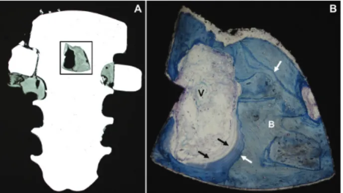
Microscopic analysis showed bone-implant interface constituted of lamellar bone tissue with various levels of radiopacity and stain-ing, typical of normal bone remodelling occurring during implant osseointegration. Necrotic bone most likely was a consequence of the placement surgery. The presence of cracks in these remnants of Figure 1. Longitudinal section through a miniplate and a screw removed
from a 13-year-old female patient after 38 months of implantation (patient no. 16). A. Microradiograph (x8). B. Enlargement of the framed area in A, observed in the stained section (x50). B: bone; V: blood vessels; white arrows: cementing lines; black arrows: osteoid (unmineralized preosseous layer).
Figure 2. Microradiograph of a transverse section through a bone sample
obtained during implant removal from a 35-year-old female patient after 35 months of implantation (×9) (patient no. 21). The right part of the bone fragment exactly corresponds to a segment of the screw circumference.
older bone, as observed in Figure 4B, was recently demonstrated in rabbit tibiae to be caused by the insertion procedure (15). This damaged bone was progressively replaced by bone remodelling, as shown by cement lines, exactly like it has been described for dental implants (16). Its persistence as well as the presence of mature lamel-lar bone after several months suggests that the implantation trauma
was limited and did not induce any massive resorption reaction. This tissue ensured primary stability while stimulating local remodelling. Indeed, new, younger bone was apposited directly on these remnants, from which it is separated by cementing lines, and can be considered the result of a moderate remodelling. The presence of osteoid on the bone surfaces completed this panel typical of bone healing around an implant (8).
Bone overgrowing over the miniplates, which contributes to increase the stability of the miniplates, also compromises the easi-ness of the removal procedure. Previous clinical investigations showed that over 25% of the miniplates are covered by bone overgrowth in more that one patient out of ten (12). Although the majority of the miniplates were easy to remove, bone healing was nevertheless extensive as attested by the presence of lamellar bone islets adhering to the screw circumference (Figure 1) as well as by the exact empty screw cast in bone fragments (Figure 2), illustrat-ing the paradox of skeletal anchoragement to be ‘temporary’ but however sometimes excessive. Integration seems to increase with time, which increases miniplates’ stability but can also impede mini-plate removal in extreme situations. Therefore, minimini-plates should be removed as soon as the objectives are obtained, so that the removal could be easy and atraumatic for the bone. In orthopaedic sur-gery, similar aspects were observed at the time of surgical removal of osteosynthesis devices. A recent clinical study describes bone encasing implants and expanding within the screw holes (17). The problem is so obvious in orthopeadic surgery that specific implant surface treatments have been tested on sheep in order to facilitate removal by decreasing bone-implant interaction (18). Bone over-growing plates and screws encourage the surgeon to remove them as soon as they are no longer necessary. This is especially true in children where plates or screws are completely surrounded by bone after a few months (19).
By contrast, miniplates removed early because of failure were surrounded by immature, woven bone and much more conjunctive tissue than found around the stable miniplates. In those miniplates, instability generally appeared during the first five weeks, prob-ably during the transition period between primary and secondary stability. Instability did not necessarily mean therapeutic failure: if miniplates were minimally mobile, they could generally achieve their anchorage purpose anyhow. This was the case for four dif-ferent patients in this study (2, 7, 9, and 19) where the miniplates were mobile but orthodontic objectives were obtained after a mean treatment duration of 16 months. These features can be related to Figure 4. Transverse section through a screw removed by core drilling from
a 13-year-old female patient after 38 months of implantation (patient no. 16). A. Microradiograph (x13). B. Enlargement of the framed area in A, observed in the stained section (x30). B: lamellar bone; c: cracks; N: necrotic bone; white arrows: cementing lines.
Figure 5. Transverse section through an unstable screw removed after one
month in a 12-year-old female patient (patient no. 8). A. Microradiograph (x20). B. Enlargement of the framed area in A, observed in the stained section (×150). F: fibroblasts.
Figure 3. Longitudinal section through a miniplate and a screw removed by core drilling from a 32-year-old male patient after 22 months of implantation (patient
no. 12). A. Microradiograph (×9). B. Enlargement of the upper framed area in A, observed in the stained section (×60). C. Enlargement of the lower framed area in A, observed in the stained section (×57). B: lamellar bone; N: necrotic bone; V: blood vessels; white arrows: cementing lines; black arrows: osteoid.
lack of primary stability, to difficulties encountered during miniplate surgery or to proximity with particular anatomical sites like sinusal cavity or dental roots (8).
In our sample, quantitatively restricted because of ethical and technical reasons (20, 21), no correlation could be established between loading duration and quantity of bone, although a longer loading period was reported to increase the possibility of lamel-lar bone development around miniscrews in humans (13). For the same reason, no correlation could be established between age of the patient and bone response in the present sample.
Conclusion
Our hypothesis was verified: the healing reactions observed in human samples in the present study are generally typical of mature lamellar bone tissue, with some bone in contact with the screw or the miniplate, and moderate remodelling activity. Few patients showed however abundant bone in contact with the screw, extending even to the miniplate surface. Finally, around the unstable miniplates, tissue reactions consisted mainly in some trabeculae of woven bone, as well as connective tissue around the screw and miniplate.
Acknowledgments
We thank C. Benoît for technical assistance with the histological sections and microradiographs.
References
1. Sugawara, J., et al. (2002) Treatment and posttreatment dentoalveolar changes following intrusion of mandibular molars with application of a skeletal anchorage system (SAS) for open bite correction. The Inter-national Journal of Adult Orthodontics and Orthognathic Surgery, 17, 243–253.
2. Costa, A., Raffainl, M. and Melsen, B. (1998) Miniscrews as orthodon-tic anchorage: a preliminary report. The International Journal of Adult Orthodontics and Orthognathic Surgery, 13, 201–209.
3. Willems, G., Carels, C.E., Naert, I.E. and van Steenberghe, D. (1999) Interdisciplinary treatment planning for orthodontic-prosthetic implant anchorage in a partially edentulous patient. Clinical Oral Implants Research, 10, 331–337.
4. Roberts, W.E., Arbuckle, G.R. and Analoui, M. (1996) Rate of mesial translation of mandibular molars using implant-anchored mechanics. The Angle Orthodontist, 66, 331–338.
5. Wehrbein, H., Feifel, H. and Diedrich, P. (1999) Palatal implant anchorage reinforcement of posterior teeth: a prospective study. American Journal of Orthodontics and Dentofacial Orthopedics, 116, 678–686.
6. Melsen, B. and Verna, C. (2005) Miniscrew implants: the Aarhus anchor-age system. Seminars in Orthodontics, 11, 24–31.
7. De Clerck, H., Geerinckx, V. and Siciliano, S. (2002) The zygoma anchor-age system. Journal of Clinical Orthodontics, 36, 455–459.
8. Cornelis, M.A., et al. (2008) Orthodontic loading of titanium miniplates in dogs: microradiographic and histological evaluation. Clinical Oral Implants Research, 19, 1054–1062.
9. Cornelis, M.A., Scheffler, N.R., Nyssen-Behets, C., De Clerck, H.J. and Tulloch, J.F. (2008) Patients’ and orthodontists’ perceptions of mini-plates used for temporary skeletal anchorage: a prospective study. American Journal of Orthodontics and Dentofacial Orthopedics, 133, 18–24.
10. Cornelis, M.A. and De Clerck, H.J. (2007) Maxillary molar distalization with miniplates assessed on digital models: a prospective clinical trial. American Journal of Orthodontics and Dentofacial Orthopedics, 132, 373–377.
11. De Clerck, H.J., Cornelis, M.A., Cevidanes, L.H., Heymann, G.C. and Tulloch, C.J. (2009) Orthopedic traction of the maxilla with miniplates: a new perspective for treatment of midface deficiency. Journal of Oral and Maxillofacial Surgery, 6, 2123–2129.
12. Cornelis, M.A., Scheffler, N.R., Mahy, P., Siciliano, S., De Clerck, H.J. and Tulloch, J.F. (2008) Modified miniplates for temporary skeletal anchorage in orthodontics: placement and removal surgeries. Journal of Oral and Maxillofacial Surgery, 66, 1439–1445.
13. Vasoglou, M., Chrysomali, E., Zinelis, S., Bitsanis, I., Haralambakis, N., Makou, M. and Eliades, G. (2013) Retrieval analysis of immediately loaded orthodontic mini-implants: material and tissue characterization. European Journal of Orthodontics, 36, 683–689.
14. Roberts, W.E. (1988) Bone tissue interface. Journal of Dental Education, 52, 804–809.
15. Lee, N.K. and Baek, S.H. (2010) Effects of the diameter and shape of orthodontic mini-implants on microdamage to the cortical bone. Ameri-can Journal of Orthodontics and Dentofacial Orthopedics, 138, 8.e1–e8; discussion 8.
16. Mori, S. and Burr, D.B. (1993) Increased intracortical remodeling follow-ing fatigue damage. Bone, 14, 103–109.
17. Georgiadis, G.M., Gove, N.K., Smith, A.D. and Rodway I.P. (2004) Removal of the less invasive stabilization system. Journal of Orthopaedic Trauma, 18, 562–564.
18. Hayes, J.S., Seidenglanz, U., Pearce, A.I., Pearce, S.G., Archer, C.W. and Richards, R.G. (2010) Surface polishing positively influences ease of plate and screw removal. European Cells & Materials, 19, 117–126.
19. Pate, O., Hedequist, D., Leong, N. and Hresko, T. (2009) Implant removal after submuscular plating for pediatric femur fractures. Journal of Pediat-ric Orthopaedics, 29, 709–712.
20. Melsen, B. and Costa, A. (2000) Immediate loading of implants used for orthodontic anchorage. Clinical Orthodontics and Research, 3, 23–28. 21. Büchter, A., et al. (2006) Load-related bone modelling at the interface of
orthodontic micro-implants. Clinical Oral Implants Research, 17, 714– 722.