With 3 figures in the text
Metacyclic Trypanosoma vivax possess a surface coat
P. R. GARDINER1, P. WEBSTER1, L. JENNP and S. K. MOLOO1
1
International Laboratory for Research on Animal Diseases, P.O. Box 30709, Nairobi, Kenya
2
Swiss Tropical Institute, Socinstrasse 57, C//-4051 Basel, Switzerland (Accepted 20 May 1985)
SUMMARY
Coated metacyclics of Trypanosoma vivax exist in the hypopharynges of infected tsetse flies and are extruded in low numbers when the flies are induced to probe onto warm slides or into medium. After extensive searching of T. vivax-mkcteA proboscides, and resort to a process for the examination of single, extruded, metacyclic trypanosomes, electron micro-scopic evidence is presented that, contrary to an earlier report, metacyclic T. vivax acquire a surface coat before contact with the mammalian host. Since T. vivax exhibits antigenic variation, the role of the surface coat in this species is likely to be functionally equivalent to the surface coat of the other tsetse-transmitted trypanosome species, T. brucei and T. congolense.
INTRODUCTION
The cycle of development that Trypanosoma (Duttonella) vivax Ziemann 1905 undergoes in its tsetse fly vector is confined to the proboscis (Bruce, Hamerton, Bateman &Mackie, 1910; Bruce, Hamerton, Bateman, Mackiefe Bruce, 1911). Earlier descriptive work (Fraser & Duke, 1912; Lloyd & Johnson, 1924; Roubaud, 1935; van Hoof, Henrard & Peel, 1937; reviewed in Hoare, 1972) suggested that ingested bloodstream trypomastigotes attach to the inner wall of the food canal of the tsetse proboscis where they differentiate into epimastigote forms (called crithidial forms in the older works). The epimastigote forms divide at their site of attachment giving rise to clusters of trypanosomes (Roubaud, 1935). Vickerman (1973) has described the ultrastructure of these attached trypanosomes in detail. Some of the epimastigotes detach from their original site and enter the hypopharynx where free-swimming metacyclic trypanosomes are seen following differentiation of some of the epimastigotes through intermediate trypomastigote stages. Roubaud (1935) stressed, however, that the appearance of trypanosomes, morphologically resembling metacyclic forms, often preceded the actual acquisition of infectivity of tsetse flies for vertebrate hosts. In T. brucei and T. congolense the acquisition of infectivity for vertebrate hosts is correlated with the assumption by metacyclic trypanosomes of an electron-dense surface coat (Vickerman, 1969,1974), the site of the variant-specific surface glycoprotein (Vickerman & Luckins, 1969). All other phenotypes in the vector are uncoated as are the epimastigotes of T. vivax (Vickerman, 1973). It was to be expected therefore that metacyclics of T. vivax would also possess a surface coat before their injection by the fly into the vertebrate host. However, Tetley, Vickerman & Moloo (1981) failed to find coated trypanosomes upon electron microscopical investigation of T. vivax-miecteA tsetse hypopharynges, and suggested that the attached, uncoated trypomastigotes in the hypopharynx represent the true metacyclic stage of T. vivax and hypothesized that the surface coat, prominent on bloodstream forms of T. vivax, as it is in the bloodstream forms of T. brucei and
P. R. GARDINER AND OTHERS
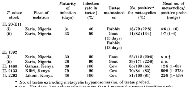
Table 1. An estimate of the number of metacyclic trypanosomes of various Trypanosoma vivax stocks extruded in salivary probes by Glossina morsitans centralis
T. vivax stock IL20-E11 (i) (») IL 1392 (i) (») IL 1480 IL 2133 IL 2292 Place of isolation Zaria, Nigeria Zaria, Nigeria Zaria, Nigeria Zaria, Nigeria Galana, Kenya Kilifi, Kenya Likoni, Kenya Maturity of infection (days) 31 33 30 26 30 79 38 Infection rate in tsetse% (%) 40 50 90 90 100 90 100 Tsetse maintained on Rabbit Goat (15 days) Rabbit (13 days) Goat Goat Cow Cow Cow No. positive* for metacyclics (%> 18/79 (22-8) 11/82(13-4) 23/112 (20-5) 39/171 (22-8) 65/100 (65) 70/84 (83) 81/100(81) Mean no. of metacyclics/ positive probe (range) 4-6(1-16) 1-7(1-4) N.D.f N.D. 12-8 (1-65) 50-9 (1-273) 22-9 (1-159) * No. of tsetse extruding metacyclic trypanosomes/no. of tsetse probed.
I N.D., Not done, but only rarely was more than 1 metacyclic present/positive probe.
% Derived by determining hypopharynx infection in 10 random flies.
We therefore undertook to examine T. vivax trypanosomes both in the hypopharynx of infected tsetse flies, and those trypanosomes extruded by tsetse flies induced to probe onto warmed slides or into medium. Because of the relative paucity of extruded trypanosomes in fly probes we evolved a technique for single cell electron-microscopy. In this paper we demonstrate that coated metacyclic trypanosomes of T. vivax exist in the tsetse vector before contact with the mammalian host.
MATERIALS AND METHODS
T. vivax stocks IL 20-E11 and IL 1392 are derivatives of IL V17 - the stock employed
in the previous study (Tetley et al. 1981) - originally isolated by Leeflang, Buys & Blotkamp (1976), as Y486 from a steer in Nigeria. T. vivax IL 1480 has been syringe passaged through 2 calves since its isolation from a steer at Galana, Kenya. T. vivax IL 2133 has been cycled twice through tsetse, once between cattle and once between a steer and a goat, since its isolation from a bovine at Kilifi, Kenya. T. vivax IL 2292 has been passaged 5 times in cattle, including 2 tsetse transmissions between cattle, since its isolation from a steer at Likoni, Kenya.
Teneral Glossina morsitans centralis, or G. m. morsitans in one experiment, from the ILRAD colony were fed on goats for the West African stocks of T. vivax, or on cattle in the case of the East African stocks, shortly after the first appearance of trypanosomes in the blood, and then fed daily (except Sundays) as shown in Table 1. Infection rates in the tsetse were estimated after 25 days by counting the number of flies showing hypopharyngeal infections when 10 tsetse were dissected at random. The tsetse were starved for 2 days and then allowed to probe onto individual wells of a warmed (37 °C) Teflon-coated slide. The dried probe was examined without staining by means of a light microscope at a final magnification of 320 x . All metacyclic trypanosomes (i.e. excluding occasional long forms) were counted in each probe.
For more thorough investigation of metacyclic morphology at the light microscopic level, fly probes were Giemsa stained by a slight modification of the method of Gray, Ross, Taylor & Luckins (1984). The modification added was pre-fixation of the probe with phosphate-buffered 2 % (v/v) formaldehyde (pH 70) for 2-5 min before rinsing in buffer alone and methanol fixation.
For direct electron microscopical examination of an infected hypopharynx the head and proboscis of a G. m. morsitans fly was fixed 11 days after feeding on the blood of a goat infected with IL 20-E11. The proboscis was processed in a manner similar to that described (Tetley et al. 1981) but the initial fixative [3 % (v/v) glutaraldehyde in 200 imi phosphate buffer] contained 2% (w/v) saccharose rather than CaCl2. All reagents were
of a grade of purity sufficient for electron microscopy. Ultrathin sections of the embedded proboscis were examined in a Zeiss EM 9 electron microscope.
For electron microscopy of extruded metacyclics, 40-50 infected tsetse flies were allowed to probe through a silicone membrane into approximately 1-5 ml of medium contained in a test-tube. The medium contained HEPES-buffered Modified Eagle's Medium with 20 % (v/v) foetal calf serum, 1 % (v/v) non-essential amino acids, 1 % (v/v) glutamine (all from Gibco Ltd., Paisley, Scotland) and 5/ig/ml gentamycin sulphate. The probed medium was fixed with an equal volume of a fixative containing 5 % (v/v) glutaraldehyde, 2-8% (w/v) formaldehyde and 0-28% (w/v) picric acid in 63 mil phosphate buffer, pH 74, and then centrifuged at 1300g for 10 min. Most of the supernatant fraction was discarded whilst the bottom 0-2 ml was retained and gently mixed by pipetting up and down. Glass cover-slips, pre-coated with a film of O'l % (w/v) gelatin and 0-5% (w/v) agarose which had been allowed to dry, were trapped between a glass slide and the filter paper of a Cytospin (Shandon Southern, Cheshire, England) and pre-spun for 2 min at 1000 r.p.m. (speed setting 7) with approximately 0-l ml distilled water to rehydrate the gelatin/agarose film. The fixed samples were then spun similarly onto the same cover-slips and the cover-slips rapidly removed and flooded with further fixative for 5—10 min. Taking care to keep them moist at all times the cover-slips were then subjected to the following processing. They were washed by constant replacement of 100 mM phosphate buffer, pH 7-4, for 5 min followed by a 1 min rinse in 0-l M sodium cacodylate buffer, pH 7-4. A 10 min fixation in cacodylate buffered 1 % (w/v) osmium tetroxide was followed by constant rinsing for 5 min in cacodylate buffer. Washing for 1 min in 0-05 M sodium maleate, pH 5-4, preceded staining for 10 min in 1 % (v/v) uranyl acetate in the same buffer. After a further rinse for 5 min in sodium maleate the gelatin/agarose films were dehydrated by 1 min step-wise processing through graded alcohols. Two rinses of 1 min each in propylene oxide preceded incubation for 10 min in a 1:1 mixture of propylene oxide/resin mixture before overnightembedmentinresin mixture (1:1 mixtureofEpon812/Araldite6005,ErnestF. Fullam, Schenectady, N.Y., USA) at 60 °C. After polymerization the resin was peeled away with the help of a scalpel blade to expose the back (i.e. not the gelatin/agarose film side) of the cover-slip. The cover-slip was then broken away after plunging the resin block into liquid nitrogen and allowing the embedded cover-slip to come to room temperature. The resin blocks were scanned with the aid of a light microscope using a 32 x long working distance objective lens. The position of individual trypanosomes was marked by scratching the resin with a syringe needle. Marked areas were removed, mounted on resin stubs with sealing wax, trimmed and then aligned to a diamond knife edge with the aid of a backlighted knife stage and block holder (Sunkay Laboratories, Tokyo, Japan) on a Sorvall MT-2B ultramicrotome (Dupont, Wilmington. DE, USA). All sections were collected on carbon-coated, single slot grids (2x1 mm), stained with
Fig. 1. Light micrograph of Uiemsa-stained trypanosomes {Trypanosoma vivax IL 1480) extruded in the salivary probe of a Glossina morsitans centralis fly. Four typical metacyclic
trypanosomes are shown. The acid treatment extracts cytoplasmic organelles but reveals the outline morphology, kinetoplast and nucleus in detail.
uranyl acetate and lead citrate and viewed in a Zeiss EM 10A electron microscope operating at 60 kV. Trypanosome profiles were detected approximately 20-30 sections
into the block, depending upon the thickness of the gelatin layer.
RESULTS
The number of metacyclic trypanosomes of the T. vivax stocks employed in this study which are extruded by G. m. centralis in salivary probes under experimental conditions is low, the naturally rodent-infective stock from Nigeria being poorer in this regard than the East African stocks (Table 1). Giemsa-stained metacyclics of IL 1480 (Fig. 1) show the large kinetoplast typical of T. vivax located terminally. The body of the trypanosome appears broad posteriorly, tapering sharply anteriorly of the nucleus. The undulating membrane is well developed and a pronounced free flagellum is characteristically present. This morphology corresponds closely with the drawings of Hoare (1970, form 0 in fig. 2.1; and 1972, form k in fig. 86) of ' metatrypanosomes in the hypopharynx'. The more slender forms depicted by Hoare (1970, form n in fig. 2.1; and 1972 forms
1 a n d j in fig. 86) were only rarely noted in this study.
The results of electron microscopy of single, extruded metacyclic T. vivax demonstrate that they possess a dense surface coat around the body and flagellum of the trypanosome (Figs. 2 and 3 A) before contact with the mammalian host. Further, profiles of coated trypanosomes are present within the hypopharynx of T. vivax-'mfected tsetse flies (Fig. 3B). The observation of uncoated trypanosomes (Fig. 3C) in the same salivary probes as coated trypanosomes, verifies that the surface coat of extruded metacyclic trypanosomes is not an artefact of the specimen preparation technique employed. These findings are at variance with an earlier report (Tetley et al. 1981) but confirm that the maturation of T. vivax in the insect vector culminates in a coated metacyclic trypanosome as is the case with T. brucei and T. congolense.
DISCUSSION
Before the present study was undertaken there was good circumstantial evidence that the forms of T. vivax injected by tsetse into mammals must possess a surface coat since
goats immunized with, and producing antibody to, an MT 83 K surface membrane
Fig. 2. Transmission electron micrograph of a section of an extruded metacyclic of
Trypanosoma vivax IL 2292 showing a surface coat over the body and flagellum of the
P. R. GARDINER AND OTHERS
Fig. 3. Transmission electron micrographs of sections of Trypanosoma vivax. A. Extruded metacyclic of T. vivax IL 2292 clearly showing a surface coat in the parts of the section cut perpendicular to the surface membrane. B. Part of a section of coated metacyclic trypanosome of T. vivax IL 20-E11 in the hypopharynx of Glossina morsitans morsitans. The arrow indicates the thickness of the surface coat. C. An extruded trypomastigote of T. vivax IL 2292 from the same sample as the trypanosomes depicted in Figs 2 and 3A. The trypanosome lacks a surface coat but shows small, surface membrane accretions thought to represent an artefact of the phosphate-buffered fixative.
protein common to T. vivax, T. congolense and T. rhodesiense were completely susceptible to T. vivax infection upon tsetse challenge (Rovis, Musoke & Moloo, 1984). If metacyclic
T. vivax lacked a surface coat (Tetley el al. 1981), thus exposing the MT 83 K membrane
protein, some degree of protection would have been expected to have been afforded the immunized goats. Similarly, the susceptibility of livestock to homologous and heterologous rechallenge by tsetse fly (Moloo & Emery, unpublished; Gardiner, unpublished), would be difficult to explain if the infecting trypanosomes presented invariant, cell membrane antigens to the host animal.
That the surface coat of metacyclic T. vivax has previously been so difficult to demonstrate is no doubt due to the paucity of these forms both in the hypopharynx of infected tsetse and in salivary probes, and perhaps to the technical aspects of fixation (phosphate-buffered fixatives give better preservation of the surface coat of T. vivax than similar fixatives buffered in cacodylate - Professor S. Ito, Harvard University, personal communication). The numbers of metacyclic trypanosomes given in Table 1 can only be an estimate, since the extent of the probe on the glass slide inevitably differs from fly to fly, but from the data presented the West African stock which was employed in a previous investigation with negative results (Tetley et al. 1981) would seem the least suitable material to examine. Nevertheless metacyclics of this stock also possess a surface coat (Fig. 3B). Other variables which might increase the number of metacyclic
T. vivax produced by tsetse flies, such as the passage history of the stock, the species
of animal on which the tsetse are maintained, the maturity of infection in the tsetse or the tsetse species itself, have not been critically examined in this study. If, however,
the figures given here for metacyclic numbers are representative of the biology of
T. vivax in the field, the bite of T. mvaz-infected tsetse will deposit fewer metacyclics in
the skin of the vertebrate host than do flies infected with T. congolense or T. brucei. It seems probable also that a mixture of coated and uncoated trypanosomes will be introduced into the skin of an animal bitten by T. vivaz-infected tsetse, since both were found in salivary probes by electron microscopj', and larger, presumably epimastigote forms, were occasionally noted in some salivary probes at the light microscope level. That uncoated trypomastigote forms are extruded by tsetse flies as well as coated metacyclic trypanosomes suggests that a process of maturation may be occurring in the trypomastigotes in the hypopharynx of the tsetse fly (Roubaud, 1935). This presumably involves reduction in the tenacity of flagellar attachment, as well as the generation of a surface coat and, perhaps, subtle modulations in the morphology of these hypo-pharyngeal forms to yield the final metacyclic trypanosome depicted in Fig. 1. Nothing is known of the biochemistry of this differentiation but Roubaud's (1935) observation that there is a delay in the assumption of infectivity after trypanosome invasion of the hypopharynx, and Tetley et al.'s (1981) finding that trypomastigotes in the proboscis are unaffected by fresh guinea pig serum (uncoated trypanosomes from 25 °C cultures of the same stock of T. vivax are both agglutinated and lysed by fresh guinea pig serum — Hirumi & Gardiner, unpublished observations), together suggest that generation of the surface coat in T. vivax may be a gradual process and that cell surface changes in trypomastigote forms occur in advance of a demonstrable surface coat on mature metacyclic trypanosomes.
T. vivax demonstrates antigenic variation (reviewed by Gray & Luckins, 1976; De
Gee, Shah & Doyle, 1979, 1980; Murray & Clarkson, 1982; Barry & Gathuo, 1984) and undergoes a cyclical development in the tsetse fly in which it first loses and then reacquires a surface coat. This life-cycle is thus functionally comparable with that of the two other major tsetse-transmitted species, T. brucei and T. congolense.
Note added by Mr L. Tetley and Professor K. Vickerman:
We happily concede that metacyclic Trypanosoma vivax does possess a surface coat and that the choice of material on which we based our 1981 paper was unfortunate, as indicated by Dr Gardiner and colleagues. Our recent detailed study of metacyclic differentiation in T. brucei (Tetley & Vickerman, 1985) makes it clear that the attached uncoated T. vivax trypomastigotes that we described from the tsetse hypopharynx correspond to the pre-metacyclic trypomastigotes of T. brucei. The latter acquire a surface coat to become 'nascent metacyclics' while still attached to the tsetse salivary epithelium, and are then released as mature metacyclics.
The authors would like to thank Ms Ravi Thatthi and staff members of the ILRAD Tsetse Vector Laboratory for technical assistance, and Dr Maureen Gray for bringing the method of staining trypanosomes in salivary probes to our attention in advance of publication. This is ILRAD publication number 336.
REFERENCES
BARRY, J. D. & GATHUO, H. (1984). Antigenic variation in Trypanosoma vivax: isolation of a serodeme.
Parasitology 89, 49-58.
BRUCE, D., HAMERTON, A. E., BATEMAN, H. R. & MACKIE, F. P. (1910). The development of trypanosomes in tsetse flies. Proceedings of the Royal Society, B 82, 382-8.
on the development ofTrypanosoma vivax in laboratory-bred Glossinapalpalis. Report of the Sleeping
Sickness Commission of the Royal Society 11, 50-3.
D E GEE, A. L. W., SHAH, S. D. & DOYLE, J. J. (1979). Trypanosoma vivax: sequence of antigenic
variants in mice and goats. Experimental Parasitology 48, 352-8.
D E GEE, A. L. W., SHAH, S. D. & DOYLE, J. J. (1980). Trypanosoma vivax: Host influence on appearance of variable antigen types. Experimental Parasitology 51, 392-9.
FRASER, A. D. & DUKE, H. L. (1912). The development of trypanosomes in Glossina palpalis. Report
of the Sleeping Sickness Commission of the Royal Society 12, 36-56.
GRAY, A. R. & LUCKINS, A. G. (1976). Antigenic variation in salivarian trypanosomes. In Biology of
the Kinetoplastida (ed. W. H. R. Lumsden and D. A. Evans) pp. 493-542. London: Academic Press.
GRAY, M. A., Ross, C. A., TAYLOR, A. M. & LUCKINS, A. G. (1984). In vitro cultivation of Trypanosoma
congolense: The production of infective metacyclic trypanosomes in cultures initiated from cloned
stocks. Ada Tropica 41, 343-53.
HOARE, C. A. (1970). The mammalian trypanosomes of Africa. In The African Trypanosomiases (ed. H. W. Mulligan), pp. 3-59. Ministry of Overseas Development. London: George Allen and Unwin.
HOARE, C. A. (1972). The Salivaria. Subgenus Duttonella Chalmers, 1918. In The Trypanosomes of
Mammals. A Zoological Monograph, pp. 401-429. Oxford: Blackwell Scientific Publications.
HOOF, L. VAN, HENRARD, C. & PEEL, E. (1937). Influence de repas preliminaires indifferents sur 1 'evolution de Trypanosoma cazalboui chez Glossina palpalis. Comptes Rendus des Seances de la Societe
de Biologie et de ses Filiales, France 126, 1249-52.
LEEFLANG, P., BUYS, J. & BLOTKAMP, J. (1976). Studies on Trypanosoma vivax: infectivity and serial maintenance of natural bovine isolates in mice. International Journal for Parasitology 6, 453-6.
LLOYD, L. & JOHNSON, W. B. (1924). The trypanosome infections of tsetse flies in northern Nigeria and a new method of estimation. Bulletin of Entomological Research 14, 265-88.
MURRAY, A. K. & CLARKSON, M. J. (1982). Characterization of stocks of Trypanosoma vivax. II. Immunological studies. Annals of Tropical Medicine and Parasitology 76, 283-92.
ROUBAUD, E. (1935). Les modalites atypiques de l'infection trypanosomienne cyclique chez les Glossines. Annales de VInstilul Pasteur (Paris) 55, 340-64.
Rovis, L., MUSOKE, A. J . & M O L O O , S . K. (1984). Failure oftrypanosomal membrane antigens to induce protection against tsetse-transmitted Trypanosoma vivax or T. brucei in goats and rabbits. Ada
Tropica 41, 227-36.
TETLEY, L. & VICKERMAN, K. (1985). Differentiation of Trypanosoma brucei: Host-parasite cell junctions and their persistence during acquisition of the variable surface coat. Journal of Cell Science 74, 1-19.
TETLEY, L., VICKERMAN, K. & MOLOO, S. K. (1981). Absence of a surface coat from metacyclic
Trypanosomavivax: possible implications for vaccination against vivax trypanosomiasis. TVarasaeiiems of the Royal Society of Tropical Medicine and Hygiene 75, 409-14.
VICKERMAN, K. (1969). On the surface coat and flagellar adhesion in trypanosomes. Journal of Cell
Science 5, 163-93.
VICKERMAN, K. (1973). The mode of attachment of Trypanosoma vivax in the proboscis of the tsetse fly Glossina fuscipes: an ultrastructural study of the epimastigote stage of the trypanosome. Journal of Protozoology 20, 394-404.
VICKERMAN, K. (1974). Antigenic variation in African trypanosomes. In Parasites in the Immunized
Host: Mechanisms of Survival. Ciba Fdn Symp. No. 25, pp. 53-80. Amsterdam: Elsevier/North
Holland/Excerpta Medica.
VICKERMAN, K. & LUCKINS, A. G. (1969). Localization of variable antigens in the surface coat of
Trypanosoma brucei using ferritin-conjugated antibody. Nature, London 224, 1125-6.