Plant & GUI Pkfiiol. 18: 1047-1055 (1977)
Aging of the photosynthetic apparatus VI. Changes in
pH dependence of JpH, thylakoid internal pH and
proton uptake and relationships to electron transport
Paul-Andre1 Siegenthaler and Franchise Dep£ryLaboratoirc de Phyiiologic Vegetalc ct Biochimic, University de Neuchatcl, 20 rue de Chantcmerle, 2000 Neuchfitel, Switzerland
(Received April 19, 1977)
The pH difference generated across the chloroplast membrane upon illumination (idpH) and the internal pH (pHi) were analyzed in aged spinach chloroplasts and in fresh chloroplasts supplemented with linolenate. In electron-flow conditions where both photosystems or either photosystem alone were functional, the ApH dropped and their optima shifted toward more acidic external pH (pHo) with a simultaneous increase in pHi. Upon aging or addition of linolenate, a decrease of pHo was therefore required to maintain the pHi in the range of 5—5.5 for maximum electron-flow activity. More-over, aging like linolenate, diminished the proton pump activity and shifted its optimum (pH 6.7 in the controls) toward higher pHo. Although J p H and pHi changes were similar in all electron-flow conditions, the sensitivity of idpH toward aging and linolenate was eventually higher under photosyitem II than photosyitcm I conditions.
In conclusion, the electron-flow activity seems to be delicately controlled by the proton pump, idpH, pHi and pHo. Unsaturatcd fatty acids which arc released during chloroplast aging damage the membrane integrity in such a way that the subtle equili-brium between these factors is disturbed.
Free fatty acids control the structure (5, 15, 16, 22, 27) and function (3, 13,
14, 28-31) of chloroplast membranes. For instance, the inhibition of photosynthetic
electron flow by unsaturated fatty acids is pH-dependent: linolenic acid causes a shift in the pH optimum toward acidity (28, 29, 31). This phenomenon was shown to be related to the thylakoid membrane integrity as suggested by the action of exogenous linolenic acid on ^pH, pHi and proton uptake (29).
Aging of isolated spinach chloroplasts results also in an acidic shift of the pH optimum for electron flow through both photosystems or through either photo-system alone (14, 31). This was correlated with the release of unsaturated fatty acids, predominantly linolenate, which takes place in the membrane environment during aging (31).
Using the same conditions as those adopted in the electron-flow studies (31), we verified that aging and linolenic acid have similar effects on light-induced ^ipH, pHi and proton uptake in thylakoid membranes. These results confirm that the factors which do regulate the aging process are mainly due to the level of free fatty acids in the thylakoid membranes.
1048 P. A. Sicgenthalcr and F. Depery
Material and methods
Spinach (Spinacia oleracea var. Nobel) was grown in a growth chamber (29). Thylakoid membranes were prepared as described previously (29) and resuspended in 25 mM HEPES (pH 7.6) and 0.35 M sucrose to 2 mg chlorophyll/ml or in the aging medium (25 HIM HEPES, pH 7 and 175 HIM NaCl) to 1 mg chlorophyll/ml. Thylakoid membranes were dark-aged at 20°C and portions were removed at various times for J p H and proton pump measurements. All isolation operations were car-ried out at 4°C and in dim light. Chlorophyll was determined spectrophoto-metrically (4).
The JpH in chloroplast membrane systems was estimated from the extent of the fluorescence quenching of 9-aminoacridine, employing the technique originally proposed by Schuldiner et al. (21). The fluorescence was measured with a spectro-fluorimeter adapted for illumination on one side of the cuvette. Actinic light (approximately 5 X 10⁶ ergs-cm^-sec"1 provided by a halogen lamp, was passed through two Calflex and a Corning CS-260 filters. Monochromatic exciting light (390 nm), provided by a Bausch and Lomb high-intensity monochromator (xenon light source combined with a visible grating), was passed through a PAR light chopper (Model 125 A, 48 aperture wheel) before reaching the 1-cm cuvette. The fluorescence emission was detected at a 90° angle with an EMI photomultiplier tube (quartz window) monitored by a Bausch and Lomb monochromator at 460 nm. The chopper and the photomultiplier tube were connected to a PAR lock-in ampli-fier (Model 128 A) and the fluorescence signal was recorded with a W + W recorder (Model 1200). The osmotic volume (V) of the chloroplast preparation was estimat-ed from the data providestimat-ed by Rottenberg et al. (19) and was found to be 32.5 /<l/mg of chlorophyll in our experimental conditions. The J p H was obtained from the relationship JpH=log [QV(1 — Q,) X 1/V] (Q,=fraction of the total fluorescence that was quenched) and the internal pH (pHi) calculated by subtracting the idpH from the external pH (pHo). The basic reaction mixture contained: 30mM N-2-hydroxy-ethylpiperazine-N-2-ethane sulfonic acid (HEPES) or N-tris (hydroxymethyl) methylglycine at various pH, 40 mM NaCl, 0.8 /m 9-aminoacridine, 0.5% ethanol or linolenic acid, and chloroplasts (20 fig chlorophyll/ml). According to the type of electron flow studied, various electron donors and acceptors were added as indicat-ed in the figure legends. When linolenic acid, DCMU, diaminodurol (dissolvindicat-ed in ethanol) and DBMIB (dissolved in methanol) were added to these reaction mixtures, the appropriate controls were made with ethanol (0.5%) and methanol. At the concentrations used, ethanol and methanol had no detectable effect on electron flow, JpH, pHi and proton uptake.
To measure the proton pump, chloroplasts were isolated as indicated previously but resuspended in 175 HIM NaCl (pH 8) without buffer. The pH changes were measured with a Metrohm pH-meter (Type E 300 B) and recorded continuously (77). The reaction mixture (5 ml) contained 35 mM NaCl, 20 fm phenazine methosulfate, 0.5% ethanol and chloroplasts (40 /ig chlorophyll/ml). Light intensity of the actinic light was approximately 5x 10s ergs-cm-2-sec~1 and the temperature was 20°C. The p equiv. H+ taken up per mg chlorophyll was estimated from the data provided by Walz et al. [see Fig. 1A in (32) ].
JpH, pHi and proton uptake in aged chloroplajti 1049
from Fluka. Dibromothymoquinone was kindly provided by Drs H. Baltscheffsky and A. Trebst.
Results
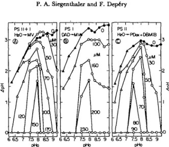
The influence of chloroplast aging on light-induced J p H is illustrated in Fig. 1 for three types of electron flows which were previously studied (31). The J p H of the controls increased as a function of pHo with a ma-rimnm around 9 for PS I I + I and PS I (Fig. 1A and B) and around 8-8.5 for PS II (Fig. 1C) conditions. The J p H values of the PS I I + I controls agree with those reported by Avron's group
(2, 19-21). However, at acid pHo, the J p H values for the PS II controls were
generally lower than those for the two other electron-flow systems. When the chloroplasts were aged, the JpH dropped for all three electron flow conditions. For example (Fig. 1A), at high pHo (9.0), the J p H decreased from 3.1 to 0 after 6 hr of chloroplast aging. At lower pHo, the extents of the J p H drop were smaller. This implies that aging caused a shift of the J p H optimum toward more acidic pHo and around pHo 7.5, had a much smaller effect on the JpH. Although the J p H changes were similar in the three types of electron-flow conditions, it is evident that the sensitivity of the proton concentration gradient to aging was eventually higher under PS II than PS I conditions (see for example the effect of 4-hr incubation in Fig. IB and 1C).
Fig. 2 shows that the effects of linolenic acid on J p H were similar to those initiated by chloroplast aging. The basic features described in Fig. 1 were again observed upon the addition of linolenic acid: (a) a J p H drop which was greater at
65 7 75 8 a5 9 pHo 65 7 75 8 85 9 pHo 65 7 75 8 85 9 pHo
Fig. 1. ApH Dependtnc* on pHo and chloroplast aging sxprtssed in hours (A) in tkre* differtnt electron-flow systems.
(A) In photosyitems II + I (from HjO to methylviologen): the baiic reaction mixture was supplement-ed with 0.15 mM methylviologen and 2 mil NaNa. (B) In, photosystem I only (from diaminodurcne to methylviologen): the basic reaction mixture was supplemented with 10 fiu DCMU, 300 /tu di-aminodurene, 2 mM sodium ascorbate, 0.15 n w methylviologen and 2 mM NaNa. (C) In photosyitem II only (from HjO to oxidized ^-phenylene diamine): the basic reaction mixture was supplemented with 0.2 mM />-phenylene diamine, 1.2 HIM KaFe(CN)« and 1 /ac DBMIB. A, 0 , controls; A . A , HEPES (pH 6.3-7.5); O , • . tricine-glycine (pH 7.5-9.3).
1050 P. A. Siegenthaler and F. Depcry l—I—r-n—I—r PS II HbO—PDw+DeMIB 6 65 7 75 8 85 9 pHo 6 6 5 7 73 B 85 9 pHo - 3 6 65 7 7.5 8 85 9 pHo
Fig. 2. ApH Dependence on pHo and linolmk acid conctrdration in the three different eltctron-fiow systems. (A) Photosystems 11 + 1 . (B) Photosystem I alone. (C) Photosyitem II alone. Conditions were ai described in Material and m e t h o d s and Fig. 1.
Fig. 3. pHi Dependence on pHo and Moroplast aging expressed in hours (A). The electron-flow syjtems and conditions were the same as in Fig. 1.
high than at low pHo, (b) a shift of the JpH optimum toward acidity, (c) a greater sensitivity of JpH under PS II (and PS II + I) than under PS I conditions, (d) a smaller inhibition of J p H in the pHo 7.5-8.0 region.
Since the rate of electron transport seems to be controlled not only by J p H but also by pHi (2), we investigated the dependence of pHi on various pHo during the aging process (Fig. 3) and compared it with the pHi of fresh thylakoids sup-plemented with linolenic acid (Fig. 4). In control experiments (closed symbols), pHi increased when pHo was raised. The maximum electron flow is known to be around pHo 8.7 in the r^O/methylviologen, 8.5 in the diaminodurol/methylviologen and 7.8 in the HaO/oxidized/>-phenylenediamine systems (31). The corresponding internal pH values were around 5.5, 5.3 and 5.1, respectively. These pHi values
, pHi and proton uptake in aged chloroplasti 1051
Fig. 4. pHi Dtptndtnu on pHo and linolenic add concmtraiioni (/*M). The electron-flow systems and conditions were the same as in Fig. 1.
agree well with those reported for other electron flow conditions (2, 19-21). Fig. 3 and 4 show also that below pHo 8, the pHi values were generally constant, probably indicating the existence of a natural buffering capacity in the acidic pHi range as suggested by Schuldiner et al. (21). Both aging (Fig. 3) and addition of increasing concentrations of linolenic acid (Fig. 4) displaced the curves toward higher pHi in the same way. For example, at pHo 8.5 in the PS I system alone, the pHi were respectively 5.3, 6.1, 8.5 after 0 (control), 4 and 8 hr of aging (Fig. 3B) and 5.2, 5.5, 6.8 and 8.5 in the presence of 0 (control), 100, 160 and 200 fut of linolenic acid (Fig. 4B). At pHo 7.5, the pHi were respectively 4.7, 5.3, 6.3 and 7.5 after 0, 4, 8 and 23 hr of aging (Fig. 3B) and 4.4, 4.7, 5.3, 6.3 and 7.5 in the presence of 0 (cont-rol), 100, 160, 200 and 230 /ZM of linolenic acid, respectively (Fig. 4B). Since a pHi around 5.2 was the optimum for photosystem I electron flow, in the course of chloroplast aging or in the presence of increasing concentration of linolenic acid, a decrease of pHo would be required to maintain pHi in the proper range (pHi 5.2) for maximum activity. This is also true for the two other types of electron-flow conditions, i.e., PS I I + I (Fig. 3A and 4A) and PS II alone (Fig. 3G and 4C). Moreover, Fig. 3 and 4 show that at pHo 7.5 ^0.5, aging and linolenic acid had smaller effects on the pHi, maybe due to a buffering capacity which was more effective in this pH range.
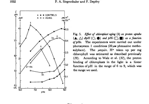
In order to test this buffering capacity, the extent of proton uptake, as well as JpH and pHi were plotted as a function of pHo and compared for fresh and aged chloroplasts. Fig. 5 shows that in the controls, the pHo optimum of proton uptake (6.7) was two to three units lower than the pHo optimum of JpH (9.0). After three hr of aging, both the proton uptake and ApYL diminished and the optima were displaced toward one another (see arrows in Fig. 5). If the extent of the proton pump activity is basically a measure of the internal buffer capacity (19), one may infer that aging, like linolenic acid (29), decreases the buffering capacity. After three hr of aging, all conditions (proton uptake, JpH, pHi and pHo) were optimal for electron-flow activity, only around pHo 7.4, as found previously in the presence of linolenic acid (29).
1052 P. A. Siegenthaler and F. Dep£ry
Fig. 5. Effect of Moroplast aging (A) on proton uptake ( A , A ) ApH ( O , • ) and pHi ( Q • ) as a function
qfpHo. The cxperimenti were carried out under
photosystem I conditions (20/tu phenazine metho-sulphate). The /xequiv. H+ taken up per mg
chlorophyll was estimated ai described previously
{29). According to Walz et al. (32), the proton
binding of chloroplasts in the light is a linear function of pH in the range of 6 to 9, which was the range we used.
Discussion
We have recently reported that both aging of isolated thylakoids and addition of linolenate to fresh thylakoids result in a similar acid shift of the pH optima for electron flows through both photosystems or cither photosystem alone (57). This phenomenon can now be explained or at least correlated with the integrity of the thylakoid membrane and its diminishing ability to create a proton gradient between the inner and outer spaces of the membrane during the aging process.
In thylakoids aged in vitro or supplemented with linolenate, the membrane deteriorates what is accompanied by decreases in the proton uptake (see Fig. 5 and ref. 29) and/lpH; the extent of the decreases depends upon aging time (Fig. 1) or linolenate concentration (Fig. 2). As a consequence, pHi increases eventually reaching pHo (Fig. 3 and 4). As these pH values are no longer optimal for electron flow, the activity is inhibited (31). Since the size of J p H is lowered, the pHo optimum for electron flow has to be shifted toward the acidic side in both thylakoids aged in vitro or supplemented with linolenate (28, 29, 31). Under these conditions, one can postulate that the fluxes of cations and anions are altered in such a way that swelling occurs (12, 23, 27) and light-induced shrinkage decreases (23, 27). Although light-induced dpH through both electron-flow conditions were affected by aging and linolenate, the ApH through PS II were much more sensitive than those through PS I conditions (Fig. 1 and 2). This finding is similar to and can be nicely correlat-ed with the observations of the electron-flow activities themselves (24, 28, 31). Moreover, the overall extent of proton uptake (internal buffering capacity) is diminished in aged thylakoids and its optimum shifts from pHo 6.7 to higher pHo as suggested by the results in Fig. 1, 3 and 5. Thus, in addition to swelling (18,
photo-, pHi and proton uptake in aged chloroplastx 1053
phosphorylation (24, 28) and electron transport activities (3, 5, 6, 9, 10, 13-15, 22,
24, 28-31, 34), the ^pH, pHi and proton uptake are affected in the same way by
aging and added linolenate. Therefore, as reported previously (27), one can simulate the aging process in thylakoids by using linolenic acid.
The factors which do regulate the aging process, especially the accumulation of free fatty acids in the thylakoid membranes, are still unknown. However, the present investigation suggests that aging is controlled, at least in part, by a pH mech-anism. Galactolipases which are bound to thylakoid membranes (1) are known to hydrolyze both mono- and digalactosyldiglyceride, the pH optima for the two substrates being respectively 7.5 and 5.9 for the spinach enzyme (7/). We can consider that in intact thylakoids, light generates a pHi of approximately 4.5 to 5.0 (Fig. 3 and 4) when pHo are respectively 7.4 and 7.9 [these values being the pH of the stroma in darkness and light (33) ]. In this pHi range, galactolipases (their localization within the membrane remain unknown) are probably almost inactive and no free fatty acids are released. On the contrary, in aged chloroplasts, the pHi increases thus creating conditions favorable for lipid hydrolysis.
Finally, we would like to mention that the fluorescence quenching of 9-amino-acridine as an indicator of the pH difference generated across the chloroplast membrane upon illumination has been questioned seriously by Fiolet et al. (7, 8). Although not incompatible with the energization theory proposed by these authors, our results fit well with the theory of Schuldiner et al. (21).
We thank M n Jana Smutny for her able technical assistance and the Swiss National Science Foundation for iti support (Grant no. 3.2470.74 to P. A. S.).
References
( / ) Anderson, M. M., R. E. McCarty and E. A. Zimmer: The role of galactolipids in spinach chloroplast lamellar membranes. I. Partial purification of a bean leaf galactolipid lipase and iti action on sub-chloroplast particles. Plant Physiol. 53: 699-704 (1974).
( 2) Bambcrger, E. S., H. Rottenberg and M. Avron: Internal pH, 4pH and the kinetics of electron transport in chloroplajtt. Em. J. Biochtm. 34: 557-563 (1973).
(3) Brody.S.S.: The effects of linolenic acid and extracts of Ricimu leaf on system I and system II. Z. Naturforsch. 25b: 855-859 (1970).
( 4 ) Bruinima, J.: A comment on the spectrophotometric determination of chlorophyll. Biochim.
Biophys. Acta 53: 576-578 (1961).
( 5) Cohen, W. S., B. Nathanson, J. E. White and M. Brody: Fatty acids as model systems for the action of Ricinus leaf extract on higher plant chloroplasts and algae. Arch. Biochtm. Biophys. 135: 21-27 (1969).
( 5 ) Constantopoulos, G. and C. N. Kenyon: Release of free fatty acids and loss of Hill activity by aging spinach chloroplasts. Plant Physiol. 43: 531-536 (1968).
( 7 ) Fiolet, J. W. T., E. P. Bakker and K. Van Dam: The fluorescent properties of acridines in the presence of chloroplasts or liposomes. On the quantitative relationship between the fluores-cence quenching and the transmembrane proton gradient Biochtm. Biophys. Acta 368: 432-445 (1974).
( 8 ) Fiolet, J. W. T., L. Van dcr Erf-Ter Haar, R. Kraayenhof and K. Van Dam: On the stimula-tion of the light-induced proton uptake by uncoupling aminoacridinc derivatives in spinach chloroplasts. ibid. 387: 320-334 (1975).
( 9 ) Fragata, M.: Effects of aging on chlorophyll fluorescence and photosystcm II electron trans-port in isolated chloroplasts. Can. J. Bot. 53: 2842-2845 (1975).
1054 P. A. Siegcnthaler and F. DepeTy
(70) Hamischfeger, G.: Photosensitized inhibitor formation in isolated, aging chloroplastj. Planta 104: 316-328 (1972).
(77) Hc.lmring, P.J.: Hydrolysis of galactolipids by enzymes in spinach leaves. Biochim. Biophys.
Acta\44: 467-470 (1967).
(12) Hoshina, S., T. Kaji and K. Nishida: Photoswelling and light-inactivation of isolated
chloro-plasts I. Change in lipid content in light-aged chloroplastj. Plant & Cell Physiol. 16: 465-474 (1975).
(13) Krogmann, D. W. and A. T. Jagendorf: Inhibition of the Hill reaction by fatty acids and metal
chclating agents. Arch. Biochem. Biophys. 80: 421-430 (1959).
(If) McCarty, R. E. and A. T. Jagendorf: Chloroplast damage due to enzymatic hydrolysis of
endogenous lipids. Plant Physiol. 40: 725-735 (1965).
(15) Molotkovsky, Y. G., I. M. Zhestkova: Morphological and functional changes in isolated
chloroplasts under the influence of oleate. Biochim. Biophys. Acta 112: 170-179 (1966). (76") Murakami, S. and P. S. Nobel: Formation of lipid peroxide in swelling chloroplasts. Plant Sf
Cell Physiol. 8: 657-671 (1967).
(17) Neumann, J. and A. T. Jagendorf: Light-induced pH changes related to phosphorylation by
chloroplasts. Arch. Biochim. Biophys. 107: 109-119 (1964).
(75) Packer, L., P. A. Siegenthalcr and P. S. Nobel: Light-induced volume changes in spinach chloroplasts. J. Cell Biol. 26: 593-599 (1965).
(79) Rottenberg, H., T. Grunwald and M. Avron: Determination of J p H in chloroplasti. I. Distribution of \}*C] methylamine. Em. J. Biochem. 25: 54-63 (1972).
(20) Rottenberg, H. and T. Grunwald: Determination of -dpH in chloroplasts. 3. Ammonium
uptake as a measure of 4pH in chloroplasts and sub-chloroplast particles, ibid. 25: 71-74 (1972).
(21) Schuldiner, S., H. Rottenberg and M. Avron: Determination of J p H in chloroplasts. 2.
Fluorescent amines as a probe for the determination of J p H in chloroplaits. ibid. 25: 64—70 (1972).
(22) Shaw, A. B., M. M. Anderson and R. E. McCarty: Role of galactolipids in spinach chloroplast
lamellar membranes. II. Effects of galactolipid depletion on phosphorylation and electron flow. Plant Physiol. 57: 724-729 (1976).
(23) Siegenthaler, P. A.: Vieillisscment de l'apparcil photosynthetique. I. Effet synergique de la
lumiere et du vieillissemcnt in vitro sur lea changcments dc volume des chloroplastcs isoles d'6pinard. Plant & CM Physiol. 10: 801-810 (1969).
(24) Siegenthaler, P. A.: Vieilliisement de l'appareil photosynth6tique II. Effct synergique dc la
lumiere et du vieillissement in vitro sur les activites photochimiques de chloroplastes isoles d'epinard. ibid. 10: 811-820 (1969).
(25) Siegenthaler, P. A.: Chloroplatt aging in vitro and relationships to fatty acids and
polyphenol-oxidase activity. Experientia 26: 1308-1310 (1970).
(26) Siegenthaler, P. A. and P. Vaucher-Bonjour: Vieillissement del'appareil photosynthetique. III.
Variations et charactiristiquej de I'activit6 o-diph£noloxydase (polyphinoloxydase) au coura du vieillissement in vitro de chloroplastes isoles d'epinard. Pltmla 100: 106-123 (1971).
(27) Siegenthaler, P. A.: Aging of the photosynthetic apparatus. IV. Similarity between the
effects of aging and unsaturatcd fatty acids on isolated spinach chloroplasts as expressed by volume changes. Biochim. Biophys. Acta 275: 182-191 (1972).
(28) Siegenthaler, P. A.: Change in pH dependence and sequential inhibition of photosynthetic
activity in chloroplast by unsaturated fatty acids, ibid. 305: 153-162 (1973).
(29) Siegenthaler, P. A. and F. Depiry: Influence of unsaturated fatty acids in chloroplasts, shift
of the pH optimum of electron flow and relations to 4pH, thylakoid internal pH and proton uptake. Eur. J. Biochem. 61: 573-580 (1976).
(30) Siegenthaler, P. A. and J. Horakova: Control of the photosynthetic electron transport by free fatty acids and Mn»+ salts. In Proc. 3rd Intern. Congress on Photosynthesis Res., Rehovot (1974), Edited by M. Avron. p. 655-664. Elsevier Sci. Publ. Co., Amsterdam, 1975.
JpH, pHi and proton uptake in aged chloroplasts 1055
(31) Siegenthaler, P. A. and A. Rawyler: Aging of the photcnynthetic apparatus. V. Change in
pH dependence of electron transport and relationships to endogenous free fatty acids. Plant
Sd. Lttt. 9: 265-273 (1977).
(32) Walz, D., L. Goldstein and M. Avron: Determination and analysis of the buffer capacity of
iiolated chloroplasts in the light and in the dark. Eur. J. Biocheiiu 47: 403-407 (1974). (53) Werdan, K. and H. W. Hcldt: Accumulation of bicarbonate in intact chloroplasti following
a pH gradient. BiocMm. Biophys. Acta 283: 430-441 (1972).
(34) Wintermans, J. F. G. M., P. J. Helmsing, B. J. J. Polman, J. Van Gisbergen and J. Gollard:
Galactolipid transformations and photochemical activities of spinach chloroplasts. ibid. 189: 95-105 (1969).