Determinants of the essential one-carbon metabolism
metabolites, homocysteine, S-adenosylmethionine,
S-adenosylhomocysteine and folate, in cerebrospinal fl uid
Desir é e E.C. Smith 1 , Yvo M. Smulders 2,3 , Henk J.
Blom 1,3 , Julius Popp 4,5 , Frank Jessen 4 , Alexander
Semmler 6 , Melinda Farkas 6 and Michael Linnebank 6, *
1 Department of Clinical Chemistry , Metabolic Laboratory,
VU University Medical Center, Amsterdam , The Netherlands
2 Department of Internal Medicine , VU University Medical
Center, Amsterdam , The Netherlands
3 Institute for Cardiovascular Research ICaR-VU , VU
University Medical Center, Amsterdam , The Netherlands
4 Department of Psychiatry , University Hospital Bonn, Bonn ,
Germany
5 Department of Psychiatry , University Hospital of Lausanne,
Lausanne , Switzerland
6 Department of Neurology , University Hospital Zurich,
Zurich , Switzerland
Abstract
Background: Disturbances in the levels of one-carbon (1C) metabolism metabolites have been associated with a wide variety of neuropsychiatric diseases. Cerebrospinal fl uid (CSF) levels of homocysteine (Hcy) and the other 1C metab-olites, nor their interrelatedness and putative determinants, have been studied extensively in a healthy population. Methods: Plasma and CSF samples from 100 individu-als free from neuropsychiatric diseases were analyzed (55 male, 45 female; age 50 ± 17 years). In blood, we measured plasma Hcy, serum folate and serum vitamin B12. In CSF, we measured total Hcy, S-adenosylmethionine (SAM), S-adenosylhomocysteine (SAH) and 5-methyltetrahydrofo-late (5-methylTHF). Highly selective analytical methods like liquid chromatography combined with either mass spectrom-etry or fl uorescence detection were used.
Results: CSF Hcy was inversely correlated with CSF 5-methylTHF and positively with plasma Hcy, independent of serum folate status. CSF SAH correlated with age, lower CSF 5-methylTHF and higher CSF Hcy. CSF 5-methylTHF showed independent negative correlations with age and
positive correlations with serum folate. CSF SAM did not correlate with any of the 1C metabolites.
Conclusions: Aging is characterized by a reduction in CSF 5-methylTHF levels and increased CSF levels of the poten-tially neurotoxic transmethylation inhibitor SAH. CSF 5-methylTHF, which is itself determined in part by systemic folate status, is a powerful independent determinant of CSF levels of Hcy and SAH.
Keywords: cerebrospinal fl uid; folate; homocysteine; S-adenosylhomocysteine; S-adenosylmethionine.
Introduction
One-carbon (1C) metabolism encompasses a series of bio-chemical reactions that involves the transfer of 1C moieties (Figure 1 ). One of its main purposes is to provide methyl groups for virtually all transmethylation reactions. One car-bon metabolism has been the focus of extensive research, particularly since increased plasma levels of one of its inter-mediates, homocysteine (Hcy), were linked to numerous diseases, such as cardiovascular disease (3) and neurological disease (4, 5) .
Studies with small sample sizes have also shown correla-tions of these neurological diseases with Hcy in cerebrospinal fl uid (CSF) (6, 7) . To date, however, several uncertainties sur-round the association between increased plasma levels of Hcy and neuropsychiatric disease. Firstly, it is unclear whether plasma or CSF concentrations of these constituents are pri-marily of interest. In addition, it remains unclear whether Hcy itself or one of the other constituents of 1C metabolism exerts detrimental effects on the neural system. Potential can-didates in this context include S-adenosylmethionine (SAM), S-adenosylhomocysteine (SAH) and B-vitamin status.
SAM is the methyl donor in many essential methylation reactions, such as DNA methylation, synthesis and inacti-vation of neurotransmitters, and myelin synthesis. SAH has opposite effects, in that it acts as a powerful inhibitor of most transmethylation reactions (8) . The SAM/SAH ratio has been used as a refl ection of methylation potential in, amongst oth-ers, the central nervous system (CNS) (9) . The potential rel-evance of SAM is supported by observations that low SAM levels in CSF are associated with depression and dementia (10) . The effect of SAH on neuropsychiatric disturbances is less well studied. However, the CSF levels of SAH are asso-ciated with CSF levels of hyperphosphorylated tau, a marker for Alzheimer pathology (11) .
*Corresponding author: PD Dr. Michael Linnebank, Department of Neurology, University Hospital Zurich, Frauenklinikstr. 26, 8091 Zurich, Switzerland
Phone: +41 44 2551111, Fax: +41 44 2554507 , E-mail: michael.linnebank@usz.ch
Received August 18, 2011; accepted February 27, 2012; previously published online March 24, 2012
One-carbon metabolism is critically dependent on the B-vitamin status (B2, B6, B12 and, predominantly, folate) (12) . Defi ciencies of B-vitamins are commonly seen in the elderly population (13, 14) . As the remethylation pathway via betaine is not expressed in the brain, the only option for remethylation of Hcy, and thus regeneration of methionine, is via 5-methyltetrahydrofolate (5-methylTHF, Figure 1). Therefore, low 5-methylTHF levels could not only increase Hcy levels but could also potentially diminish the methy-lation capacity of the brain (15) . Additionally, folates are involved in DNA synthesis; also low folate levels could potentially impair DNA stability and structure, for example by the misincorporation of uracil in DNA (16) . The literature on folate status as an independent risk factor for neuropsy-chiatric diseases indicates that low levels of 5-methylTHF in CSF are found in patients suffering from a wide spectrum of neuropsychiatric diseases, such as Alzheimer, depression and autism (17 – 20) . Additionally, the absence of 5-methylTHF in CSF in rare inborn errors of metabolism causes progressive neurological decline (21) .
In short, concentrations in the CNS of Hcy, but also of other 1C metabolites, in particular SAM, SAH and 5-methylTHF, may be of relevance for the development of neuropsychiatric diseases. A better understanding of the determinants of these intermediates is important. This paper focuses on identifi ca-tion of determinants of Hcy, SAM, SAH and 5-methylTHF
Protein MAT SAM SAH B6 B6 Homocysteine Cystathionine Cysteine Protein B12 5,10-MethyleneTHF THF 5-MethylTHF SH MS MTHFR CBS Compound Methylated compound Methionine
Figure 1 Overview of the 1 carbon metabolic pathway in humans. Homocysteine (Hcy) can follow two pathways.
It could be remethylated to methionine and subsequently converted to the universal methyl donor S-adenosylmethionine (SAM). Disturbed remethylation infl uences methylation reactions, such as the methy-lation of neurotransmitters or DNA. Alternatively, Hcy can be irre-versibly converted to cystathionine and further to cysteine. However, whether this transsulfuration pathway is active in the brain, is still debated (1) . Elevated Hcy levels could also infl uence SAH levels by the equilibrium reaction. High SAH is an inhibitor of a wide variety of methyltransferases (2) . MS, methionine synthase; MTHFR, methy-lene tetrahydrofolate reductase; CBS, cystathionine β -synthase; MAT, methionine adenosyl transferase; SH, SAH hydrolase.
in CSF of individuals unaffected by neuropsychiatric disease. Additionally, the reference values in this paper will offer an indication for the sample size estimation of future studies.
Materials and methods
Subjects
Ninety-eight subjects, who were subjected to a lumbar puncture (for different indications, such as exclusion of CNS infl ammation, exclu-sion of aneurysmal subarachnoid hemorrhage or excluexclu-sion of men-ingitis, all negative) at the Department of Neurology (University of Bonn), were enrolled in this study. Except for the symptoms they presented with, majorly acute, but not chronic cepahlgia, none of the enrolled subjects showed evidence or history of cognitive impair-ment or any other symptoms of possible neurological or psychiatric disease; which were exclusion criteria. Additional exclusion cri-teria for this study were: chronic or unstable medical illness (e.g., symptomatic cardiac disease, renal or hepatic dysfunction, insulin-dependent diabetes mellitus, untreated thyroidal dysfunction), ex-cessive alcohol intake, vitamin supplementation, disturbance of the blood brain-barrier (defi ned as CSF whole protein content > 500mg/ dL), or infl ammation of the central nervous system (i.e., more than 5 leucocytes/mm 3 or intrathecal immunglobulin production) or an
ab-normal result in the Mini Mental State Examination (MMSE), which was done for all subjects (22) . The study was approved by the Local Ethics Committee, written informed consent was obtained from all study participants.
Sample collection
Diagnostic lumbar punctures were performed at the Department of Neurology, University of Bonn. A standardized technique with a 20G “ atraumatical ” spinal needle and a sitting or lying position for the patient was applied. Approximately 10 – 14 mL of CSF was removed for analysis. Depending on the indication of the lumbar puncture, the fi rst fractions were used for diagnostic purposes, the third or fourth for the current study. CSF samples were frozen immediately on dry ice.
Fasting venous blood was collected in lithium heparin vacutainers and immediately placed on ice. Plasma was isolated by centrifuga-tion for 10 min at 2000 g at 4 ° C. Light protected sera were used for the folate and vitamin B12 measurements; these were centrifuged for 10 min at 3300 g at room temperature. All samples were stored at – 80 ° C until analysis.
Methods
Plasma total Hcy concentrations were determined by means of fully automated particle-enhanced immunonephelo metry with a BN II System (Siemens Healthcare Diagnostics GmbH, Eschborn, Germany). The intraassay and interassay coeffi cient of variation (CV) of the homocysteine assay were 3.4 % and 5.6 % , respectively.
Serum vitamin B12 and folate concentrations were deter-mined by means of a competitive chemiluminescent immu-noassay with an Access ™ Immuimmu-noassay System (Beckman Coulter, Krefeld, Germany). The intraassay and inter-assay CVs of the vitamin B12 inter-assay were 3.8 % and 6.3 % ,
respectively. The intraassay and interassay CVs of the folate assay were 2.8 % and 4.8 % , respectively.
CSF SAM and SAH concentrations were determined by liquid chromatography - tandem mass spectrometry (LC-MS/ MS) (API3000, Applied Biosystems, Foster City, CA, USA). The intraassay and interassay CVs for SAM were 6.8 % and 4.2 % , respectively. The intraassay and interassay CVs for SAH were 6.9 % and 5.5 % , respectively (23) .
CSF folate vitamers were determined by LC-MS/MS (API3000, Applied Biosystems). Intraassay and interassay CVs for 5-methylTHF were 1.2 % and 2.8 % , respectively. Intraassay and interassay CVs for non-methylTHF as a group were 1.6 % and 1.5 % , respectively (24) .
CSF total Hcy concentrations were measured by HPLC using fl uorescence detection (Waters, Milford, MA, USA). An adaptation of a previously published method was used (25) . In order to be able to measure the nanomolar homocysteine concentration in 100 μ L of CSF accurately, the reaction volume was minimized and a fl ushing gradient (30 % ace-tonitrile) was added after each injection to create a more sta-ble HPLC baseline. The intraassay and interassay CVs were 2.2 % and 3.6 % , respectively.
Statistical analysis
All statistical analyses were performed using SPSS 17.0 for Windows. Most of the measured parameters showed posi-tively skewed distributions (plasma Hcy, CSF Hcy, serum folate, serum B12, CSF SAH, CSF SAM/SAH), so the data were log transformed prior to regression analysis. As outlined in the Introduction, the main dependent variables of interest in CSF were Hcy, SAM, SAH, SAM/SAH and 5-methylTHF. We performed separate analyses to identify determinants of these CSF constituents.
In order to reduce the risk of fi nding associations by chance, a priori hypotheses regarding plausible determinants of the dependent variables of interest were formulated using avail-able knowledge of 1C metabolism and epidemiology. For CSF Hcy, we fi rst assessed traditional determinants known to infl uence plasma Hcy, namely age and B-vitamin status (in both plasma and CSF). In order to determine whether serum folate status would determine CSF Hcy via its correlation with CSF folate status, serum folate and CSF 5-methylTHF were analyzed in a multiple regression model. In addition, we analyzed the correlation between plasma vs. CSF Hcy, and adjusted associations between serum folate status and CSF Hcy for plasma Hcy. All signifi cant determinants of CSF Hcy were fi nally entered into a single multiple regression model.
For CSF SAM, SAH and their ratio, the following poten-tial determinants were analyzed: age, serum B vitamin status, plasma and CSF Hcy and CSF 5-methylTHF. Positive corre-lations were analyzed for independence, again with multiple regression analysis
Finally, for CSF 5-methylTHF, we assessed whether its concentration independently correlated with age and/or serum folate status and whether the CSF 5-methylTHF/serum folate ratio correlated with age.
Table 1 Study population characteristics.
Age, years 50 ± 17
Gender, male/female, n 55/45
Plasma homocysteine, μ M 11 (4 – 34) Serum total folate, nmol/L 11 (2 – 39)
Serum vitamin B12, pg/mL 325 ± 126 CSF homocysteine, nmol/L 61 (13 – 303) CSF 5-methylTHF, nmol/L 42 ± 14 CSF non-methylTHF, nmol/L 0 (0 – 12) Non-methylTHF, % 0 (0 – 31) CSF SAM, nmol/L 205 ± 41 CSF SAH, nmol/L 22 (11 – 49) CSF SAM/SAH 9 (3 – 20)
Data are presented as mean (SD) or, in case of skewed distributions, as median (range).
As most of the measured parameters were log transformed, interpretation of the effect size of a given determinant on the investigated 1C metabolite is diffi cult. In order to display the effect sizes more clearly, we divided the non-logarithmic transformed levels of the investigated metabolites into tertiles, repeated the multivariate analyses and plotted the increase in the dependent variable in tertiles, with the fi rst tertile as reference.
Results
Characteristics of included individuals are listed in Table 1 . CSF values for Hcy, folate, SAM, and SAH compared well with a previous reported study (9) . Some individuals showed hyperhomocysteinemia in plasma, which coincided with low serum folate levels. Subjects were not excluded for this rea-son. With regard to the comparison of 1C metabolites levels in CSF to plasma, CSF Hcy levels were a 100-fold lower than the plasma levels. Remarkably, CSF folate levels were sub-stantially higher than plasma levels. SAM and SAH levels in CSF were comparable to plasma levels, previously determined
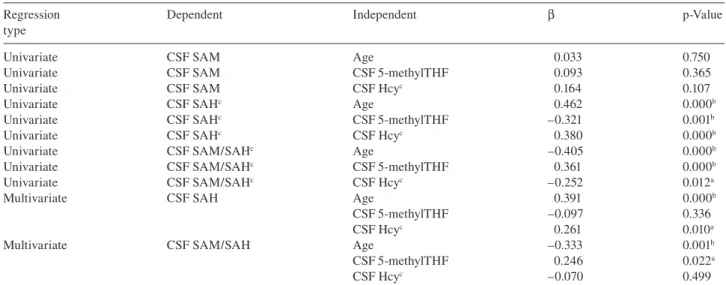
Table 2 Univariate and multivariate regression models of determinants of CSF Hcy.
Regression type
Dependent Independent β p-Value
Univariate CSF Hcy c Age 0.181 0.075
Univariate CSF Hcy c CSF 5-methylTHF – 0.495 0.000 b
Univariate CSF Hcy c Plasma Hcy c 0.331 0.001 b
Univariate CSF Hcy c Serum folate c – 0.244 0.019 a
Univariate CSF Hcy c Serum B12 c – 0.176 0.094
Univariate CSF Hcy c CSF SAH c 0.380 0.000 b
Multivariate 1 CSF Hcy c CSF 5-methylTHF – 0.499 0.000 b
Serum folate c 0.008 0.943
Multivariate 2 CSF Hcy c Plasma Hcy c 0.279 0.016 a
Serum folate c – 0.109 0.339
Multivariate 3 CSF Hcy c CSF 5-methylTHF – 0.338 0.001 b
Plasma Hcy c 0.204 0.027 a
CSF SAH c 0.265 0.004 b
a Correlation is signifi cant at the 0.05 level (2-tailed), b correlation is
using similar analytical methods (26) . Of note, folate existed in CSF predominantly in the 5-methylTHF form, as was pre-viously described for plasma (27) . With regard to gender, no signifi cant correlations with any of the parameters were found (data not shown).
Determinants of CSF Hcy
For the potential determinants of CSF Hcy listed in the Methods section, the univariate correlations were deter-mined. The results are listed in Table 2 . CSF Hcy levels were positively correlated with plasma Hcy and CSF SAH, then negatively correlated with serum folate and CSF folate. Age, a traditional determinant of plasma Hcy, just failed to reach signifi cance for CSF Hcy (p = 0.075).
Multiple regression analysis was used to determine inter-dependence of the univariate correlations. As is evident from both multivariate regression model 1 and 2 in Table 2, serum folate is not an independent determinant of CSF Hcy. Rather, its correlation with CSF Hcy is predominantly explained by its effect on CSF 5-methylTHF and plasma Hcy.
As explained in Figure 1, the metabolic fate of Hcy is determined by several pathways, implying that its concentra-tion could be independently correlated to parameters related to these pathways. Regression model 3 indeed identifi ed CSF 5-methylTHF (remethylation), plasma Hcy (import/export across the blood-brain barrier) and CSF SAH (SAH hydro-lase activity) as independent determinants of CSF Hcy levels. When age and vitamin B12 were forced into the multiple regression model, they were again not signifi cantly related to
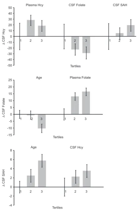
Plasma Hcy Plasma Folate Age Age 50 40 30 20 10 0 -10 1 2 3 2 1 3 1 2 3 2 1 3 2 1 3 2 1 Tertiles Tertiles Tertiles 3 1 2 3 -20 -30 -40 25 20 15 10 5 0 -5 -10 -15 8 6 4 2 0 -2 -4 -50 CSF Folate CSF SAH CSF Hcy Δ CSF Hcy Δ CSF Folate Δ CSF SAH
Figure 2 Change in CSF Hcy, CSF folate and CSF SAH, in respective tertiles (multivariate regression model). Effect sizes were adjusted for the variables as indicated in Table 2.
Table 3 Univariate and multivariate regression models of determinants of CSF SAM, SAH and SAM/SAH. Regression
type
Dependent Independent β p-Value
Univariate CSF SAM Age 0.033 0.750
Univariate CSF SAM CSF 5-methylTHF 0.093 0.365
Univariate CSF SAM CSF Hcy c 0.164 0.107
Univariate CSF SAH c Age 0.462 0.000 b
Univariate CSF SAH c CSF 5-methylTHF – 0.321 0.001 b
Univariate CSF SAH c CSF Hcy c 0.380 0.000 b
Univariate CSF SAM/SAH c Age – 0.405 0.000 b
Univariate CSF SAM/SAH c CSF 5-methylTHF 0.361 0.000 b
Univariate CSF SAM/SAH c CSF Hcy c – 0.252 0.012 a
Multivariate CSF SAH Age 0.391 0.000 b
CSF 5-methylTHF – 0.097 0.336
CSF Hcy c 0.261 0.010 a
Multivariate CSF SAM/SAH Age – 0.333 0.001 b
CSF 5-methylTHF 0.246 0.022 a
CSF Hcy c – 0.070 0.499
a Correlation is signifi cant at the 0.05 level (2-tailed), b correlation is signifi cant at the 0.01 level (2-tailed), c log transformed data. All serum
(Hcy, folate and B12) correlations were not signifi cant.
CSF Hcy (data not shown). Figure 2 demonstrates the mul-tiple adjusted impact of plasma Hcy, CSF 5-methylTHF and CSF SAH, on CSF Hcy.
Determinants of CSF SAM, SAH and SAM/SAH
As shown in Table 3 , CSF SAM showed no signifi cant cor-relation with any of the potential determinants studied. CSF SAH was independently positively correlated with age and CSF Hcy, and negatively correlated with CSF 5-methylTHF (Figure 2). The CSF SAM/SAH ratio was independently posi-tively correlated with CSF 5-methylTHF and negaposi-tively cor-related with age and CSF Hcy. None of the serum parameters (Hcy, folate and B12) showed any signifi cant correlation with CSF SAM, SAH or the SAM/SAH ratio (data not shown).
Determinants of CSF 5-methylTHF
As shown in Table 4 , CSF 5-methylTHF is negatively correlated with age and positively correlated with serum folate concentra-tions. A multivariate regression model for both age and serum folate was applied, showing that both parameters independently infl uence CSF 5-methylTHF (Figure 2). Additionally, the ratio CSF 5-methylTHF/serum folate was negatively correlated with age ( R 2 = 0.076, ß = – 0.276, p = 0.008).
Discussion
These are the salient fi ndings of our study in healthy indi-viduals. Firstly, older age and lower serum folate status are independently associated with lower CSF 5-methylTHF availability. This may be unfavorable, as we also observed that a low CSF folate status adversely affects CSF Hcy and methylation potential, at least insofar as the latter is refl ected by concentrations of SAH. Secondly, aging also appears to
directly adversely affect methylation potential, independent of CSF folate status. Finally, CSF Hcy appears to be at least partly determined by plasma Hcy, suggesting a degree of blood-brain equilibration. In the highest tertile of CSF Hcy, the contribution of plasma Hcy is less, suggesting that higher CSF Hcy is predominantly related to intracerebral metabo-lism, rather than to equilibration with plasma Hcy. CSF Hcy is independently correlated to the concentration of the trans-methylation inhibitor SAH, which may represent a mecha-nism of untoward effects of CSF Hcy.
The fate of Hcy in CSF will depend on many different factors (remethylation, export from brain tissue, trans-port across the blood-brain barrier, equilibrium with SAH, etc). From the perspective of Hcy metabolism (Figure 1), the positive correlation of CSF Hcy with both plasma Hcy and CSF SAH, and the negative correlation with CSF 5-methylTHF seem logical (9) . However, given the strong degree of inter-relatedness of 1C metabolites, analyzing sta-tistical independency of these associations, as we did in the current study, is crucial.
The methylation potential of cells is conceivably refl ected by SAM and SAH (and their ratio), as SAM is a universal methyl donor and SAH is an inhibitor of most transmethyla-tion reactransmethyla-tions. However, in line with previously reported data, CSF SAM levels did not correlate with any of the other stud-ied potential determinants (9) . From a teleological perspec-tive, this could mean that keeping CSF SAM levels stable is vitally important, which would be in keeping with its central role not only as a methyl donor, but also as a regulator of enzyme activity in both the remethylation and transsulfuration pathway. The fact that signifi cantly lower amounts of SAM were measured in CSF of patients suffering from depression and dementia, may offer insight into how disturbances in 1C metabolism and this type of diseases are linked (10) . It may be that intracellular concentrations in brain tissue obtained from specifi c regions will offer more insight into these matters,
as CSF SAM does not correlate well with intracellular SAM concentrations in brain (28, 29).
Levels of the methyltransferase inhibitor SAH were adversely associated with higher age and CSF Hcy (9) . Age seems to be a strong determinant of the methylation potential in the brain. The underlying mechanism remains unclear.
In our study, CSF folate levels were approximately fi ve times higher than serum folate levels. Another study (18) showed similar results, while a further one (9) observed higher folate serum levels than in our population and lower CSF levels. However, this last study did not use multi vitamin intake as an exclusion criteria. This may account for the higher plasma values. Perhaps folate demand in the brain is higher than in other organs, but exactly how the brain main-tains its high folate status is mechanistically unknown. The relation between increased age and diminished serum folate levels has been well documented (14) . This correlation has been attributed to the decreased ability for folate uptake in the elderly population (13, 14) . Our study also showed that age was an independent determinant of CSF folate levels, a fact that has not been described before to our knowledge.
As CSF folate levels have been shown not to follow serum folate levels outside the normal range ( ≥ 45 nmol/L) (9) , an additional determinant of CSF folate is of importance. Choroid plexus epithelial cells express high levels of folate receptor α , suggesting that a role for it in folate traffi cking (30) . Perhaps transport of folate over the blood-brain bar-rier is also a determinant of CSF folate and might be the rate limiting step. The observation that the CSF/serum folate ratio diminishes with age may suggest that this transport alters with age. Supplementation might increase the often reduced plasma folate levels of the elderly. However, whether this supplemen-tation will lead to desired increase in CSF folate has yet to be determined. Additionally, this observation of hindered trans-port of folate across the blood-brain barrier could provide insight to the origin of the low CSF folate levels observed in cerebral folate defi ciency, a treatable neurological abnor-mality with genetic, acquired or even unknown etiology (31) . Whether defi cient CSF folate refl ects an intracellular folate defi ciency in the brain is not well understood. Because of the strong observed correlation between CSF folate and CSF Hcy, CSF Hcy could be considered to be a more practical bio-marker (compound stability) of cerebral folate defi ciency.
Obtaining CSF samples from healthy volunteers is diffi -cult. Even though individuals taking medication or suffering
Table 4 Univariate and multivariate regression models of determinants of CSF 5-methylTHF.
Regression type
Dependent Independent β p-Value
Univariate CSF 5-methylTHF Age – 0.242 0.016 a
Univariate CSF 5-methylTHF Serum folate c 0.504 0.000 b
Multivariate CSF 5-methylTHF Age – 0.256 0.004 b
Serum folate c 0.511 0.000 b
a Correlation is signifi cant at the 0.05 level (2-tailed), b correlation is
signifi cant at the 0.01 level (2-tailed), c log transformed data.
from diseases known to infl uence the 1C metabolism were omitted, a certain degree of bias cannot be excluded.
Having identifi ed the determinants of 1C metabolites in CSF, it will be interesting to investigate whether the same determinants play a role in various neurodegenerative diseases and whether various intervention strategies can alter CSF concentrations of the relevant 1C metabolites, offering path-ways to prevention and/or treatment.
In conclusion, CSF 5-methylTHF levels decrease while CSF SAH levels increase with age, which may contribute to CNS ageing and neurodegeneration. Higher serum folate con-centrations were associated with higher CSF concon-centrations of 5-methylTHF and with lower levels of the neurotoxic agents Hcy and SAH, arguing that folate defi ciency may accelerate ageing and neurodegeneration.
Confl ict of interest statement
Authors ’ confl ict of interest disclosure: The authors stated that there are no confl icts of interest regarding the publication of this article.
Research funding: None declared. Employment or leadership: None declared. Honorarium: None declared.
References
1. Griffi ths R, Tudball N. Observations on the fate of cystathionine in rat brain. Life Sci 1976;19:1217 – 24.
2. James SJ, Melnyk S, Pogribna M, Pogribny IP, Caudill MA. Elevation in S-adenosylhomocysteine and DNA hypomethyla-tion: potential epigenetic mechanism for homocysteine-related pathology. J Nutr 2002;132:2361S – 6S.
3. Homocysteine Studies Collaboration. Homocysteine and risk of ischemic heart disease and stroke: a meta-analysis. J Am Med Assoc 2002;288:2015 – 22.
4. Obeid R, Herrmann W. Mechanisms of homocysteine neuro-toxicity in neurodegenerative diseases with special reference to dementia. FEBS Lett 2006;580:2994 – 3005.
5. Stanger O, Fowler B, Piertzik K, Huemer M, Haschke-Becher E, Semmler A, et al. Homocysteine, folate and vitamin B12 in neu-ropsychiatric diseases: review and treatment recommendations. Expert Rev Neurother 2009;9:1393 – 412.
6. Isobe C, Murata T, Sato C, Terayama Y. Increase of total homo-cysteine concentration in cerebrospinal fl uid in patients with Alzheimer ’ s disease and Parkinson ’ s disease. Life Sci 2005; 77:1836 – 43.
7. Selley ML, Close DR, Stern SE. The effect of increased concen-trations of homocysteine on the concentration of (E)-4-hydroxy-2-nonenal in the plasma and cerebrospinal fl uid of patients with Alzheimer ’ s disease. Neurobiol Aging 2002;23:383 – 8.
8. Finkelstein JD. Metabolic regulatory properties of S-adenosyl-methionine and S-adenosylhomocysteine. Clin Chem Lab Med 2007;45:1694 – 9.
9. Obeid R, Kostopoulos P, Knapp JP, Kasoha M, Becker G, Fassbender K, et al. Biomarkers of folate and vitamin B12 are related in blood and cerebrospinal fl uid. Clin Chem 2007;53: 326 – 33.
10. Bottiglieri T, Godfrey P, Flynn T, Carney MW, Toone BK, Reynolds EH. Cerebrospinal fl uid S-adenosylmethionine in
depression and dementia: effects of treatment with parenteral and oral S-adenosylmethionine. J Neurol Neurosurg Psychiatry 1990;53:1096 – 8.
11. Popp J, Lewczuk P, Linnebank M, Cvetanovska G, Smulders Y, Kolsch H, et al. Homocysteine Metabolism and Cerebrospinal Fluid Markers for Alzheimer ’ s Disease. J Alzheimers Dis 2009;18:819 – 28.
12. Selhub J. Folate, vitamin B12 and vitamin B6 and one carbon metabolism. J Nutr Health Aging 2002;6:39 – 42.
13. Serot JM, Barb é F, Arning E, Bottiglieri T, Franck P, Montagne P, et al. Homocysteine and methylmalonic acid concentrations in cerebrospinal fl uid: relation with age and Alzheimer ’ s disease. J Neurol Neurosurg Psychiatry 2005;76:1585 – 7.
14. Brattstrom L, Lindgren A, Israelsson B, Andersson A, Hultberg B. Homocysteine and cysteine: determinants of plasma levels in middle-aged and elderly subjects. J Intern Med 1994;236: 633 – 41.
15. Surtees R, Leonard J, Austin S. Association of demyelination with defi ciency of cerebrospinal-fl uid S-adenosylmethionine in inborn errors of methyl-transfer pathway. Lancet 1991;338: 1550 – 4.
16. Linhart HG, Troen A, Bell GW, Cantu E, Chao WH, Moran E, et al. Folate defi ciency induces genomic uracil misincorporation and hypomethylation but does not increase DNA point muta-tions. Gastroenterology 2009;136:227 – 35.
17. Serot JM, Christmann D, Dubost T, Bene MC, Faure GC. CSF-folate levels are decreased in late-onset AD patients. J Neural Transm 2001;108:93 – 9.
18. Hagnelius NO, Wahlund LO, Nilsson TK. CSF/serum folate gradient: physiology and determinants with special reference to dementia. Dement Geriatr Cogn Disord 2008;25:516 – 23. 19. Taylor MJ, Carney SM, Goodwin GM, Geddes JR. Folate for
depressive disorders: systematic review and meta-analysis of randomized controlled trials. J Psychopharmacol 2004;18: 251 – 6.
20. Smach MA, Jacob N, Golmard JL, Charfeddine B, Lammouchi T, Ben OL, et al. Folate and homocysteine in the cerebrospinal fl uid of patients with Alzheimer ’ s disease or dementia: a case control study. Eur Neurol 2011;65:270 – 8.
21. Ramaekers VT, Blau N. Cerebral folate defi ciency. Dev Med Child Neurol 2004;46:843 – 51.
22. Folstein MF, Robins LN, Helzer JE. The mini-mental state examination. Arch Gen Psychiatry 1983;40:812.
23. Struys EA, Jansen EE, de Meer K, Jakobs C. Determination of S-adenosylmethionine and S-adenosylhomocysteine in plasma and cerebrospinal fl uid by stable-isotope dilution tandem mass spectrometry. Clin Chem 2000;46:1650 – 6.
24. Smith DE, Kok RM, Teerlink T, Jakobs C, Smulders YM. Quantitative determination of erythrocyte folate vitamer distribu-tion by liquid chromatography-tandem mass spectrometry. Clin Chem Lab Med 2006;44:450 – 9.
25. Ubbink JB, Hayward Vermaak WJ, Bissbort S. Rapid high-performance liquid chromatographic assay for total homo-cysteine levels in human serum. J Chromatogr 1991;565: 441 – 6.
26. Smulders YM, Smith DE, Kok RM, Teerlink T, Gellekink H, Vaes WH, et al. Red blood cell folate vitamer distribution in healthy subjects is determined by the methylenetetrahydrofolate reductase C677T polymorphism and by the total folate status. J Nutr Biochem 2007;18:693 – 9.
27. Fazili Z, Pfeiffer CM, Zhang M. Comparison of serum folate species analyzed by LC-MS/MS with total folate measured by microbiologic assay and Bio-Rad radioassay. Clin Chem 2007;53:781 – 4.
28. Weir DG, Molloy AM, Keating JN, Young PB, Kennedy S, Kennedy DG, et al. Correlation of the ratio of S-adenosyl-L-methionine to S-adenosyl-L-homocysteine in the brain and cerebrospinal fl uid of the pig: implications for the determina-tion of this methyladetermina-tion ratio in human brain. Clin Sci (Lond) 1992;82:93 – 7.
29. Morrison LD, Smith DD, Kish SJ. Brain S-adenosylmethionine levels are severely decreased in Alzheimer ’ s disease. J Neurochem 1996;67:1328 – 31.
30. Wollack JB, Makori B, Ahlawat S, Koneru R, Picinich SC, Smith A, et al. Characterization of folate uptake by choroid plexus epithelial cells in a rat primary culture model. J Neurochem 2008;104:1494 – 503.
31. Steinfeld R, Grapp M, Kraetzner R, Dreha-Kulaczewski S, Helms G, Dechent P, et al. Folate receptor alpha defect causes cerebral folate transport defi ciency: a treatable neurodegenera-tive disorder associated with disturbed myelin metabolism. Am J Hum Genet 2009;85:354 – 63.