Bulletin of Entomological Research (1994) 84, 065-090 65
The parasitoids of the African white rice
borer, Maliarpha separatella Ragonot
(Lepidoptera: Pyralidae)
A. Polaszek
Department of Entomology, Wageningen Agricultural University,
The Netherlands/International Institute of Entomology, London, UK
M.G. Fitton
Department of Entomology, The Natural History Museum,
London, UK
G. Bianchi
Eidgenossische Technische Hochschule, Zurich, Switzerland
T. Huddleston
Department of Entomology, The Natural History Museum,
London, UK
Abstract
A key is provided for the recognition of the hymenopterous parasitoids of the
African white rice borer, Maliarpha separatella Ragonot, a pest of rice in Africa and
Madagascar. Five species are described as new: Braconidae: Chelonus maudae
Huddleston, Rhaconotus carinatus Polaszek; Ichneumonidae: Pristomerus bullis Fitton,
Pristomerus cans Fitton, Venturia jordanae Fitton. The following synonyms are
proposed: Goniozus indicus Muesebeck, G. natalensis Gordh and G. procerae Risbec
are synonymized with Goniozus indicus Ashmead. Phanerotoma major Brues is
synonymized with Phanerotoma saussurei Kohl. Lectotypes are designated for
Goniozus procerae Risbec, Rhaconotus scirpophagae Wilkinson and Garouella ovicida
Risbec. The known distributions, biologies and alternative hosts of each parasitoid
are provided, and their use as biological control agents or components of integrated
pest management programmes are discussed.
Introduction pest of cultivated rice (Oryza sativa and O. glaberrima). It
. , , , , ,, o , , I, , ,, also occurs on the wild rices O. longistammata and O. Maliarpha separatella Ragonot, commonly called the , , / D ., i i ir«*-.\ A L r /-T. LU
. c . lf-L • L • l i L i i punctata (Breniere et«/., 1962). Apart from Oryza spp. it has African white rice borer, is known almost exclusively as a f , , , c ', ., i A i
been reported only trom the wild grasses Andropogon tectorum and Echinochloa holubu (Anon., 1970, 1977). Correspondence: Dr A. Polaszek, Dept of Entomology, Wagenin- Maliarpha separatella is distributed throughout sub-gen Agricultural University, PO.B. 8031, 6700 EH Wasub-geninsub-gen, saharan Africa and Madagascar, and published records The Netherlands from outside these regions (Martin, 1958; CIE, 1970; Sandhu
66 A. Polaszek et al & Chander, 1975, Li, 1985) are of doubtful validity.
How-ever, there remain several problems concerning the genus Maliarpha. Taxonomic studies show that in West Africa two Maliarpha spp. may be present, the pan-African (and Madagascan) M. separatella, and a second species, possibly undescribed. The possible existence of a second Maliarpha sp. in West Africa was suggested by Viette (in Ehenne, 1977), more recently by M. Shaffer (pers. comm.), and by studies of sex pheromones (Cork et al, 1991). The most recent taxonomic treatment of Maliarpha by Martin (1958) is rather cursory and needs careful reassessment in order to establish exactly which species are present, their status and distribution. The biology and ecology of M. separatella have been thoroughly reviewed by Breniere et al (1962), Appert (1970), Pollet (1981), Ho et al. (1983) and Bianchi et al. (1990). It is unlikely that any of these studies concern a species other than M. separatella itself.
The impact of M. separatella on rice yield is difficult to assess because damage to the plant only rarely results in the appearance of 'dead hearts' or 'white heads', which are the most apparent signs of damage by other stem-boring species. Some assessments of yield loss have been under-taken (Pollet, 1981; Ho et al, 1983; Akinsola, 1984; Ho, 1986; Anon., 1989; Baumgartner et al, 1990; Bianchi et al, in press), but remain neglected for most African and Madagascan rice agroecosystems. As a major pest M. separatella is therefore something of a borderline case, but its importance may increase with the intensification of rice production. Control using chemical insecticides has been thoroughly investigated, but is not widely practised because of the high costs involved (Akinsola & Agyen-Sampong, 1984). Alternative methods are currently being developed, and biological control represents the most economically efficient and ecologically least harmful prevention method. Biological control of rice stem borers through the introduction of exotic parasitoids has been attempted pre-viously in Madagascar (Appert et al, 1969; Appert, 1970, 1971; Anon., 1975) and Senegal (Roudeillac, 1972, Vercambre, 1977a), as well as in several parts of Asia. In Madagascar parasitoids were usually introduced either from East Africa or from India with the primary intention of controlling stem borers in sugarcane or maize monocultures (Appert et al, 1969; Betbeder-Matibet, 1971). In most cases field testing and release of parasitoids against M. separatella were carried out incidentally during other biological control projects, and with natural enemies that probably were not well adapted to M. separatella. Often, species were used which had never been reared from M. separatella, either from the field or in the laboratory.
Until now there has been no thorough investigation of the natural enemy complex of M. separatella. In order to attempt to fill this gap a new research programme was initiated to complete previous surveys in different African and Madagascan rice agroecosystems (Bousses & Rahalivavololona, 1990; Bianchi et al, 1991). These surveys have revealed a large complex of parasitoids associated with this pest, and in order to facilitate correct identification of these parasitoids, and to provide access to information concerning their biology, the current study was initiated.
Methods
Field work was conducted in Cameroon, Ivory Coast, Kenya, Madagascar, Senegal and Tanzania. Wherever
poss-ible, upland, irrigated and flooded rice ecosystems were investigated. Rice stems were collected and dissected for larvae or pupae of M separatella and egg masses were collected from leaves Parasitoids were reared by placing the excised part of the plant containing the apparently para-sitized borer larva, pupa or eggs in a small plastic vial kept at 25 °C. After parasitoid emergence head capsules of host larvae were examined to confirm the host's identity. Egg masses of M. separatella are distinctive and easily recogniz-able, even when parasitized (Breniere et al, 1969, figs 12-14).
The most important previous surveys of M. separatella parasitoids were carried out in Ghana (Scheibelreiter, 1972; Agyen-Sampong, 1977) Ivory Coast (Pollet, 1981), Mada-gascar (Appert, 1973), Senegal (Roudeillac, 1972; Etienne, 1987) and Sierra Leone (Jordan, 1966; Agyen-Sampong, 1980), and a largely successful attempt was made to retrieve all voucher specimens from these surveys. Reared specimens from various insect collections in Europe and Africa were also examined, and this resulted in the discovery that many published records of parasitoids have been based on mis-identifications. Six species, belonging to different genera, examined during the current survey were each represented by between one and three specimens only. Positive identifi-cation of these as either previously described species or species new to science was not possible, and they have therefore each been designated 'species A' Material exam-ined is listed only for species newly described and those designated 'species A'. For previously described species it can be assumed that all material currently present in the collections of the BMNH, MNHN and WAU has been examined. Specimens of all the species in this study are deposited in the collections of The Natural History Museum, London, UK. All abbreviations can be found under Acknowledgments.
Unpublished reports cited in this paper are deposited in the library of the Department of Entomology, Wageningen Agricultural University, The Netherlands.
Key to the parasitoids of M. separatella, males and females
The following key is designed to be used for specimens examined with a stereo binocular ('dissecting') microscope at magnifications of up to at least X60 For the correct identification of some species it is necessary to make slide preparations of certain parts of the body for subsequent examination with a compound microscope at higher mag-nifications. The range of morphological diversity of M. separatella parasitoids extends from relatively large species (up to 10 mm), such as some Ichneumomdae, down to Trichogrammatidae (less than 1 mm), and includes groups from the section Aculeata (Bethylidae) and Parasitica (remaining families). For specimen preparation and morpho-logical terminology the user is therefore referred to Gauld & Bolton (1988) or Borror et al. (1989).
This key is designed exclusively for the identification of specimens which are known to have been reared from M. separatella. Many of the genera mentioned in this study contain groups of closely related species which attack stem borer pests in Africa and Madagascar. Some of these species attack hosts whose ecology may be very similar to that of M. separatella, but are not, as yet, known from M separatella. For this reason it is important that the host is correctly
Parasitoids of the African white rice borer Table 1 Checklist of Maharpha separatella parasitoids
67
Genus/species Family Subfamily referenceFage
Amauromorpha sp. A Bracon testaceorufatus Chelonus maudae Cotesia ruficrus Elasmus sp. A Eurytoma oryzwora Gonwzus mdicus Lathromens oviada Macroneura sp A Mesobraconoides psolopterus Norbanus sp. A Phanerotoma saussuret Pnstomerus afncator Pnstomerus bulks Pnstomerus cans Psilochalas soudanensis Rhaconotus carmatus Rhaconotus scirpophagae Telenomus him Temelucha sp. A Tetrastichomyia sp. A Tropobracon antennatus Vadonma ?mmbipenms Venhma jordanae Ichneumomdae Bracomdae Braconidae Bracomdae Elasmidae Eurytomidae Bethyhdae Tnchogrammatidae Eupelmidae Braconidae Pteromalidae Braconidae Ichneumonidae Ichneumomdae Ichneumonidae Chalcididae Braconidae Braconidae Scelionidae Ichneumonidae Eulophidae Braconidae Ichneumonidae Ichneumonidae Phygadeuonhnae Braconinae Cheloninae Microgastrinae Eurytominae Bethyhnae Braconinae Pteromalinae Cheloninae Cremastinae Cremastinae Cremastinae Doryctinae Doryctinae Telenominae Cremastinae Tetrastichinae Braconinae Phygadeuontinae Campopleginae p 84 P 77 p 78 p 80 p 81 p 82 p 68 p 85 p 82 p 78 p 84 p 79 p 83 p 83 p 83 p 81 p 79 p 80 p 85 p 83 p 81 p 78 p 84 p 82
identified. Reference to the diagnosis or description con-tained in each species treatment is essential. It is hoped that the authors have succeeded in excluding the possibility of misidentifications by providing diagnoses for previously described species and complete descriptions for new species. In doubtful cases, specimens may be sent to the first author for checking.
In addition to the species known to have been reared from M. separatella, two additional species have been included in the key for reasons given below: Ceraphronidae-Aphanogmus fijiensis (Ferriere) and Ichneumonidae: Pimplinae Itoplectis naranyae (Ashmead).
1. Fore wing without closed cells, or brachypterous (figs 11-14) 2 - Fore wing with at least one closed cell (figs 15 — 17, 33,
34, 44-46, 65) 11 2. Tarsi 3-segmented; antennae with fewer than 8 seg-ments (excluding anelli, fig. 10). Fore wing as in fig. 11
Lathromeris oviada
- Tarsi 4- or 5-segmented; antennae with more than 8 segments 3 3. Pronotum reaching tegulae (fig. 1); prepectus (see fig. 2) absent 4 - Pronotum not reaching tegulae, prepectus present be-tween pronotum and tegula (fig. 2) 5 4. Stigmal vein distinctly curved, fore wing venation as in fig. 12 (hyperparasitoids, emerging from coccoons)
Aphanogmus fijiensis
- Fore wing venation different, stigmal vein straight (fig. 13); (egg parasitoids) Telenomus bmi 5. Tarsi 4-segmented 6
- Tarsi 5-segmented 7 6. Hind coxa greatly expanded and flattened (fig. 3) Elasmus sp. A
- Hind coxa not greatly expanded and flattened Tetrastichomyia sp. A 7. Hind femur greatly expanded and toothed (fig. 4) Psilochalas soudanensis - Hind femur not greatly expanded or toothed 8 8. Mesopleuron forming a strongly convex shield (fig. 5), brachypterous species Macroneura sp. A ('?$) - Mesopleuron different 9 9. Pronotom rectangular in dorsal view, with coarse punctures (fig. 6) Eurytoma oryzivora - Pronotum different 10 10. Notauli incomplete (fig. 8). Antenna 12-segmented (?), consisting of scape, pedicel, 2 small ring segments, a 6-segmented funicle and a rather obscurely 2-seg-mented club with a point (fig. 9), $ unknown, antennae may be 12- or 13-segmented Norbanus sp. A - Notauli complete. Antenna 12-segmented, consisting of scape, pedicel, a single ring segment, 6-segmented funicle and three segmented club without a point Macroneura sp. A ^c? 11. Hind wing without veins, fore wing venation as in fig. 15; propodeum as in fig. 81 Goniozus mdicus - Hind wing with veins, fore wing venation and
propodeum different 12 12. Fore wing with a single recurrent vein (that is, 2m-cu absent; figs 16, 17, 33) first cubital cell almost always separated from the discoidal cell by a vein (Braconidae, except Chelonus, fig. 34) 13 - Fore wing with two recurrent veins (that is, 2m-cu present; figs 44-46, 65); first cubital cell always fused with the discoidal cell (Ichneumonidae) 20 13. Hypoclypeal depression present, deep and wide (fig. 24) 14 - Hypoclypeal depression absent 18 14. Posterior flange of propleuron absent; first metasomal tergite coarsely rugose (figs 19, 21). Fore tibia without a row of pegs or thick spines 15
68 A. Polaszek et al - Posterior flange of propleuron present (figs 28, 31); first
metasomal tergite longitudinally striate (figs 29, 32) Fore tibia with a row of pegs or thick spines 17 15. Wings strongly infumate (darkened)
Mesobraconoides psolopterus - Wings largely hyaline (clear) 16 16 Mesosoma entirely orange. Antennae elongate, about 1.25 x the body length (excluding ovipositor). Mesoscutum with fine, reticulate sculpture (fig. 20) Tropobracon antennatus - Mesosoma with some brown or black markings.
Anten-nae shorter, about equal to, or less than, the body length (excluding ovipositor). Mesoscutum smooth, shining (fig. 18) Bracon testaceorufatus 17. Pterostigma uniformly pale (fig. 16). Head and
meso-soma with dense setae (figs 25, 27, 28)
Rhaconoius sarpophagae - Pterostigma dark (fig. 17) at least in its anterior half.
Head and mesosoma less densely setose (figs 26, 30, 31) Rhaconohis cannatus 18. Tergites 1-3 of metasoma immovably joined (figs 23,
35) and forming a carapace. Vein SRI present and enclosing a marginal cell (fig. 34) 19 - Tergites 1—3 of metasoma articulating, not forming a carapace. Vein SRI absent, and fore wing venation as in fig. 33 Cotesia ruficrus 19. Body entirely yellow. Tergites of metasoma clearly
distinguishable (fig. 23) Phanerotoma saussurei - Body largely dark. Metasoma forming an undivided
carapace Chelonus maudae 20. Segment 1 of the metasoma rather wide, with the
spiracles on the anterior half (fig. 39). Edge of eye strongly indented opposite antennal socket (fig. 38) Hoplectis naranyae - Segment 1 of the metasoma more slender (fig. 40), with
the spiracles on the posterior half (fig. 42). Edge of eye at most only weakly sinuate opposite antennal socket (fig. 41) 21 21. Fore wing with pterostigma long and slender (fig. 44—46). Tergite 2 of metasoma granulate or punctate 22 - Fore wing with pterostigma broad and triangular (fig. 65). Tergite 2 of metasoma very finely, longitudinally aciculate (figs 73-75) 24 22. Clypeus not distinctly separated from face, the two together forming a continuous, weakly convex surface (fig. 66). Fore wing with areolet (absent in a few aberrant specimens) petiolate (2rs-m and 3rs-m arising together from Rs) (fig. 44) Ventuna jordanae - Clypeus separated from face, clypeus and face each
forming a distinct, curved surface (figs 67, 68). Fore wing with areolet present and almost quadrate (the sections of Rs and M between 2rs-m and 3rs-m almost equal in length) (fig. 46) or areolet absent (fig. 45) 23 23. Fore wing with areolet present, although 3rs-m weakly or not pigmented (fig. 46). Frons with a differentiated trans-striate area above the antennal socket (fig. 67) Vadonina ?nimbipenms - Fore wing with areolet absent (fig. 45), although the
position which 3rs-m would occupy may be indicated by the configuration of Rs and M. Frons without a differentiated area (although it is less punctate) above the antennal sockets (fig. 68) Amauromorpha sp. A
24. Tergite 2 of metasoma without thyndia. Hind femur of males and females without a tooth on the ventral surface Temelucha sp A - Tergite 2 of metasoma with thyridia (figs 73 — 75). Hind femur of males, but not of all females with a distinct tooth on the ventral surface (figs 62-64) 25 25. Eyes larger in proportion to size of head: face narrower (male fig. 49, female fig. 50), gena (in lateral view) narrower (male fig. 58, female fig. 55). Male with ocelli not quite touching eyes (fig. 60). Female with hind femur lacking a distinct tooth on ventral surface (fig. 61) Pristomerus cans - Eyes smaller in proportion to size of head: face wider
(male figs 47, 48, female figs 51, 52), gena (in lateral view) wider (male figs 56, 57, female figs 53, 54). Male with ocelli separated from eyes by more than 0.6 of their diameter (fig. 59). Female with hind femur with a distinct tooth on ventral surface (fig. 62) 26 26. Clypeus wider and less convex (male fig. 47, female fig. 51). Female with ovipositor shorter, less than 1.9 (the visible portion 1.3) times as long as the hind tibia. Male with hind femur stouter (fig. 63) [East Africa] Pristomerus bullis - Clypeus narrower and more convex (male fig. 48,
female fig. 52). Female with ovipositor longer, more than 2.3 (the visible portion 1.4) times as long as the hind tibia. Male with hind femur more slender (fig. 64) [West Africa] Pristomerus afncator
BETHYLIDAE
Goniozus indicus Ashmead (figs 15, 78, 79, 81)
Gonwzus indicus Ashmead, 1903: 2 Holotype +, India Calcutta [ex Scirpophaga} (USNM) [examined],
Gonwzus mdicus Muesebeck, 1940- 121. Holotype +, INDIA. Coimbatore V.26 36 (P. Israel) ex Scirpophaga on sugarcane (USNM) [examined] syn. nov.
Gonwzus procerae Risbec, 1956b. 157 Lectotype + (here desig-nated), [CAMEROON] Garoua (Descamps 173) (MNHN) [exam-ined] syn. nov.
Gonwzus natalensis Gordh, 1986: 257 Holotype +, SOUTH
AFRICA: Natal, Ngwavuma 2O.xi 1982 (H. Hastings) ex Eldana sacchanna (PPRI) [holotype not examined: paratypes examined]
syn. nov.
Goniozus sp Descamps, 1956, Harris, 1962, Jordan, 1966, Appert, 1973: Agyen-Sampong, 1980: Agyen-Sampong & Fannah, 1987 Diagnosis. Length 3.0—6.0 mm Entirely chestnut brown to black, with the following usually paler- antennae, all tibiae and tarsi, fore femora. Head prognathous as in all Bethylidae, body appreciably dorso-ventrally flattened. Head (V) in dorsal view elongate, dis-tance from the hind margin of the eyes to the posterior end of the head slightly greater than the maximum length of the eyes Ocelli forming an approximately equilateral triangle, separated from the posterior end of the head by about the maximum length of the ocellar triangle Clypeus evenly rounded, not pointed or truncate Sculpture of the head and body smooth, shining, with a few shallow punctures Fore femora enlarged. Fore wing typical for the genus (fig. 15), with both pterostigma and prostigma present, without a closed areolet Propodeum without any trace of a transverse carina separating the dorsal (horizontal) surface from the posterior (vertical) surface: with fine, reticulate sculpture apart from
Parasitoids of the African white nee borer 6 9
Figs 1-9. 1, Aphanogtnus fi)iensis mesosoma, lateral (m=mesopleuron, p=pronotum, t=tegula); 2, chalcidoid mesosoma, lateral (p = prontotum, t=tegula); 3, Elasmus sp. mesosoma, lateral (c=hmd coxa); 4, Psilochahs sodanensis hind femur; 5, eupelmid (?) mesosoma, lateral (m=mesopleuron); 6, Eurytoma oryzwora mesosoma, dorsal (p = pronotum); 7, £. oryzwora mesosoma, ventral (sa=subpleural area);
70 A Polaszek et al.
10 11
16
Figs 10—17. 10, Lathromens ovicida antenna; I I , L oviada fore wing; 12, Aphanogmus fijiensis fore wing; 13, Telenomus him fore wing; 14,
Psilochalas soudanensis fore wing; 15, Gonwzus mdicus fore wing; 16, Rhaconotus scirpophagae fore wing; 17, Rhaconotus carmatus fore wing.
Figs 18—23. 18—19, Bracon testaceorufatus- 18, mesosoma, dorsal; 19, metasoma, basal tergites; 20—21, Tropobracon antennatus. 20, mesosoma dorsal, 21, metasoma, basal tergites, 22—23, Phanerototna saussurer 22, mesosoma, dorsal; 23, metasoma, dorsal
Parasitoids of the African white rice borer 71
Figs 24—32. 24, Rhaconotus sp head, anterior (h=hypoclypeal depression), 25, Rhaconotus scirpophagae head, dorsal, 26, R. cannatus head, dorsal; 27-29, R scirpophagae. 27, mesosoma, dorsal, 28, mesosoma, lateral; 29, metasoma, basal tergites; 30-32, R cannatus. 30, mesosoma,
72 A. Polaszek et al.
33
40
39
Figs 33—41. 33, Cotesia ruficrus fore wing; 34—37, Chelonns maudae: 34, fore wing; 35, metasoma, lateral; 36, head, dorsal; 37, head, anterior; 38—39, Iioplechs naranyae: 3&, head, anterior; 39, metasoma, tergite I, 40—41, Venturia jordcmae: 40, metasoma, tergite I, 41, head, anterior.Parasitoids of the African white nee borer 73
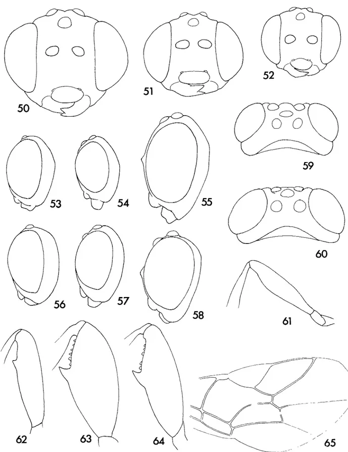
49
Figs 42—49. 42—44, Ventuna jordanae- 42, mesosoma, lateral 43, head, dorsal; 44, fore wing; 45, Amauromorpha sp. A fore wing; 46, Vadonina
74 A. Polaszek et al.
62
65
Figs 5 0 - 6 5 . 50, Pristomerus cans ($) head, anterior; 51, P. bulks (?) head, anterior; 52, P. afncator (+) head, anterior; 53, P bulks (+) head,
lateral; 54, P. afncator ($) head, lateral, 55, P. cans (£) head, lateral; 56, P. b«//i's (3) head, lateral; 57, P afncator (o) head, lateral, 58, P cans (o) head, lateral; 59, P. fcn//is {3) head, dorsal, 60, P. cans (rj) head, dorsal; 61, P. cans (+) hind femur, 62, P. fctW/is (4-) hind femur;
Parasitoids of the African white rice borer 75
Figs 66-72 66, Veniuna jordanae head; 67, Vadomna ? nimbipennis head; 68, Amauromorpha sp A head; 69, Veniuna jordanae propodeum; 70, Pristomerus bulks propodeum; 71, P cans propodeum; 72, P. afncaior propodeum.
76 A. Polaszek et ah
— t
Figs 7 3 - 7 5 . 73, Pristomerus cans tergite 2; 74, P. bulks tergite 2; 75, P afncator tergite 2
78
80
Figs 76—80. 76, Tetrastichomyia sp A m e s o s o m a , dorsal; 77, Aphanogmus fijiensis (^) gemtalia (from Dessart, 1971), 78—79, GOUIOITHS mdtcus (3Y- 78, subgenital plate; 79, gemtalia, 80, Telenomus him (,j) gemtalia.
Parasitoids of the African white rice borer 77
Fig 81. Gomozus mdicus propodeum, dorsal.
a smooth, triangular area tapering antero-postenorly on the dorsal surface (fig. 81) Gemtalia and sub genital plate of male as in figs 78, 79
Alternative hosts. Pyrahdae Chilo infuscatellus Snellen, C. partellus
(Swinhoe), C. sacchanphagus (Bojer), C. suppressahs (Walker) (labora-tory host), C. zaccomus Bleszynski, Chlo sp., Comesta ignefusalis (Hampson), Corcyra cephalonica (Stainton) (laboratory host),
Di-atraea centrella Moeschler, D hneolata (Walker), Eldana sacchanna
(Walker), Emmalocera depressella (Swinhoe), Sarpophaga excerptahs (Walker), 5. incertulas (Walker), S nivella (F.), 5. ocadentelk (Walker),
Sarpophaga sp. (see Biology, below).
Distribution All of sub-Saharan Africa, Mauritius and Madagascar.
Asia- Bangladesh, India, Pakistan
Biology Like most Gomozus, a gregarious larval ectoparasitoid,
usually attacking concealed larvae of Lepidoptera. Oviposition follows temporary anaesthesia, and is into the dorsum of the larva The biology of G. mdicus (as G procerae) under laboratory conditions is given in detail by Ly (1976), Ndoye (1980) and Coquard (1987)
Gordh & Moczar (1990) list Cryptophlebia carpophaga (Walsingham) and Cnaphalocroas medinahs (Guenee) among the hosts of G. mdicus We have examined Gomozus specimens reared from C. medinahs and from Cryptophlebia sp., from India They are not G mdicus, and these species seem unlikely to be (natural) hosts
of G. mdicus.
Remarks Type material of G. mdicus Ashmead, G. mdicus
Muese-beck, G. procerae Risbec and G. natalensis Gordh was compared for this study Several hundred specimens of G. mdicus from Africa and Asia were also examined No important differences were found, and these species are therefore here regarded as conspecihc
The syntype series of G procerae consisted originally of 7'5 and 2 5 mounted on three microscope slides One female, on a slide with a partially dissected (by Risbec) male was clearly marked 'type' in Risbec's writing. This female has since been remounted
on a card-point and is now labelled 'lectotype'. The following are now labelled as paralectotypes- the first slide previously containing the lectotype, now containing a remaining male (partially dis-sected); a second slide containing the gemtalia of this male (used for Risbec's original figure) and two females; a third slide contain-ing the gemtalia (which are aberrant and asymmetrical) of the second male of the series, and five specimens ( 1 ^ , 4$) mounted on two card squares. In the original description, nine females and two males were mentioned The remaining two females, if they ever existed, have not been located. Also in the original descrip-tion, the hosts were mentioned as 'Adelpherupa sp. et Salurea sp. (Pyrahdae)' The type series is labelled (by Risbec) 'ex Proceras
africana' (=Chdo zaccomus) No specimen was located in Risbec's
collection with the names of the former genera, neither has any specimen ever been recorded from these hosts since the descrip-tion. It seems highly probable that the names of these hosts were published in error.
A distinct species of Gomozus (Parasierola -group) is also widespread on lepidopterous cereal stem borers in Africa (Girling, 1977; Conlong & Graham, 1988), which Gordh (1986) suggested might be the same as G. natalensis. This species is known to us so far only in association with maize.
Gomozus mdicus has been the subject of several studies in
relation to biological control of the stem borer E. sacchanna in South Africa (Conlong et «/., 1988; Graham & Conlong, 1988, as G. natalensis). It is also a well-known parasitoid of the millet stem borer C. ignefusahs in the West African Sahel (Ndoye, 1980; as G.
procerae). Gomozus mdicus was introduced into Madagascar (where
it may well have already been present) from Senegal in 1973, for the attempted biological control of M. separatella (Appert, 1975), and into Reunion in 1972 (Betbeder-Matibet, 1977). Gomozus
mdicus was imported into Trinidad for the biological control of
sugarcane borers in 1960. It successfully attacked two Diatraea species (see hosts, above), but it is not known whether it became established (Bennett, 1965).
BRACONIDAE: Braconinae
Bracon testaceorufatus Granger
(figs 18, 19)
Bracon testaceorufatus Granger, 1949- 71 Holotype f,
MADAGAS-CAR. Bekily, (reg. sud de l'lle) vn 1936 (A. Seyng) (MNHN) [examined]. Bracon testaceorufatus- Breniere et ah, 1962; Appert, 1967, 1973, Anon, 1989.
Braconidae [sp. indet.] Tran, 1977.
Bracon quadratinotatus Granger- Bordat, 1979 (misidentification).
Diagnosis. Length 2.0-4.5 mm, excluding antennae and ovipositor
sheaths. Deep orange-brown, with the following dark brown-black, antennae, stemmaticum, most of the wing venation, ovipositor sheaths, fifth tarsal segments; mesoscutum centrally, the sides varying from entirely without dark pigmentation to deeply pigmented; propodeum, first metasomal tergum, second metasomal tergum centrally and third and fourth terga broadly pigmented. Antenna (female) with 26—36 segments Head transverse, head and mesoscutum smooth and shining, notauli smooth, shallowly impressed. Propodeum coarsely sculptured, especially distally. Sculpture of first metasomal tergum, and second tergum centrally, strongly rugose (fig. 19). Male as female but habitus somewhat narrower, antennae with a few more segments and first metasomal tergum often paler than in female.
78 A Polaszek et al.
Alternative hosts. Pyralidae: Chilo diffusthneus (J- de Joannis), C. zacconius, Chilo sp., Scirpophaga sp.
Distribution. Cameroon, Cote d'lvoire, Ghana, Kenya, Madagascar,
Mali, Mauritania, Mozambique, Nigeria, Senegal, Tanzania, Uganda.
y. A gregarious larval ectoparasitoid (6—14 individuals per
host) with an average fecundity of 25 eggs and a developmental time of 12 days. The larvae pupate within the rice stem, a few centimeters below the host's remains. Emergent adults leave the stem via the aperture made by the host larva. In Madagascar, at least part of the population undergoes larval diapause whereas the remainder is active throughout the year (Bianchi et al, 1991).
Remarks. In areas of Madagascar where this species was abundant
it had no important role in regulating populations of M. separatella (Appert, 1967). During September and October 1989 B
testaceor-ufatus was the most abundant parasitoid of M. separatella at Lac
Alaotra, Madagascar (Anon., 1989) In Bouake, Cote d'lvoire, B.
testaceorufatus was abundant on irrigated rice from May to June
(Tran, 1977).
Mesobraconoides psolopterus (Wilkinson)
Mesobracon psolopterus Wilkinson, 1931: 394. Holotype -f, SIERRA
LEONE Njala, ex coffee branch borer, em. 4 xn.30 (E. Hargreaves) (BMNH) [examined].
Mesobraconoides psolopterus (Wilkinson): Sarhan & Quicke, 1990
221
Diagnosis. Length 6.0—7 0 mm. Antennae with about 55—60
flagel-lomeres, the median flagellomeres considerably longer than wide. Wings largely infumate, pterostigma yellow to orange, hind leg and metasoma largely orange to red.
Alternative hosts. Coleoptera Scolytidae Xyleborus sp. Distribution Nigeria, Sierra Leone.
Biology Unknown, probably an ectoparasitoid, attacking the final
larval instar of the host (Sarhan & Quicke, 1990).
Remarks The inclusion of this species is based on two published
records of its attacking M. separatella in Sierra Leone (Sarhan & Quicke, 1990) No material reared from M. separatella was exam-ined during the present study.
Tropobracon antennatus (Granger)
(figs 20, 21)Habrobracon tnangulans Szepligeti, 1911 405. Holotype + [Kenya]
Mombasa (Hildebrandt) (HMB) [examined].
Bracon antennatus Granger, 1949- 61. Replacement name for H. triangularis Szepligeti.
Tropobracon antennatus (Granger) Etienne, 1987 Habrobracon sp.. Risbec, 1950.
Mesobracon sp.: Ingram, 1958.
Shirakia sp.: Appert, 1973, Badawy, 1967, Jordan, 1966. Diagnosis. Length 2.5—6 5 mm (excluding antennae and ovipositor
sheaths). Body entirely deep orange except the following dark areas- antennae, stemmaticum, claws and base of fifth tarsal segment, ovipositor. Occasionally the triangle on T2 bordered with dark pigmentation which extends to a median longitudinal stripe the length of the metasoma. Pterostigma uniformly pale
brown. Antenna with 50-65 segments Sculpture of mesosoma as in figure 20, mesoscutum with evident reticulate sculpture. Dor-sally visible part of ovipositor+sheaths about half the length of the metasoma Male as female except for genitahc characters and the following, habitus narrower than in female and triangle on T2 usually complete at its apex.
Alternative hosts. Diptera: IDiopsis curva Bertolim (Risbec, 1956a)
Noctuidae: Sesamia crehca Lederer, Sesamia sp.; Pyralidae Chilo
zacconius, Chilo sp.; Coniesta ignefusalts; Scirpophaga sp.
Distribution. Cameroon, Cote d'lvoire, Kenya, Madagascar, Malawi,
Mali, Mozambique, Niger, Nigeria, Senegal, Sierra Leone, Somalia, South Africa, Sudan, Togo, Uganda.
Biology. A gregarious larval parasitoid, T. antennatus is also known
from stem borers of millet and wheat According to Jordan (1966) this species makes a small cocoon inside the stem on which its host has been feeding, usually an inch or so above the host's remains. The host record from Diptera. Diopsidae requires confirmation.
Remarks Three other closely related species of Tropobracon are
known to attack lepidopterous cereal stem borers in the Palaeo-tropics. Tropobracon antennatus can be most easily distinguished from these by the reticulate sculpture of the mesoscutum (fig. 20). A key to Tropobracon spp. is provided by van Achterberg (1993).
In Madagascar, this species is uncommon on M separatella or other nee borers (P Bousses, pers comm.)
BRACONIDAE: Cheloninae
Chelonus maudae Huddleston sp. n.
(figs 34-37)
Description. Length 6 - 8 mm. Female- antenna with 28-29
seg-ments, filiform; basal flagellar segment at least 3 x as long as broad, each succeeding segment slightly shorter. Head strongly rounded behind eyes (fig. 36) Eye slightly longer than temple in dorsal view Frons strongly depressed behind antennae, coarsely rugose, sometimes reticulate. Vertex coarsely reticulate-rugose at sides but with strong transverse rugae medially, behind ocelli Ocelli mod-erately large, 0 0 = 2 . 0 - 2 . 5 O D , almost on line. Eyes large, not protuberant Face at least twice as broad as high (fig. 37), only weakly convex, coarsely rugose with a reticulate element laterally. Clypeus distinctly narrower than face, moderately convex, densely, finely punctate; apical border produced but truncate medially Malar space marked by a band of fine rugulose sculpture. Mandibles large, moderately twisted. Thorax (mesosoma) rather depressed, in lateral view almost twice as long as high. Pronotum projecting distinctly in front of mesonotum, reticulate laterally, transversely striate dorsally. Notauh deeply impressed, foveolate Mesonotum generally coarsely reticulate-rugose except for small, smooth punctate areas laterally. Precoxal suture indistinguishable from the coarsely reticulate-rugose sculpture of the mesopleuron. Propodeum coarsely reticulate-rugose, divided by a strong trans-verse medial canna that is raised laterally into blunt dentate flanges. Carapace elongate, oval in dorsal view, ventral opening not reaching apex; posteroventrally strongly impressed medially Ovipositor long (at least half as long as carapace), narrow Hind coxa rugose dorsally, densely finely punctate ventrally
Colour black, but antennae brown; fore leg yellow except coxa and trochanter brown, mid leg brown except apex of femur, base of tibia and sometimes tarsus yellow, hind leg brown but always with a medial pale band on tibia, and often also base of tarsus yellow. Carapace with lateral pale areas anteriorly. Sternites and
Parasitoids of the African white nee borer 79 ovipositor sheaths brown, sometimes pale Male same as female
except antennae 31-segmented, carapace more parallel-sided in dorsal view with smaller pale areas anterolaterally and no strong medial impression posteroventrally.
Material examined. Holotype x, SENEGAL: Djibelor, 15.x.1979
(J Etienne) [ex larva Maharpha separatella on rice] (MNHN). Paratypes 3 J , 2$, same data as holotype (BMNH, MNHN). 1$ Senegal, Kagnout, 13.xi.1969 (B Vercambre) [ex M. separatella] (MNHN).
Alternative hosts. None known Distribution. Senegal.
Biology. A solitary egg-larval endoparasitoid. Quartey (1975)
referred to a 'genus near Chelonus ?sp' attacking M. separatella in Ghana which had a 7—29% parasitization rate. It is not unlikely that he was observing C. maudae, although we have not been able to recover the original material on which these observations were based.
Remarks Chelonus maudae is most closely related to C. capensis
Cameron; the carapace of C. capensis, however, has a much shorter ventral opening and the posteroventral impressed part is therefore much longer. The temples of C. capensis are less strongly rounded and the sculpture on most parts of the body is less well developed
Phanerotoma saussurei Kohl (figs 22, 23)
Phanerotoma saussurei Kohl, 1906: 125. Holotype % (mistakenly
given as 3 in original description), MADAGASCAR- Tamatave (NMW) [examined].
Phanerotoma major Brues, 1926- 266. Holotype J, [KENYA] British
East Africa: Masai Reserve (BMNH) [examined] syn. n.
Phanerotoma major: Jordan, 1966; Sampong, 1980;
Agyen-Sampong & Fannah, 1987
Phanerotoma saussurei: Appert, 1967 Phanerotoma sp: Etienne, 1987
Diagnosis. Length 6.0-8.0 mm. Entirely orange/brown; antennae,
stemmaticum and occasionally the posterior metasoma brown, but otherwise without any distinct brown or black markings. Pterostigma uniformly orange. Body dorsoventrally flattened. Metasoma distinctly longer than mesosoma. Propodeum coarsely reticulate, without distinct cannae. Face with stnate sculpture, tentonal pits deep. Reared always in association with nee stem borers.
Alternative hosts Pyralidae: Chilo zacconius, 7Sarpophaga sp.
Distribution Kenya, Madagascar, Mali, Senegal, Sierra Leone,
Tanzania.
Biology A solitary egg-larval endoparasitoid, with a fecundity of
90 to 130 eggs. The life cycle is synchronized with that of its host, and death of the host occurs at the host's prepupal stage, with the emergence of the final instar parasitoid larva. Pupation occurs in the vicinity of the host remains within a bottle-shaped cocoon, about 14 mm long. Adult emergence occurs 12—14 days later (Bianchi et al, 1991) In Madagascar this species is active through-out the year at Marovoay, from the end of December on the plateau but enters diapause at Lac Alaotra between July and October (Bianchi et al, 1991).
Remarks. In the BMNH collection there are three specimens of P. saussurei labelled 'S.E. Asia, L. Caresche CIE A1745'. These
specimens were part of a donation by Dr Caresche in 1971, which
included many specimens from Cambodia and Vietnam, but it is doubtful whether the Phanerotoma specimens were collected from either of those countries. Dr Caresche was himself involved with classical biological control of stem borers in Madagascar in the 1960s, a more probable origin for these specimens. According to Appert (1967) P. saussurei was ineffective in controlling M
separatella in Madagascar.
BRACONIDAE: Doryctinae
Rhaconotus carinatus Polaszek sp. n.
(figs 17, 26, 30-32)
Rhaconotus niger (Szepligeti)- Appert, 1967, 1973; Bremere et al.,
1962; Bianchi et al, 1991 (misidentifications).
Rhaconotus nr sudanensis Wilkinson: Jordan, 1966. Rhaconotus sp.. Etienne, 1987.
Rhaconotus nr sciron Nixon: Alam, 1992.
Diagnosis. Length 3.5—6.0 mm Apex of fifth metasomal tergum
simple, not dentate or sinuate. T2+T3 of the metasoma with a single division (fig. 32). Fore wing with pterostigma dark (fig. 17), each antenna with about 30 segments. Mesoscutum with a median longitudinal carina posteriorly (fig. 30).
Description. Female with ground colour dark red/brown with the
following dark brown/black- an ill-defined inverted triangle on the vertex, most of the mesoscutum, propodeum, lateral areas of metasomal terga and ovipositor. Head matt with fine reticulate sculpture, moderately setose, the setae directed posteriorly. An-tenna with 28-33 segments. Mesoscutum as in figure 30, with reticulate sculpture except the median part posteriorly with an irregular, longitudinal carina having several short lateral branches. Only the notauh and edges of the mesoscutum with setae, the mid and side lobes of the mesoscutum asetose centrally. Propodeum as in figure 30, sculpture largely finely reticulate dorsally, with a central longitudinal carina and two shorter cannae laterally. Posteriorly sculpture becoming more rugose. Fore wing (fig. 17) with the pterostigma very distinctly infuscate. Metasoma with the fused T 2 + T 3 divided by a single groove; the longitudinal striae on T2 continuing posteriorly across this groove into the anterior part of T3, without being interrupted by an irregular transverse carina (as in R. sudanensis Wilkinson). Dorsally visible part of ovipositor about 2/3 the length of the metasoma. Male largely as for female except for genitalia characters; habitus somewhat narrower.
Alternative hosts. Pyralidae: Chilo zacconius.
Distribution. Cameroon, Ghana, Madagascar, Nigeria, Senegal,
Sierra Leone, Tanzania, Togo
Biology. A gregarious larval ectoparasitoid (6-27 individuals per
host), with a fecundity of 10 to 30 eggs. Developmental time is about 20 days, and adult females show a preference for attacking fourth or fifth instar larval hosts. The cocoons are white and can be distinguished from those of B. testaceorufatus by being more closely aggregated and by the smoother texture of the silk. At Lac Alaotra this species undergoes a winter diapause to reappear during October (Bianchi et al, 1991)
Material examined. Holotype $, Cameroon, near Santchou, Plaine
de Mbo 250 km North Douala, i.1991, (G. Bianchi)/8 (BMNH). Paratypes (2(J 28%) same data as holotype (BMNH, MNHN, RMNH).
80 A. Polaszek et al.
Remarks. Type material of those African and Asian Rhaconotus
described by Granger, Nixon and Szepligeti which come at all close to R. cannatus has been examined.
Rhaconotus scirpophagae W i l k i n s o n
(figs 16, 25, 2 7 - 2 9 )
Rhaconotus scirpophagae Wilkinson, 1927. 34. Lectotype V (here
designated): INDIA: Pusa, Bihar 18.iii.19I4 'ex Scirpophaga aunflita in sugar stem' (BMNH) [examined].
Rhaconotus sp nr oryzae Jordan, 1966. Rhaconotus sp. Mathez, 1972
Rhaconotus sp nr scirpophagae Alam, 1992.
Diagnosis Length 4.0—7.0 mm Head and mesosoma densely hairy,
the hairs on the mesosoma arranged as in figure 27. Hind margin of pronotum usually obscured by a ridge, mesosoma narrow, its sculpture and setation as in figure 27 T2 + T3 of metasoma divided by a simple slightly curved groove. Antenna of female with more than 40 segments. Pterostigma uniformly pale, colouration and venation of fore wing as in figure 16
Alternative hosts Noctuidae: Busseola fusca, Pyralidae: Chilo partellus, Chilo sp., Scirpophaga lexcerpiahs, S. nwella, Scirpophaga sp. Distribution. Africa: Cote d'lvoire, Ghana, Kenya, Nigeria, Senegal,
Sierra Leone, Tanzania (Wilkinson, 1927). Asia. India, Java, Pak-istan.
Biology. A gregarious larval ectoparasitoid.
Remarks. The holotype of Rhaconotus caudatus (Szepligeti) differs in
very few respects from R scirpophagae, and may well be conspe-cific. We have refrained from synonymizing the two species pending a thorough taxonomic revision of Rhaconotus from the Old World tropics.
In addition to the two Rhaconotus species treated here, at least five other species are recorded as attacking other stem borer species in the Old World tropics These are the following: R
cauhcola Muesebeck, R. oryzae Wilkinson, R. roshnensis Lai, R. schoenobivorus (Rohwer) and R. signipennis (Walker).
Rhaconotus scirpophagae was described from Scirpophaga aunflua
on sugarcane. Scirpophaga aunflua is a junior synonym of S. nwella, a rice stem borer. According to Lewvanich (1981), records of this species from sugarcane are probably misidentificahons of S.
excerptahs.
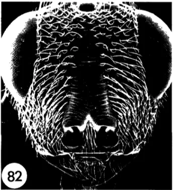
Fig 82. Psilochalas soudanensis head, anterior
Alternative hosts. An extremely polyphagous species. Additional
African cereal stem borer hosts not mentioned by Nixon (1974) are the following: Noctuidae: Sesamia nonagrioides Lefebvre, Pyralidae:
Chilo zacconius
Distribution. Cosmopolitan in the Palaeotropics and Palaearctic. Biology. A gregarious endoparasitoid.
Remarks Reared from M separatella at Kotiessou, Cote d'lvoire, by
Dr A Pollet (1J, 2^, CIRAD, examined)
CERAPHRONIDAE
BRACONIDAE: Microgastrinae
Cotesia ruficrus (Haliday)
(fig. 33)Microgaster ruficrus Haliday, 1834: 253 [Whereabouts of syntypes
unknown]. For full synonymy see Nixon, 1974.
Diagnosis. Length 2 0—2 5 mm Ocelli in a high triangle. Antenna
as long as body, the two preapical segments each about 1 7 x longer than wide. Mesoscutum shining, coarsely punctate Scutellum deeply and strongly punctate. Phragma of scutellum concealed by postscutellum. Hind coxae dull, rugose, Inner spur of hind tibia not reaching beyond middle of basitarsus. Tergite 2 + 3 highly polished distally to basal field, setae restricted almost to a single row in the middle, frequently double at the sides. Hypopy-gium short, roundly truncate at apex as seen in profile (Nixon, 1974). Colour: legs bright red/yellow, hind femur dark distally. Metasomal tergite 2 + 3 with yellow markings.
Aphanogmus fijiensis (Ferriere)
(figs 1, 12, 77)Calliceras fijiensis Ferriere, 1933: 106. Holotype '$, FIJI: Taveuni, xi
1931 (R.W. Paine) ex Apanteles hrathabae (BMNH) [examined]
Aphanogmus fipensis (Ferriere). Dessart, 1971: 98.
Diagnosis. Length 0.9—2.2 mm Wing venation as m figure 12
Mesopleuron almost entirely smooth (fig I), genitalia as in figure
77.
Hosts Aphanogmus fiitensis attacks a wide range of hymenopterous
primary parasitoids, mostly Bracomdae It is especially commonly recorded from cocoons of Apanteles spp., Cotesia spp. and
Dolicho-genidea spp (Apanteles sensu lato) and is often reared from braconid
primary parasitoids of cereal stem borers in Africa
Distribution Pantropical, accidentally introduced into many parts of
its range (Dessart, 1971).
Parasitoids of the African white nee borer 81
Remarks. Aphanogmus finensis has never, to our knowledge,
been recorded in association with M separatella, but it seems likely that it eventually will be, and is therefore included here
CHALCIDIDAE
Psilochalcis soudanensis (Steffan)
(figs 4, 14, 82)
Hyperchaladia soudanensis Steffan, 1951- 67 Invreia soudanensis (Steffan) Boucek, 1988 53. Psdochalas soudanensis (Steffan)- Narendran, 1989- 184
Diagnosis. Length 3 0—7 0 mm Head, hind femur and wing
vena-tion as in figures 4, 14 and 82.
Alternative hosts Noctuidae. Busseola fusca (Fuller), Pyrahdae. Chilo auncihus Dudgeon, C. mfuscatellus, C partellus, C zaccomus, Contesta ignefusahs, Eldana sacchartna
Distribution Africa- Cameroon, Ghana, Kenya, Nigeria, Mali, Niger,
Senegal, Sudan, Uganda. Asia. India, Pakistan
Biology. Solitary larval-pupal endoparasitoid
Remarks Despite the widespread occurrence of P. soudanensis, we
have not seen any specimens from Madagascar A single male of a very closely related (and probably undesenbed) species, almost certainly reared from M. separatella at Tanandava, Madagascar, has been examined by Y Jongema (pers. comm ) It seems wise to wait until further, reliably reared, material becomes available before treating this species. Appert (1973) referred to the proposed introduction of P soudanensis into Madagascar and the Comoros against Chilo sp. and Maharpha separatella. Presumably this intro-duction never took place
ELASMIDAE
Elasmus sp. A
Diagnosis. Length 1 90 mm Hind coxa enlarged and flattened as in
figure 3. Wings uniformly infuscate (brown) head and body with a contrasting pattern of orange and black areas, the following black: most of head, pronotum centrally, central mesoscutum proximally, axillae, postscutellum and propodeum centrally, hind coxae and most of metasoma dorsally.
Alternative hosts. None known. Distribution Senegal.
?v. Not known.
Material examined. 3V SENEGAL: Casamance iv—v.90 (G Bianchi) ex M. separatella (specimen labelled as follows: Elasmus sp A Bull.
Ent Res 1994, Polaszek et al.) (BMNH)
Remarks Elasmus spp. are known both as primary parasitoids,
mostly of Lepidoptera, but also as hyperparasitoids Elasmus
zehntnen is a primary parasitoid of stem borers in Asia. The species
treated here is known from only three specimens reared from M.
separatella, and it is not known whether they were primary or
secondary parasitoids
EULOPHIDAE
Tetrastichomyia sp. A
(fig. 76)
Diagnosis. Length 1 70 mm Wing venation reduced with the
stigmal vein straight (as in figs 11 & 13) Tarsi 4-segmented, the hind coxa not expanded and flattened. Dorsum of mesosoma as in figure 76, dorsellum of mesosoma divided by a median longitudinal ridge.
82 A. Polaszek et al.
Alternative hosts. None known.
Distribution. Uganda.
Biology. Presumably a primary parasitoid
Material examined. 1$ UGANDA: Toro (Tetrashchomyia sp. A Bull
Ent Res 1994, Polaszek et al.) (BMNH).
Remarks. This species differs from Tetrashchomyia chswcampae, a
widespread species (Italy and USA), which is also known to attack cereal stem borers (Graham, 1991; J. LaSalle, pers. comm.).
EUPELMIDAE
Macroneura sp. A
Diagnosis (%.). Length 3.50 mm. Mesopleuron convex as in figure
5, wings reduced. Pronotal dorsum with two prominent tufts of thick dark setae. Brown/black with blue/purple irridescence.
Alternative hosts. None known.
Distribution. Senegal.
Biology. Not known.
Material examined. 2$ SENEGAL- Casamance iv-v.90 (G. Bianchi) ex Maharpha separatella (Macroneura sp. A Bull. Ent Res 1994,
Polaszek et al.) (BMNH).
Remarks. This species is included here on the basis of two females
reared from Maharpha separatella in Senegal.
EURYTOMIDAE
Eurytoma oryzivora Delvare
(figs 6, 7, 83, 84)
Eurytoma sp.. Jordan, 1966; Sampong, 1980;
Agyen-Sampong & Fannah, 1987; Etienne, 1987.
Eurytoma oryzivora Delvare, 1988: 130. Holotype $,
CAMEROON- Yagoua, U 9 5 5 (Descamps) 'ex borer du Riz' (MNHN) [examined].
Diagnosis. Length 3.2—3.6 mm almost entirely black, with some
areas reddish-brown. Metasoma petiolate (fig 83), petiole longer than wide. Mesopleuron with a distinct subpleural area, defined anteriorly by an epicnemial carina (fig. 7). Clypeus without a deep medial depression; face with foveolate/striate sculpture (fig. 84).
Alternative hosts Chilo sp.
Distribution. Cameroon, Senegal, Sierra Leone, Tanzania.
Biology. Assumed to be a primary parasitoid.
Remarks. The only other Eurytoma species commonly reared from
cereal stem borers in Africa is the polyphagous hyperparasitoid £.
braconidis Ferriere, which can be distinguished from E. oryzivora
by the diagnosis given above, particularly by the lack of an elongate petiole and presence of a clypeal depression. Eurytoma
braconidis has never, to our knowledge, been reared in association
with M. separatella.
ICHNEUMONIDAE: Campopleginae
Venturia jordanae Fitton sp. n.
(figs 40-44, 66, 69)
Scenocharops sp.: Jordan, 1966 [misidentification].
Cassarma [sic] sp.: Njokah & Okhoba, 1985 [misspelling of Casinana; misidentification]
Venturia crassicaput (Morley)- Agyen-Sampong & Fannah, 1987
[misidentification].
Diagnosis. Length 7.0—10.0 mm Slender, with the metasoma
later-ally compressed. Colour as described below. Characteristic campo-plegine combination of confluent face and clypeus, black in colour, with coarse granulate sculpture and clothed with conspicuous silver hairs.
A closely related species of Venturia attacks Sesamia spp. boring in rice (Jordan, 1966). It can be distinguished from V. jordanae by its slightly less slender proportions and, in the female, by its much longer ovipositor, which is more than 2.7 times as long as the hind tibia. The visible part of the ovipositor, the length of which equals the ovipositor sheaths, is more than twice as long as the hind tibia.
Description. Fore wing length 4.3-5.7 mm. Head comparatively
buccate, in dorsal view (fig. 43) gena about 0.9 times as long as eye. Mesosoma elongate, profile as in figure 42. Propodeal carinae as in fig 69. Mesosoma, including entire upper lateral area of pronotum and metapleuron, coarsely and closely punctate, with coarse granulate sculpture between the punctures. Fore wing with cu-a meeting Cu at the point of divergence of M and Cu (fig. 44). Metasoma elongate; segment 1 in profile as in figure 42; tergite 2, 2.3-2.5 times as long as wide posteriorly Ovipositor about 1.6 (the visible portion, which equals the ovipositor sheaths, about 1.0) times as long as hind tibia, with the tip slightly upcurved.
Colour: mainly black and dark brown, with underside of antenna, mouthparts, tegula, fore and mid legs, hind trochantelli, and part or all of metasomal tergites 3—7 reddish or yellowish.
Alternative hosts. Jordan's data (1966) suggest that the species is
restricted to M separatella.
Distribution. Cote d'lvoire, Kenya, Sierra Leone.
Biology. A solitary koinobiont endoparasitoid of the larva, killing
the host in its pupal chamber or sometimes before the pupal chamber has been made. In the first case the adult V. jordani emerges through the 'window' prepared by the larval lepidopteran for the escape of the adult moth. In the latter case V jordam has to make its own emergence hole (Jordan, 1966). In Sierra Leone the first adults emerge in November, rising to peak numbers in February and then declining until August Oordan, 1966).
Material examined Holotype $, Sierra Leone: Rokupr, 1964-65,
reared from M. separatella in rice (Jordan) (BMNH, London). Paratypes, llcJ, 12$. Sierra Leone. 5$, 11$, same data as holotype (BMNH, London). Sierra Leone: 2 ^ , 1$, Rokupr, 1979-80 ex
Maliarpha sp. on rice (WARDA) (BMNH, London). Ivory Coast
lcJ, Man, iv-v.90, ex M. separatella (Bianchi) (BMNH). Kenya: l£, Ahero, 22+23.ix.71, ex M. separatella in nee (Greathead) (KPCRS); l^J, Ahero Irrigation Scheme, near Kisumu, 350 km W of Nairobi, 1.1991 (Bianchi) (BMNH).
Remarks. Of the described African species of Venturia this species
and the related parasitoid of Sesamia come closest to V crassicaput (Morley), of which the lectotype has been examined
Parasitoids of the African white nee borer
ICHNEUMONIDAE: Cremastinae
Pristomerus africator Aubert & Shaumar
(figs 48, 52, 54, 57, 64, 72, 75)
Pristomerus sp.: Jordan, 1966.
Pristomerus pallidus africator Aubert & Shaumar, 1978. 18. Holotype 5, IVORY COAST: Bouake, 2811977 [P Cochereau] (Aubert
collection) [not examined].
Pristomerus africator- Horstmann, 1990. 16
Diagnosis. Length 6.5-8.5 mm. Colour as in P. bulks sp. n.
brownish-yellow overall, with face paler and antenna, wing veins and stigma, hind tarsi and a patch on metasoma tergite 2 darker. The two species can be separated as shown in the key.
Alternative hosts. IChilo on rice. Two specimens reared in Senegal
may be P. africator, but this requires further consideration in conjunction with a revision of Pristomerus in Africa.
Distribution. Cote d'lvoire, ?Egypt, Senegal, Sierra Leone. The
record from Egypt is doubtfully correct (Horstmann, 1990)
Biology. A solitary, koinobiont, larval endoparasitoid. Jordan (1966)
reports the host being killed as a larva and two adult males emerging in May and June. Dates (months) associated with other adult specimens examined are February, March, April and Decem-ber, but some of these may refer to rearing under laboratory conditions.
Remarks. The species of Pristomerus are in need of revision. There
are six described Afrotropical species, but there are many more in collections. Of the three species attacking M. separatella, P. africator and P bulks are superficially similar and may be related to P
cunctator Tosquinet (of which the lectotype has been examined),
while P. cans sp. n clearly belongs to another species-group. As noted above, the identity of some material reared from nee and other stem borers is currently in doubt.
Pristomerus bullis Fitton sp. n.
(figs 47, 51, 53, 56, 59, 62, 63, 70, 74)
Description. Length 6.3—8.1 mm Fore wing length 4.5—5.6 mm
Clypeus wider and less convex than in P. africator and with face less strongly widened ventrally (figs 47, 51, compare figs 48, 52). Malar space narrow, about 0.5 (slightly less in male) of basal width of mandible Female with mesoscutum strongly punctured, male with centre of mesoscutum with only a few strong punctures. Propodeum with area superomedia shaped as in figure 70. Tergite 2 of metasoma 1.6—2.0 times as long as broad posteriorly and thyridia shaped as in figure 74. Hind femur stout in male, with a well developed tooth ventrally (fig. 63), relatively slender in female and with a small tooth (fig 62). Ovipositor about 1.8 (the visible portion, which equals the ovipositor sheaths, about 1.2) times as long as hind tibia, with the apex sinuous
Colour: brownish-yellow overall, with face paler and antenna, wing veins and stigma, hind tarsi and a patch on metasoma tergite 2 darker.
Alternative hosts. None known Distribution Tanzania.
Biology. Probably a solitary, koinobiont, larval endoparasitoid like
other cremastines.
Material examined. Holotype 9, Tanzania, Morogoro region,
Mkindo, i.1991, reared from M. separatella in rice (Bianchi)
(BMNH). Paratypes 2o, 2'^, Tanzania: 1Q, 15, same data as holotype (BMNH). Tanzania: 1$, Zanzibar, 14.1.1986, reared from
M separatella (Feijen) (RMNH). Tanzania- l^J, Zanzibar, Ungo]a,
Bumbw[e]: irrigated] rice, 3.vi.l985 (Feijen) (RMNH).
Remarks. See remarks under P. africator.
Pristomerus carts Fitton sp. n.
(figs 49, 50, 55, 58, 60, 61, 65, 71, 75)
Description. Length 4.4—5.8 mm. Fore wing length 3.4—4.6 mm
Head with eyes relatively large, gena narrow (figs 55, 58, compare figs 53, 54, 56, 57), face relatively narrow and strongly widened ventrally in male (fig. 49), male with enlarged ocelli, very close to eyes laterally (fig. 60). Mesoscutum strongly punctured. Propodeum with area superomedia shaped as in figure 71. Tergite 2 of metasoma 1 7-2.1 times as long as broad posteriorly and thyridia shaped as in figure 7i. Hind femur relatively slender in female and without a distinct tooth ventrally (fig. 61) (tooth present in male) Ovipositor 2.2-2 5 (the visible portion, which equals the ovipositor sheaths, 1.5—1 8) times as long as hind tibia; with the apex sinuous
Colour- brownish orange overall, with antennae and tergites 1 and 2 of metasoma blackish, and the head pale cream, except the face centrally brown, extending across the frons and back of the head as a broad black area. Male with the remainder of the metasoma infuscate and the female with the hind tarsi and ovipositor sheaths blackish.
Alternative hosts. None known.
Distribution. Madagascar.
Biology. Probably a solitary, koinobiont, larval endoparasitoid like
other cremastines.
Material examined. Holotype ?, Madagascar- Lac Alaotra, Stn Cala,
12.il 1988, almost certainly reared from M. separatella (P. Bousses) (BMNH). Paratypes, 2$, 2$, same data as holotype (BMNH)
Remarks. See remarks under P. africator
Temelucha sp. A
Diagnosis. Length 4.3 mm. A small species with a contrasting
black-and-yellow colour pattern, the yellow areas varying to a lighter creamy colour and the black areas to a dark brown. The head, mesosoma and ventral surface of the metasoma are all extensively yellow.
Alternative hosts. None known.
Distribution. Madagascar.
Biology. Probably a solitary, koinobiont, larval endoparasitoid like
other cremastines.
Material examined. 1? MADAGASCAR: Lac Alaotra, Station Cala
15 02.88 ex M. separatella (Temelucha sp. A Bull. Ent. Res. 1994, Polaszek et al.) (BMNH).
Remarks. The single known specimen, a female, very probably
represents an undescribed species. However, it seems prudent to wait until more material is available before giving it a name.
84 A. Polaszek et al.
ICHNEUMONIDAE: Phygadeuontinae
Amauromorpha sp. A
(figs 45, 68)Diagnosis Length 8.5 mm. The females, especially, of this and Vadonma Inimbipennis Seyrig have a heavier build than most of the
other ichneumonid species. Amauromorpha sp. A is mainly brown-ish orange in colour with the end of the metasoma black with a white band and with the head and antennae black, the latter with a white band in females.
Alternative hosts. None known. Distribution. Togo (but see below)
Biology. Unknown, but possibly a solitary, ldiobiont ectoparasitoid. Material examined. 1$ TOGO. 22.ii.80 (M. Tore) ex M. separatella
CIE A12209 (Amauromorpha sp. A Bull. Ent. Res. 1994, Polaszek
et al) (BMNH).
Remarks. The single reared specimen, almost certainly represents an
undescnbed species. Two much larger female Amauromorpha in the BMNH collection—one from Malawi (not reared) and one from Nigeria (from a nee stem)—may be the same species. It seems prudent to wait until more material is available before deciding.
Vadonina Inimbipennis Seyrig
(figs 46, 67)Vadonina mmbipennis Seyrig, 1952: 181. Holotype $,
MADAGAS-CAR. Marsansitra (MNHN) [examined] Gen. near Isotima sp.: Jordan, 1966.
Isotima mmbipennis: Townes & Townes, 1973- 105.
Menafona spp.: Agyen-Sampong & Fannah, 1987
[misidentifica-tion].
Diagnosis. Length 6.5—11 mm. See note under Amauromorpha.
Colour brownish with the mesosoma and anterior metasoma more reddish and the posterior metasoma, head and antennae darker, often almost black. The posterior tip of the metasoma and, in females, a band on the antennae, white.
Alternative hosts. Not known, see remarks below.
Distribution. Ghana, Kenya, Madagascar, Sierra Leone, Swaziland,
Tanzania, Togo, Uganda.
Biology. Reported by Jordan (1966) to be a larval-pupal parasitoid,
but with no data supporting the contention that oviposition is into or onto the larva. Data attached to other specimens indicate they were reared from pupae Further investigation is obviously desir-able in view of the wide geographic range and relative abundance of the species. Quartey (1975) recorded what is almost certainly this species from pupae of M. separatella in Ghana. At Lac Alaotra this species is found only rarely, but most frequently between September and December. Dates (months) associated with adults are January to May, August, November and December (but some of these may refer to laboratory rearings)
Remarks. Townes (1970) included V. mmbipennis within Isotima Isohma as recognized by Townes is a moderately large genus with
an Afrotropical and Oriental distribution. However, V nimbipennis and most (but not all) of the African species of this group lack the key character used by Townes to distinguish it (frons with a semi-circular canna above each antennal socket) As well as lacking
frontal carinae V. mmbipennis and related African forms have a distinctive habitus They are rather more 'robust' than Isohma spp from the Oriental region Considering the state of the systematics of the group it seems best at present to regard them as separate genera.
Vadonina includes only one described species Study of the
material available, much of it reared from lepidopterous cereal stem borers, reveals a range of variation in a number of characters. Species limits are not obvious and since material reared from M.
separatella does not differ very significantly from the holotype of V. nimbipennis it is tentatively identified as that species until a
revision of the genus is undertaken. Other material of Vadonina examined includes specimens from the following additional host and crops- Chilo partellus, sorghum and maize.
ICHNEUMONIDAE: Pimplinae
Itoplectis naranyae (Ashmead)
(figs 38, 39)
Nesopimpla naranyae Ashmead, 1906- 180 Holotype V, JAPAN:
Sapporo ex Naranga dtffusa (USNM) [not examined].
Itoplectis narangae Appert, 1973- 85.
Diagnosis. Length 7 0—10.5 mm. The metasoma is rather broad
(somewhat depressed) and mainly orange in colour. The head, the mesosoma usually, and the tip of the metasoma are black The legs are orange-yellow with conspicuous dark bands
Itoplectis naranyae is very close to, and may even be the same
as, the Palaearctic species /. melanocephala (Gravenhorst) The latter differs mainly in having the legs (apart from the claws) entirely orange-yellow.
Hosts. Not yet recorded from M separatella, but known from a very
wide range of hosts, mainly Lepidoptera, but also Ichneumonidae (Hymenoptera) and Chrysomelidae (Coleoptera) (Gupta, 1987). Included here for the reasons given below.
Distribution. An eastern Asian species, introduced into several
parts of the world to control various lepidopterous pests Stock of Japanese origin was introduced into Madagascar to control
M. separatella in 1972 (Appert, 1973), but probably did
not establish An attempt to introduce this species into Senegal against M separatella (Vercambre, 1977b) also appears to have failed.
Biology. A solitary idiobiont endoparasitoid usually of cocooned or
weakly concealed pupae, but sometimes of pharate pupae or even pharate adults. Acting as a facultative pseudohyperparasitoid when it attacks ichneumonids in their own cocoon or their host's cocoon or pupa.
Remarks We have seen no material of this species from the
Afrotropical region. Even if established in the region it may not attack M. separatella and is included in our key only in case it does
PTEROMALIDAE
Norbanus sp. A
(figs 8, 9)
Diagnosis Wing venation reduced, with the stigmal vein straight,
as in figures 11, 13. Tarsi 5-segmented Mesopleuron divided, not in the form of a convex shield. Antenna of female with an elongate,