Cardiovascular toxicity induced by chemotherapy,
targeted agents and radiotherapy: ESMO Clinical
Practice Guidelines
†
G. Curigliano
1, D. Cardinale
2, T. Suter
3, G. Plataniotis
4, E. de Azambuja
5, M. T. Sandri
6,
C. Criscitiello
1, A. Goldhirsch
1, C. Cipolla
2& F. Roila
7, on behalf of the ESMO Guidelines Working
Group*
1
Department of Medicine, Division of Medical Oncology;2
Division of Cardiology, Cardiology Unit, European Institute of Oncology, Milan, Italy;3
Swiss Cardiovascular Center, Inselspital, Bern University Hospital, Bern, Switzerland;4
Department of Oncology, Aberdeen Royal Infirmary Foresterhill, Aberdeen, UK;5
Institut Jules Bordet and Université Libre de Bruxelles, Brussels, Belgium;6
Division of Laboratory Medicine, European Institute of Oncology, Milan, Italy;7
Department of Medical Oncology, S. Maria Hospital, Terni, Italy
Cardiovascular (CV) toxicity is a potential short- or long-term complication of various anticancer therapies. Some drugs, such as anthracyclines or other biological agents, have been implicated in causing potentially irreversible clinically important cardiac dysfunction. Although targeted therapies are considered less toxic and better tolerated by patients compared with classic chemotherapy agents, rare but serious complications have been described, and longer follow-up is needed to determine the exact profile and outcomes of related cardiac side-effects. Some of these side-effects are irreversible, leading to progressive CV disease, and some others induce reversible dysfunction with no long-term cardiac damage to the patient. Assessment of the prevalence, type and severity of cardiac toxicity caused by various cancer treatments is a breakthrough topic for patient management. Guidelines for preventing, monitoring and treating cardiac side-effects are a major medical need. Efforts are needed to promote strategies for cardiac risk prevention, detection and management, avoiding unintended consequences that can impede development, regulatory approval and patient access to novel therapy. These new ESMO Clinical Practice Guidelines are the result of a multidisciplinary cardio-oncology review of current evidence with the ultimate goal of providing strict criteria-based recommendations on CV risk prevention, assessment, monitoring and management during anticancer treatment.
introduction
The rationale for Cardiology–Oncology Clinical Practice Guidelines has a strong basis which can be outlined in several issues: (i) the awareness of the toxicity of anthracyclines and newer targeted agents and the planning of the optimal treatment regimens that reduce cardiotoxicity without compromising anticancer efficacy; (ii) the detection of potential cardiovascular (CV) effects needs to be an integral part of treatment when potential cardiotoxic agents are used. This begins with careful clinical assessment, paying attention to subtle signs and symptoms such as minor impairment of exercise capacity and a resting tachycardia; (iii) the prevention of CV side-effects is a major issue. A careful CV work-up should be undertaken before the initiation of chemotherapy known to be associated with significant cardiotoxicity. Strict attention should be paid to patient comorbidities, especially
coronary artery disease and hypertension, in those patients receiving multitargeted agents, and these comorbidities should be robustly managed before and during therapy; (iv) the identification of areas of uncertainties related to (a) the heterogeneity of the treated population in clinical trials; (b) the fragmentation and limitation of prospective on long-term survival data, treatment strategies and monitoring and (c) the absence of information on elderly patients.
The purpose of these guidelines is to summarize the current state of knowledge regarding CV complications, such as left ventricular (LV) dysfunction (LVD), myocardial ischemia, hypertension and QTc prolongation, associated with commonly used anticancer drugs and radiotherapy (RT). Following this assessment, the multidisciplinary board provided recommendations on: (a) CV risk assessment and prevention in cancer patients; (b) optimal screening and monitoring of cardiac function during cancer treatment (considering costs, feasibility and outcomes); (c) active management of pre-existing cardiac disease to promote the most effective cancer therapy; (d) active management of chemotherapy, targeted agents or RT-induced cardiac toxicity. These guidelines summarize the deliberations of a cross-disciplinary working group, but do not bring together
†Approved by the ESMO Guidelines Working Group: February 2010, last update July
2012. This publication supersedes the previously published version—Ann Oncol 2010; 21(Suppl 5):v277–v282.
*Correspondence to: ESMO Guidelines Working Group, ESMO Head Office, Via L. Taddei 4, CH-6962 Viganello-Lugano, Switzerland; E-mail: [email protected]
clinical
pr
a
ctice
guidelines
© The Author 2012. Published by Oxford University Press on behalf of the European Society for Medical Oncology. All rights reserved. For permissions, please email: [email protected].
stakeholders involved in basic, translational and pharmaceutical science.
nonreversible damage and reversible dysfunction The use of many targeted agents, such as signaling inhibitors, induces a cardiac dysfunction that resolves for most patients over time. Suter and Ewer [1] proposed a system to identify drugs that have the potential to cause irreversible damage (Type I) versus drugs that predominantly induce reversible dysfunction (Type II). This classification system does have limitations; for example, trastuzumab, a Type II drug, can trigger irreversible cardiac damage in patients with severe preexisting cardiac disease or potentiate anthracycline Type I cardiotoxicity. In Type I cardiotoxicity, usually
pathophysiology is related to cell loss, and in Type II cellular dysfunction (mitochondrial and protein alterations) underlies the reversible damage. While nonreversible damage can induce progressive CV disease, a reversible dysfunction is usually temporary, with no injury marker release and will be recovered with normalization of CV function. This is a seminal concept that can impact‘go no go’ decisions in the case of LVD induced by targeted agents and that identifies Type I (such as
anthracyclines) and type II (such as trastuzumab) agents. LV dysfunction
One of the most common manifestations of cardiotoxicity associated with exposure to anticancer therapies is the development of LVD and overt heart failure (HF). The definition of LVD has been proposed by the Cardiac Review and Evaluation Committee supervising trastuzumab clinical trials [2]. According to this definition, LVD is characterized by
a‘(1) decrease in cardiac LV ejection fraction (LVEF) that was either global or more severe in the septum; (2) symptoms of congestive heart failure (CHF); (3) associated signs of CHF, including but not limited to S3 gallop, tachycardia, or both; and (4) decline in LVEF of at least 5% to less than 55% with accompanying signs or symptoms of CHF, or a decline in LVEF of at least 10% to below 55% without accompanying signs or symptoms’. LVD and HF have been defined by the Common Terminology Criteria for Adverse Events (CTCAE) for the purposes of uniform reporting [3]. The CTCAE criteria, however, have changed over the years; and in the recently updated version 4, HF and more specialized testing were introduced (such as echocardiography and biomarker testing) to provide a framework for a more sophisticated detection of toxicity with newer chemotherapeutic and targeted agents. Recent definitions have varied and include a greater change in LVEF to below the lower limit of normal (LLN) or LVEF <50%. As a consequence, at present, a consensus definition for cardiotoxicity is still lacking.
anthracyclines and cytotoxics with cumulative dose-related cardiotoxicity: Type I agents
Anthracyclines are a class of chemotherapeutics widely used in the management of multiple malignancies, most prominently in adjuvant therapy of breast cancer, as well as systemic treatment of sarcomas, lymphomas and leukemia [4]. Since most studies and registries have not specifically analyzed
anthracycline-induced cardiomyopathy (CMP) among the several possible causes of chronic HF, formal estimates of the worldwide prevalence of anthracycline cardiotoxicity are lacking [5]. Anthracycline-induced cardiotoxicity has been categorized into acute, early-onset chronic progressive and late-onset chronic progressive. Acute cardiotoxicity occurs in <1% of patients immediately after infusion of the anthracycline and manifests as an acute, transient decline in myocardial
contractility, which is usually reversible. The early-onset chronic progressive form occurs in 1.6%–2.1% of patients, during therapy or within thefirst year after treatment. Late-onset chronic progressive anthracycline-induced cardiotoxicity occurs at least 1 year after completion of therapy in 1.6%–5% of patients. Early- and late-onset chronic progressive
cardiotoxicity typically present as dilated CMP in adults, which can be progressive. Late-occurring cardiotoxicity may not become clinically evident until 10–20 years after the first dose of cancer treatment. This classification, however, dates back to the early 1990s, and it is based on several, retrospective, small studies, reporting the occurrence of symptoms of HF also many years after the completion of anthracycline therapy in childhood cancer survivors’ populations. Moreover, the clinical usefulness and significance of such a classification is unclear, particularly when the same classification is applied to adult populations, even if, at present, no prospective studies on long-term cardiac effects of anthracycline chemotherapy are available. Actually, the incidence and the timing of occurrence of anthracycline-induced cardiotoxicity are, as yet, not well defined. Risk factors for anthracycline toxicity include cumulative dose, intravenous bolus administration; higher single doses; history of prior irradiation; the use of other concomitant agents known to have cardiotoxicity including cyclophosphamide, trastuzumab and paclitaxel; female gender; underlying CV disease; age (young and elderly); increased length of time since completion of chemotherapy; increase in cardiac biomarkers, as troponins and natriuretic peptides, during and after administration [6–9]. The risk of clinical cardiotoxicity increases with a cumulative dose. Studies evaluating cumulative probability of doxorubicin-induced HF have found rates in the range of 3%–5% with 400 mg/m2
, 7%–26% at 550 mg/m2and 18%–48% at 700 mg/m2
. The recommended maximum lifetime cumulative dose for doxorubicin is 400–550 mg/m2. After treatment with anthracyclines, it is important to reassess cardiac function of all patients to identify asymptomatic patients who are experiencing increased cardiac damage; if LVEF has decreased by either 15 percentage-points, or 10 percentage-points to a value below 50 and a repeat assessment after 3 weeks confirms thefinding; or if troponin or BNP are elevated, alternative chemotherapeutic options should be discussed, as continuing treatment with an anthracycline carries increased risk for cardiotoxicity. Considerable variation exists in the
measurement of LVEF in that factors unrelated to the cancer drug may have a substantial impact on cardiac function. Treatment of anthracycline-induced cardiac dysfunction warrants aggressive intervention with standard modalities consistent with treatments for other forms of HF. No clear consensus regarding the duration of the follow-up for asymptomatic patients exists. A reasonable schedule might
include a measurement of systolic function at 6 months after the conclusion of treatment, annually for 2 or 3 years thereafter, and then at 3- to 5-year intervals for life. Any CV occurrence during the follow-up warrants more stringent surveillance. High-risk patients, e.g. those with underlying CV disease or those who have received >300 mg/m2of doxorubicin or equivalent, may be monitored more frequently, although data to support an outcome advantage resulting from such monitoring has not been reported.
alkylating agents
LVD has been associated with cyclophosphamide therapy in 7%–28% of patients. In addition, there are, as well, reports of pericardial effusions and myopericarditis [10,11]. The risk of cardiotoxicity appears to be dose related (≥150 mg/kg and 1.5 g/m2/day). Another alkylating agent, ifosfamide, can induce the onset of HF, with a dose–response trend (doses ≥12.5 g/ m2) [12].
inhibitors of microtubule polymerization
Paclitaxel and docetaxel are widely used in the treatment of multiple malignancies. The incidence of HF associated with taxanes according to retrospective analysis is relatively low. In the Breast Cancer International Research Group trial 001, the overall incidence of CHF (including that during follow-up) was 1.6% among patients treated with docetaxel–doxorubicin– cyclophosphamide and 0.7% for those treated with 5-flourouracil–doxorubicin–cyclophosphamide (P = 0.09) [13].
monoclonal antibodies and targeted agents not associated with cumulative dose-related cardiotoxicity: type II agents
Monoclonal antibodies represent the paradigm of targeted oncologic therapy and are widely used in the management of multiple malignancies. In breast cancer,∼15% of all tumors overexpress the cell surface receptor HER2 and traditionally are clinically defined by aggressive behavior and worse prognosis. Accordingly, the presence of the HER2 has served as an
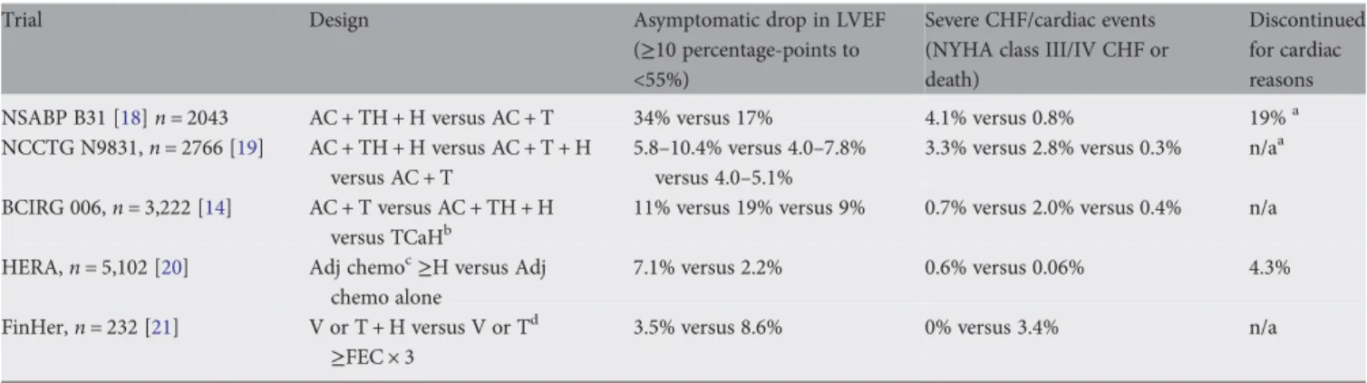
optimal target for biologic therapies. The use of the humanized monoclonal antibody trastuzumab (directed against the HER2 receptor) has revolutionized the treatment of HER2-positive breast cancer, with landmark adjuvant phase III trials demonstrating a 50% reduction in recurrence of disease and a 33% improvement in survival [14–18]. Rates of cardiac toxicity reported in the adjuvant trials of trastuzumab have been variable and reflect differences in trial design, chemotherapy administration and definitions of cardiac events, as described in Table1.
Bevacizumab, a humanized monoclonal antibody directed against vascular endothelial growth factor (VEGF), has demonstrated substantial antitumor activity when combined with chemotherapy, leading to regulatory approval for several advanced solid tumors, including breast, lung, colorectal and renal carcinomas. With greater use of bevacizumab, data are emerging regarding potential cardiac toxicity. To date, the rates of cardiac toxicity associated with bevacizumab therapy appear to be relatively low. In the major phase 3 trials in metastatic breast cancer, the reported rates of Common Terminology Criteria for Adverse Events CTCAE grade 3/4 CHF were 0.8%–2.2% in mostly anthracycline-pretreated population [22]. Overall, clinical trial data to date do not suggest significant increases in cardiac toxicity during treatment with
bevacizumab, even in the setting of concurrent treatment with other cardiotoxic agents. However, long-term monitoring of patients who have completed bevacizumab therapy has not been carried out; therefore, the safety of adjuvant bevacizumab in the setting of cancer survivorship is unknown. The risk factors for trastuzumab-associated cardiotoxicity identified from clinical trials are: prior treatment with anthracycline chemotherapy; a borderline LLN LVEF; prior treatment with antihypertensive medication; older age; and a poorly understood result found in one trial, a body mass index >25 kg/m2. In all adjuvant clinical trials, a commonfinding was that cardiac dysfunction and HF occurred predominantly during the trastuzumab treatment and was frequently
Table 1. Cardiac toxicity induced by trastuzumab
Trial Design Asymptomatic drop in LVEF
(≥10 percentage-points to <55%)
Severe CHF/cardiac events (NYHA class III/IV CHF or death)
Discontinued for cardiac reasons NSABP B31 [18] n = 2043 AC + TH + H versus AC + T 34% versus 17% 4.1% versus 0.8% 19%a
NCCTG N9831, n = 2766 [19] AC + TH + H versus AC + T + H versus AC + T
5.8–10.4% versus 4.0–7.8% versus 4.0–5.1%
3.3% versus 2.8% versus 0.3% n/aa BCIRG 006, n = 3,222 [14] AC + T versus AC + TH + H
versus TCaHb
11% versus 19% versus 9% 0.7% versus 2.0% versus 0.4% n/a HERA, n = 5,102 [20] Adj chemoc≥H versus Adj
chemo alone
7.1% versus 2.2% 0.6% versus 0.06% 4.3% FinHer, n = 232 [21] V or T + H versus V or Td
≥FEC × 3
3.5% versus 8.6% 0% versus 3.4% n/a
A, anthracycline; C, cyclophosphamide; T, taxane; H, trastuzumab; Ca, carboplatin; V, vinorelbine; F: 5-flourouracil; E, epirubicin; n/a, information not available.
a
6.7% did not receive H after A due to unacceptable drops in LVEF.
bIncluded a nonanthracycline arm. c
96% of chemotherapy was A containing.
dNo prior anthracycline before H exposure; H exposure limited to 9 weeks.
reversible. Longer follow-up surveillance is needed in order to better confirm this observation. Optimal surveillance for patients treated with Type II agents is not well established. Patients who have received both anthracyclines and anti-HER2 agents who develop cardiac failure should be treated and monitored as patients with an irreversible cardiac toxicity. Those who develop cardiac dysfunction during or following treatment with type II agents in the absence of anthracyclines can be observed if they remain asymptomatic and LVEF remains≥40%. Persistently low or further declines in LVEF or development of symptoms should trigger discussion of risk and benefit with the treating oncologist, as well as
consideration for pharmacologic cardiac treatment.
tyrosine kinase inhibitors and other targeted agents
Lapatinib, an oral receptor tyrosine kinase inhibitor (TKI) of HER2 and EGFR, has an approved role in combination with capecitabine chemotherapy in the treatment of patients with trastuzumab-resistant breast cancer. Experience to date in a relatively small studied population suggests relatively low rates of symptomatic cardiac failure (1.4%), specifically in a population with prior exposure to anthracycline and
trastuzumab [23]. Multiple small molecule TKIs of the VEGF receptor (VEGFR) have been developed, including sunitinib and sorafenib, with cross activity against other growth factor receptors including platelet-derived growth factor (PDGF), c-kit and BRAF. In contrast to bevacizumab, VEGFR antagonists appear to have a more profound effect on cardiac function. Initial reports of sunitinib in renal cell carcinoma suggested a 10% incidence of asymptomatic drop in LVEF to >10% LLN, with full recovery when treatment was completed [24]. early detection of anticancer drug-induced LVD At present, the most frequently used modality for detecting cardiotoxicity is the periodic measurement of LVEF by using either echocardiography or multigated acquisition scanning. To date, however, there are no evidence-based guidelines for cardiotoxicity monitoring during and after anticancer therapies in adults, while guidelines in pediatric oncology are subject to debate. Although several guidelines are available, none specify how often, by what means, or how long cardiac function should be monitored during and after cancer treatment [24]. Serial evaluation of LVEF is recommended for patients treated with trastuzumab [25,26]. However, the LVEF measurement is a relatively insensitive tool for detecting cardiotoxicity at an early stage. This is largely because no considerable change in LVEF occurs until a critical amount of myocardial damage has taken place, and only comes to the forefront after
compensatory mechanisms are exhausted. In addition, the measurement of LVEF presents a number of challenges related to image quality, assumption of LV geometry, load dependency and expertise. Multiple-gated acquisition (MUGA) scan can reduce interobserver variability with the disadvantages of including the exposure to radioactivity and the limited information than can be obtained on cardiac structure and diastolic function. Magnetic resonance imaging (MRI) is considered the gold standard for the evaluation of LV volumes, mass and function. However, its lack of availability and high cost limit its routine use. Novel ultrasound imaging techniques,
such as contrast echocardiography and real-time three-dimensional (3D) echocardiography that allows for an improvement in the accuracy of calculating LVEF, are under investigation. Small studies examining tissue Doppler and strain rate imaging appear to be promising in detecting early subclinical changes in cardiac performance that anticipate a decrease in conventional LVEF, even if long-term data on large populations, confirming the clinical relevance of such changes, are not available yet [27].
In the last decade, a new approach, based on the use of cardiac biomarkers, in particular troponins, has emerged, and has proven to be a more sensitive and more specific tool for early, real-time identification, assessment and monitoring of anticancer drug-induced cardiac injury [7,8]. Strong data indicate that troponin detects anticancer drug induced-cardiotoxicity in its earliest phase, long before any reduction in LVEF has occurred. Its evaluation during high-dose chemotherapy allows for the early identification of patients at risk of developing cardiac
dysfunction, the stratification of risk of cardiac events after chemotherapy and the opportunity for a preventive therapy in selected high-risk patients [6–8]. In patients treated with trastuzumab, troponin might help us to distinguish between reversible and irreversible cardiac injury by identifying myocardial cell necrosis [28]. The measurement of troponin immediately before and immediately after each cycle of cancer therapy seems to be effective enough, and is also transferable from clinical research to real-world routine assessment.
treatment of anticancer drug-induced LVD All patients with cancer who are treated with potentially cardiotoxic therapy represent a high-risk group for the development of HF. These patients have been excluded from large randomized trials evaluating the effectiveness of angiotensin-converting enzyme inhibitors (ACE-I) and beta-blocking agents (BB). The use of ACE-I and BB may be highly effective in this setting of patients. Recentfindings reported in a large population of anthracycline-induced CMP patients demonstrated that the time elapsed from the end of
chemotherapy to the start of HF therapy (time-to-treatment), with ACE-I and, when tolerated, with BB, is a crucial variable for recovery of cardiac dysfunction [29,30]. Indeed, the likelihood of obtaining a complete LVEF recovery is higher in patients in whom treatment is initiated within 2 months from the end of chemotherapy. Although promising data have been published, convincing evidence from large randomized and prospective trials is still needed. Treatment of trastuzumab-related cardiotoxicity (TIC) is a more controversial issue. Despite new, updated guidelines for monitoring patients receiving adjuvant trastuzumab being periodically published, they are specifically focused on the continuation/withdrawal/ resuming of trastuzumab therapy. No evidence-based recommendations for the treatment of patients developing cardiac dysfunction after trastuzumab therapy have been proposed. The evidence that support the use of ACE-I and BB in this setting is limited to case series. Despite evidence, the potential efficacy of ACE-I and BB in improving LVEF in patients receiving trastuzumab remains uncertain.
prevention of anticancer drug-induced LVD According to the American College of Cardiology and American Heart Association guidelines, patients receiving chemotherapy may be considered a Stage A HF group, namely those with an increased risk of developing cardiac dysfunction [24]. Carvedilol may prevent cardiac damage induced by doxorubicin due to its antioxidant activity. The effect of
carvedilol was confirmed in a randomized study in which prophylactic use of carvedilol in a small population of patients treated with anthracycline prevented LVD and reduced mortality [31]. Nakamae et al. [32] have demonstrated that valsartan, an angiotensin receptor blocker (ARB), given concurrently to anthracycline-containing regimens, prevents cardiac damage. Dexrazoxane, an iron-chelating agent,
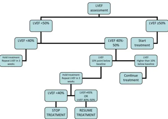
Figure 1. Algorithm for the management of cardiotoxicity in patients receiving anthracyclines.
Figure 2. Algorithm for continuation and discontinuation of trastuzumab based on LVEF assessments.
significantly reduces anthracycline-related cardiotoxicity in adults with different solid tumors and in children with acute lymphoblastic leukemia and Ewing’s sarcoma [33].
Dexrazoxane is not routinely used in clinical practice and it is recommended as a cardioprotectant by the American Society of Clinical Oncology only for patients with metastatic breast cancer who have already received more than 300 mg/m2of doxorubicin.
Figure1reports the algorithm for the management of cardiotoxicity in patients receiving anthracyclines. Figure2
reports the algorithm for continuation and discontinuation of trastuzumab based on LVEF assessments. Table2reports recommendations for CV evaluation before anticancer treatment. Table3reports recommendations for CV monitoring during and after anticancer treatment with potential cardiotoxicity (including anti-HER2 agents). Table4
reports on treatment of LVD induced by anticancer treatment (including anti-HER2 agents).
management of trastuzumab cardiotoxicity Management of trastuzumab-related cardiotoxicity has two distinct aspects: withdrawal of trastuzumab therapy and treatment of cardiac dysfunction.
The‘stopping/restarting’ rules used in the adjuvant trials were effective and are recommended, with some modifications regarding recommendations for a cardiology consult or treatment of cardiac dysfunction (or both) when appropriate.
Symptomatic LVD must be treated with HF treatment: • All patients with HF and an LVEF <40% should be
treated with an ACE-I in combination with a BB unless a specific contraindication exists [I, A].
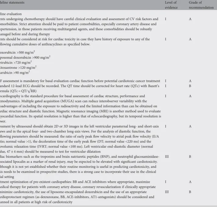
Table 2. CV evaluation before anticancer treatment with potential nonreversible (Type I) or reversible (type II) cardiotoxicity
Guideline statements Level of
evidence
Grade of recommendation Baseline evaluation
Patients undergoing chemotherapy should have careful clinical evaluation and assessment of CV risk factors and comorbidities. Strict attention should be paid to patient comorbidities, especially coronary artery disease and hypertension, in those patients receiving multitargeted agents, and these comorbidities should be robustly managed before and during therapy
I A
Patients should be considered at risk for cardiac toxicity in case they have history of exposure to any of the following cumulative doses of anthracyclines as specified below.
• Doxorubicin >500 mg/m2 • Liposomal doxorubicin >900 mg/m2 • Epirubicin >720 mg/m2 • Mitoxantrone >120 mg/m2 • Idarubicin >90 mg/m2 I A
LVEF assessment is mandatory for basal evaluation cardiac function before potential cardiotoxic cancer treatment I A A standard 12-lead ECG should be recorded. The QT time should be corrected for heart rate (QTc) with Bazett’s
formula (QTc = QT/√RR)
I B
Echocardiography is the standard procedure for basal assessment of cardiac structure, performance and hemodynamics. Multiple gated acquisition (MUGA) scan can reduce interobserver variability with the disadvantages of including the exposure to radioactivity and the limited information than can be obtained on cardiac structure and diastolic function. Magnetic resonance imaging (MRI) is another method used to evaluate myocardial function. Its spatial resolution is higher than that of echocardiography, but its temporal resolution is lower.
I A
Assessment by ultrasound should obtain 2D or 3D images in the left ventricular parasternal long- and short-axis views and in the apical four- and two-chamber long-axis views. For the analysis of diastolic function, the following parameters should be measured: the ratio of early peakflow velocity to atrial peak flow velocity (E/A ratio; normal value >1), the deceleration time of the early peakflow (DT; normal value <220 ms) and the isovolumic relaxation time (IVRT; normal value <100 ms). Left ventricular end-diastolic diameter (normal value, 47 ± 4 mm) should be measured to test for ventricular dilatation
I A
Cardiac biomarkers such as the troponins and brain natriuretic peptides (BNP), and neutrophil glucosaminidase-associated lipocalin as a marker of renal injury, may be expected to be elevated with significant cardiotoxicity. Although it is not yet established whether their routine monitoring is useful in predicting cardiotoxicity, and this needs to be examined in prospective studies, there is a strong case to incorporate their use in the clinical trial setting
III B
Treatment optimization of pre-existent cardiopathies: BB and ACE inhibitors where appropriate, maximize medical therapy for patients with coronary artery disease, coronary revascularization if clinically appropriate
I A
To minimize cardiotoxicity, the use of liposome-encapsulated doxorubicin and the use of an appropriate cardioprotectant regimen (as dexrazoxane, BB, ACE-inhibitors, AT1-antagonists) should be considered and planned in all patients at high risk of cardiotoxicity
III B
• Some members of the panel also felt that, to prevent further degradation of LVEF or the development of clinical HF, an ACE-I should be considered if the patient’s LVEF is between 40% and 50%. Asymptomatic LVD should be treated:
• ACE-Is should be used in all asymptomatic patients with LVD and an ejection fraction <40% [I, A for ejection fraction <35%; I, B for ejection fraction 35%–40%]. • Also, an ACE-I should be considered if LVEF is <50%. • BB should be considered in all patients with
asymptomatic LVD and an LVEF <40% [if prior myocardial infarction I, B; if no myocardial infarction II, C].
cardiac ischemia
Although cardiac ischemia related to chemotherapy
administration is an unusual occurrence, an increased risk of acute coronary syndrome has been associated with
administration of cytotoxic and targeted agents for cancer treatment.
antimetabolites. The incidence of cardiac events associated with 5-FU varies in the literature ranging anywhere from 1% to 68% [34]. Cardiac toxicity typically occurs with early onset (within 2–5 days of starting therapy). Ischemic
electrocardiogram (ECG) changes have been reported in up to 68% of patients, although in the majority evidence of cardiac
Table 3. CV monitoring during and after anticancer treatment with potential non-reversible (Type I) or reversible (type II) cardiotoxicity
Guideline statements Level of
evidence
Grade of recommendation Cardiac monitoring
Patients receiving anthracyclines and/or trastuzumab in the adjuvant setting should perform serial monitoring of cardiac function at baseline, 3, 6 and 9 months during treatment, and then at 12 and 18 months after the initiation of treatment. Monitoring should be repeated during or following treatment as clinically indicated. Limited data are available for elderly patients: increased vigilance is recommended for patients≥60 years old
I A
Patients treated for metastatic disease: LVEF should be monitored at baseline and then infrequently in the absence of symptoms
II A
Troponin I or BNP concentrations seem to identify patients at risk of cardiotoxicity, specifically in case of administration of type I agents (such as anthracyclines). Performing baseline assessment of biomarker concentrations and periodic measurements during therapy (every each cycle) may identify patients who need further cardiac assessment
III B
Assessment of cardiac function is recommended 4 and 10 years after anthracycline therapy in patients who were treated at <15 years of age, or even at age >15 years but with cumulative dose of doxorubicin of >240 mg/m2or
epirubicin >360 mg/m2
II B
LVEF reduction of≥15% from baseline with normal function (LFEV ≥ 50%) is an indication to continue anthracyclines and/or trastuzumab. LVEF decline to <50% during anthracyclines containing regimens necessitate reassessment after 3 weeks. If confirmed, hold chemotherapy, consider therapy for LVD and further frequent clinical and echocardiographic checks. In case of LVEF decline to <40% stop chemotherapy, discuss alternatives and treat LVD
II B
LVEF decline to <50% during trastuzumab therapy (post-anthracyclines) necessitate reassessment after 3 weeks. If confirmed, continue trastuzumab and consider therapy for LVD and further frequent clinical and
echocardiographic checks. In case of LVEF decline to <40% stop trastuzumab and treat LVD
Aggressive medical treatment of those patients, even asymptomatic, who show LVD at DEcho after anthracycline therapy is mandatory, especially if the neoplasia could have a long-term survival; it consists of ACE inhibitors and b-blockers and the earlier HF therapy is begun (within 2 months from the end of anthracycline therapy), the better the therapeutic response
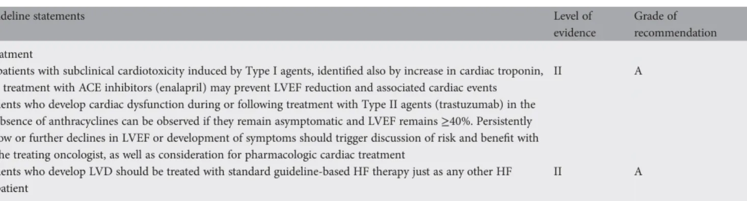
Table 4. Treatment of LVD induced by anticancer treatment with non-reversible (Type I) or reversible (Type II) cardiotoxicity
Guideline statements Level of
evidence
Grade of recommendation Treatment
In patients with subclinical cardiotoxicity induced by Type I agents, identified also by increase in cardiac troponin, a treatment with ACE inhibitors (enalapril) may prevent LVEF reduction and associated cardiac events
II A
Patients who develop cardiac dysfunction during or following treatment with Type II agents (trastuzumab) in the absence of anthracyclines can be observed if they remain asymptomatic and LVEF remains≥40%. Persistently low or further declines in LVEF or development of symptoms should trigger discussion of risk and benefit with the treating oncologist, as well as consideration for pharmacologic cardiac treatment
Patients who develop LVD should be treated with standard guideline-based HF therapy just as any other HF patient
II A
myocyte damage including elevation in serum cardiac markers is lacking [34]. Coronary artery thrombosis, arteritis and vasospasm secondary to drug exposure have been proposed as the most likely underlying mechanisms.
inhibitors of microtubule polymerization. Paclitaxel administration has been associated with cases of myocardial ischemia and infarction. A retrospective study reviewed cardiac events in four clinical trials, and reported manifestations of cardiac ischemia in 5% of patients [35].
endocrine agents. Aromatase inhibitors (AIs) are an established component of the treatment of postmenopausal hormone receptor-positive breast cancer. Cardiac events, including myocardial infarction and cardiac failure, have been reported at low frequency in the major adjuvant trials comparing the use of AIs to a control arm of 5 years of tamoxifen. A meta-analysis of over 19 000 participants in adjuvant studies suggests a relative risk of 1.31 (95% CI 1.01– 1.60, P = 0.007) for a cardiac adverse event associated with exposure to an AI compared with tamoxifen, although the absolute risk remains low,∼0.5% [36]. Differential changes in lipid profile have been proposed as an etiology for these observations; however, a strong signal linking AIs and relevant changes in lipid levels are lacking. The long-term clinical significance of these clinical observations remains unclear. targeted agents. Whether novel molecular therapies increase the risk of cardiac ischemia is not clear. In an evaluation of the VEGF receptor antagonist sunitinib, 18% of 68 patients studied were noted to have modest increases in cardiac troponins, possibly serving as a biomarker of myocyte damage [37]. clinical recommendations
Ischemic events following or during antimetabolites or paclitaxel infusion
Baseline ECG evaluation is recommended [III/IV, A] Frequent vital sign monitoring is recommended during chemotherapeutic agent infusion, particularly with 5-FU or paclitaxel [III/IV, A]. Monitoring of BNB and troponin I should be recommended in patients with anamnesis of cardiac ischemia [III/IV, C].
A collaborative decision should then be made as to whether more advanced cardiac testing (e.g. stress testing and coronary angiography) is needed and whether the benefits of resuming therapy with aggressive supportive care outweigh the risk.
hypertension
Multiple investigative and clinical observations have demonstrated hypertension to be a common class effect resulting from treatment with VEGF inhibitors. The rates of substantial hypertension appear to depend on the
antiangiogenic agent used, the tumor type and patient-related factors including age and comorbidity. Angiogenesis inhibitor-related hypertension is typically manageable with early initiation of pharmacologic therapy to reach accepted blood pressure (BP) targets. Preferred antihypertensive agents for angiogenesis inhibitor-associated hypertension include ACE-I and dihydropyridine calcium channel blockers, although there
are minimal data to suggest superiority of a single class of agents. Early and aggressive initiation of antihypertensive therapy appears to help maintain treatment schedule and reduce the risk of substantial complications, including malignant hypertension and reversible posterior leukencephalopathy.
The exact mechanism underlying the hypertension associated with angiogenesis inhibitors is not well defined. However, several theories have been proposed, including an imbalance in neurohumoral factors, the development of vascular rarefaction or an alteration in vascular nitric oxide balance [38,39]. The alternative hypothesis of vascular rarefaction, the process of reduced microvascular density, has been proposed to result in hypertension through increases in systemic vascular resistance. Prospective evaluation of patients receiving anti-VEGF agents has demonstrated reductions in capillary microdensity and alterations in capillary endothelial function in the setting of documented hypertension support this event as a contributing factor [40,41].
clinical recommendations for assessment and management of patients candidate to
antiangiogenic agent inducing hypertension Individuals should be considered at risk in case of: systolic BP ≥160 mmHg or diastolic BP ≥100 mmHg; diabetes mellitus; established CV disease including any history of ischemic stroke, cerebral hemorrhage or transient ischemic attack; myocardial infarction, angina, coronary revascularization, or HF; peripheral artery disease; subclinical organ damage previously documented by ECG or echocardiogram revealing left ventricular hypertrophy; cigarette smoking; dyslipidemia.
Repeated BP measurements are recommended.
Aggressive management of BP elevations is recommended to prevent clinically limiting complications.
There are no evidence-based guidelines for follow-up echocardiograms in asymptomatic patients receiving antiangiogenic agents. Although some LVEF-based dose modification algorithms have been used in clinical trials that called for periodic echocardiogram/MUGA assessments, at the time of writing, there are no data on which to base general recommendations for antiangiogenic agents dose adjustment.
QT prolongation
Prolongation of the QT interval can lead to life-threatening cardiac arrhythmias, including‘torsade de pointes’ (TdP). Although prolongation of the QT interval is not the best predictor of proarrhythmic risk, it represents the principal clinical surrogate marker by which to evaluate the arrhythmic risk of a drug, and it has led to withdrawal of several
anticancer drugs from the market. Although drugs leading to prolonged QT may possess substantial risks of serious adverse events, the clinical benefit of therapy in the oncologic setting, including the possibility of cure for a cancer patient, may outweigh the potential risks of QTc prolongation, even when the prolongation is significant [42].
arsenic trioxide. Arsenic trioxide (ATO) presents an
interesting example of successful risk management, supporting a decision for a patient to accept, or a physician to administer,
an anticancer drug with established liabilities of QTc prolongation. Although this drug is known to provoke QTc prolongation and TdP, it is also uniquely effective in an otherwise fatal disease, relapsed acute promyelocytic leukemia [43]. Therefore, until alternative therapy becomes available, ATO remains a drug of choice, despite its potential for causing arrhythmia. In patients receiving multiple courses, QTc intervals may return to pre-treatment levels before the second course, implying that serial ATO administration does not permanently prolong the QTc interval; however, documented episodes of TdP have been observed beyond thefirst month of treatment, presumably due to drug accumulation in cardiac tissue [43].
clinical recommendation for patients receiving drugs prolonging QTc interval
Patients with a history of QT interval prolongation, patients who are taking antiarrhythmics, or patients with relevant CV disease, bradycardia, thyroid dysfunction or electrolyte disturbances should be screened and monitored. Periodic monitoring with on-treatment ECGs and electrolytes should be considered.
cardiac toxicity induced by RT
A considerable amount of literature supports evidence of radiation-related heart injury after RT to the chest. The most appropriate groups of patients where radiation-associated cardiac injuries are of clinical importance are those with curable malignancies irradiated at a relatively younger age, so there is enough time to develop clinically significant late cardiac injury. Such malignancies are mainly Hodgkin’s lymphoma and early-stage breast cancer, while there is an increasing number of lung and esophageal cancer patients with long-term controlled disease who could develop post-RT cardiac sequelae. Radiation-associated CV toxicity may be progressive. Complex, combined disease of heart coronary arteries, valves, myocardium and conduction system as well as diastolic dysfunction may be seen [44,45]. Estimates of relative risk of fatal CV events after mediastinal irradiation for
Hodgkin’s disease ranges between 2 and 7 and after irradiation for left-sided breast cancer from 1.0 to 2.2.
Risk factors for radiation-associated heart damage include:
• dose >30–35 Gy
• dose per fraction >2 Gy
• large volume of irradiated heart
• younger age at exposure
• longer time since exposure
• use of cytotoxic chemotherapy
• endocrine therapy or trastuzumab
• presence of other risk factors such as diabetes, hypertension, dyslipidaemias, obesity, smoking etc.
evidence from breast cancer patients In the last update of the Early Breast Cancer Trialists’
Collaborative Group (EBCTCG) meta-analysis on locoregional RT, comparison of CV mortality between patients treated with and without RT has shown a statistically significant relative
risk of 1.27. A similar latent time was estimated in an overview of trials started before 1975.
In a Swedish study that included 55 000 patients, a mortality ratio of all CV disease for left versus right side in a period of >10 years after treatment was 1.10 [95% confidence interval (CI) 1.03–1.18] for all CV diseases and 1.13 (95% CI 1.03– 1.25) for deaths from ischemic disease. Other studies confirm those results.
evidence from Hodgkin’s lymphoma patients
Mediastinal RT can cause a variety of CV complications such as pericarditis, myocardialfibrosis, coronary artery disease, valvular abnormalities and conduction disturbances [46]. Restrictive CMP, valvular defects and conduction defects, persistent tachycardia and blunted hemodynamic responses to exercise are usually diagnosed. Nevertheless, myocardial infarction is the major cause of higher long-term mortality in survivors of Hodgkin’s disease. It is very important to note that death due to cardiac causes is estimated to be responsible for about one-quarter of the mortality from reasons other than Hodgkin’s disease itself, which equals 2%–5% of overall mortality in those with Hodgkin’s disease.
cardiac structures affected by radiation
Injury of the various structures and tissues in the heart can cause a spectrum of radiation-induced CV diseases. Arteritis of the endothelium of coronary arteries can cause premature coronary artery disease and atherosclerosis mainly in left anterior descending and right coronary artery. The time of appearance is 10–15 years after RT.
• Acute pericarditis and symptomatic (hemodynamic compromise with constriction or tamponade) or
asymptomatic chronic pericardial effusion appears usually 6– 12 months following RT.
• Myocarditis and CHF due to nonspecific diffuse interstitial fibrosis.
• Valvular stenosis and regurgitation mainly of mitral and aortic valves.
• Fibrosis of the conduction system and disturbed heart rate and complete or incomplete heart block.
• Some indirect implications on the heart may result from irradiation of adjacent structures. Lung and mediastinal fibrosis may result in respiratory insufficiency, pneumonic hypertension and may complicate any potential heart surgery. Hypothyroidism may affect the lipid profile and CV function. Mediastinal venous and lymph vessel obstruction may cause pericardial effusion or chylothorax.
Radiation tolerance doses for the above late-effects have been estimated to be between 30 and 40 Gy.
recommendations to reduce cardiac toxicity in patients treated by RT
There is some evidence that newer irradiation techniques seem to decrease the risk of RT-induced cardiac disease, but a longer follow-up time is needed to confirm it. Modern RT techniques include 3D treatment planning with dose volume histogram for
accurate heart volume and dose calculation. Linear accelerator photons and multiplefield conformal or intensity-modulated RT (IMRT) are desirable for chest irradiation.
Normal tissue complication probability (NTCP) is a method of reporting radiation dosing in RT, by taking into account the dose and the volume of normal tissues that receive the corresponding dose [47]. The NTCP model estimates predict that a V25Gy <10% (i.e. a dose of 25 Gy (in 2 Gy per fraction) in <10% of the whole volume of heart) will be associated with 1%– 2% probability of cardiac mortality within 15 years after RT.
For (left) breast/chest wall RT, 6 MV or occasionally higher energy photons (for large breasts) from a linear accelerator should be used. The introduction of cardiac-sparing lead block during standard simulator planning will result in cardiac irradiation being at least partially avoided in many patients. The use of a four-field IMRT technique can offer better sparing than the partial shielding technique as the maximum heart depth is increased. The IMRT plans also showed improved dose homogeneity within the Planning Target Volume (PTV) but may be associated with increased irradiation of the contralateral breast.
It has been proposed that maximum heart distance (MHD), i.e. the maximal distance between anterior cardiac contour and posterior tangentialfield edges as seen on the beam’s eye view, is a reliable predictor of the mean heart dose in left-tangential breast or chest wall irradiation, and may be useful in centers where 3D cardiac dose assessment is not routinely available. A strong linear correlation was found between the MHD and the mean heart dose: for every 1-cm increase in MHD, the mean heart dose increased by 2.9% on average (95% CI 2.5–3.3).
Electron beams can be used for the treatment of superficial structures such as in the internal mammary lymph nodes or the boost dose on the breast after tumorectomy, in the treatment of breast cancer.
For mediastinal RT, high-energy photons from a linear accelerator should be used to treat patients with equal weighting of anterior and posterior portals (instead of anterior weighting), allfields should be treated on each RT fraction, use of a subcarinal block after a dose of 30 Gy and use of shrinking-field technique are the most important parameters to minimize heart exposure. As Adams et al. has stated, although permanent complications tend to occur less frequently under a total dose of 40 Gy, it is not a good idea to systematically limit treatment, which may be inadequate to control the neoplastic disease.
treatment of RT-related heart complications Radiation-induced heart diseases are treated as nonradiation-related ones, but with special attention to the changes radiation causes to the heart and other structures of the chest.
monitoring of cardiac function after chest RT Patients at high risk for RT-induced complications are those treated as children or young adults for Hodgkin’s lymphomas with a mediastinal/heart dose of >30 Gy, mainly with what are called outdated RT techniques (see above). Those patients should be informed and followed up closely [III,A]. Radiation-related heart toxicity is extremely rare during RT.
Breast cancer patients treated by cytotoxic chemotherapy or monoclonal antibodies should be monitored. Patients treated by postoperative breast RT (with or without adjuvant endocrine treatment) are not regularly monitored for cardiac problems although RT should be considered as a risk factor when heart disease is diagnosed in those patients [III, B].
Data to support definitive recommendations on various tests and their frequency do not exist. However, RT-induced risk is ongoing and requires long-term follow-up. Screening and monitoring of heart function is identical to the standard tests and procedures cardiologists use for other patients, and consequently the follow-up protocols are based on departmental or personal experience and on each patient’s needs and clinical picture. There is therefore a need for both oncologists and cardiologists to be aware of the risks and underlying pathophysiology of RT-related heart complications.
Apart from clinical examination and medical history, tests usually requested depend on the studied abnormality.
• Coronary artery disease: lipid profile, exercise stress test, radionuclide, angiography, echocardiogram, ECG.
• Pericarditis: ECG, chest X-ray and echocardiogram.
• CMP: ECG, echocardiogram, and radioisotopic angiography.
• Arrythmias: ECG and 24-h ECG.
• Valvular disease: echocardiogram, cardiac catheterization.
conclusions
Within the oncologic toolbox are both traditional and novel anticancer agents with the potential to induce cardiac toxicity. Oncologists thus have had to develop the ability to identify and manage complicated cardiac risks in clinical investigations and in general practice. Many highly effective agents in
contemporary oncology, including anthracyclines and trastuzumab, are associated with well-described risks of short-and long-term cardiac events. As these agents are often used with curative intent, maximizing the benefits while reducing cardiac risks has become a priority in oncologic management, as well as monitoring for late-term toxicity. Additionally, given the influx of novel biologic therapies designed to fulfill unmet medical needs, efforts are needed to promote strategies for risk detection and management to avoid dangerous toxicities which may impede development of and patient access to new agents. In principle, the risk–benefit ratio for an agent must be interpreted in the context of the nature and severity of the disease, and restrictive approaches have the potential to delay or prevent access to innovative treatment. More research is needed to assess and manage the CV safety of patients treated with anticancer agents, beginning with a dynamic partnership between oncologists and cardiologists, and the development of a new generation of‘cardio-oncologic’ investigators. A thoughtful risk management plan generated by an organized collaboration between oncologists, cardiologists and regulatory agencies can support developmental programs essential for anticancer agents with cardiac safety concerns.
con
flict of interest
Dr. Roila has reported consultancy/honoraria: Merck Sharp & Dohme, Roche; research funding to department: Bristol-Myers
Squibb, Merck Sharp & Dohme, GlaxoSmithKline, Sanofi Aventis, AstraZeneca. Dr Suter has reported research support from Hoffmann La Roche and speakers’ bureau for
Robopharm. Dr de Azambuja has reported research support from GlaxoSmithKline; honoraria from Roche; travel grants from GlaxoSmithKline and Roche.
Dr. Goldhirsch has not reported any potential conflicts of interest. The other authors have reported no potential conflicts of interest.
references
1. Suter TM, Ewer MS. Cancer drugs and the heart: importance and management. Eur Heart J. 2012. [Epub ahead of print ].
2. National Cancer Institute. Cancer therapy evaluation program. [on-line]http ://ctep.cancer.gov/protocoldevelopment/electronic_applications/docs/ ctcv20_4e30e992.pdf.
3. Bird BR, Swain SM. Cardiac toxicity in breast cancer survivors: review of potential cardiac problems. Clin Cancer Res. 2008; 14: 14–24. 4. Von Hoff DD, Layard MW, Basa P et al. Risk factors for doxorubicin-induced
congestive heart failure. Ann Intern Med. 1979; 91: 710–717.
5. Cardinale D, Sandri MT, Martinoni A et al. Left ventricular dysfunction predicted by early troponin I release after high-dose chemotherapy. J Am Coll Cardiol. 2000; 36: 517–522.
6. Cardinale D, Sandri MT, Martinoni A et al. Myocardial injury revealed by plasma troponin I in breast cancer treatment with high dose chemotherapy. Ann Oncol. 2002; 13: 710–715.
7. Cardinale D, Sandri MT, Colombo A et al. Prognostic value of Troponin I in cardiac risk stratification of cancer patients undergoing high-dose chemotherapy. Circulation. 2004; 109: 2749–2754.
8. Pichon MF, Cvitkovic F, Hacene K et al. Drug-induced cardiotoxicity studied by longitudinal B-type natriuretic peptide assays and radionuclide ventriculography. In Vivo. 2005; 19: 567–576.
9. Braverman AC, Antin JH, Plappert MT et al. Cyclophosphamide cardiotoxicity in bone marrow transplantation: a prospective evaluation of new dosing regimens. J Clin Oncol. 1991; 9: 1215–1223.
10. Goldberg MA, Antin JH, Guinan EC et al. Cyclophosphamide cardiotoxicity: an analysis of dosing as a risk factor. Blood. 1986; 68: 1114–1118.
11. Quezado ZM, Wilson WH, Cunnion RE et al. High-dose ifosfamide is associated with severe, reversible cardiac dysfunction. Ann Intern Med. 1993; 118: 31–36. 12. Martin M, Pienkowski T, Mackey J et al. Adjuvant docetaxel for node-positive
breast cancer. N Engl J Med. 2005; 352: 2302–2313.
13. Slamon D, Eiermann W, Robert N et al. Adjuvant trastuzumab in HER2-positive breast cancer. N Engl J Med. 2011; 365(14): 1273–1283.
14. Romond EH, Perez EA, Bryant J et al. Trastuzumab plus adjuvant chemotherapy for operable HER2-positive breast cancer. N Engl J Med. 2005; 353: 1673–1684.
15. Piccart-Gebhart MJ, Procter M, Leyland-Jones B et al. Trastuzumab after adjuvant chemotherapy in HER2-positive breast cancer. N Engl J Med. 2005; 353: 1659–1672.
16. Slamon DJ, Leyland-Jones B, Shak S et al. Use of chemotherapy plus a monoclonal antibody against HER2 for metastatic breast cancer that overexpresses HER2. N Engl J Med. 2001; 344: 783–792.
17. Tan-Chiu E, Yothers G, Romond E et al. Assessment of cardiac dysfunction in a randomized trial comparing doxorubicin and cyclophosphamide followed by paclitaxel, with or without trastuzumab as adjuvant therapy in node-positive, human epidermal growth factor receptor 2-overexpressing breast cancer: NSABP B-31. J Clin Oncol. 2005; 23: 7811–7819.
18. Perez EA, Suman VJ, Davidson NE et al. Cardiac safety analysis of doxorubicin and cyclophosphamide followed by paclitaxel with or without trastuzumab in the
North Central Cancer Treatment Group N9831 adjuvant breast cancer trial. J Clin Oncol. 2008; 26: 1231–1238.
19. Suter TM, Procter M, van Veldhuisen DJ et al. Trastuzumab-associated cardiac adverse effects in the Herceptin adjuvant trial. J Clin Oncol. 2007; 25: 3859–3865. 20. Joensuu H, Kellokumpu-Lehtinen PL, Bono P et al. Adjuvant docetaxel or
vinorelbine with or without trastuzumab for breast cancer. N Engl J Med. 2006; 354: 809–820.
21. Miller K, Wang M, Gralow J et al. Paclitaxel plus bevacizumab versus paclitaxel alone for metastatic breast cancer. N Engl J Med. 2007; 357: 2666–2676.
22. Perez EA, Koehler M, Byrne J et al. Cardiac safety of lapatinib: pooled analysis of 3689 patients enrolled in clinical trials. Mayo Clin Proc. 2008; 83: 679–686.
23. Motzer RJ, Hutson TE, Tomczak P et al. Sunitinib versus interferon alfa in metastatic renal-cell carcinoma. N Engl J Med. 2007; 356: 115–124. 24. Eschenhagen T, Force T, Ewer M et al. Cardiovascular side effects of cancer
therapies: a position statement from the Heart Failure Association of the European Society of Cardiology. Eur J Heart Fail. 2011; 13: 1–10. 25. Jones AL, Barlow M, Barrett-Lee PJ et al. Management of cardiac health in
trastuzumab-treated patients with breast cancer: updated United Kingdom National Cancer Research Institute recommendations for monitoring. Br J Cancer. 2009; 100: 684–692.
26. Martin M, Esteva FJ, Alba E et al. Minimizing cardiotoxicity while optimizing treatment efficacy with trastuzumab: review and expert recommendations. Oncologist 2009; 14: 1–11.
27. Geisberg CA, Sawyer DB. Mechanisms of anthracycline cardiotoxicity and strategies to decrease cardiac damage. Curr Hypertens Rep. 2010; 12: 404–410.
28. Cardinale D, Colombo A, Torrisi R et al. Trastuzumab-induced cardiotoxicity: clinical and prognostic implications of troponin I evaluation. J Clin Oncol. 2010; 28: 3910–3916.
29. Cardinale D, Colombo A, Sandri MT et al. Prevention of high-dose chemotherapy-induced cardiotoxicity in high-risk patients by angiotensin-converting enzyme inhibition. Circulation. 2006; 114: 2474–2481. 30. Cardinale D, Colombo A, Lamantia G et al. Anthracycline-induced
cardiomyopathy. Clinical relevance and response to pharmacologic therapy. J Am Coll Cardiol. 2010; 55: 213–220.
31. Kalay N, Basar E, Ozdogru I et al. Protective effects of carvedilol against anthracyclines-induced cardiomyopathy. J Am Coll Cardiol. 2006; 48: 2258–2262.
32. Nakamae H, Tsumura K, Terada Y et al. Notable effects of angiotensin II receptor blocker, valsartan, on acute cardiotoxic changes after standard chemotherapy with cyclophosphamide, doxorubicin, vincristine, and prednisolone. Cancer. 2005; 104: 2492–2498.
33. Huh WW, Jaffe N, Durand JB et al. Comparison of doxorubicin cardiotoxicity in pediatric sarcoma patients when given with dexrazoxane versus continuous infusion. Pediatr Hematol Oncol. 2010; 27: 546–557.
34. Van Cutsem E, Hoff PM, Blum JL et al. Incidence of cardiotoxicity with the oral fluoropyrimidine capecitabine is typical of that reported with 5-fluorouracil. Ann Oncol. 2002; 13: 484–485.
35. Rowinsky EK, McGuire WP, Guarnieri T et al. Cardiac disturbances during the administration of taxol. J Clin Oncol. 1991; 9: 1704–1712.
36. Cuppone F, Bria E, Verma S et al. Do adjuvant aromatase inhibitors increase the cardiovascular risk in postmenopausal women with early breast cancer? Meta-analysis of randomized trials. Cancer 2008; 112: 260–267.
37. Schmidinger M, Zielinski CC, Vogl UM et al. Cardiac toxicity of sunitinib and sorafenib in patients with metastatic renal cell carcinoma. J Clin Oncol. 2008; 26: 5204–5212.
38. Kamba T, McDonald DM. Mechanisms of adverse effects of anti-VEGF therapy for cancer. Br J Cancer. 2007; 96: 1788–1795.
39. Verheul HM, Pinedo HM. Possible molecular mechanisms involved in the toxicity of angiogenesis inhibition. Nat Rev Cancer. 2007; 7: 475–485.
40. Veronese ML, Mosenkis A, Flaherty KT et al. Mechanisms of hypertension associated with BAY 43–9006. J Clin Oncol. 2006; 24: 1363–1369.
41. Mourad JJ, des Guetz G, Debbabi H et al. Blood pressure rise following angiogenesis inhibition by bevacizumab. A crucial role for microcirculation. Ann Oncol. 2008; 19: 927–934.
42. Curigliano G, Spitaleri G, Fingert HJ et al. Drug-induced QTc interval prolongation: a proposal towards an efficient and safe anticancer drug development. Eur J Cancer. 2008; 44(4): 494–500.
43. Barbey JT, Soignet S. Prolongation of the QT interval and ventricular tachycardia in patients treated with arsenic trioxide for acute promyelocytic leukemia. Ann Intern Med. 2001; 135: 842–843.
44. Heidenreich PA, Hancock SL, Lee BK et al. Asymptomatic cardiac disease following mediastinal irradiation. J Am Coll Cardiol. 2003; 42: 743–749. 45. Heidenreich PA, Hancock SL, Vagelos RH et al. Diastolic dysfunction after
mediastinal irradiation. Am Heart J. 2005; 150: 977–982.
46. Galper SL, Yu JB, Mauch PM et al. Clinically significant cardiac disease in patients with Hodgkin lymphoma treated with mediastinal irradiation. Blood 2011; 117(2): 412–418.
47. Gagliardi G, Constine LS, Moiseenko V et al. Radiation dose-volume effects in the heart. Int J Radiat Oncol Biol Phys. 2010; 3(Suppl): S77–S85.