Cystathionine-γ-lyase expression is associated with mitochondrial respiration during sepsis-induced acute kidney injury in swine with atherosclerosis
Texte intégral
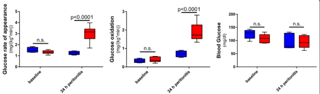
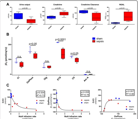
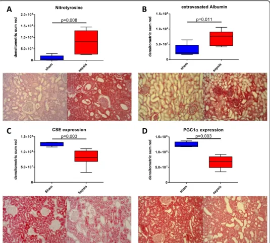
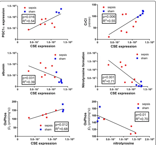
Figure
Documents relatifs
If you were to add this navigation scheme to your Web site manually, you would assemble button graphics you want to use, insert them into your pages, and then add links to them
We prove that, if 7 is a simple smooth curve in the unit sphere in C n, the space o pluriharmonic functions in the unit ball, continuous up to the boundary, has a
[r]
[r]
5 – Pour les constructions qui ne sont pas destinées à de l’habitation et qui s’implantent sur des terrains disposant d’une façade sur l’Avenue du Maréchal de
Nous ne devons plus accepter cette injustice profonde, mais exiger des mesures de rattrapage im- médiat pour toutes celles qui ont vu leur carrière bloquée du fait de grossesses ou
[r]
[r]
