Obstructive and Non-Obstructive Conditions.
by
Harish S. Lecamwasam Submitted to the Department of
Mechanical Engineering in Partial Fulfillment of the Requirements for the
Degree of
MASTER OF SCIENCE in Mechanical Engineering
at the
Massachusetts Institute of Technology June 1995
0 1995 Massachusetts institute of Technology All rights reserved
Signature of Author... ...
/Z" . Department of Mechanical Engineering
June 9, 1995
C ertified by ... ... ... ... Professor Ernest G. Cravalho
Thesis Supervisor
Accepted by . ...- . ... ... ...
Professor Ain A. Sonin Chairman, Graduate School Committee
;:.,ASSACHUSETTS INSTITUTE OF TECHNOLOGY
AUG 31 1995
LIBRARIES
CANINE URINARY TRACT UNDER OBSTRUCTIVE AND NON-OBSTRUCTIVE CONDITIONS
by
HARISH S. LECAMWASAM
Submitted to the Department of Mechanical Engineering on June 9, 1995 in partial fulfillment of the requirements for the Degree of Master of Science in Mechanical
Engineering
ABSTRACT
Benign prostatic hyperplasia (BPH) is typically a disease of men aged 40 years, and above. By the age of 50 years, approximately 20% of men are affected, while by the age of 75 years, approximately 75% of men are affected. Given this incidence, and the
aging population, BPH has become a major health care issue. The pathology of BPH is induced by a hyperplastic prostate occluding the bladder outlet. How an outlet
obstruction impacts both the detrusor function and urethral flow must, therefore, be fully understood before an optimal diagnosis or treatment of BPH can be formulated. This
study uses a canine model to evaluate how the passive urethral resistance, the detrusor internal work (W, = VAp) and the detrusor external work (W, = pQdt) respond to a
changing outlet obstruction. The experimental data is also used to define a urethral pressure-area relation, and to characterize urethral flows in terms of a Reynolds number and an entry length.
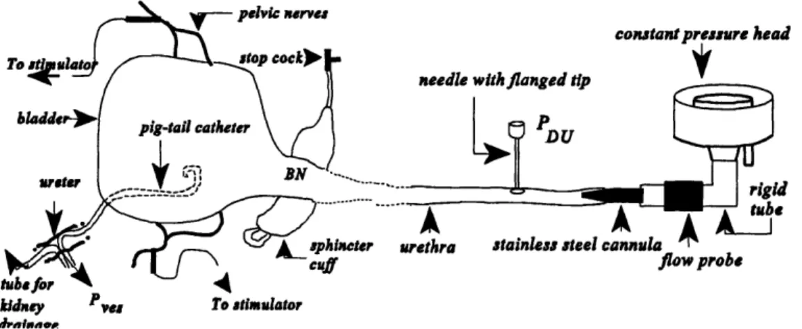
The detrusor function, outlet resistance and urethral flows of five female canines were assessed under non-obstructive (phase I) and obstructive (phase II) conditions. All urodynamic analyses were performed on a surgically exposed urinary tract. Solid state pressure transducers were used to measure intravesical and distal urethral pressures. An ultrasonic flowmeter was used to obtain an instantaneous measurement of the urethral
flowrate. An electrical stimulation (30V, 50Hz, 0. msec and 10sec duration) of the pelvic nerves was used to induce bladder contractions. All outlet obstructions were modeled using an inflatable sphincter cuff. During the phase II studies, an outlet was assumed
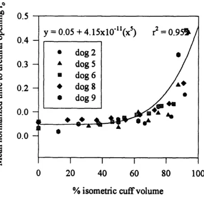
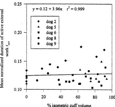
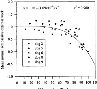
non-obstructed at a 0% cuff volume and completely occluded at a 100% cuff volume. The mean outlet resistances of the phase I studies ranged between 0.07-1.7 cmH2Osml' 2. As outlet obstruction was increased, the normalized internal work, normalized external work and normalized outlet resistance (all normalized with respect to the corresponding mean phase I result) remained approximately constant until a threshold cuff volume - 60% for the work parameters, and 75% for the resistance - was exceeded. As obstruction was increased beyond the respective thresholds, the normalized outlet resistance and the normalized internal work increased from their (non-obstructed) unity references to 400 and 4 respectively. The normalized external work decreased from unity
external work remained approximately equal. At volumes exceeding 60%, however, the internal work increased while the external work decreased. The concept of the internal work as an elastic energy that is fully recovered during voiding thus appears valid only at low outlet obstructions. As the normalized resistance was increased from unity to 60, the normalized internal work increased exponentially, while the normalized external work decreased exponentially. At normalized resistances exceeding 100, both normalized work parameters were plateaued at their maximum and minimum values of 4 and 0 respectively. Given this exponential correlation, at low outlet obstructions, the internal and external work appear more sensitive to changes in the outlet obstruction than the outlet resistance.
Under non-obstructive conditions, the peak Reynolds numbers R,Pand entry length
to diameter ratios li/D obtained ranged between 500 <R,P< 1500, and 30 < /D< 90. The non-obstructed urethral diameters D computed ranged between 1.5mm < D <2.5mm, while the urethral lengths I used for the urodynamic assessments ranged between 75mm < I < 95mm. The non-obstructed phase I flows were thus typically laminar entry length flows. The peak Reynolds numbers for the obstructed phase II studies also remained within the boundaries of laminar flow.
1 Introduction ...
1.1 Overview
...
7
1.2 The Lower Urinar 1.2.1 The Urinar 1.2.3 The Urethr 1.2.3.1 1.2.3.2 1.3 Micturition 1.3.1 The Mecha 1.3.1.1 1.3.1.2 1.4 Benign Prostatic Hyl 1.4.1 Pathology 1.4.2 Incidence 1.4.3 Diagnosis 1.4.4 Treatment 1.5 Current status of r 1.6 Significance ....
yTract
...
8
yBladder
...
8
a ... 10The Male Urethra ... 11
The Female Urethra ... 12
...
13
nics of Micturition ... 14
Energy approach ... 16
Distensible tube approach ... 31
perplasia (BPH) ... 37
of Benign Prostatic Hyperplasia ... 37
...
38
...
40
...
40
esearch
...
41
...
48
2 Materials and Methods ... 2.1 Surgical procedure ... 2.2 Functional evaluation ... 3 Results and Discussion ... 3.1 Outlet resistance and detrusor dynamics ... 3.1.2 Power and energy considerations ... 3.2 Characterization of urethral flow ... 3.2.1 The Reynolds number ... 3.2.1.1 Flow in the inlet length of a circular pipe ... 3.2.2 Developing and fully developed flow in tubes ... 4 Conclusions. 5 Bibliography ... .6 50 51 53 55 57 64 76 82 84 97 102
...
106
1 Introduction
The primary goal of this thesis is to provide new insights into the evaluation of benign prostatic hyperplasia (BPH) and to spur new directions in its management. For this, a canine model will be used to elucidate the physiologic impact of a bladder outlet obstruction, as induced by BPH, on detrusor function, outlet resistance and urethral flows. The reader should keep in mind that although many similarities exist between the human and canine urinary systems, the anatomical and pathological information presented in the subsequent introductory material strictly apply only to humans. If specific information regarding canine anatomy is required, the reader is directed to a canine anatomy text. The analytical methodologies discussed in assessing outlet obstruction are however equally applicable to both humans and canines.
1.1
Overview
Anatomically, the urinary system can be divided into an upper urinary tract composed of the two kidneys and ureters; and a lower urinary tract composed of the urinary bladder and urethra. Functionally, this system is responsible for urine formation, collection, transportation and micturition (the voluntary expulsion of urine via the urethra).
Figure 1.1 An anterior view of the human urinary system.
This thesis is exclusively dedicated to an analysis of the micturitional function of the urinary system. All subsequent discussions and analyses are therefore limited to the lower urinary tract.
1.2
The Lower Urinary Tract
The lower urinary tract is composed of the urinary bladder and the urethra. The bladder is connected to each kidney by a long, muscular ureter, along which filtered urine is propelled by peristalsis. The lower urinary tract can be modeled mechanically as a pump (the bladder), a piping system (the urethra), valves (sphincters), and a nozzle (the external meatus)'. Traditionally, the majority of studies investigating voiding function have
focused on the urethra. However, during a voiding cycle, the urethra has been shown to play a passive role, only modulating the relation between the bladder pressure and the voiding flowrate. Conversely, voiding patterns under normal and obstructive conditions are dominated by the dynamics of a bladder contraction and its adaptations to varying outflow conditions. Thus, an appreciation of the bladder's role within a voiding cycle is pivotal to the proper understanding of both the physiology and the pathophysiology of micturition.
1.2.1
The Urinary Bladder
The bladder is a hollow, muscular sac that functions as a passive reservoir for urine. An empty adult bladder has an internal diameter of approximately 5cm. It lies retroperitoneally, and is positioned posterior to the pubic symphysis. Anatomically, a bladder can be divided into a dome, a base, and a neck. The dome, in turn, can be divided into an apex, a superior surface, and two anterolateral surfaces. The bladder neck lies. most inferiorly, and is connected to the urethra. During filling, the bladder dome rises into the pelvic cavity, while the position of the bladder neck remains approximately stationary. The maximum capacity of a normal adult bladder ranges between 300-500ml.
The bladder contractile tissue can be divided into the detrusor and trigone musculature. Both are comprised of smooth muscle and demonstrate the slow response and fatigue rates characteristic of all smooth muscles. Their physiology is however unique in that conscious control of a bladder contraction, i.e. of micturition, can be learned.
The detrusor refers to the smooth musculature extending over the bladder dome. Around the region of the bladder outlet, the detrusor consists of distinct inner, middle, and outer muscular coats. The inner and outer layers are composed of longitudinal fibers. The former extends over the bladder outlet and into the urethra, while the latter forms an almost complete sheet of circumferentially oriented muscle bundles above the level of the internal meatus. The middle layer consists of circularly oriented muscle bundles that sweep outward along the sides of the bladder from the bladder neck. Despite their circular orientation, these fibers do not provide any sphincteric action. In the remaining regions of the dome, muscles bundles cross each other freely with no definite orientation. Any layering in these regions is thus unclear.
The trigone is a triangular shaped muscle layer, extending over the bladder base between the ureterovesicle junction, and the lip of the internal meatus. It consists of two layers: the superficial layer, and the deep layer. Muscle bundles from the superficial layer pass over the internal meatal lip, and into the proximal urethra. The deep layer forms a dense sheet at the bladder base, and anchors the ureterotrigonal unit. The muscle cells of this layer are largely indistinguishable from those of the detrusor muscle proper.
The bladder is well innervated by the autonomic nervous system. The sympathetic supply via the hypogastric nerves originates at the thoracic and lumbar segments T 1-T12, and L1-L2. The parasympathetic supply originates at the sacral segments S2-S4 (the pelvic splanchnic nerves). The primary motor nerve supply to the detrusor and the bladder base (including the trigone) is parasympathetic, and sympathetic respectively.
The bladder vasculature is derived from branches of the internal illiac artery and veins. The interior of the bladder is lined with a transitional epithelium.
Figure 1.2 Schematic diagram of sections through the male bladder and urethra,
viewed (a) from the front, and (b) from the left side. D, detrusor smooth muscle; T, tigone; SM, urethral smooth muscle; DS, distal intrinsic urethral sphincter; PS, periurethral sphincter; BN, bladder neck; P, prostate gland; MU, membranous urethra; PU, penile urethra; EM, external meatus; E, ejaculatory duct; 0, ureteral orifices. (After Gosling 1979). From: Urodynamics, The Mechanics and Hydrodynamics of the Lower
Urinary Tract, Griffiths, D.J., Adam Hilger Ltd, 1980, p 7.
1.2.3
The Urethra
The urethra is a muscular tube connecting the bladder to the external environment, and extends between the bladder outlet and the external meatus. The urethral musculature and epithelial lining are continuous with that of the bladder. At zero flow, the urethra is collapsed, and completely dry. The collapsed walls create a slit-like cross sectional area. The open regions of this cross section are filled with a viscous fluid secreted by the urethral lining, creating a watertight seal. During micturition, the urethra opens to a
.1 AIM...d
maximum diameter of approximately 3 mm. The exact diameter of the urethral lumen at any given location depends upon the local distensibility and static pressure, both of which change along the urethral length.
The striated sphincter urethrae muscle encircles the urethra to form an external voluntary sphincter (refer figure 1.2) at the level of the urogential diaphragm. In the male this sphincter is positioned around the membranous urethra, and in the female, around the mid-urethral segment. In both sexes, the urethra is innervated by the same autonomous nervous supply as the bladder. The external striated sphincter is innervated by the pudendal nerve (S2,3,4) originating from the sacral plexus.
The male and female urethras demonstrate some structural and functional dissimilarities, and are briefly discussed below.
1.2.3.1
The Male Urethra
The male urethra functions as a conduit for both the urinary and the genital systems. The external meatus is positioned at the tip of the glans penis. On average, an adult male urethra is approximately 20 cm in length.
The male urethra can be divided into a prostatic, membranous, and penile region. The prostatic segment is about 3 cm in length. It is surrounded by the prostate gland and contains openings for the ejaculatory ducts. (The circularly oriented muscle bundles of a pre-prostatic region helps prevent any back flow of ejaculate into the bladder.) The membranous urethra is the thickest, least distensible segment, and is about 2.5 cm in
length. It involves both striated musculature - the external sphincter - and smooth musculature. The former is oriented in a ring shaped structure, extending between the prostatic apex and the penile bulb. The penile urethra is the longest segment (about 15 cm in length), and is contained within the corpus spongiosum. The most proximal part of this segment is the widest urethral region, and is surrounded by the penile bulb. At its
terminus - the external meatus - the penile urethra narrows to open into the environment. This is the narrowest region of the urethra.
urethra
enile urethra
Figure 1.3 Schematic of male urethra. From Campbells Urology, Anatomy of the Lower Urinary Tract, Tanagho, E.A., p 53.
1.2.3.2
The Female Urethra
The adult female urethra is about 4 cm in length, 6 mm in diameter, and is a conduit only for urinary flow. It is positioned along the anterior vaginal wall, with the external meatus lying within the vaginal vestibule about 2.5 cm posterior to the glans clitoris. A glandular structure homologous to the male prostate can be seen along the distal end of the female urethra. Although this structure is called the female prostate, it does not acquire the functional or structural complexity of the male prostate. The entire female urethral length is rich in elastin and collagen fibers, and is more distensible than the male urethra.
,rnal mpatus ~I·_. .,_, ... _._
1.3
Micturition
Micturition, or voiding, is defined as the intermittent, voluntary expulsion of urine from the bladder. The elastic and viscoelastic properties of a bladder allow it to
accumulate urine with a minimal rise in bladder pressure during filling. Normally, the bladder fills at a rate of about 1 ml/min. During filling, motor impulses from the sympathetic system prevent any leakage into the urethra by both maintaining a closed urethra (by stimulating a urethral muscle contraction) and inhibiting a bladder contraction. As filling progresses, a sensation of "fullness" will originate from stretch receptors in the bladder wall. If necessary, this urge to void can be ignored up to a critical threshold. Beyond that, a voiding detrusor contraction will begin.
A voiding bladder contraction is caused by the activity of efferent postganglionic parasympathetic nerves. Voiding itself is accomplished by a contraction of the bladder musculature in conjunction with a relaxation of the urethral musculature.
Mechanically, a bladder contraction is represented by a rise in bladder pressure, while a urethral relaxation is represented by a drop in urethral pressure and vice versa. The urethra, which remains in a collapsed state during bladder filling, will open only when the bladder pressure exceeds a minimum urethral opening pressure'. Once voiding is complete, the bladder pressure will gradually decrease. As the pressure falls below a critical closure pressure, the urethra will return to its collapsed state starting from its distal regions and continuing proximally up to the bladder neck. The critical urethral opening and closing pressures are not necessarily identical.
During a bladder contraction, motor impulses from the parasympathetic system
Because of its shorter length, a lower resistance is associated with the female urethral than the male urethra. The minimum urethral opening pressure for the female is thus lower than for the male.
will inhibit any urethral muscular contraction. These efferent nerves, along with the afferent nerves conveying information about the bladder state, constitute a micturitional loop. The contraction of the bladder with the simultaneous relaxation of the urethra is termed the micturition reflex. Voluntary control of micturition (within limits) becomes possible once control of the spinal center co-ordinating this reflex is learned.
1.3.1
The Mechanics of Micturition
Micturition is a process under neuromuscular control, and results from an interplay between the expulsive action of the bladder, and the impedance offered to flow by the urethra (refer Figure 1.4). The former can be represented by the pressure of urine within the bladder, the intravesical pressure (p,). p,, is composed of an intrinsic detrusor component p (the detrusor pressure), and an extrinsic abdominal component p (the abdominal pressure).
p,(t) = p(t)
+
p(t)
(1)
In general, p, is determined predominantly byp,,. The mechanical properties of the bladder are thus governed by the detrusor. In all subsequent discussions, the detrusor and intra-vesical pressures will therefore be considered synonymous.
The detrusor pressure itself arises primarily from the active contraction of the detrusor muscle (and to a lesser extent from the viscoelastic properties of the bladder wall and muscle tone). At any given instant, pdt depends upon: (a) the intrinsic strength of the detrusor muscle (b) the level of (parasympathetic) neural stimulation (c) the volume of urine in the bladder, and (d) the outflow rate of urine from the bladder (Q(t)).
That is, if we
know the rate
Bladder
presatells
thetsand
vice versaFigure 1.4 Meaning of urethral impedance (From Griffiths, D.J., Mechanics of Micturition. Neurology and Urodynamics. p 96, 19882)
As with any other muscle, the detrusor, when stimulated, can either shorten or develop force. This compromise between the velocity of shortening (related to the urine flow rate) and the force developed (related to the detrusor pressure) can be depicted using
a trade-off curve.
Detrusor pressure
v ra
As can be seen, the same detrusor contraction will produce vastly different
detrusor pressures in individuals afflicted with different conditions (e.g. the same detrusor contraction will produce a large p,, in obstructed individuals, and a small pat in individuals with low impedance)i.
Urine from the bladder will enter the urethra only after a critical value ofp,t has been attained. This is because the urethra provides a finite impedance to the bladder
output. A high impedance (low Q for highpat) implies obstruction. Low impedance (high Q for lowpdt) implies no obstruction. Two alternate approaches can be used to describe this concept of urethral impedance.
1.3.1.1
Energy approach
This approach describes micturition as follows: The detrusor pressure, at a given bladder volume, is a potential energy source for driving flow through a urethra. As flow occurs, a fraction of this potential energy will be dissipated by viscous and inertial effects, while the remainder will appear as the kinetic energy of the exit stream2 (strictly speaking, it will appear as the time integral of the kinetic energy of the exit stream, as will be shown below).
A finite control volume approach can be used to apply the fundamental laws of mechanics and thermodynamics to a finite region of space containing a finite amount of fluid (i.e. a control volume), and explicitly derive the relation between the detrusor
pressure (or, alternately, the flow work) and the flow kinetic energy. The following derivation assumes a basic understanding of control volume theory. An in-depth
i Anyp,, measurement corresponding to non-zero flow rates alone is thus an inadequate estimate of
detrusor contractility. The detrusor pressure at zero flow, the isometric (or isovolumetric) detrusor pressure,
is a better indicator of contractility. However, since it is invalid during the period of voiding proper, the
usefulness of the isovolumetric pressure is also limited.
discussion of control volumes and their applications, if required, is available in
Engineering Thermodynamics, Smith, J.L, Cravalho, E.G. pp 350 - 358.
tin - -
Control
volume
Bladder
.-
I-Figure 1.6 The control volume used to derive the relation between the detrusor pressure (or, equivalently, the flow work) and the flow kinetic energy. The bladder and urethra are indicated by the thin line. The thick-dashed line is used to demarcate the borders of a fixed control volume enclosing a fixed mass of fluid.
As shown in Figure 1.6, a system containing a constant mass of fluid (urine) can be defined within a fixed control volume located within the bladder, and extending into the urethra. The first law of thermodynamics states that the rate of change of a system's intrinsic energy (dE,,,,,dt) is equal to the net rate of energy transfer to the system by heat
(lH/dt) and work (W/dt). Mathematically, this can be expressed as:
dEystem 8H_ W
dt
dt
dt
(2)
Here, the sign convention adopted implies that heat added to a system is positive, and work done on the system is negative. This basic expression can be extended to yield the following first law of thermodynamics for a control volume3:
+j
.(h+-z+gz) d3 dt dt dt dt- 2(3)Where, dn/dt represents the rate of mass inflow/outflow, h the property enthalpy,
v the velocity of fluid at the inflow/outflow ports, g the gravitational acceleration, and z
the elevation from some arbitrary reference. The two summation terms in equation (3) have been added to the basic first law expression (equation (2)) to account for the net energy convected to the system by bulk flow across the control surface. The work represented by the &W,h/dt term in equation (3) represents the shear work transfer, and includes all work transfers for the system except those resulting from a pressure and normal displacement of the system boundary (i.e. pdVwork transfers). All ofthispdV work transfer is accounted for in the enthalpy h, defined in terms of a specific internal energy u, fluid pressurep and fluid density p.
h=P+u
h=P ~~~~+U ~(4)Since the control volume defined in Figure 1.6 is fixed, all unsteady effects can be neglected. Further, since a control volume containing a constant fluid mass is defined, the rate of energy change within the control volume is zero (i.e. within a fixed system, energy can neither be created nor destroyed). It should be noted that since the length I of the control volume within the bladder can be made infinitesimally small (without invalidating the following derivation), the assumption of a system of constant mass is applicable through the entire voiding cycle. Equation (3) can be simplified as follows:
Since the mass within the control volume is constant for all time, the mass flowrate
dm/dt into the control volume will equal the mass flowrate out of the control volume. If
constant through the control volume yielding the following expression for the conservation of mass for the system:
din outflow
~-p=c=*A and vin(o)' , A o~
dt =Q=cAinflow'vinflow =AO and, Vlfo w A )
Since, Afl >> Ao X vi,, is negligible compared to v,. Also, since all flow occurs along a horizontal axis for the system in Figure 1.6, the contribution to the first law from changes in elevation are negligible. If a zero heat transfer is also assumed, equation (3) can be re-written as (with dm/dt = pQ):
6Whear 2
0=- dt °= +E PQ(-+)inflowC PQ(++ p V (6)(6)
Equation (6) can be further simplified by noting that the control volume in Figure 1.6 contains a single inflow and a single outflow port, and that the inflow pressure is simply the detrusor pressurepd,, yielding:
aw 2
shear outlow (7) PQ= dt +P Q+p 2 Q+PQ(o- u'wnw)
Any kinetic energy dissipated within the system will transform into an increase in the specific internal energy u through the dissipative process. The term pQ(uoo - uS,) in equation (7) should thus reflect any flow kinetic energy dissipated within the urethra by
an increase in uo,, relative to ui,,. However, because the coupling of the kinetic and specific internal energies in fluids is weak, the increase in the specific internal energy from any kinetic energy dissipation will be minimal. This allows the approximations u w
2
(PMt-P°1Vlw)Q=d+P 2 Q (8)
Equation (8) is the final expression of the first law of thermodynamics for the system defined in Figure 1.6. Hydrodynamically, the productpQ represent fluidic power (i.e. rate of work done, or energy consumed). However, since the concepts of work and energy (i.e. pdVas opposed to its time derivative) are more physiologically relevant, equation (8) is best interpreted by examining its time integral:
f(PdctPouw)H dt=f(PdtP w)dV= -Whear+fP 2df (9)
dtf 2
Physiologically, equation (9) states that the difference between the detrusor flow work (defined as the integral fpdV) at the inflow and outflow ports is equal to the sum of the shear work transfer and the time integral of the kinetic energy at outflow port (with the kinetic energy represented by p(v2o,,,2)Q). The placement of the outflow port as shown in Figure 1.6 is arbitrary. If the intra-urethral arm of the control volume is extended such that it is adjacent to the external meatus, and the subject voids into the atmosphere, p,,,, =P pn. The difference P,, - puff thus becomes the gage detrusor pressure (i.e. detrusor pressure measured relative to atmospheric pressure) that is
routinely measured using a pressure transducer. The first law expression then effectively reduces to the functional expression of the energy approach:
pdtdV= Wshear +fp Qdt=Whear+fp- dV (10)
with v, representing the velocity of the exit stream. The time integral of the kinetic energy has also been simplified by noting that Q=dV/dt. Figure 1.7 is a graphical representation of this relation as drawn by Griffiths2.
Inravesicle | energy dissipat i
in urethra
depending on depending on
Q 2 Q2
Figure 1.7 Energy balance during voiding according to energy approach. (From Griffiths, D.J. Mechanics of Micturition, Neurology and Urodynamics. p 99, 1988)
Several issues regarding the correspondence between the functional and graphical forms of equation (10) merit a brief mention. First, it should be noted that Figure 1.7 explicitly considers fluidic losses in the urethra. It thus implies that the term pQ(uoo
-u,,,o) in equation (7) must be considered when computing a urethral energy balance. However, urethral losses have been characterized as minimal4. Also, as discussed, the
contribution of any urethral losses to the term pQ(u.o - u,. ) in equation (7) is
negligible. (The reader is reminded that the first law of thermodynamics uses the term
pQ(uo, - u,,) to account for any kinetic energy losses in the urethra). Equation (10) will thus still be considered an adequate representation of the urethral energy balance. Figure 1.7 also essentially ignores any biochemical energy expended by a detrusor when raising intravesical pressure against a closed outlet. (Each voiding contraction proper is preceded by an isometric contraction where the detrusor pressure is increased against a closed urethra with zero volume voided. This will be discussed in detail in section
1.3.1.2). Equation (10) accounts for this nonpdV energy transfer with the dW ./dt
term. Second, the proportional relation between the kinetic energy, the (flowrate)2 and
geometry of the external meatus as shown in Figure 1.7, although not explicit in equation (10), becomes evident by substituting Q=vA, yielding /2Jpv2.dV = /2p(Q2/A.,2)dV, with
A,i, the cross sectional area of the external meatus. Also, according to Figure 1.7 all
fluidic losses incurred are proportional to
Q
2. This implicitly assumes that flow throughthe urethra is turbulent, and that losses are predominantly due to inertial effects - with laminar flow, the pressure drop due to frictional losses AP is linearly related to the
flowrate Q as expressed by Poiseuille's equation. However at high flowrates, the pressure drop is more accurately described by Rhorer's equation: AP = k,Q + k2Q2, with the Q2
term indicating the effect of turbulence. When turbulence is present, losses are incurred by eddies and mixing, with the associated pressure drop depending upon the fluid density (as opposed to fluid viscosity, as with Poiseuille flow). The precise characteristics of urethral flows along with the relevant tube laws are treated in detail within the Results section, and will not be discussed further at this juncture.
Hence, given all of the assumptions discussed above, the precise statement of the energy approach to urethral flow is that any difference in the detrusor flow work between a proximal and a distal point along the flow stream, will be equal to the sum of a shear (non pdV) work term and the time integral of the flow kinetic energy evaluated at the distal point (refer equation (9)).
Thus far, attempts to use this information in evaluating outlet resistance have been largely unsatisfactory. Since the distensible tube approach discussed next is extremely powerful in this context, the application of the energy approach to urethral impedance will not be discussed further. It is however fundamental to an appreciation of the detrusor function during voiding, as is discussed below. The most convenient parameters that can be used to characterize detrusor dynamics are the detrusor pressure p,,(), and the urinary flowrate Q(t). (Here, a measurement of the kinetic energy of the outlet stream is not essential since the urethral flowrate is always determined within the proximal urethra - see section 1.3.1.2) . The detrusor is the primary energy source of micturition. During a voiding cycle, the detrusor converts stored biochemical energy into mechanical work by expelling a volume of urine against an outlet resistance within a finite time period. The
i The velocity of the exit stream is however dependent on the geometry of the external meatus. The exit
bladder has also been shown to store a limited quantity of energy for contracting, and "recharge" between successive contractions. Functionally, the bladder is thus a limited energy storage device. The quantity of energy stored (and thus the ability of the detrusor to do work, or generate power) has been shown to increase with bladder fillings.
The conversion of biochemical energy to mechanical work in the detrusor is not ideal, and some mechanical and thermal losses are incurred. The mechanical work generated during a contraction can be divided into two components: the external work, and the internal work. The external work is defined as the work that can be measured in terms of a detrusor pressure and a volume voided (i.e. the flow work). The internal work is defined as the work done to elongate the elastic elements in series within the detrusor wall (Schafer'), and is described only during the isometric portion of a detrusor
contraction. (An isometric contraction refers to the period between the initiation of a contraction and the commencement of flow - i.e. when the detrusor pressure rises with zero flow). In a healthy bladder, the duration and strength of an isometric contraction vary in proportion to the outlet resistance. If losses are assumed negligible4, the total bladder work can be defined as the sum of the internal work Wmt and external work W,~.
Wto = WW + Wt (11)
As shown in equation (9), the external work is related to the kinetic energy of the urethral flow at any given point along the urethra. The external work can be easily calculated from the external voiding power P.,t(t), defined as the product of the instantaneous (and corresponding) detrusor pressures p(t) and flow rates Q(t),
Pxt(t) = p~(t)Q(t) (12)
with the external voiding work defined as the time integral from time t = 0 to the terminus of voiding t = tf of the external voiding power.
I
W=fp(tO)Q(tdt
o
(13)
However, since no flow occurs during the isometric portion of a bladder contraction, this relation reduces to:
ti
W=fp,(t)Q(t)dt
t,
(14)
where, t is the time at which flow commences. This relation between the detrusor pressure, the flowrate, the external voiding power, and the external voiding work is
schematically shown in Figure 1.8.
-.-- isometricp det ... voiding p det - flow rate Q Pext Pdet Q W t = Pet Q dt tv Figure 1.8 Schematic representation of relation between detrusor pressure
Pd,, flow rate Q, external
voiding power P., and external voiding work W,. Adapted from Schafer, W. Detrusor as the Energy Source of Micturition. Benign Prostatic Hyperplasia. F.
Schafer5 has proposed that the internal voiding power P(t) can be estimated from the pressure change dp/dt at a given volume V(t) as:
Pi,t
(t) ~ V (t) dp/dt (15)The internal work W,;t (t) can thus be estimated as the time integral from t = 0 to the time of flow commencement t = t, of the internal voiding power:
tv
Witerna(t)= v(t) dPt (16)
0
Since the bladder volume is constant during an isometric contraction, equation (10) can be reduced to:
Pv
nternal= v f dp= V(P -Po) (17)
Po
where, Vo is the filled bladder volume, p, is the bladder pressure at the
commencement of flow, andp, is the bladder pressure at the initiation of the contractions. This formulation of the internal work merits a closer analysis. Recently, a continuous occlusion study6 showed that during an isometric contraction, the detrusor pressure rises to a maximum, and then declines towards baseline. Such a contraction is schematically
shown in Figure 1.9.
Here, the isometric contraction extends from time t=O and a baseline detrusor pressure p,,° to time t=t
2 and a pressure pdt*, with the peak isometric pressurepd,,"'
i Since this work is computed from the product of a volume and a pressure difference (as opposed to the product of a pressure and a volume difference), strictly speaking, it represents a complementary work term.
occurring at time t=t,. If one attempts to compute the internal work done between t=O and t=t2using equations (16) or (17), the internal work appears simply as the product
Vo(APdt A) p* Pdet Pdet … I /- - - . \ .
,
A +
X.---I nt1wA_
-' tL
Figure 1.9 Schematic of the detrusor pressure behavior during an isometric contraction as characterized by the continuous occlusion study. The contraction begins at time t=O and continues to time t=t2. The time t represents the time to first reach the
detrusor pressure p,,*.
By considering only the beginning and end pressure points Pd. and Pd.,
equations (16) and (17) effectively consider only the work required tofirst reach ,,t* (i.e.
the work done from time t=O to time t=t,) and ignore all work done between t, and t2. It
is possible to formulate the concept of internal work in this manner only if the detrusor work done to increase intravesical pressure between the times t=t, and t=tk is stored as elastic energy that can be fully recovered between the times t=t,,, and t=t2. Equations
(16) and (17) thus implicitly assume that a detrusor isometric contraction can be modeled as shown in Figure 1.10.
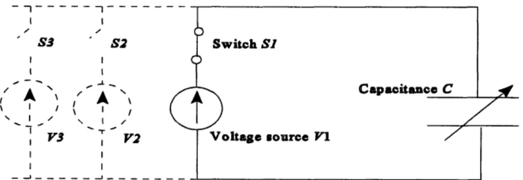
Here, an isometrically contracting detrusor is modeled as a voltage source V charging a variable capacitance C (the capacitance is used to model the bladder tissue that is assumed to store elastic energy). Since all stored energy is assumed to be fully
recoverable, the connections between the voltage source and capacitance are modeled as having a zero resistance. An increasing detrusor contractile force can be modeled by
r--- - - - --%N.
individually switching on voltage sources with successively higher voltages V1, V2... using the corresponding switches SI, S2... (The reader should note that the voltage Vand electrical charge q in the electrical domain are equivalent to the pressure p and volume Vo in the fluidic domain respectively).
I I S3 S2 I I I I I II I V3 I V2 I I I _lI _- _
Figure 1.10 The isometrically contracting detrusor as modeled by equations (16)
and (17). The contracting detrusor is modeled as a voltage source (attached across Switch SI) charging a variable capacitance C through a resistanceless wire. An increasing detrusor contractile force can be modeled by individually switching on voltage sources with successively higher voltages , V2... using the corresponding switches SI, S2 ... The energy available to drive urethral flow at a given detrusor pressure (i.e. the elastic energy as referred to by equations ( 6) and ( 7)) is modeled by the electrical energy E stored in the capacitor C at a given voltage V This stored energy is defined as E
= (q'I). Given the identity q=Crelating the capacitance C, voltage V, and electrical charge q, this energy can be written as E = Y2 (4 I)
That Figure 1. 10 models equations (16) and (17) can be verified as follows: Since an isometric contraction is being considered, the bladder volume must remain constant for all detrusor pressures. The capacitor in Figure 1. 10 used to model the bladder must therefore maintain a constant electric charge q for all applied voltages (a variable
capacitor is used in this model since the identity q=CV must be satisfied when the charge q is held constant for a varying voltage ). The change in the stored electrical energy AEE
between two voltages V, and V2(V2 > V,) can now be computed as AE = l/2q(V-V,) =
1/2q A V. Given the equivalency between the fluidic and electrical variables described
previously, this relation translates into the fluidic domain as AE Vp, which is the
functional format of equations (16) and (17).
Although this formulation is very convenient, whether it adequately represents the contractile process within a detrusor smooth muscle cell must be examined. The smooth muscle contractile process is summarized below. If a more detailed description is
required, the reader is directed to a medical physiology text such as Guyton, A.C.,
Textbook ofMedical Physiology.
Smooth muscle cells use actin and myosin filaments for their contractile function. In smooth muscle, actin and myosin filaments are not arrayed as serial sarcomeres as in skeletal and cardiac myocytes. Rather, the actin filaments are attached to dense bodies with the myosin filaments interspersed between the actin filaments (the dense bodies are equivalent to the Z disks of the skeletal myocytes). The membrane depolarization of a smooth muscle cell is dependent on the activation of slow L-type calcium channels (as opposed to the fast sodium channels in skeletal muscle). Since these channels typically generate depolarizations with a slow upstroke, and since the impulse propagation velocity is proportional to the rate of change of the upstroke, the velocity of impulse propagation in smooth muscle is relatively slow.
Smooth muscle cells typically use calcium as the major second messenger in the excitation-contraction coupling process. Here, cytoplasmic calcium (accumulated by the calcium influx through the L-type channels) activates the calcium dependent regulatory protein calmodulin. Calmodulin in turn activates the enzyme myosin kinase which phosphorylates the regulatory chain on myosin heads allowing the myosin heads to form
cross bridges with adjacent actin filaments (skeletal muscle uses the
Troponin-Tropomyocin regulatory complex for this process). During this cross bridge formation, myosin heads typically have bound ADP. Once cross bridges have formed, the myosin heads bend, moving the myosin filament relative to the actin filaments, and thus potentiate a myocyte contraction. Once bent, the myosin heads release ADP and bind ATP. ATP
bound myosin heads have a reduced affinity for their actin binding sites resulting in a gradual dissociation of the cross bridges. The free myosin heads will then hydrolyze ATP to ADP and use the resultant energy to straighten themselves (from their previously post-contractile bent configuration). Since the straightened myosin heads will again have bound ADP (residual from the preceding ATP hydrolysis) the contractile cycle can now repeat. The cycling frequency of cross bridges in smooth muscle is slower than in skeletal muscle. Since this cycling rate effectively defines the rate of ATP consumption, smooth muscle cells are able to contract for relatively longer periods (with respect to skeletal myocytes) on a given ATP store.
At contraction termination, the L-type calcium channels close while energy
dependent calcium ATPase pumps remove calcium from the smooth muscle cytoplasm and thus deactivate calmodulin. The regulatory enzyme myosin phosphatase will also become active and dephosphorylates the regulatory light chain inhibiting the formation of cross bridges. The sequence of contractile events will thus terminate, and the cell will return to its resting state.
The ability of a smooth muscle cell to maintain a contraction, is, therefore,
dependent upon the presence and interaction of numerous biochemical factors. Of these, the single factor most susceptible to depletion is ATP. Given the preceding information, one can expect that if the rate of ATP consumption during a contraction exceeds the rate of its generation, a myocyte will be unable to contract, and fatigue. Since ATP is required for the release of cross bridges, one would also predict that with ATP depletion myocyte relaxation will be impaired resulting in a rigor like low compliance (i.e. inelastic)
condition. The electrical model described in Figure 1.10 is simply unable to account for this complex behavior. Specifically, by assuming that all energy expended in increasing
detrusor pressure can be recovered, the model implicitly assumes that the isometrically contracting detrusor muscle cells will always be perfectly elastic. This is equivalent to assuming that the myocytes will not fatigue. This assumption is not physiologically valid, since all myocytes fatigue after a period of continued contraction. In fact, the decline in the detrusor pressure from a peak value as described in the continuous occlusion study
(and schematically shown in Figure 1.9) represents such a gradually fatiguing detrusor. The concept of a fully recoverable internal work thus cannot represent an isometric contraction progressing beyond a peak detrusor pressure and into fatigue conditions. With the onset of fatigue, the energy available to drive flow through the urethra at a given detrusor pressure and the work done in increasing the detrusor pressure to that level are not equal. It is therefore inappropriate to describe an isometric contraction in terms of an internal work. Rather, an isometric contraction should be described using a more
generalized energy available to drive flow through the urethra. The reader should note that in reality, it is this energy, and not the internal work that is accounted for by the shear work term in the first law of thermodynamics.
A modified version of the model depicted in Figure 1.10 incorporating the effects of fatigue is suggested below. Here, an internal resistance R has been inserted in series with the capacitance C, while the voltage source has been replaced with a battery B. The
internal resistance is used to model the effects of fatigue, while the battery is used to model a detrusor smooth muscle cell with a limited stock of ATP.
- -I -…--I- - --I I I S3 S2 I I I I -_ J_ B3 B2 I I - --- _ _ _ _ _ -_ . - _ _ _ _ -_ -_
Figure 1.11 Modified model for assessing the energy available to drive urethral flow at a given detrusor pressure. The internal resistance R is used to model the effects of fatigue, while the batteries B], B2... are used to model the detrusor smooth muscle cells with a limited ATP energy store.
The formulation of the energy available to drive urethral flow or "internal work" as formulated by equations (16) and (17) is thus neither ideal nor absolute. Ideally, it would be possible to explicitly account for this energy in terms of the moles of ATP hydrolyzed or oxygen consumed during the entire isometric contraction. At present however, such a formulation is not available. Schafer's approach will thus be used in the subsequent analyses as a first approximation. The term "internal work" will also be retained in describing an isometric contraction. The reader should however keep in mind the
limitations of equations (16) and (17), and this terminology when reviewing the relevant results in section 3.1.2.
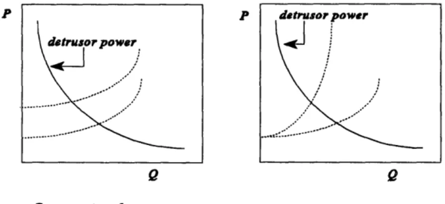
Within the limits of a maximum isometric pressure (at zero flow) and a maximum flow rate at zero pressure, the relation betweenpd, and Q for a given bladder power level is inverse. Lower Q and higherp,, are characteristic of outlet obstruction. This
phenomenon was initially postulated as an active compensatory action by the bladder (in response to the increased outlet resistance). It has also been argued5 to result purely from the passive inverse relation betweenp, and Q (refer figure 1.5). The definite mechanism has however not yet been fully elucidated.
Because of the depressed flowrates, an obstructed bladder must perform more work to expel a given volume of urine than would an unobstructed bladder. However, the bladder is a limited energy storage device. Therefore, for a given bladder volume, micturition from an obstructed bladder can be expected to terminate before the bladder is completely emptied. Post void residual urine is thus also characteristic of an obstructed bladder.
1.3.1.2
Distensible tube approach
Here, the urethral impedance is interpreted from a plot of the detrusor pressure vs. urine flowrate. The graph showing the relation between the instantaneous and
(PQ) curve. An idealized PQ curve is shown below:
Detrusor pressure
Figure 1.12 An idealized pressure-flow (PQ) curve
Urethral flow will commence only after the detrusor pressure has exceeded a critical urethral opening pressure P,. Similarly, at the terminus of voiding, flow will cease once the detrusor pressure has fallen below a critical urethral closure pressure p,. If the urethra behaves as a perfectly elastic tube, and if no muscular relaxation and/or passive viscoelastic relaxation of the bladder outlet is present, the p,, will equal the p,.
The urethral resistance relation (URR) represents the initial effort to use the distensible tube model to describe outlet resistance. However, since the URR considered pressure-flow relations through the entire voiding cycle, its interpretation was complex, and hence did not present a convenient diagnostic tool. An alternate method for outlet resistance computation is the passive urethral resistance relation (PURR). By basing its resistance computation upon the pressure-flow relation from peak flow to voiding terminus, the PURR is simple and convenient to both construct and interpret, and has become the modality of choice for the clinical evaluation of outlet resistance. The PURR, its derivation and applications, are discussed further in section 1.5.
A fundamental characteristic of collapsible (distensible) tube flow is the existence of a choke point termed a flow controlling zone. The flow controlling zone of the urethra normally lies within the membranous urethra. Within this zone, the urine flowrate is maximized via a process termed flow limitation. Once flow limitation has occurred, the flowrate becomes essentially effort independent. That is, until flow limitation is reached, straining (i.e. increased voiding effort) will produce increased flowrates. However, once flow limitation is reached, added effort will not produce any increase in flow. This phenomenon can be quantified as follows:
The transmural pressurep, along the urethra is defined as the difference between the pressure inside the urethrap, and outside the urethrap,. For the membranous urethra, this outside pressure is, to a first approximation, the abdominal pressurep, Thus, the transmural pressure at any point along the membranous urethra is:
Pt ,,=Pt -Pbd (18)
The Bernoulli equation can be used to relate the detrusor pressure pt, to the p, in terms of a proximal fluid velocity vp,, and density p as shown below. (It should be noted that since flow within the bladder itself is approximately zero, pat is effectively a
stagnation pressure).
1 2
PdtPi+2 PVprox (19)
This expression assumes steady flow through a tube of constant cross sectional area, with no viscous or other losses present. (Viscous effects can be accounted for by adding a frictional pressure drop 4p to the right-hand-side of equation (19), but will not be considered here since flow limitation occurs even if frictional losses are not explicitly considered, as will be shown below).
Equation (19) can be re-written in terms of the urinary flowrate Q by substituting Q = vaA, with A, the local cross sectional area. Thus,
1 Q 2
Pdd =Pi+P A2 (20)
and,
Q=A 2P (21)
Equation (21) can also be re-written as:
Q=VprA
=A
[(dt
Pabd)- (ItPab2d)](H-p)
(22)
with H defining the total pressure head relative to the abdominal pressure. Thus, the problem of flow limitation can be reduced to an estimation of the largest possible attainable flowrate for a given total pressure head relative to abdominal pressure, as effort is increased. (In this context, H can be considered a local driving pressure). Hence, flow limitation will occur when:
I
H=COt~r (23)With Q = vp,A, this condition can be evaluated as follows:
dQ =O-A_ +Vpr d4 =0 (24)
dptm-=
dp
tM
p(24)
Now, by using equation (22),
-d=2~ [ (2H- p1)]( 2)(- )=- v-(25)
Pt, 2p P PVprx
The evaluation of the dA/dp, term requires the introduction of a new variable, the speed of pressure wave transmission along the urethral wall c, which is a function of fluid inertia (i.e. density p) and local urethral specific compliance (1/A)dA/dp,,. The specific functional expression of c is as follows:
c= 11 dA
]_6
c4 p[ ~ A ]-'Up,,,,(26)
pA dp
and, d4 A dp pc2 (27)Thus, equation (24) reduces to:
2
dO 1 A AV
Q=0-"-A-
+V 2 pc ) (28)'4t'
PVprox pC 2 CFrom equation (28), it is evident that the maximum flowrate Q., will occur when,
prox =C (29)
Thus, the flowrate will be maximized (i.e. flow limitation will occur) once the flow velocity in the membranous urethra vp,, is equal to the wave speed c. The functional expression of the maximum flowrate Q,,. therefore becomes:
Q.=cA = A(dA -(30)
Functionally, the flow controlling zone will govern the impedance presented to the bladder, and isolate the bladder from the influences of more distal regions of the urethra.
flow controlling
point
Figure 1.13 Energy balance during voiding according to the distensible tube approach. When a flow controlling zone is present, p, is the total pressure needed locally to overcome the elasticity of the flow controlling zone. p depends upon the relevant flow rate, and the mechanical properties of the controlling zone. The distal parts of the urethra,
and the external stream (dotted) have no effect on either the proximal urethra orp, and thus do not contribute to urethral impedance.
The distensible tube approach classifies a urethra as obstructed if its impedance is above a value considered normal. Under obstructive conditions, urethral impedance will be dominated by the obstruction (i.e. the region of obstruction will contain the new flow controlling zone, with its local area and compliance defining
Qm,).
Urodynamic techniques such as a Micturitional Urethral Pressure Profile (MUPP) can be used both to detect the location of an obstruction and to estimate its severity - the obstruction will be located at the site of the static (or total) pressure drop, while the severity of obstruction will be proportional to the magnitude of the pressure drop.
Cr,7 - comnressive zone: obstructed
distance
Figure 1.14 Expected micturitional urethral pressure profiles for a normal and proximally obstructed male.
1.4 Benign Prostatic Hyperplasia (BPH)
1.4.1
Pathology of Benign Prostatic Hyperplasia
The prostate is a compact organ related to the male genito-urinary tract. Anatomically, it is positioned at the bladder neck, and surrounds the prostatic urethra (refer Figure 1.3). BPH refers to the non-cancerous growth of prostatic mass. Strictly speaking, such growth may occur from either the hypertrophy, or hyperplasia of stromal and glandular cells. However, in general, the terms hyperplasia and hypertrophy are used interchangeably in describing BPH. The glandular and stromal hyperplasia associated with BPH, while substantially increasing prostatic tissue volume, will also alter the
geometry and architecture of the prostate gland. The elastic properties of the prostatic capsule will determine whether BPH will compress the prostatic urethra. Prostates with predominantly fibromuscular components may cause outlet obstruction by altering urethral compliance, and impeding urethral flow7.
Enlarged lateral prostate lobes will compress the urethral lumen into an elongated, broad, ribbon like channel. Growth of the median lobe may, however, be confined by the urethral musculature. If so, a growing median lobe will expand in a sub trigonal direction, elevating or angulating the internal urethral meatus forward. If not, the median lobe may push into the bladder lumen, and ultimately result in a "ball-valve" obstruction of the internal meatus. BPH associated urethral obstruction itself will result from the elongation, tortuosity and compression of the posterior urethra.
1.4.2
Incidence
The prostate does not maintain a uniform growth rate throughout the life time of an individual. Prostatic mass increases slowly from birth to puberty, accelerates between puberty and the thirtieth year, and then remains constant for about a decade.
Hyperplasia/hypertrophy of the prostate could begin around the age of 40 years. If so, prostate mass will begin a rapid growth until an individual's death. (Alternately, the prostate may even begin to atrophy, and progressively decrease in size). BPH is characteristically a disease of men aged over 40 years. By the age of 75 years, the probability of prostatism increases to 75%.
50 -40 Volume. cC 30 20 -10 ' 0 0 I I I I I I 1 9 10 20 30 40 50 60 70 80
Figure 1.15 Mean volume of the prostate by age group. Hollow circles represent the mean volume of all prostates in all age groups, shaded circles represent the mean volumes of the normal prostates, and black circles represent those of hyperplastic prostates. (From Swyer, G.I.M, Anat., 1944)
100I 80 60-40 20' 40-49 60-69 80 + 50-59 70-79
Figure 1.16 Incidence ratio of benign nodular hyperplasia (BNH) with the age in
206 consecutive autopsies. (From Harbitz, T.B., and Hangen, O.A.,: Acta Pathol. Microbiol. Scand. [A], 80:766, 1972).
I 24 1 1 15 16d i5 18 16 12 1 . 9
.9
_
` 9
7 4 9 I1 1 1 1 1 1 11.4.3
Diagnosis
The most commonly used tools for BPH diagnosis include physical examinations, patient symptom questionnaires (AUASI, I-PSS), and urodynamic assessments such as cystometry, uroflometry and voiding profilometry (MUPP). For example, the site (and degree) of an obstruction can be estimated using a Micturitional Urethral Pressure Profile (MUPP); and urethral impedance can be estimated using pressure-flow relations. Despite these techniques and a host of related technologies, some confusion regarding the accurate diagnosis of obstructive BPH vs. irritative prostatism still persists. This can be attributed to a number of reasons. For example, a patient may be under medications that directly or indirectly influence the function of the bladder and its outlet. Alternately, a patient may suffer from hidden subclinical neurogenic conditions such as diabetic autonomic
neuropathy, or cerebrovascular disease that overtly mimic BPH. Moreover, the effects of aging on the mechanics of the bladder at present are not well understood. The diagnostic value of the prostatism complex in detecting obstructive BPH is also marginal since its symptoms do not necessarily indicate prostate enlargement or outlet obstruction. Even those symptoms considered unequivocal evidence of outlet obstruction such as acute urinary retention and post void residual volume, may be a result of detrusor dysfunction as opposed to BPH. At present, the most efficient methods of BPH diagnosis are
urodynamic assessments such as the MUPP.
1.4.4
Treatment
The symptoms of adult male voiding dysfunction (prostatism), either obstructive or irritative in nature, are highly non-specific, and do not necessarily indicate the underlying pathophysiology of the voiding dysfunction. Nonetheless, at present, prostatism is mostly
associated with BPH induced outlet obstruction. Hence, the most common treatment for prostatism is the Trans Urethral Resection of the Prostate (TURP), a procedure where sections of the enlarged prostate are surgically removed. In the United States alone, approximately 500,000 TURPs are performed annually at a cost of about $5 billion. In a health interview study on 471 patients, prostatic surgery was found to be effective in 93% of severely symptomatic patients, but effective in only 79% of the moderately symptomatic patients9. A long term follow-up (5-8 years) study of patients with prostatism revealed that 24% of those who had elective prostatectomies regained their symptoms'°. Both
studies indicate that a significant portion of TURPs performed fail to alleviate the
symptoms of prostatism. Surgical intervention must thus be more judiciously applied than at present. This becomes extremely critical in the present era of spiraling health care costs and dwindling funding. Alternate conservative treatment methods such as watchful waiting or pharmacological management must also be designed and attempted. But for all this, a more comprehensive understanding of the pathophysiology of prostatism than presently available is required.
1.5 Current status of research
Morphological changes such as a denervation of the bladder wall, and an infiltration of connective tissue between detrusor muscle bundles, and physiological changes such as detrusor instability and urinary retention have been observed in human males with outlet obstruction. Suitable animal models have been used to investigate the sequence and inter-relations of these pathologies"',2. Much work has also been performed to assess micturition and urethral impedance from a fluid dynamics stand point.
Several urodynamic parameters have been used to characterize detrusor function. Of these the detrusor pressure p,, and the urinary flowrate Q can be measured the most
accurately, and are the most accessible. Voiding dynamics are thus best characterized usingpt,, (orp,,) and Q data as in the pressure-flow (PQ) curve introduced in section
1.3.1.2.
Many attempts have been made to describe, and explain the relationship between
Pd,t and Q. All have attempted to do so by defining some form of urethral resistance. The
early attempts treated micturition as a simple hydrodynamic process, and concentrated on reducing the complex voiding dynamics to a single number using urethral resistance factors. The general form of a resistance factor is:
n=0.5 or 1;m= 1 or2.
(31)
Q m
These factors are, however, unsuitable to describe urethral resistance for a variety of reasons. For example, resistance factors are based on rigid pipe theories that do not extend to distensible tubes. They also treat only the point of peak flow, and thus minimize the information that can be extracted from a PQ curve. Physiologically, the practice of reducing voiding dynamics to a single number can also be highly misleading. The URR corrected some of these deficiencies, but was wanting in its complexity (refer section
1.3.1.2).
At present, bladder outlet resistance is defined, quantitatively and qualitatively, using a passive urethral resistance relation PURR. Here, outlet resistance is estimated from the (time independent) slope of a PQ curve from peak flow to voiding terminus (i.e. zero flow). This period will be termed the passive duration of voiding (a discussion of the implications of this terminology will be postponed until section 3). By limiting itself to this region, a PURR eliminates much of the complex, time dependent pressure-flow relations
associated with active changes in the bladder outlet. Further, by constructing the PURR from the path of least (relative) resistance, it can be assumed that the PURR will be very closely related to the effective morphology of a perfectly relaxed bladder outlet.
Detruaorprussure
P/'max
PURR
kh P. Q
Flowrate amax Flowrate
(b)
(a) (b)
Figure 1.17 (a) Idealized PURR for a voiding cycle. (b) The trend in the local slope of a PQ curve provides information regarding the behavior of the urethra at high pressures and flow rates. If the slope increases as from A to B, rigid tube flow (constant cross sectional area) is implied. If the slope decreases as from A to C, distensible tube flow is implied. (Adapted from Schafer, W. The Contribution of the Bladder Outlet to the Relation Between Pressure and Flow Rate During Micturition. Benign Prostatic
Hyperplasia. F. Hinman Jr. (Ed). p 474).
The slope of the PQ curve (dP/dQ) can either be positive or negative. A positive slope occurs during a period of either a constant urethral resistance or a slow passive change. A steeper slope indicates a higher relative resistance. If the slope increases with increasing pressure and flow rates, rigid tube flow (i.e. a constant cross sectional area) at these pressures and flowrates is implied. Conversely, if the slope decreases, the urethra will be as distensible at high pressures and flow rates as it is at low pressures and flow rates. Since a negative slope most often implies an active contraction of the bladder outlet musculature, only positive slopes are incorporated into the PURR.
As mentioned previously, flow will commence only once detrusor pressure has risen above a critical urethral opening pressure p. The effective driving pressure of the urine flow can thus be described as the difference between the detrusor pressurepdt and
thep.
At this juncture, two issues must be addressed. First, present literature refers to the pressure at which the PURR intersects the pressure axis as the minimum urethral opening pressurepm. Physiologically however, this pressure is a closure pressure (i.e.
the critical closure pressurepd defined in section 1.3.1.2). Thus, to define thep,,o equal to thep.o is to constrain the critical urethral opening and closure pressures to be equal (or alternately, define the urethra as a perfectly elastic tube - refer section 1.3.1.2). Since this criterion is not necessarily valid, the p,, (i.e. the p) and the p. will be considered distinct entities in the subsequent discussions. Second, although not explicitly shown, the static fluid pressure in the proximal urethra contains an abdominal pressure component p (refer equation (18)).
The Bernoulli equation (assuming steady, non-viscous flow) can be used to express the proximal flow velocity vp,, in terms of the pdp, and the fluid density p:
Pdp =Pd Pmuo=(P)Vprx . (32)
The continuity equation can be used to relate the flow rate Q, the proximal velocity at the compressive zone vf, and the effective cross sectional area of the flow controlling zone (Asf):
Q=vfczAcz (33)
The external voiding power as expressed by equation (12) is:
P.,
=pe
OQ
(12)
It should be noted that equations (33) and (12) together, relate the force of contraction in terms ofpd, to the contraction velocity in terms of flow velocity v,, and thus define the muscle mechanics requirement of micturition.
the bladder pressure is linearly proportional to the (flow rate)', and is of the form:
Pdet=p,,o+RQ2 (34)
where R is a constant representing the slope of the PURR, and can be obtained analytically. This constant R represents the outlet resistance as computed by the PURR. Combining equations (32), (33) and (34):
v
f
2 RQ- - R(VsJ f )2 (35)The cross sectional area of the flow controlling zone Af,= can now be expressed as:
Afcz = {P (36)
The cross sectional area of the flow controlling zone itself, as described by equation (36), is defined by the time independent parameters p and R. Thus, the PURR model describes a (opened) flow controlling zone as a rigid tube with a cross sectional area Afs. By assuming a constant Af, the PURR further asserts that the static pressure within the flow controlling zone will remain constant at p. The PURR is thus a graphical
representation of the pressure-flow characteristic of a fully opened flow controlling zone, that effectively dominates the passive urethral resistance of the entire urethra with its R and p, values (i.e. the compressive zone exhibits the highest p and R values of all urethral segments, and thus governs the PURR).
A normal PURR is described by a lowpo and a low R value. These parameters together define the relatively low bladder energy per unit volume voided (i.e. relatively high flow rates for relatively low bladder pressures) characteristic of normal voiding. Since the PURR is intimately associated with a passive outlet morphology, any change in


![Risiko- & [und] Schutzfaktoren der psychischen Gesundheit humanitärer Einsatzhelfer : eine systematische Literaturübersicht](data:image/gif;base64,R0lGODlhAQABAIAAAP///wAAACH5BAEAAAAALAAAAAABAAEAAAICRAEAOw==)