HAL Id: hal-02905657
https://hal.archives-ouvertes.fr/hal-02905657
Submitted on 29 Sep 2020
HAL is a multi-disciplinary open access
archive for the deposit and dissemination of
sci-entific research documents, whether they are
pub-lished or not. The documents may come from
teaching and research institutions in France or
abroad, or from public or private research centers.
L’archive ouverte pluridisciplinaire HAL, est
destinée au dépôt et à la diffusion de documents
scientifiques de niveau recherche, publiés ou non,
émanant des établissements d’enseignement et de
recherche français ou étrangers, des laboratoires
publics ou privés.
Micro-RNAs miR-29a and miR-330-5p function as
tumor suppressors by targeting the MUC1 mucin in
pancreatic cancer cells
Solange Tréhoux, Fatima Lahdaoui, Yannick Delpu, Florence Renaud,
Emmanuelle Leteurtre, Jérôme Torrisani, Nicolas Jonckheere, Isabelle van
Seuningen
To cite this version:
Solange Tréhoux, Fatima Lahdaoui, Yannick Delpu, Florence Renaud, Emmanuelle Leteurtre, et al..
Micro-RNAs miR-29a and miR-330-5p function as tumor suppressors by targeting the MUC1 mucin
in pancreatic cancer cells. Biochimica et Biophysica Acta - Molecular Cell Research, Elsevier, 2015,
�10.1016/j.bbamcr.2015.05.033�. �hal-02905657�
UNCORRECTED PR
OOF
1Q4
Micro-RNAs miR-29a and miR-330-5p function as tumor suppressors by
2
targeting the MUC1 mucin in pancreatic cancer cells
3Q5
Solange Tréhoux
a,b,c, Fatima Lahdaoui
a,b,c, Yannick Delpu
d, Florence Renaud
a,b,c,e, Emmanuelle Leteurtre
a,b,c,e,
4
Jérôme Torrisani
d, Nicolas Jonckheere
a,b,c, Isabelle Van Seuningen
a,b,c,⁎
5 a
Inserm, UMR-S1172, Jean-Pierre Aubert Research Center, Team“Mucins, Epithelial Differentiation and Carcinogenesis”, Rue Polonovski, 59045 Lille cedex, France
6 b
Université de Lille 2, 42 rue Paul Duez, 59000 Lille, France
7 c
Centre Hospitalier Régional et Universitaire de Lille, 59037 Lille cedex, France
8 d
Inserm, UMR1037, Cancer Research Center of Toulouse, 1 avenue Jean Poulhes, 31432 Toulouse cedex 4, France
9 eInstitut de Pathologie, Centre de Biologie Pathologie, Boulevard du Professeur Jules Leclercq, 59037 Lille, France
a b s t r a c t
1 0
a r t i c l e i n f o
11 Article history:
12 Received 26 January 2015
13 Received in revised form 11 May 2015
14 Accepted 28 May 2015 15 Available online xxxx 16 Keywords: 17 MUC1 mucin 18 Pancreas 19 Cancer 20 MiRNA 21 Chemoresistance 22
MUC1 is an oncogenic mucin overexpressed in several epithelial cancers, including pancreatic ductal
adenocar-23
cinoma, and is considered as a potent target for cancer therapy. To control cancer progression, miRNAs became
24
very recently, major targets and tools to inhibit oncogene expression. Inhibiting MUC1 using miRNAs appears
25
thus as an attractive strategy to reduce cancer progression. However, potent miRNAs and associated mechanisms
26
regulating MUC1 expression remain to be identified. To this aim, we undertook to study MUC1 regulation by
27
miRNAs in pancreatic cancer cells and identify those with tumor suppressive activity. MiRNAs potentially
28
targeting the 3′-UTR, the coding region, or the 5′-UTR of MUC1 were selected using an in silico approach. Our
29
in vitro and in vivo experiments indicate that miR-29a and miR-330-5p are strong inhibitors of MUC1 expression
30
in pancreatic cancer cells through direct binding to MUC1 3′-UTR. MUC1 regulation by the other selected miRNAs
31
(miR-183, miR-200a, miR-876-3p and miR-939) was found to be indirect. MiR-29a and miR-330-5p are also
32
deregulated in human pancreatic cancer cell lines and tissues and in pancreatic tissues of KrasG12Dmice. In
33
vitro, miR-29a and miR-330-5p inhibit cell proliferation, cell migration, cell invasion and sensitize pancreatic
can-34
cer cells to gemcitabine. In vivo intra-tumoral injection of these two miRNAs in xenografted pancreatic tumors
35
led to reduced tumor growth. Altogether, we have identified miR-29a and miR-330-5p as two new tumor
sup-36
pressive miRNAs that inhibit the expression of MUC1 oncogenic mucin in pancreatic cancer cells.
37 © 2015 Elsevier B.V. All rights reserved.
38 39 40 41
42 1. Introduction
43 Pancreatic Ductal Adenocarcinoma (PDAC) is the fourth leading cause
44 of death by cancer in Western countries and has a very poor prognosis
45 due to a late diagnosis and a lack of efficient treatment. The five year
sur-46 vival rate is lower than 5%[1]and less than 20% of patients are entitled to
47 surgical resection[2]. The remaining 80% of patients present a locally
ad-48 vanced metastatic PDAC and may benefit from palliative chemotherapy
49 based either on gemcitabine or FOLFIRINOX[3]. Despite this, PDAC
prog-50 nosis remains very poor and is highly resistant to chemotherapeutic
51 treatments. It is thus mandatory tofind early biomarkers, to better
under-52 stand the molecular mechanisms underlying the disease and to identify
53
new therapeutic targets/tools to allow better disease management and
54
reduce pancreatic cancer progression.
55
The mucin MUC1 is a membrane-bound glycoprotein expressed at
56
the apical pole of normal polarized epithelial cells. In most epithelial
57
cancers including PDAC [4,5], MUC1 becomes oncogenic as it is
58
overexpressed, circumferentially delocalized around the tumor cell
sur-59
face and involved in several oncogenic pathways[6,7]. These properties
60
are the consequence of MUC1 interaction with the epidermal growth
61
factor receptor (EGFR)[8]which leads to the activation of several
onco-62
genic pathways (MAPK, Wnt/β-catenin) and increased cell proliferation
63
and survival[5,9,10]. We recently showed that MUC1 regulates human
64
PDAC cell proliferation via p42–44 MAPK andβ-catenin pathways, de- Q6
65
creases cell migration and invasion via MMP13 and sensitizes PDAC
66
cells to 5-fluoro-uracil (5-Fu) and gemcitabine[5]. Others showed that
67
MUC1 may also regulate PDAC cell invasion through activation of
68
Stat3[11,12]or PDGFR-β[13]. MUC1 also induces epithelial
mesenchy-69
mal transition (EMT)[14]and increases resistance to gemcitabine via
70
mechanisms dependent or not of Akt[15]. Finally, MUC1 was found as
71
one robust predictive marker of PDAC survival[16]. For all these Biochimica et Biophysica Acta xxx (2015) xxx–xxx
Abbreviations: IC50, concentration giving half-maximal inhibition; miRNA, microRNA;
PDAC, pancreatic ductal adenocarcinoma; UTR, untranslated region
⁎ Corresponding author at: Inserm, UMR-S1172, Jean-Pierre Aubert Research Center, Team“Mucins, Epithelial Differentiation and Carcinogenesis”, Rue Polonovski, 59045 Lille cedex, France.
E-mail address:isabelle.vanseuningen@inserm.fr(I. Van Seuningen).
http://dx.doi.org/10.1016/j.bbamcr.2015.05.033
0167-4889/© 2015 Elsevier B.V. All rights reserved.
Contents lists available atScienceDirect
Biochimica et Biophysica Acta
UNCORRECTED PR
OOF
72 reasons, MUC1 appears as an attractive target to slow-down pancreatic
73 tumorigenesis.
74 MUC1 is also translocated to the nucleus and associated with
co-75 factors and/or transcription factors on gene promoters to modulate
76 their expression. It was recently shown that MUC1 regulates miRNA
ex-77 pression by binding with ZEB1 to the promoter of miR-200c resulting in
78 its inhibition and increased cell invasion and EMT[17]. Since miRNAs
79 regulate a large number of human genes, targeting regulatory
mecha-80 nisms mediated by miRNAs appears as an attractive strategy to inhibit
81 oncogenic proteins and propose new therapeutic approaches[18,19].
82 MiRNAs are 18 to 25 nucleotide long small non-coding RNAs which
83 regulate gene expression by binding to the 3′-UTR, 5′-UTR or coding
re-84 gions of their target mRNAs[20]. MiRNAs can also inhibit the translation
85 of their target or lead to the degradation of the mRNA and thereby
pro-86 tein expression[21]. MiRNAs are frequently deregulated in cancer,
in-87 cluding PDAC. Several studies have investigated the expression profile
88 of miRNAs in PDAC but few of them have studied their biological role
89 and/or biological relevance[22,23]. We recently showed that miRNA
90 can effectively inhibit oncogenic mucin MUC4 in PDAC through the
91 overexpression of tumor suppressive miRNA[23] and the mucin
92 MUC13 was also shown to be directly regulated by miRNA in PDAC
93 [24]. This highlights the interest in targeting oncogenic mucins in
94 PDAC which are known to mediate PDAC progression but so far no
95 miRNAs have been identified as targeting the oncogenic mucin MUC1
96 in PDAC.
97 In this report, we undertook to identify miRNAs directly binding to
98 MUC1 regulatory regions including the 5′-UTR, the coding region and
99 the 3′-UTR. Using in vitro and in vivo approaches we show that
miR-100 29a and miR-330-5p are direct negative regulators of MUC1 expression
101 with tumor suppressive activity in pancreatic cancer. These two miRNAs
102 appear thus as potential new actors to reduce pancreatic cancer
103 progression.
104 2. Material and methods
105 2.1. Cell culture
106 Capan-2, Capan-1, MiaPaCa-2 and BxPC-3 cell lines were purchased
107 from ATCC and Panc-1 from ECACC. PDAC cell lines were cultured as
de-108 scribed previously[5,23,25,26]. HPDE (Human Pancreatic Duct Epithelial)
109 cells were obtained from Dr M.S. Tsao (UHN, Toronto, Canada) and were
110 cultured as described previously[23].
111 2.2. Establishment of stable cell lines
112 MiaPaCa-2 stable cell lines overexpressing miR-29a, miR-330-5p or
113 miR-neg (Table 1) were generated using lentiviral particles and
miR-114 330-5p, miR-29a or miR-Neg pLenti4/TO/GFP-vectors and ViraPower
115 lentiviral packaging mix (Life Technologies) as described before[22,23].
116 2.3. Transient cell transfections
117 Transfections of pre-miR™ (30 nM) miRNA precursors (Ambion) in
118 the cell lines were performed in 6-well plates during 48 h using
119 siPORT™ NeoFX™ Transfection Agent (Ambion) and OptiMEM®
120 (Life Technologies) according to the manufacturer's instructions.
Pre-121 miR™ miRNA precursor Negative Control #1 (Ambion) was used as
122Q7 control. Total RNAswere extracted using miRNeasy Mini Kit with
123 Qiazol® (Qiagen). Total proteins were extracted as above. Three
inde-124 pendent experiments were performed in triplicate. For loss-of-function
125 studies in stable cell lines overexpressing miR-29a or miR-330-5p,
126 transfection of anti-miR™ (60 nM) miRNA inhibitors (Ambion) was
127 performed as indicated above. Anti-miR™ miRNA Inhibitor Negative
128 Control #1 (Ambion) was used as control.
129
2.4. Cell proliferation, migration and invasion assays
130
Transfected cells or stable cell lines were seeded at 105cells per well
131
in 6-well plates. Cells were counted every day using a Malassez
132
counting chamber using Trypan Blue exclusion dye (Life Technologies)
133
during 72 h for transfected cells and during 96 h for stable cell lines[5].
134
Invasion and migration assays were respectively performed using
24-135
well Boyden chambers (8μm pores) coated or not with Matrigel™
136
(Pharmingen, BD Biosciences). Briefly, 5 × 10⁴
to 105cells were seeded
137
on the top chamber and FBS 10% (v/v) was used as a chemoattractant in
138
the bottom chamber for 24 h to 48 h[5]. Three independent
experi-139
ments were performed in triplicate.
140
2.5. Cytotoxicity assay
141
104cells were seeded in 96-well plates during 24 h. Medium was
142
refreshed with gemcitabine, 5-Fu, oxaliplatin or SN-38 for 72 h. The
vi-143
ability of cells was determined using
3-(4,5-dimethylthiazol-2-yl)-2,5-144
diphenyltetrazoliumbromide assay (MTT, Sigma-Aldrich, Saint Quentin
145
Fallavier, France) as previously described[5]. Formazan crystals were
146
solubilized in dimethylsulfoxide (Sigma-Aldrich) and analyzed at
147
570 nm with a microplate reader (Bio-Rad).
148
2.6. Human pancreatic tissues
149
Human PDAC samples were obtained from the Lille University
Hos-150
pital with the approval of the Institutional Review Board. No patient
re-151
ceived either chemotherapy or radiotherapy before the surgical
152
resection except one patient.
153
2.7. Pdx-1-Cre; LStopL-KrasG12D mouse model of early pancreatic
154
carcinogenesis
155
Pdx1-Cre mice were obtained from the Mouse Models of Human
156
Cancer Consortium (MMHCC, USA). LStopL-KrasG12Dmice were
obtain-157
ed from Dr D. Tuveson (Cambridge Research Institute, England). All
pro-158
cedures are in accordance with the guidelines of animal care ethical
159
committee (Comité Ethique Expérimentation Animale Nord
Pas-de-160
Calais, #AF042008)[23].
161
2.8. Subcutaneous xenografts
162
Subcutaneous (SC) xenografts (2 × 10⁶cells in 100μl of RPMI 1640
163
or DMEM media) of miR-neg, miR-29a or miR-330-5p cell lines were
164
carried out with 100μl of Matrigel™ (BD Biosciences) into the flank of
165
severe-combined immunodeficient (SCID) male mice (CB-17, Charles
166
Rivers) that were bred and maintained under pathogen-free conditions
167
(6 mice/group). Tumor development was followed weekly. The tumor
168
volume (mm3) was determined by calculating V = W2× L in which W
169
corresponds to the width (mm) and L to the tumor length (mm). Mice
170
were sacrificed 28 days after inoculation. All procedures were
per-171
formed in accordance with the guidelines and approved by the animal
172
care ethical committee (Comité Ethique Expérimentation Animale
173
Nord Pas-de-Calais, CEEA #122012)[5]. For intra-tumoral injection of
174
miRNAs, 2 × 10⁶cells for Capan-2 and 3 × 10⁶cells for MiaPaCa-2
175
were xenografted. After 10 days, 20μg of miRNAs or miR-neg cloned
176
into the pCDNA6.2emGFP vector was injected into the SC tumors
177
(~ 300 mm3) using Exgen 500 (Euromedex) reagent and glucose 5%
178
(v/v) as described by the manufacturer's instructions. Tumors were
179
followed twice a week and mice were sacrificed 20 days after miRNA
180
injection.
181
2.9. Cloning of MUC1 3′-UTR, MUC1 5′-UTR/first exon and luciferase assays
182
Human MUC1 3′-untranslated region (UTR) or MUC1 first exon
con-183
UNCORRECTED PR
OOF
184 (Promega) using XbaI or HindIII restriction sites (Biolabs) (Table 1).
Mu-185 tations were performed using the QuickChange® XL site-directed
muta-186 genesis kit (Stratagene, Agilent Technologies) (Table 1). Luciferase
187 activity and protein assays were performed as previously described
188 [23,26]. Luciferase assays were carried out three times in triplicate.
189 2.10. Biotin pull-down assay
190 106cells were transfected with miR-29a, miR-330-5p or scramble
191 3′-biotinylated miRNA (100 nM) (Dharmacon, Thermo Scientific) as
de-192 scribed above. Biotin pull-down assays were performed in Capan-1,
193 Capan-2 and MiaPaCa-2 cell lines in triplicate in three independent
se-194 ries of experiments as previously described[23,27]. Pulled-down RNA
195 was directly extracted from the beads or from the input RNA using the
196 miRNeasy Mini kit (Qiagen). Reverse transcription and qRT-PCR were
197 then performed.
198 2.11. Quantitative reverse transcription-polymerase chain reaction
199 (qRT-PCR)
200 Total RNA including miRNAs from cells and formaldehyde-fixed
201 paraffin-embedded tissues was extracted using miRNeasy Mini Kit
202 with Qiazol® (Qiagen) and RecoverAll™ Total Nucleic Acid Isolation
203 Kit (Ambion, Life Technologies) according to the manufacturer's
in-204 structions. RNA (1μg) was reverse transcribed with the QuantiMiR
Re-205 verse Transcription Kit (System Biosciences, Ozyme). For mRNA, cDNA
206 was prepared as previously described[5,26]. qPCR was performed
207 using SsoFast Evagreen® Supermix (Bio-Rad) and the CFX96 real time
208 PCR system (Bio-Rad). Expression levels of MUC1 and miRNAs of
inter-209 est were respectively normalized to GAPDH and human U6/mouse u6
210Q8 (Table 1). MiRNA universal primers were provided with the
211
quantiMiR™ kit (System Bioscience, Ozyme). Expression levels were
212
calculated using the 2−ΔΔCtmethod. Three independent experiments
213
were performed.
214
2.12. Protein extraction and Western-blot analysis
215
Total proteins were extracted, electro-transferred, immunostained,
216
and visualized as described before[5,23,26,28]. Antibodies used are
217
MUC1 (M8, 1/250, from Pr D. Swallow);β-actin (A5441 AC15, 1/5000)
218
from Sigma-Aldrich; cyclin D1 (sc-718, 1/250) and CDK6 (sc-177, 1/
219
250) from Santa Cruz; and EGFR (#4267s, 1/500), Bcl-2 (#2872, 1/250),
220
phospho P53 (#9284, 1/500), P53 (#9282, 1/500), phospho p42–44
221
MAPK (#9101, 1/500), p42–44 MAPK (#9102, 1/500), phospho Stat3
222
(#9145, 1/250), Stat3 (#9139, 1/500),β-catenin (#8480s, 1/1000),
223
phospho AKT (#4060, 1/500), andAKT (#4691s, 1/500) from Cell Signal- Q9
224
ing, Ozyme. The signal was detected using LAS 4000 apparatus (Fujifilm).
225
Densities of bands were integrated using image J analysis software and
226
represented as histograms.
227
2.13. Immunohistochemistry
228
Human pancreatic tissues, tumor xenografts and mousse tissues were
229
fixed in 10% (w/v) buffered formaldehyde, paraffin-embedded, cut at
230
4μm thickness and applied on SuperFrost® slides (Menzel-Glaser,
Ther-231
mo Scientific). Histology was assessed by staining tissues with
Hematox-232
ylin–Eosin. Manual immunohistochemistry (IHC) was carried out as
233
described[30]and automatic IHC was performed with an automated
234
immunostainer (ES, Ventana Medical System, Strasbourg, France) as
de-235
scribed[5,31]. Detection of MUC1 in human tissues and xenografts was
236
carried out with anti-MUC1 M8 Mab (1/50)[5]and in mouse tissues t1:1 Table 1
t1:2Q3 Characteristics of the primers used for qRT-PCR, cloning and site-directed mutagenesis.
t1:3 Gene Forward/reverse Sequences 5′ → 3′
t1:4 MUC1 Forward TGCCGCCGAAAGAACTACG
t1:5 Reverse TGGGGTACTCGCTCATAGGAT
t1:6 GAPDH Forward CCACATCGCTCAGACACCAT
t1:7 Reverse CCAGGCGCCCAATACG
t1:8 U6 Forward CGCAAGGATGACACGCAA
t1:9 u6 Forward TGGCCCCTGCGCAAGGATG
t1:10 hsa-miR-330-5p Forward TCTCTGGGCCTGTGTCTTAGGC
t1:11 hsa-miR-29a Forward TAGCACCATCTGAAATCGGTTA
t1:12 hsa-miR-183 Forward TATGGCACTGGTAGAATTCACT
t1:13 hsa-miR-200a Forward TAACACTGTCTGGTAACGATGT
t1:14 hsa-miR-876-3p Forward TGGTGGTTTACAAAGTAATTCA
t1:15 hsa-miR-939 Forward TGGGGAGCTGAGGCTCTGGGGGTG
t1:16 MUC1 3′-UTR XbaI Forward CGCTCTAGATCTGCCAACTTGTAG
t1:17 Reverse CGCTCTAGATTGGCGCAGTGGGAGA
t1:18 MUC1 3′-UTR mut miR-330-5p #1 Forward CCCAGGAGGACTGAAGCAACAAGCCCTGAGATAGC
t1:19 Reverse GCTATCTCAGGGCTTGTTGCTTCAGTCCTCCTGGG
t1:20 MUC1 3′-UTR mut miR-29a Forward CTGTTTGGGCTGGTGAGCTGGGAGTCACGGTGGGCCAATCACAGCCTCCTT
t1:21 Reverse AAGGAGGCTGTGATTGGCCCACCGTGACTCCCAGCTCACCAGCCCAAACAG
t1:22 MUC1 3′-UTR mut miR-330-5p #2 Forward CTCAGGTTCTTCAGGGCGCCGGCCCCTGCACCCTGTTT
t1:23 Reverse AAACAGGGTGCAGGGGCCGGCGCCCTGAAGAACCTGAG
t1:24 MUC1 5′UTR HindIII Forward CGCAAGCTTCCACCTCTCAAGCAGCCA
t1:25 Reverse CGCAAGCTTCTGTAAGCACTGTGAGGAGC
t1:26 MUC1 5′-UTR mut miR-876-3p Forward CCTCCCCACCCATTTCACTCTAACCATGACACCGGGCACC
t1:27 Reverse GGTGCCCGGTGTCATGGTTAGAGTGAAATGGGTGGGGAGG
t1:28 MUC1 5′-UTR mut miR-939 Forward GTGGTGGTGAAATGGGTTATAAGGGGGCAGAACAGATTCAGGCAG
t1:29 Reverse CTGCCTGAATCTGTTCTGCCCCCTTATAACCCATTTCACCACCAC
t1:30 LV-hsa-miR-29a Forward TGCTGTAGCACCATCTGAAATCGGTTAGTTTTGGCCACTGACTGACTAACCGATTTCATGGTGCTA
t1:31 Reverse CCTGTAGCACCATGAAATCGGTTAGTCAGTCAGTGGCCAAAACTAACCGATTTCA
t1:32 LV-hsa-miR-330-5p Forward TGCTGTCTCTGGGCCTGTGTCTTAGGCGTTTTGGCCACTGACTGACGCCTAAGACACACCCAGAGA
t1:33 Reverse CCTGTCTCTGGGTGTGTCTTAGGCGTCAGTCAGTGGCCAAAACGCCTAAGACACAGGCCCAGAGAC
t1:34 LV-hsa-miR-neg Forward AAATGTACTGCGCGTGGAGACGTTTTGGCCACTGACTGACGTCTCCACGCAGTACATTT
UNCORRECTED PR
OOF
237 with anti-MUC1 Ab5 (MH1, 1/200) (HM-1630P1, Neomarkers Labvision,
238 Thermo Scientific).
239 2.14. Statistical analyses
240 Statistical analyses were performed using Graphpad Prism 4.0
soft-241 ware (Graphpad softwares Inc.). Data are presented as mean ± SD or
242 ± SEM. Differences in the mean of samples were analyzed by the
243 student's t test or one way ANOVA test with selected comparison
244 using Tukey's HSD post-hoc test or Wilcoxon signed-rank test.
Differ-245 ences less than 0.05 were considered significant and were indicated
246Q10 with an“*”. ** indicates pb 0.01, and *** indicates p b 0.001.
247 3. Results
248 3.1. In vitro regulation of MUC1 by miRNAs in PDAC cell lines
249 Using miRanda, miRWalk and mirBase softwares, we found one
250 potential binding site for miR-29a and two potential binding sites
251 for miR-330-5p on MUC1 3′-UTR (Fig. 1A). Additionally, we found
252 one binding site for miR-876-3p and miR-939 on MUC1 5′-UTR and
253 one binding site in the 3′-UTR and the first exon of MUC1 for
miR-254 183 and miR-200a. Overexpression of these miRNAs in PDAC cells
in-255 duced a strong decrease of MUC1 expression compared to miR-neg
256 (Fig. 1B). Co-transfection of MUC1 3′-UTR and the miRNAs of interest
257 led to a significant decrease of luciferase activity in the presence of
258 miR-29a or miR-330-5p (Fig. 1C). Mutation of miR-29a or
miR-330-259 5p binding sites into MUC1 3′-UTR led to luciferase activity
restora-260 tion for miR-29a and miR-330-5p site #2 but not for miR-330-5p
261 site #1 (Fig. 1C). No significant variation of luciferase activity was
262 found with any other miRNAs. To further validate the direct
interac-263 tion between miR-29a, miR-330-5p and MUC1 mRNA we used an
264 affinity-based approach with biotinylated miRNAs. In Capan-2 cells,
265 we show a 1.5 fold enrichment of MUC1 mRNA for both miR-29a
266 and miR-330-5p pulldown (Fig. 1D). The same fold enrichments
267 were found in Capan-1 cell line (1.8 for miR-29a pulldown and 1.5
268 for miR-330-5p pulldown, not shown). In MiaPaCa-2 cell line,
en-269 richments were stronger with a 2.5-fold enrichment of MUC1
270 mRNA for miR-29a pulldown and 4.4-fold for miR-330-5p pulldown
271 (Fig. 1D). Altogether these results demonstrate that miR-29a and
272 miR-330-5p (site #2) negatively regulate MUC1 expression by
273 interacting directly with its 3′-UTR.
274 3.2. Expression of miR-29a, miR-330-5p and MUC1 in human and mouse
275 PDAC cells and tissues
276 Having shown in vitro that miR-29a and miR-330-5p are direct
neg-277 ative regulators of MUC1 expression in PDAC cells, we undertook to
278 study their expression in human PDAC cell lines and ex vivo in human
279 and mouse pancreatic tissues. A decreased expression of miR-29a was
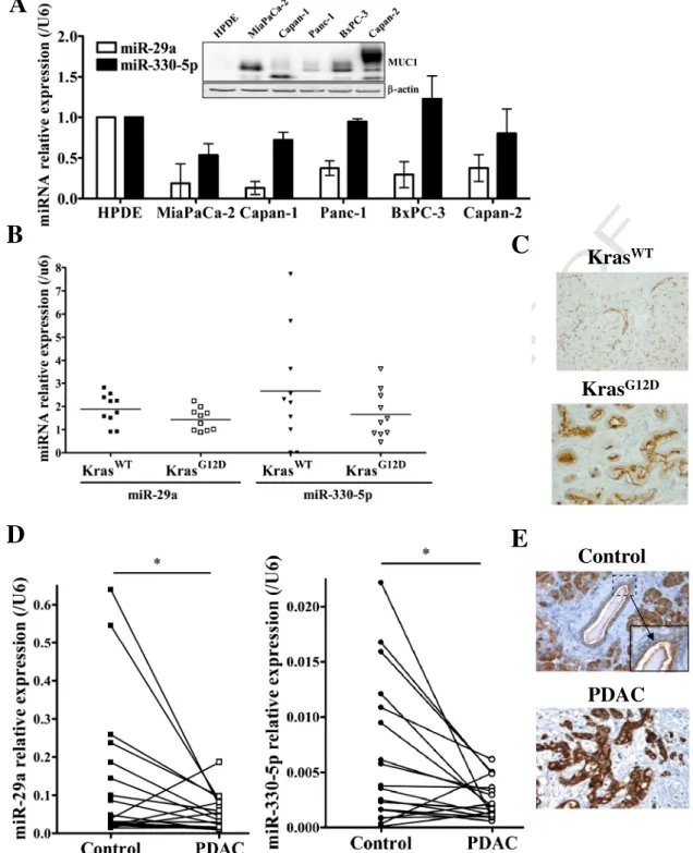
280 found in thefive PDAC cell lines (MiaPaCa-2, Capan-1, Panc-1, BxPC-3,
281 and Capan-2) compared to normal HPDE cells (Fig. 2A). MiR-330-5p
ex-282 pression was decreased in MiaPaCa-2 and Capan-1 cells (Fig. 2A).
Ex-283 pression of MUC1 mucin in the different cell lines is also shown
284 (Fig. 2A, insert). Pancreatic expression was then studied in a
pre-285 clinical mouse model of pancreatic carcinogenesis (KrasG12D)
develop-286 ing PanIN preneoplastic lesions in which we found a mild decreased of
287 miR-29a and miR-330-5p expression compared to control mice
288 (KrasWT) (Fig. 2B) whereas Muc1 was overexpressed (Fig. 2C). In
289 human PDAC samples, a significant decreased expression of miR-29a
290 and miR-330-5p was found compared to non-tumoral adjacent tissues
291 (Fig. 2D). This was correlated to overexpression and cytoplasmic
delo-292 calization of MUC1 (Fig. 2E). Altogether these results indicate a
de-293 creased expression of miR-29a or miR-330-5p, associated with MUC1
294 overexpression in human and mouse PDAC tissues.
295
3.3. Biological properties of PDAC cells transiently overexpressing miR-29a
296
and miR-330-5p
297
To further study the functional activity of miR-29a and miR-330-5p,
298
transient overexpression of these miRNAs was realized in Capan-2,
299
MiaPaCa-2 and Capan-1 cell lines (Fig. 3A). MiRNAoverexpression led Q11
300
to a significant decreased expression of both MUC1 mRNA (Fig. 3B)
301
and protein (Figs. 1B and3C) levels in all cell lines. Simultaneous
over-302
expression of both miR-29a and miR-330-5p did not potentiate the
ef-303
fects. Overexpression of miR-29a or miR-330-5p in cells alone or in
304
combination induced a significant decrease of cell proliferation
305
(Fig. 3D), cell migration (Fig. 3E) and cell invasion (Fig. 3F). Altogether
306
these results indicate that both miR-29a and miR-330-5p alter PDAC
307
cell proliferation, migration and invasion.
308
3.4. Biological properties of PDAC cells stably overexpressing miR-29a
309
and miR-330-5p
310
Having shown that miR-29a, miR-330-5p and MUC1 had an
in-311
verse profile of expression in PDAC, and that these two miRNAs
al-312
tered biological properties of PDAC cells, we established stable cell
313
lines overexpressing either miR-29a or miR-330-5p in MiaPaCa-2
314
cells in order to further assess their biological effects (Fig. 4A). A
315
strong decrease of MUC1 mRNA and protein expression was
ob-316
served in stable cell lines compared to control cells (Fig. 4B & C).
317
Cells overexpressing miR-29a or miR-330-5p showed a statistically
318
significant decrease of cell proliferation (Fig. 4D), migration and
in-319
vasion (Fig. 4E). Accordingly to the decreased of cell proliferation,
320
we observed a decrease of phospho p42–44 MAPK, cyclin D1 and
321
β-catenin expression in the two cell lines and a decrease of CDK6
322
and EGFR in cells overexpressing miR-29a (Fig. 4C). Finally, we
per-323
formed loss-of-function studies using anti-miRNAs and measured
324
impact on cell proliferation (Fig. 4F) and cell migration/invasion
325
(Fig. 4G). The results indicate a statistically significant increase of
326
cell proliferation (Fig. 4F), migration and invasion (Fig. 4G) for
327
both anti-miRNAs. Altogether, these results indicate that PDAC cells
328
overexpressing transiently or stably miR-29a or miR-330-5p harbor
329
an altered PDAC cell behavior that is less tumorigenic.
330
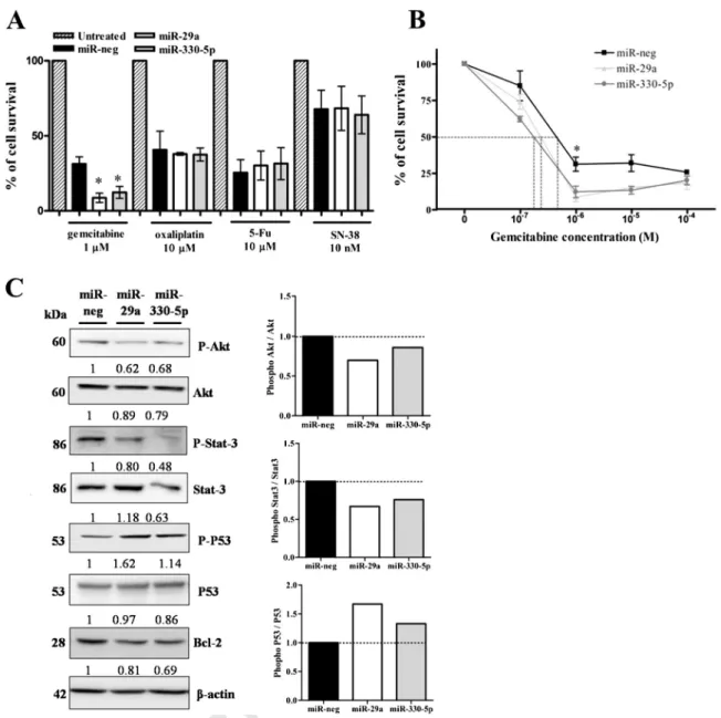
3.5. Effect of miR29a and miR-330-5p on PDAC cell chemosensitivity,
331
survival and apoptosis
332
PDAC being an extremely chemoresistant cancer, we undertook
333
to evaluate miR-29a and miR-330-5p effectson PDAC cell chemo- Q12
334
sensitivity. Measurement of cell survival following drug treatment
335
indicated that overexpression of miR-29a or miR-330-5p led to a
336
significant decrease of cell viability following treatment with
337
gemcitabine whereas no significant variation was found with drugs
338
of the FOLFIRINOX protocol (oxaliplatin, 5-Fu or SN-38) (Fig. 5A).
339
IC50determination also indicated that stable cell lines
overexpress-340
ing miR-29a (~ 383 nM) or miR-330-5p (~ 239 nM) were more
sensi-341
tive to gemcitabine than miR-neg cells (~ 646 nM) (Fig. 5B). We then
342
measured the activation of major signaling markers/pathways
in-343
volved in cell survival or cell apoptosis in these cellular models. We
344
found an increased level of apoptotic marker phospho-P53 and a
de-345
crease of anti-apoptotic marker Bcl-2 together with a decrease of
346
phospho-Akt and phospho-Stat3 (Fig. 5C). Altogether, these results
347
indicate that PDAC cells overexpressing miR-29a or miR-330-5p are
348
more sensitive to gemcitabine,survival less and are more apoptotic. Q13
349
3.6. In vivo biological effects of miR-29a and miR-330-5p on PDAC
350
tumor properties
351
Having shown in vitro that miR-29a and miR-330-5p possessed
352
tumor suppressive activities, we undertook to evaluate their activity
353
UNCORRECTED PR
OOF
354 MiaPaCa-2 cells were performed in SCID mice followed by an
intra-355 tumoral injection of the miRNA of interest. A significant decrease of
356 Capan-2 (pb 0.05) and MiaPaCa-2 (p b 0.001) tumor growth was
357
found when compared to control miR-neg (Fig. 6A). Tumor weights
358
were also significantly decreased when injected with miR-29a in
359
Capan-2 (pb 0.05) or both miRNAs in MiaPaCa-2 (p b 0.05) (Fig. 6B).
A
B
~200
42
β-actin
MUC1
kDa
miR-neg
miR-29a
miR-330-5p
miR-neg
miR-183
miR-200a
miR-neg
miR-876-3p
miR-939
C
D
miR-neg
miR-29a
miR-330-5p
0
1
2
*
*
MU
C
1
m
R
N
A
f
o
ld
en
ri
ch
m
en
t
(/G
A
P
D
H
)
miR-neg
miR-29a
miR-330-5p
0
2
4
6
*
*
MU
C
1
m
R
N
A
f
o
ld
en
ri
ch
m
en
t
(/
GAP
D
H)
Fig. 1.Q1 MUC1 regulation by miRNAs in PDAC cell lines
Q2 . (A) Sequence of MUC1 3′-UTR and position of miR-29a, miR-330-5p, miR-183, and miR-200a potential binding sites (left) and se-quence of MUC1first exon containing its 5′-UTR and position of miR-876-3p, miR-939, miR-183, and miR-200a potential binding sites (right). (B) Study of the expression of MUC1 in Capan-2 cells transfected with the miRNA of interest or miR-neg by Western-blot. (C) Measurement of luciferase activity in Capan-2 cells co-transfected with miRNAs of interest and MUC1 3′-UTR or MUC1 Exon1 containing the 5′-UTR cloned into pGL3 promoter luciferase-reporter vector. The neg luciferase activity is arbitrarily set to 1. (D) Biotinylated miR-29a or miR-330-5p or scramble miRNAs (miR-neg) were transfected in Capan-2 (left panel) and MiaPaCa-2 (right panel) cells. Enrichment of MUC1 mRNA in miRNA pulldown was assessed by qRT-PCR. The MUC1/GAPDH ratio was calculated and normalized to the input.
UNCORRECTED PR
OOF
360 SC xenografts of the MiaPaCa-2 cells stably overexpressing miR-29a or
361 miR-330-5p were also carried out, in which a significant decrease of
362 tumor growth was observed (pb 0.01) (Fig. 6C). This was correlated
363 to a strong decrease of MUC1 expression (Fig. 6D). Altogether these
re-364 sults indicate that in vivo miR-29a and miR-330-5p possess tumor
sup-365 pressive activities on PDAC tumors.
366
4. Discussion
367
Pancreatic Ductal Adenocarcinoma suffers from a very poor
progno-368
sis and absence of early detection and efficient therapy[2,19]. Recently,
369
miRNAs have emerged as very attractive molecules to target cancer
370
since they regulate many genes encoding oncogenic proteins[20]. In
A
B
D
C
Kras
WTKras
G12DE
Control
PDAC
Fig. 2. MiRNA expression in PDAC cells and tissues. (A) Expression of miRNAs in HPDE, MiaPaCa-2, Capan-1, Panc-1, BxPC-3, and Capan-2 cell lines was studied by qRT-PCR. MiRNA ex-pression was normalized to control U6 RNA. MiRNA level in normal pancreatic ductal cells HPDE was arbitrarily set to 1. Insert: exex-pression of MUC1 mucin by Western-blotting. (B) MiRNA expression in control KrasWT
and KrasG12D
mice studied by qRT-PCR. MiRNA expression was normalized to control u6 RNA. Expression in KrasWT
mice was arbitrarily set to 1. (C) MUC1 expression by IHC in control KrasWTand in KrasG12Dmice. (D) MiR-29a and miR-330-5p expression in a cohort of 18 patients with PDAC by qRT-PCR. MiRNA expression was normalized to
control U6 RNA and expressed as a ratio of PDAC/non-tumoral adjacent tissues (control). (E) MUC1 expression by IHC in non-tumoral adjacent tissues (control) and in tumors (PDAC). As an illustration, results with patient #1 are shown.
UNCORRECTED PR
OOF
371 PDAC, several studies have described the pattern of expression of
372 miRNAs but their exact biological role and the identification of their
tar-373 gets remain largely unknown[22,23]. In this report, we have identified
374 miR-29a and miR-330-5p as two new miRNAs directly targeting the 3′
375 UTR of MUC1 oncogenic mucin in PDAC cells and demonstrate that
376 these two miRNAs display tumor suppressive activities in PDAC both
377
in vitro and in vivo. We thus propose miR-29a and miR-330-5p as
ap-378
propriate targets to inhibit MUC1 expression in order to slow-down
379
pancreatic carcinogenesis and develop new potential therapeutic tools.
380
MiR-29a and miR-330-5p were shown to inhibit MUC1 expression
381
by directly binding to its 3′-UTR but interestingly we have also
identi-382
fied additional potential binding sites for miR-29a and miR-330-5p
Fig. 3. Biological properties of PDAC cells transiently overexpressing miR-29a or miR-330-5p. (A) Expression of miR-29a and miR-330-5p was measured after transient overexpression in Capan-2, MiaPaCa-2 and Capan-1 cells by qRT-PCR. MiRNA expression was normalized to control U6 RNA. The miR-neg expression was arbitrarily set to 1. (B) Analysis of MUC1 mRNA expression in Capan-2, MiaPaCa-2 and Capan-1 cells transfected with pre-miRNAs of interest or neg by qRT-PCR. MUC1 expression was normalized to control GAPDH. The miR-neg expression was arbitrarily set to 1. (C) Analysis of MUC1 protein expression in MiaPaCa-2 and Capan-1 cells transfected with pre-miRNAs of interest or miR-miR-neg by Western-blot. (D) Cell proliferation, (E) cell migration and (F) cell invasion were measured in Capan-2, MiaPaCa-2 and Capan-1 cells transfected with pre-miRNAs. (E, F) Results are expressed as a per-centage of migration or invasion in miR-transfected cells compared to the control miR-neg (100%).
UNCORRECTED PR
OOF
A
C
B
D
E
F
G
0 24 48 72 96 0 10 20 30 40 anti-miR-neg anti-miR-330-5p**
*
Time (h) N u m b er of c e ll s ( x 10 5) 0 24 48 72 96 0 2 4 6 8 anti-miR-neg anti-miR-29a*
*
Time (h) N u m b e r of c e ll s ( x 10 5 )anti-miR-neg anti-miR-330-5panti-miR-neg anti-miR-330-5p 0 50 100 150 200 Migration Invasion
*
% o f cel ls / con tro lanti-miR-neg anti-miR-330-5panti-miR-neg anti-miR-330-5p 0 50 100 150 200
*
**
Migration Invasion % o f cel ls / con tro lUNCORRECTED PR
OOF
383 into MUC1 coding regions suggesting that the inhibition of MUC1 could
384 also involve the binding of these two miRNAs to additional regions. The
385 validation of these sites will be of particular interest especially because
386 it was shown that MUC4 and MUC16 oncogenic membrane-bound
mu-387 cins are regulated by miRNAs through their coding regions[32]. This
388 could thus represent a general mechanism of mucin gene regulation
389 by miRNAs.
390 In breast cancer cells, MUC1 is regulated by miR-145[33], miR-1226
391 [34]and miR-125b[35]. In this report we show that, MUC1 is directly
392 regulated by miR-29a and miR-330-5p and indirectly regulated by
393 miR-876-3p, miR-939, miR-183 and miR-200a in PDAC cells. MiR-183
394 and miR-200 families
Q14 are EMT inhibitors and favor epithelial
395
differentiation[36]by inhibiting the Wnt/β-catenin signaling pathway
396
[37,38]. The membrane-bound mucin MUC1 is an epithelial marker of
397
polarized epithelial cells[4,6,7]but aberrant overexpression of MUC1
398
in cancer cells is correlated with an induction of EMT through the
399
Wnt/β-catenin signaling pathway [14]. The indirect inhibition of
400
MUC1 by miR-183 and miR-200a could be an additional mechanism
401
to inhibit EMT in PDAC. The role of miR-876-3p and miR-939 is hardly
402
known in cancer. The indirect mechanisms leading to MUC1 inhibition
403
will have to be identified in future studies. The identification of
predict-404
ed partners such as EGFR, cyclin D1, and Stat3, indirectly regulating
405
MUC1, that are potential targets of miR-876-3p or miR-939 (personal
406
communication) suggests that targeting these miRNAs could represent
Fig. 4. Biological properties of PDAC cells stably overexpressing miR-29a or miR-330-5p. (A) Expression of miR-29a and miR-330-5p in stable cell lines was measured by qRT-PCR. MiRNA expression was normalized to control U6 RNA. The miR-neg expression was arbitrarily set to 1. (B) Analysis of MUC1 mRNA expression by qRT-PCR. MUC1 expression was normalized to control GAPDH. The miR-neg expression was arbitrarily set to 1. (C) Analysis of MUC1, EGFR,β-catenin, cyclin D1, phospho-p42–44 MAPK, p42–44 MAPK, and CDK6 expression by West-ern-blot. Density of each marker was measured and protein of interest/β-actin ratio is indicated. Expression in miR-Neg cells was arbitrarily set to 1. (D) Measurement of cell proliferation and (E) cell migration and invasion. (E) Results are expressed as a percentage of migration or invasion of cells compared to the control miR-neg or Mock cells (100%). Measurement of cell proliferation (F) and cell migration/invasion (G) in MiaPaCa-2 stable cell lines overexpressing miR-29 (top panel) or miR-330-5p (bottom panel) treated with anti-miR-neg, anti-miR-29a or anti-miR-330-5p at 60 nM.
Fig. 5. Chemosensitivity of PDAC cells overexpressing miR-29a or miR-330-5p. (A) Cells were treated for 72 h with different concentrations of gemcitabine, 5-Fu, oxaliplatin or SN-38 be-fore cell survival measurement. (B) IC50rates after 72 h of gemcitabine treatment. (C) Analysis of phospho-AKT, AKT, phospho Stat3, Stat3, phospho-P53, P53 and Bcl-2 expression by
Western-blot. Density of each marker was measured and protein of interest/β-actin ratio is indicated. Expression in miR-Neg cells was arbitrarily set to 1. Phospho Akt/Akt, phospho Stat3/Stat3, and phospho P53/P53 ratios were determined and represented as histograms.
UNCORRECTED PR
OOF
407 an interesting approach to slow-down carcinogenesis. We cannot also
408 exclude that miR-145, miR-1226 and miR-125b known to regulate
409 MUC1 in breast cancer cell lines may also be MUC1 regulators in
pancre-410 atic cancer cells. Some preliminary data obtained in our laboratory seem
411Q15 to indicate that these miRNAsalso downregulate MUC1 in pancreatic
412 cancer cells (personal communication).
413 We also demonstrate that miR-29a and miR-330-5p inhibit MUC1
414 expression and deregulate signaling pathways as well as PDAC cell
pro-415 liferation, migration, invasion and tumor growth. It is well-established
416 that MUC1 is involved in these pathways[5,39]. These mechanisms
417 may involve p42–44 MAPK, cyclin D1 and β-catenin as we recently
418 showed that inhibiting MUC1 in PDAC cells altered these pathways
419 [5]. It was also proposed that the overexpression of miR-29a decreased
420 β-catenin expression and cell proliferation in non-small cell lung cancer
421 [40]. The same effect was observed in miR-29a overexpressing PDAC
422 cells and in PDAC cells lacking MUC1[5]. The overexpression of
miR-423 330-5p in colorectal cancer cells was previously shown to decrease
424
cell proliferation through direct regulation of Cdc42 and decrease
ex-425
pression of cyclin-D1[41]. The same effect was observed in PDAC cells
426
overexpressing miR-330-5p. Moreover, the fact that miR-29a alters
pro-427
liferation more profoundly than miR-330-5p suggests that both miRNAs
428
have additional targets. In that matter, we observed the decreased
ex-429
pression of CDK6 and Bcl-2 two known targets of miR-29a when it
430
was overexpressed[42,43]. Altogether these data are in favor of a
431
tumor suppressor activity for these two miRNAs in PDAC cells.
432
Contrarily to miR-330-5p, overexpression of miR-29a had a modest
433
effect on PDAC cell invasion. This could result from the inhibition of
434
CDK6 that we observed in these cells, loss of CDK6 being a marker of
435
several PDAC cell line invasiveness[44]. This may also be due to other
436
properties of miR-29a that depend on the cellular context: increased
in-437
vasion through MMP2, E-cadherin and KLF4 in colorectal cancer cells
438
[45], decreased invasion in gastric[46], lung and pancreatic cancer
439
cells[47], or decreased invasion and metastasis probably through the
440
inhibition of CEACAM6 in pancreatic cancer[48].
Fig. 6. In vivo effects of miR-29a and miR-330-5p on PDAC tumor properties. Subcutaneous (SC) xenografts of Capan-2 (left panel) and MiaPaCa-2 (right panel) cells in SCID mice. Intra-tumoral injection of miR-neg, miR-29a or miR-330-5p was performed 10 days post-xenografting (~300 mm3). (A) Tumor growth (mm3) was measured for 20 days after miRNA injection.
(B) Tumor weight (g) was measured at the sacrifice in Capan-2 (left panel) and MiaPaCa-2 (right panel) tumors injected with miR-neg, miR-29a or miR-330-5p. (C) SC xenografts of MiaPaCa-2 cells stably overexpressing either miR-neg, miR-29a or miR-330-5p in SCID mice. Tumor growth (mm3
) was measured during 20 days. (D) MUC1 protein expression by IHC in SC tumors.
UNCORRECTED PR
OOF
441 Additionally, we found that overexpression of miR-29a or
miR-330-442 5p sensitizes PDAC cells to gemcitabine and induces a decrease of Akt
443 pathway activation. Interestingly, MUC1 is known to mediate PDAC cell
444 chemoresistance to gemcitabine by regulating MRP1 gene expression
445 through the PI3K/Akt pathway[5,15]. Although, miR-29a overexpression
446 was previously shown to induce gemcitabine resistance in other PDAC
447 cells (BxPC-3 and Panc-1) through the Wnt/β-catenin signaling pathway
448 [49], other results in lung cancer cells indicate that overexpression of
449 miR-29a inhibits Wnt/β-catenin signaling and β-catenin expression[40].
450 The same effects were described in PDAC cells lacking MUC1[5].
Interest-451Q16 ingly, it was also shown that miR-29a inhibits Akt expression[50]and
452 sensitizes ovarian cancer cells to cisplatin[51], and that miR-330-5p
453 overexpression decreased Akt phosphorylation in colorectal cancer[41].
454 The decrease of Akt expression suggests a MUC1/Akt dependent
mecha-455 nism mediating miR-29a/miR-330-5p effects on MiaPaCa-2 cell
sensitiv-456 ity to gemcitabine. Altogether, these data highlight the complex role of
457 these miRNAs in chemoresistance processes triggered by cancer cells
458 that most likely are cell-specific. Further investigation will be needed to
459 decipher the mechanisms underlying the sensitivity of PDAC cells to
che-460Q17 motherapeutic drugs in relation with MUC1, miRNAand Akt expression.
461 Moreover, we also showed that overexpression of miR-29a or
miR-330-462 5p increases P53 activity as previously proposed for miR-29a[52]and
de-463 creases Bcl-2 expression like it was previously proposed in PDAC cells
464 lacking MUC1[5]suggesting an involvement of these two miRNAs in
465 cell apoptosis.
466 We also show that miR-29a and miR-330-5p are deregulated in
467 human PDAC cell lines and tissues, and in the KrasG12Dpre-clinical
468 mouse model of pancreatic cancer. Expression studies in a cohort of 18
469 human PDAC tissues showed inhibition of miR-29a and miR-330-5p
470 which is in accordance with previous studies showing that miR-29a is
fre-471 quently deregulated in cancerous tissues. This confirms the potential of
472 miR-29a as a target for cancer therapy as it was previously proposed
473 [53]and highlights the interest for miR-330-5p in PDAC.
474 5. Conclusion
475 In conclusion, we show (i) that MUC1 is regulated by miRNAs in
476 PDAC cells by direct and indirect mechanisms, (ii) that miR-29a and
477 miR-330-5p are deregulated in PDAC and that PDAC cells
overexpress-478 ing transiently or stably either miR-29a or miR-330-5p possess similar
479 biological properties: in vitro decreased proliferative, migrating, and
in-480 vasive properties and in vivo decreased tumor growth, and (iii) that
481 miR-29a and miR-330-5p sensitize PDAC cells to gemcitabine. Based
482 on that, we propose that tumor suppressive miR-29a and miR-330-5p
483 may represent attractive targets to inhibit the expression of the
onco-484 genic MUC1 mucin and decrease pancreatic cancer progression.
485 Author contribution
486 Solange Tréhoux conceived and designed the project, performed
ex-487 periments, data analysis and interpretation, and wrote the paper. Fatima
488 Lahdaoui performed in vitro experiments, data analysis, interpretation,
489 and revised the manuscript. Isabelle Van Seuningen conceived and
de-490 signed the project, providedfinancial support, data analysis and
interpre-491 tation and wrote the paper. Nicolas Jonckheere performed in vivo
492 experiments in mice models, data analysis and interpretation, and
re-493 vised the manuscript. Florence Renaud and Emmanuelle Leteurtre
per-494 formed anatomopathological analyses and revised the manuscript.
495 Yannick Delpu and Jérôme Torrisani raised stable cell lines
overexpress-496 ing miR-29a and miR-330-5p and revised the manuscript.
497 Conflicts of interest
498 Authors declare no conflict of interest.
499Q18 Uncited reference 500 [29] 501 Acknowledgments 502
We thank D. Tuveson (Cambridge Research Institute, England) for the
503
kind gift of LStopL-KrasG12Dmouse model; M. S. Tsao (University Health
504
Network, Toronto, Canada) for the kind gift of HPDE cells; D. Swallow
505
(MRC, London, UK) for providing MUC1 M8 antibody, and A. Lansiaux
506
and S. Meignan (Centre Oscar Lambret and Inserm UMR837, team
507
3) for the chemotherapeutic drugs. We are grateful to B. Barbot and
508
B. Duchêne (Inserm UMR837, team 5) and M.H. Gevaert and R. Siminsky
509
(Department of Histology, Faculty of Medicine, University of Lille 2) for
510
their excellent technical help. We thank the IFR114/IMPRT (University
511
of Lille 2) facilities for luciferase measurements (A.S. Drucbert) and
ani-512
mal experimentation (D. Taillieu).
513
Funding: Solange Tréhoux is a recipient of a PhD fellowship of the
514
University of Lille 2. Isabelle Van Seuningen is the recipient of a“Contrat
515
Hospitalier de Recherche Translationnelle” (AVIESAN/CHRT 2010).
516
Fatima Lahdaoui is a recipient of a SIRIC ONCOLille fellowship (Grant
517
INCa-DGOS-Inserm 6041 (IVS)). This work was supported by grants
518
from la Ligue Nationale Contre le Cancer (Equipe Labellisée Ligue
519
2011–2013, IVS), from SIRIC ONCOLille, Grant INCa-DGOS-Inserm Q19
520
6041 (IVS) and from “Contrat de Plan Etat Région” CPER Cancer
521
2007–2013(IVS). Q20Q21
522
References
523
[1] A. Jemal, F. Bray, M.M. Center, J. Ferlay, E. Ward, D. Forman, Global cancer statistics,
524
CA Cancer J. Clin. 61 (2011) 69–90.
525
[2] A. Vincent, J. Herman, R. Schulick, R.H. Hruban, M. Goggins, Pancreatic cancer, Lancet
526
378 (2011) 607–620.
527
[3] T. Conroy, F. Desseigne, M. Ychou, O. Bouché, R. Guimbaud, Y. Bécouarn, A. Adenis, J.-L.
528
Raoul, S. Gourgou-Bourgade, C. de la Fouchardière, et al., FOLFIRINOX versus
529
gemcitabine for metastatic pancreatic cancer, N. Engl. J. Med. 364 (2011) 1817–1825.
530
[4] N. Jonckheere, I. Van Seuningen, The membrane-bound mucins: how large
O-531
glycoproteins play key roles in epithelial cancers and hold promise as biological
532
tools for gene-based and immunotherapies, Crit. Rev. Oncog. 14 (2008) 177–196.
533
[5] S. Tréhoux, B. Duchêne, N. Jonckheere, I. Van Seuningen, The MUC1 oncomucin
reg-534
ulates pancreatic cancer cell biological properties and chemoresistance. Implication
535
of p42–44 MAPK, Akt, Bcl-2 and MMP13 pathways, Biochem. Biophys. Res.
536
Commun. 456 (2015) 757–762.
537
[6] M.A. Hollingsworth, B.J. Swanson, Mucins in cancer: protection and control of the
538
cell surface, Nat. Rev. Cancer 4 (2004) 45–60.
539
[7] D.W. Kufe, Mucins in cancer: function, prognosis and therapy, Nat. Rev. Cancer 9
540
(2009) 874–885.
541
[8] J.A. Schroeder, M.C. Thompson, M.M. Gardner, S.J. Gendler, Transgenic MUC1
inter-542
acts with epidermal growth factor receptor and correlates with mitogen-activated
543
protein kinase activation in the mouse mammary gland, J. Biol. Chem. 276 (2001)
544
13057–13064.
545
[9] D.M. Besmer, J.M. Curry, L.D. Roy, T.L. Tinder, M. Sahraei, J. Schettini, S.-I. Hwang, Y.Y.
546
Lee, S.J. Gendler, P. Mukherjee, Pancreatic ductal adenocarcinoma mice lacking
547
mucin 1 have a profound defect in tumor growth and metastasis, Cancer Res. 71
548
(2011) 4432–4442.
549
[10] H. Rajabi, R. Ahmad, C. Jin, M. Kosugi, M. Alam, M.D. Joshi, D. Kufe, MUC1-C
550
oncoprotein induces TCF7L2 transcription factor activation and promotes cyclin
551
D1 expression in human breast cancer cells, J. Biol. Chem. 287 (2012)
552
10703–10713.
553
[11] R. Ahmad, H. Rajabi, M. Kosugi, M.D. Joshi, M. Alam, B. Vasir, T. Kawano, S.
554
Kharbanda, D. Kufe, MUC1-C oncoprotein promotes STAT3 activation in an
555
autoinductive regulatory loop, Sci. Signal. 4 (2011) ra9.
556
[12] J. Gao, M.J. McConnell, B. Yu, J. Li, J.M. Balko, E.P. Black, J.O. Johnson, M.C. Lloyd, S.
557
Altiok, E.B. Haura, MUC1 is a downstream target of STAT3 and regulates lung cancer
558
cell survival and invasion, Int. J. Oncol. 35 (2009) 337–345.
559
[13] P.K. Singh, Y. Wen, B.J. Swanson, K. Shanmugam, A. Kazlauskas, R.L. Cerny, S.J.
560
Gendler, M.A. Hollingsworth, Platelet-derived growth factor receptorβ-mediated
561
phosphorylation of MUC1 enhances invasiveness in pancreatic adenocarcinoma
562
cells, Cancer Res. 67 (2007) 5201–5210.
563
[14] L.D. Roy, M. Sahraei, D.B. Subramani, D. Besmer, S. Nath, T.L. Tinder, E. Bajaj, K.
564
Shanmugam, Y.Y. Lee, S.I.L. Hwang, et al., MUC1 enhances invasiveness of pancreatic
565
cancer cells by inducing epithelial to mesenchymal transition, Oncogene 30 (2011)
566
1449–1459.
567
[15] S. Nath, K. Daneshvar, L.D. Roy, P. Grover, A. Kidiyoor, L. Mosley, M. Sahraei, P.
568
Mukherjee, MUC1 induces drug resistance in pancreatic cancer cells via
upregula-569
UNCORRECTED PR
OOF
570 [16] J.M. Winter, L.H. Tang, D.S. Klimstra, M.F. Brennan, J.R. Brody, F.G. Rocha, X. Jia, L.-X.
571 Qin, M.I. D'Angelica, R.P. DeMatteo, et al., A novel survival-based tissue microarray
572 of pancreatic cancer validates MUC1 and mesothelin as biomarkers, PLoS One 7
573 (2012) e40157.
574 [17] A.M. Mohr, J.M. Bailey, M.E. Lewallen, X. Liu, P. Radhakrishnan, F. Yu, W. Tapprich,
575 M.A. Hollingsworth, MUC1 regulates expression of multiple microRNAs involved
576 in pancreatic tumor progression, including the miR-200c/141 cluster, PLoS One 8
577 (2013) e73306.
578 [18] A.L. Kasinski, F.J. Slack, MicroRNAs en route to the clinic: progress in validating and
579 targeting microRNAs for cancer therapy, Nat. Rev. Cancer 11 (2011) 849–864.
580 [19] M. Humeau, J. Torrisani, P. Cordelier, miRNA in clinical practice: pancreatic cancer,
581 Clin. Biochem. 46 (2013) 933–936.
582 [20] M.V. Iorio, C.M. Croce, MicroRNA dysregulation in cancer: diagnostics, monitoring
583 and therapeutics. A comprehensive review, EMBO Mol. Med. 4 (2012) 143–159.
584 [21] D.P. Bartel, MicroRNAs: genomics, biogenesis, mechanism, and function, Cell 116
585 (2004) 281–297.
586 [22] Y. Delpu, H. Lulka, F. Sicard, N. Saint-Laurent, F. Lopez, N. Hanoun, L. Buscail, P.
587 Cordelier, J. Torrisani, The rescue of miR-148a expression in pancreatic cancer: an
588 inappropriate therapeutic tool, PLoS One 8 (2013) e55513.
589 [23] F. Lahdaoui, Y. Delpu, A. Vincent, F. Renaud, M. Messager, B. Duchêne, E. Leteurtre, C.
590 Mariette, J. Torrisani, N. Jonckheere, I. Van Seuningen, miR-219-1-3p is a negative
591 regulator of the mucin MUC4 expression and is a tumor suppressor in pancreatic
592 cancer, Oncogene 34 (2015) 780–788.
593 [24] S. Khan, M.C. Ebeling, M.S. Zaman, M. Sikander, M.M. Yallapu, N. Chauhan, A.M.
594 Yacoubian, S.W. Behrman, N. Zafar, D. Kumar, et al., MicroRNA-145 targets MUC13
595 and suppresses growth and invasion of pancreatic cancer, Oncotarget 5 (2014)
596 7599–7609.
597 [25] N. Jonckheere, M. Perrais, C. Mariette, S.K. Batra, J.-P. Aubert, P. Pigny, I. Van
598 Seuningen, A role for human MUC4 mucin gene, the ErbB2 ligand, as a target of
599 TGF-beta in pancreatic carcinogenesis, Oncogene 23 (2004) 5729–5738.
600 [26] N. Skrypek, B. Duchêne, M. Hebbar, E. Leteurtre, I. van Seuningen, N. Jonckheere, The
601 MUC4 mucin mediates gemcitabine resistance of human pancreatic cancer cells via
602 the Concentrative Nucleoside Transporter family, Oncogene 32 (2013) 1714–1723.
603 [27] A. Lal, M.P. Thomas, G. Altschuler, F. Navarro, E. O'Day, X.L. Li, C. Concepcion, Y.-C.
604 Han, J. Thiery, D.K. Rajani, et al., Capture of microRNA-bound mRNAs identifies the
605 tumor suppressor miR-34a as a regulator of growth factor signaling, PLoS Genet. 7
606 (2011) e1002363.
607 [28] N. Jonckheere, N. Skrypek, J. Merlin, A.F. Dessein, P. Dumont, E. Leteurtre, A. Harris,
608 J.-L. Desseyn, C. Susini, F. Frénois, et al., The mucin MUC4 and its membrane partner
609 ErbB2 regulate biological properties of human CAPAN-2 pancreatic cancer cells via
610 different signalling pathways, PLoS One 7 (2012) e32232.
611 [29] G. Piessen, N. Jonckheere, A. Vincent, B. Hémon, M.-P. Ducourouble, M.-C. Copin, C.
612 Mariette, I. Van Seuningen, Regulation of the human mucin MUC4 by
613 taurodeoxycholic and taurochenodeoxycholic bile acids in oesophageal cancer
614 cells is mediated by hepatocyte nuclear factor 1alpha, Biochem. J. 402 (2007) 81–91.
615 [30] M. Van der Sluis, M.H.M. Melis, N. Jonckheere, M.-P. Ducourouble, H.A. Büller, I.
616 Renes, et al., The murine Muc2 mucin gene is transcriptionally regulated by the
617 zinc-finger GATA-4 transcription factor in intestinal cells, Biochem. Biophys. Res.
618 Commun. 325 (2004) 952–960.
619 [31] C. Mariette, M. Perrais, E. Leteurtre, N. Jonckheere, B. Hémon, P. Pigny, et al.,
Tran-620 scriptional regulation of human mucin MUC4 by bile acids in oesophageal cancer
621 cells is promoter-dependent and involves activation of the phosphatidylinositol
3-622 kinase signalling pathway, Biochem. J. 377 (2004) 701–708.
623 [32] P. Radhakrishnan, A.M. Mohr, P.M. Grandgenett, M.M. Steele, S.K. Batra, M.A.
624 Hollingsworth, MicroRNA-200c modulates the expression of MUC4 and MUC16 by
625 directly targeting their coding sequences in human pancreatic cancer, PLoS One 8
626 (2013) e73356.
627 [33] M. Sachdeva, Y.-Y. Mo, MicroRNA-145 suppresses cell invasion and metastasis by
di-628 rectly targeting mucin 1, Cancer Res. 70 (2010) 378–387.
629 [34] C. Jin, H. Rajabi, D. Kufe, miR-1226 targets expression of the mucin 1 oncoprotein
630 and induces cell death, Int. J. Oncol. 37 (2010) 61–69.
631
[35] H. Rajabi, C. Jin, R. Ahmad, C. McClary, M.D. Joshi, D. Kufe, MUCIN 1 oncoprotein
632
expression is suppressed by the miR-125b oncomirR, Genes Cancer 1 (2010)
633
62–68.
634
[36] U. Wellner, J. Schubert, U.C. Burk, O. Schmalhofer, F. Zhu, A. Sonntag, B. Waldvogel,
635
C. Vannier, D. Darling, A. zur Hausen, et al., The EMT-activator ZEB1 promotes
tu-636
morigenicity by repressing stemness-inhibiting microRNAs, Nat. Cell Biol. 11
637
(2009) 1487–1495.
638
[37] H. Xia, W.K.C. Cheung, J. Sze, G. Lu, S. Jiang, H. Yao, X.-W. Bian, W.S. Poon, H. Kung,
639
M.C. Lin, miR-200a regulates epithelial–mesenchymal to stem-like transition via
640
ZEB2 andβ-catenin signaling, J. Biol. Chem. 285 (2010) 36995–37004.
641
[38] C. Chen, H. Xiang, Y.-L. Peng, J. Peng, S.-W. Jiang, Mature miR-183, negatively
regu-642
lated by transcription factor GATA3, promotes 3T3-L1 adipogenesis through
inhibi-643
tion of the canonical Wnt/β-catenin signaling pathway by targeting LRP6, Cell.
644
Signal. 26 (2014) 1155–1165.
645
[39] D.W. Kufe, MUC1-C oncoprotein as a target in breast cancer: activation of signaling
646
pathways and therapeutic approaches, Oncogene 32 (2013) 1073–1081.
647
[40] M. Tan, J. Wu, Y. Cai, Suppression of Wnt signaling by the miR-29 family is mediated
648
by demethylation of WIF-1 in non-small-cell lung cancer, Biochem. Biophys. Res.
649
Commun. 438 (2013) 673–679.
650
[41] Y. Li, X. Zhu, W. Xu, D. Wang, J. Yan, miR-330 regulates the proliferation of colorectal
651
cancer cells by targeting Cdc42, Biochem. Biophys. Res. Commun. 431 (2013)
652
560–565.
653
[42] J.-J. Zhao, J. Lin, T. Lwin, H. Yang, J. Guo, W. Kong, S. Dessureault, L.C. Moscinski, D.
654
Rezania, W.S. Dalton, et al., microRNA expression profile and identification of
miR-655
29 as a prognostic marker and pathogenetic factor by targeting CDK6 in mantle
656
cell lymphoma, Blood 115 (2010) 2630–2639.
657
[43] Y. Xiong, J.-H. Fang, J.-P. Yun, J. Yang, Y. Zhang, W.-H. Jia, S.-M. Zhuang, Effects of
658
microRNA-29 on apoptosis, tumorigenicity, and prognosis of hepatocellular
carcino-659
ma, Hepatology 51 (2010) 836–845.
660
[44] F. Liu, M. Korc, Cdk4/6 inhibition induces epithelial–mesenchymal transition and
661
enhances invasiveness in pancreatic cancer cells, Mol. Cancer Ther. 11 (2012)
662
2138–2148.
663
[45] W. Tang, Y. Zhu, J. Gao, J. Fu, C. Liu, Y. Liu, C. Song, S. Zhu, Y. Leng, G. Wang, et al.,
664
MicroRNA-29a promotes colorectal cancer metastasis by regulating matrix
metallo-665
proteinase 2 and E-cadherin via KLF4, Br. J. Cancer (2013) Q22
666
[46] J. Gong, J. Li, Y. Wang, C. Liu, H. Jia, C. Jiang, Y. Wang, M. Luo, H. Zhao, L. Dong, et al.,
667
Characterization of microRNA-29 family expression and investigation of their
mech-668
anistic roles in gastric cancer, Carcinogenesis (2013) bgt337.
669
[47] M.K. Muniyappa, P. Dowling, M. Henry, P. Meleady, P. Doolan, P. Gammell, M.
670
Clynes, N. Barron, MiRNA-29a regulates the expression of numerous proteins and
671
reduces the invasiveness and proliferation of human carcinoma cell lines, Eur. J.
672
Cancer 45 (2009) 3104–3118.
673
[48] J. Chen, Q. Li, Y. An, N. Lv, X. Xue, J. Wei, K. Jiang, J. Wu, W. Gao, Z. Qian, et al.,
674
CEACAM6 induces epithelial–mesenchymal transition and mediates invasion and
675
metastasis in pancreatic cancer, Int. J. Oncol. 43 (2013) 877–885.
676
[49] H. Nagano, Y. Tomimaru, H. Eguchi, N. Hama, H. Wada, K. Kawamoto, S. Kobayashi,
677
M. Mori, Y. Doki, MicroRNA-29a induces resistance to gemcitabine through the
678
Wnt/β-catenin signaling pathway in pancreatic cancer cells, Int. J. Oncol. 43
679
(2013) 1066–1072.
680
[50] W. Wei, H.-B. He, W.-Y. Zhang, H.-X. Zhang, J.-B. Bai, H.-Z. Liu, J.-H. Cao, K.-C. Chang,
681
X.-Y. Li, S.-H. Zhao, miR-29 targets Akt3 to reduce proliferation and facilitate
differ-682
entiation of myoblasts in skeletal muscle development, Cell Death Dis. 4 (2013)
683
e668.
684
[51] P.-N. Yu, M.-D. Yan, H.-C. Lai, R.-L. Huang, Y.-C. Chou, W.-C. Lin, L.-T. Yeh, Y.-W. Lin,
685
Downregulation of miR-29 contributes to cisplatin resistance of ovarian cancer cells,
686
Int. J. Cancer 134 (2014) 542–551.
687
[52] J.-K. Park, E.J. Lee, C. Esau, T.D. Schmittgen, Antisense inhibition of microRNA-21 or -221
688
arrests cell cycle, induces apoptosis, and sensitizes the effects of gemcitabine in
pancre-689
atic adenocarcinoma, Pancreas 38 (2009) e190–e199.
690
[53] Y. Wang, X. Zhang, H. Li, J. Yu, X. Ren, The role of miRNA-29 family in cancer, Eur. J. Cell Biol. 92 (2013) 123–128.