Cell-mediated Contraction & Induced Regeneration
Of the Injured Peripheral Nerve
by
MASSACHUby
OF TE JULEric C. Soller
LIB
S.M., Mechanical Engineering,Massachusetts Institute of Technology, 2005
ARCHIVES
S.B., Mechanical Engineering,Rose-Hulman Institute of Technology, 2003
Submitted to the Department of Mechanical Engineering in Partial Fulfillment of the Requirements for the Degree of
Doctor of Philosophy in Mechanical Engineering at the
MASSACHUSETTS INSTITUTE OF TECHNOLOGY
June 2011
Signature
© 2011 Massachusetts Institute of Technology. All rights reserved
of Author. ... . . ...
=
::-=
...Department of Mechanical Engineering
A. May 20, 2011
C ertified by ... ... . ... .
Ioannis V. Yannas Professor of Mechanical Engineering, Polymer Science & Biological Engineering Thgsfupervisor A ccepted by...
Douglas P. Hardt
Professor of Mechanical Engineering Chairman, Department Committee on Graduate Students
SETTS INSTITUTE CHNOLOGY
29 2011
|
Document Services Room 14-0551 77 Massachusetts Avenue Cambridge, MA 02139 Ph: 617.253.2800 Email: docs@mit.edu http://libraries.mit.edu/docs
DISCLAIMER OF QUALITY
Due to the condition of the original material, there are unavoidable flaws in this reproduction. We have made every effort possible to provide you with the best copy available. If you are dissatisfied with this product and find it unusable, please contact Document Services as soon as possible.
Thank you.
There are some duplicate numbered pages in this document due to a pagination error by the author.
Cell-mediated Contraction & Induced Regeneration
Of the Injured Peripheral Nerve
by
Eric C. Soller
Submitted to the Department of Mechanical Engineering on May 20, 2011 in Partial Fulfillment of the Requirements for the Degree of
Doctor of Philosophy in Mechanical Engineering
ABSTRACT
Cell-mediated mechanical forces drive closure of severe wounds in adult mammalian organs, including the sciatic nerve following neurotmesis. Without experimental intervention the defect closes rapidly via contraction of transected nerve stumps by a thick, cohesive capsule of myofibroblasts (MFB) and subsequent collagen synthesis (scar), leading to a painful neuroma. Despite considerable progress in regenerating injured peripheral nerves with biomaterials, adequate recovery is generally limited to inter-stump gap lengths of about 20-30 mm in humans. Observations of successful induced regeneration in adults coincide with reduced MFB formation, yet the direct effect of the MFB capsule on nerve regeneration is unknown. According to the pressure cuff theory, a transected peripheral nerve could heal by regeneration, rather than MFB-mediated contraction and scar formation, provided the MFB capsule size (and corresponding cellular forces applied) are reduced.
A well-characterized library of type I collagen tubular scaffolds with identical
chemical composition and pore size, and varying degradation rate was used to evaluate the ability of a porous scaffold to block MFB contraction after injury in a demanding model of peripheral nerve regeneration in the adult rat. At 9 weeks post-neurotmesis, the MFB capsule thickness, 5, around the regenerating nerve was measured and the correlation with several quantitative measures of the quality of nerve regeneration,
Q,
was evaluated.
A negative, statistically significant association was observed between the
contractile capsule thickness, 6, and the quality of axonal regeneration,
Q,
(consisting of measures of regenerate area, the number of myelinated fibers, and the number of large-diameter fibers) at 9 weeks post-sciatic neurotmesis. This constitutes the strongest evidence to date that capsules of contractile MFB antagonize induced regeneration of severely injured peripheral nerves in the adult mammal. Collagen devices of intermediate degradation rate minimized 5 and maximizedQ.
Reduced contractile cell presence and disrupted organization inside moderately cross-linked scaffolds that consequently degraded at an intermediate rate, but not in highly cross-linked scaffolds that degraded at very low rate, support the hypothesis of cell escape from the wound (making use of scaffold permeability) as a mechanism for scaffold regenerative activity. Thesis Supervisor: loannis V. YannasThis page was left blank intentionally.
Acknowledgements
I am grateful for the guidance, enthusiasm, and support of the following
individuals, without whom this work would not have been possible.
Prof. loannis V. Yannas, my advisor and mentor
Prof. Myron Spector and Prof. Lorna Gibson, my doctoral committee members Dr. Hu-Ping Hsu, a talented surgeon and friend
Drs. Soller (n=2), my parents The Soller Boys (n=5)
Past & present members of the Yannas Lab for Regenerative Biomaterials, particularly Dimitrios Tzeranis
The many friends who made my time here in Cambridge truly enjoyable.
I dedicate this work to my grandfather James Camillo Flanagan, P.E., who first
put me to work as an engineer some 15 years ago analyzing artesian water wells in Central Texas.
Eric Soller Cambridge, MA
This page was left blank intentionally.
TABLE OF CONTENTS
A B ST R A C T ... 3
BIOGRAPHICAL NOTE ... ERROR! BOOKMARK NOT DEFINED. IN T R O D U C T IO N ... 15
1.1 A CLINICAL NEED FOR IMPROVED PERIPHERAL NERVE REGENERATION...15
1.2 IRREVERSIBLE INJURY IN SKIN AND PERIPHERAL NERVES...16
1.2.1. MACROSCOPIC OUTCOMES OF WOUND HEALING: REPAIR VS. REGENERATION... 16
1.2.2. REGENERATIVE SIMILARITY OF THE TISSUE TRIAD ... ... ... 17
1.3 EXPERIMENTAL CONSIDERATIONS...20
1.3.1 IMPORTANCE OF AN ANATOMICALLY WELL-DEFINED DEFECT .. ... ... 20
1.3.2 SYNTHETIC PROTOCOL: IN VITRO OR IN VIVO? ... ... 21
1.4 OVERVIEW OF INDUCED ORGAN REGENERATION... 21
1.4.1. E M PIRICA L E V ID EN CE ... 21
1.4.2. T H E D EF CT C LO SU RE R U LE ... 26
1.4.3 PREVALENCE OF CONTRACTION DURING SPONTANEOUS HEALING ... 27
1.4.4 ANTAGONISTIC RELATION BETWEEN CONTRACTION AND REGENERATION ... ... 28
1.4.5. REPAIR: M ECHANISM OF CONTRACTION ... 32
1.4.6. Structural Determinants of Scaffold Regenerative Activity... 34
1.5 THE PERIPHERAL NERVOUS SYSTEM (PNS): A MODEL ORGAN FOR INDUCED REGENERATION STUDIES .40 1.5.1. STRU CTU RE A N D FU N CTIO N ... 40
1.5.1.1 P arenchym al C om ponents...40
1 .5 .1 .2 S tro m a ... 4 3 1.5.2. REGENERATIVE MICROENVIRONMENT AFTER NEUROTMESIS ... 44
1.6. THEORIES OF PERIPHERAL NERVE REGENERATION & THE PRESSURE CUFF THEORY...45
1.7. R ESEA R C H G O A L ... 46
1.7.1 EXPERIM EN TA L A PPRO A CH ... 46
1.7 .2 S IG N IFIC A N C E ... 4 7 1.7.3. IN NOVATION / M AJOR FINDINGS ... 48
1.7.4. T H ESIS O RGA N IZA TIO N ... 48
2. CELL-MEDIATED CONTRACTION IMPAIRS AXONAL REGENERATION AFTER SCIATIC
N EU R O T M ESIS...51
2.1 IN TR O D U C TIO N ... 51
2.1.1 PROJECT G OAL & M AJOR FINDINGS ... 52
2.2 MATERIALS AND METHODS... 53
2.2.1 SYNTHESIS AND CHARACTERIZATION OF A HOMOLOGOUS SERIES OF COLLAGEN TUBES THAT VARY IN D EG RA D A TIO N R A TE ... 53
C ollagen T ube Sy n thesis ... 53
C ross-lin king T reatm en ts ... 54
2.2.2 TU BE C H A RA CTERIZA TIO N ... 55
2.2.3 A N IM A L M O D EL ... 56
2.2.4 H ISTO M O RPH O M ETRY ... 57
2 .2 .5 S TA T ISTIC S ... 6 0 2.3 R E SU LT S... 60
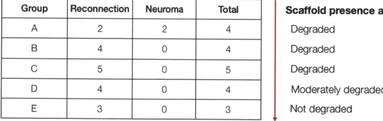
2.3.1 G EN ERA L O BSERVA TIO N S ... 60
2.3.2. PORE SIZE CHARACTERIZATION OF COLLAGEN TUBES... 61
2.3.3. IN VITRO DEGRADATION RATE OF COLLAGEN TUBES ... ... 62
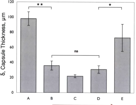
2.3.4. C O N TRACTILE C A PSU LE ... 63
2.3.4.1. M ean C apsule Thickness, 6 ... 63
2.3.4.2. C ontractile C apsule H istology ... 64
2.3.5. QUALITY OF AXONAL REGENERATION, Q... 68
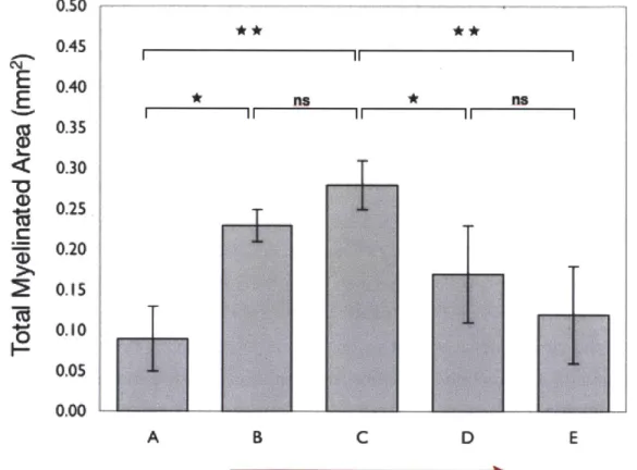
2.3.5.1 . Total M yelinated A rea... 68
2.3.5.2. M ean M yelinated Fiber D iam eter ... 74
2.3.5.3. N um ber and Percentage of A -fibers ... 74
2 .3 .5 .4 N -ra tio ... 7 6 2.3.6 RELATIONSHIP BETWEEN CAPSULE THICKNESS, A, AND QUALITY OF REGENERATION, Q ... 80
2.3.6.1 Correlation between 6 and Total Myelinated Area... 80
2.3.6.2 Correlation between 3 and Total Number of Myelinated Fibers ... ... 81
2.3.6.2 Correlation between 5 and Total Number of A-fibers ... 82
2.3.6.3 Correlation between 3 and Other Q metrics ... ... 83
2.3.6.4 Summary of Correlation between 3 and Q metrics.... ... 83
2.3.7 CAPSULE THICKNESS, A AND REGENERATE AREA AS A FUNCTION OF DISTANCE ... 84
2.3.8 CELL-SCAFFOLD INTERACTIONS APPEAR TO COINCIDE WITH DECREASED A ... 89
2.3.8. PRELIMINARY RESULTS FROM 7 DAYS AFTER INJURY ... 93
2.3.8.1. MFB-Scaffold Interactions Appear to Coincide with Reduced 3 ... ... ... 93
2.3.8.2. Thick MFB Capsules Appear to Compress Transected Nerve Stumps ... 93
2.4 DISCUSSION & CONCLUSIONS... 96
2.4.1. CONTRACTILE CAPSULE THICKNESS, A, HAS A LARGE NEGATIVE ASSOCIATION WITH THE QUALITY OF IN D U CED R EG EN ERA TIO N , Q . ... 96
2.4.2 COLLAGEN TUBES OF INTERMEDIATE CROSS-LINKING MINIMIZE CONTRACTILE CAPSULE THICKNESS, A, AND REDUCE CAPSULE "CONTRACTILITY" ... 97
2.4.3. COLLAGEN TUBES OF INTERMEDIATE CROSS-LINKING MAXIMIZE HISTOMORPHOMETRIC QUALITY OF R EG EN ER A TIO N , Q ... 102
2.4.4. THE PRESSURE CUFF THEORY AND THE HOMOLOGOUS SERIES... ... . . ... 103
2 .4 .5 . FU TU R E W O R K ... 104
2.5 LITERATUR E CITED ... 105
3. A PILOT STUDY OF Y-27632, A SOLUBLE INHIBITOR OF ACTIN-MYOSIN CONTRACTILITY, AND ITS EFFECT ON THE EARLY RESPONSE TO SCIATIC NEUROTMESIS...111
3.1 INTRODUCTION...111
3.1.1 INTRACELLULAR PATHWAYS LEADING TO CONTRACTION ... 111
3 .1 .2 . R h o G T P ases ... 1 12 3 .1.3 C on traction O verview ... 112
3.1.4 Inhibition of R O CK and Contraction ... 114
3.1.2. P roject G oal & M ajor Findings: ... 116
3.2. MATERIALS AND METHODS ... 116
3.2.1. DESIGN AND FABRICATION OF A BIODURABLE NERVE GUIDE, CONTINUOUS DRUG DELIVERY SYSTEM. ... 1 1 6 3.2.2. "T TUBE" TESTING & STERILIZATION ... 118
3.2.3. IN VIVO TESTING OF DRUG DELIVERY SYSTEM ... 118
3.2.4. SU RGICA L P RO CED U RE ... 118
3.2.5. TISSUE PROCESSING AND HISTOMORPHOMETRIC PROCEDURES (EDIT) ... 123
3.2.5.1. a-SM A Staining of C apsule ... 124
3.2.5.2. Immunohistochemical Staining of Phosphorylation targets of ROCK ... 125
3.3. R E SU LTS ... 125
3.3.1. G EN ERA L O BSERVA TIO N S...125
3.3.2. CONFIRMATION OF VEHICLE DELIVERY IN VIVO... 126
... 1 2 6 3.3.3. EFFECT OF Y-27632 ON CAPSULE HISTOLOGY, THICKNESS... 127
3 .3 .3 .1 C apsu le H istology ... 12 7 3.3.5. EFFECT OF Y-27632 ON IMMUNOHISTOCHEMICAL STAINING OF PHOSPHORYLATION TARGET OF ROCK ... 1 2 9 3.3.4. EFFECT OF Y-27632 ON TISSUE CABLE DIAMETER ... 131
3.4. DISCUSSION & CONCLUSIONS...132
3.5 LITER A TU R E C ITED ... 135
4.14 ... 141
C O N C LU SIO N S ... 141
4.1 NOVEL CONTRIBUTIONS AND MAJOR FINDINGS...141
4.1.1. NEGATIVE ASSOCIATION BETWEEN CAPSULE THICKNESS 5, AND QUALITY OF REGENERATION, Q. ... 141
4.1.2. DEGRADATION RATE MEDIATES CONTRACTION-BLOCKING AND REGENERATIVE ACTIVITY OF COLLAGEN SC A FFO L D S ... 14 1 4.1.3. CONTRACTILE CELL PERMEABILITY IS A POSSIBLE MECHANISM OF SCAFFOLD REGENERATIVE ACTIVITY 141 4.1.4. NOVEL METHODOLOGY TO STUDY SOLUBLE FACTORS IN BIODURABLE NERVE GUIDE ... 142
4.2.1 PREVALENCE OF SPONTANEOUS CELL-MEDIATED CONTRACTION OF SKIN AND NERVE WOUNDS...143
4.2.2. CELL-MEDIATED CONTRACTION ANTAGONIZES SCAFFOLD-INDUCED REGENERATION...143
4.2.3. BIOMATERIALS THAT MINIMIZE CONTRACTION MAXIMALLY INDUCE REGENERATION... 144
4.2.6. IMPLICATIONS FOR FUTURE DESIGN OF BIOMATERIALS TO INDUCE REGENERATION ... 145
APPENDIX A: SYNTHESIS OF TYPE I COLLAGEN NERVE GUIDES WITH VARIABLE PORE SIZE ... 152
APPENDIX B.1 5% COLLAGEN TUBE FA-BRICATION PROTOCOL ... 155
B.2 CROSS-LINKING TREATMENT OF COLLAGEN TUBES...160
B.3 IN VITRO CHARACTERIZATION OF COLLAGEN TUBES...162
B.4. A N IM A L SU R G ER Y...166
B.5 POST-OPERATIVE CARE AND SUPERVISION PROTOCOL ... 170
B.6 ANIMAL SACRIFICE AND TISSUE PROCESSING PROTOCOL ... 134
B.8 PARAFFIN EMBEDDING PROTOCOL ... 136
B.9 TOLUIDINE BLUE STAININING ... 136
B.10 IM M U N O STA IN IN G ... 136
LIST OF FIGURES
FIG. 1.1 THE TISSUE TRIAD STRUCTURE IN SKIN AND PERIPHERAL NERVES... ... ... 18
FIG. 1.2. EVIDENCE FOR INDUCED REGENERATION OF SKIN... ... 24
FIG. 1.3. EVIDENCE FOR INDUCED REGENERATION OF PERIPHERAL NERVES.. ... ... 25
FIG. 1.4. INDUCED REGENERATION OF SKIN COINCIDES WITH SIGNIFICANT REDUCTION OF CONTRACTION AS A MODE OF W OUND CLOSURE IN THE ADULT GUINEA PIG. ... 30
FIG. 1.5. THE TWO-STAGE MODEL OF MYOFIBROBLAST DIFFERENTIATION... 34
FIG. 1.6. HISTOLOGICAL CONTRAST BETWEEN UNGRAFTED AND GRAFTED FULL-THICKNESS SKIN WOUND IN THE G U IN E A PIG . ... 3 8 FIG. 1.7. STRUCTURAL DETERMINANTS OF SCAFFOLD CONTRACTION-BLOCKING ACTIVITY IN SKIN.. ... 39
FIG. 1.8. THE H UM AN N ERVOU S SYSTEM... 41
FIG. 1.9. N ORMAL PERIPHERAL N ERVE ANATOMY... 42
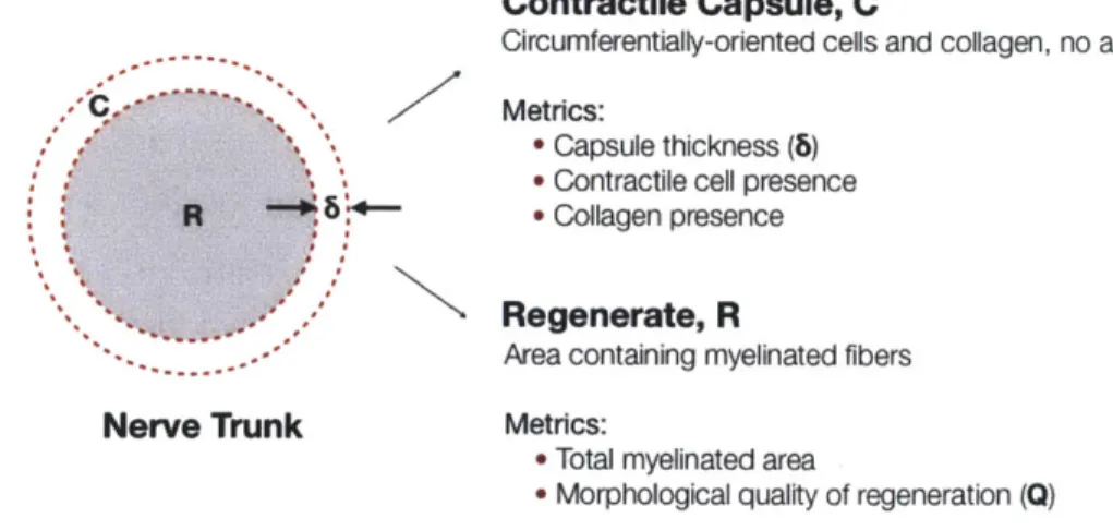
FIG. 2.1 OVERVIEW OF METRICS USED TO EVALUATE CONTRACTION AND QUALITY OF REGENERATION. ... 57
FIG. 2.2 STRUCTURE OF TYPE I COLLAGEN TUBES... 62
FIG. 2.3: IN VITRO MEASUREMENTS OF DEGRADATION RATE OF HOMOLOGOUS SERIES OF COLLAGEN DEVICES IN RESPONSE TO BACTERIAL COLLAGENASE.. ... 63
FIG. 2.6. INTERMEDIATELY CROSS-LINKED COLLAGEN DEVICES YIELD THINNER CAPSULES WITH BOTH DIMINISHED COLLAGEN CONTENT AND FEWER CONTRACTILE CELLS... 67
FIG. 2.7. COLLAGEN TUBES OF INTERMEDIATE DEGRADATION RATE MAXIMIZE TOTAL MYELINATED AREA.. ... 69
FIG. 2.8. MORPHOLOGY OF NERVE FIBERS REGENERATED USING THE HOMOLOGOUS SERIES OF COLLAGEN TUBES. 70 FIG. 2.9. DENSITY OF MYELINATED FIBERS FOR HOMOLOGOUS SERIES OF COLLAGEN TUBES ... 72
FIG. 2.10. TOTAL NUMBER OF MYELINATED NERVE FIBERS FOR HOMOLOGOUS SERIES OF COLLAGEN TUBES... 73
FIG. 2.11. MEAN MYELINATED FIBER DIAMETER FOR HOMOLOGOUS SERIES OF COLLAGEN TUBES... 75
FIG. 2.12. PERCENTAGE OF A-FIBERS FOR HOMOLOGOUS SERIES OF COLLAGEN TUBES... 77
FIG. 2.13. TOTAL NUMBER OF A-FIBERS FOR HOMOLOGOUS SERIES OF COLLAGEN TUBES. ... 78
FIG. 2.14. N-RATIO FOR HOMOLOGOUS SERIES OF COLLAGEN TUBES (MEAN ± SEM) AT 9 WEEKS POST-IM PL A N T A T IO N . ... 7 9 FIG. 2.15. SCATTER PLOT OF TOTAL MYELINATED AREA OF REGENERATE AS A FUNCTION OF CAPSULE THICKNESS 81 FIG. 2.16. SCATTER PLOT OF TOTAL NUMBER OF MYELINATED FIBERS AS A FUNCTION OF CAPSULE THICKNESS... 82
FIG. 2.17. SCATTER PLOT OF A-FIBERS AS A FUNCTION OF CAPSULE THICKNESS. ... 83
FIG. 2.18 INTERMEDIATELY CROSS-LINKED COLLAGEN DEVICES (DEVICE C) DECREASE IN REGENERATE AREA AND CAPSULE THICKNESS MEASURED FROM THE SITE OF TRANSECTION ... ... 86
FIG. 2.19 HIGHLY CROSS-LINKED COLLAGEN DEVICES (DEVICE E) DECREASE DRAMATICALLY IN REGENERATE DIAMETER BUT MAINTAIN RELATIVELY CONSTANT CAPSULE THICKNESS FROM THE SITE OF TRANSECTION... 88
FIG. 2.20. COLLAGEN TUBES OF INTERMEDIATE DEGRADATION RATE DISRUPT CAPSULE FORMATION. ... 90
FIG. 2.22 COLLAGEN TUBES OF INTERMEDIATE DEGRADATION (DEVICE D) APPEAR TO HAVE EXTENSIVE
MYOFIBROBLAST - SCAFFOLD INTERACTIONS AT 9 WEEKS-POST-NEUROTMESIS... 92
FIG. 2.23. SPECULATIVELY, MYOFIBROBLAST-SCAFFOLD INTERACTIONS COINCIDE WITH DECREASED 8 7 DAYS A FTE R IN JU R Y ... 94
FIG. 2.24 THICK CONTRACTILE CAPSULES APPEAR TO COMPRESS NERVE STUMPS SEVEN DAYS AFTER INJURY. ... 95
FIG. 2.25. OBSERVATIONS OF CONTRACTILE CAPSULE REDUCTION BY DEGRADABLE COLLAGEN DEVICES. ... 99
FIG. 3.1: INTRACELLULAR PATHWAYS LEADING TO SYNTHESIS OF a-SMOOTH MUSCLE ACTIN (a-SMA)... 119
FIG. 3.2 A CUSTOM -BUILT SILICONE "T TUBE". ... 123
FIG. 3.4 SECTIONING OF EXPLANTS FOR HISTOLOGICAL ANALYSIS... ... ... 130
FIG. 3.4 IN VIVO VEHICLE DELIVERY OF MINIPUMP VERIFIED WITH 1% METHYLENE BLUE DYE. ... 132
FIG. 3.5. IMMUNOHISTOCHEMICAL DETECTION OF a-SM A... 134
FIG. 3.6. IMMUNOHISTOCHEMICAL STAINING OF ROCK TARGETS. . ... 136
FIG. 3.5: GROSS DIAMETER OF NERVE REGENERATE AT SPECIFIED DISTANCES FROM PROXIMAL SITE OF T R A N SE C T IO N ... 13 8 FIG. 4.1 REGENERATIVELY-ACTIVE SCAFFOLDS IN SKIN AND PERIPHERAL NERVES DISRUPT THE SPONTANEOUS MFB CAPSULE THAT FORMS DURING WOUND HEALING IN BOTH ORGANS... 144
1
Course of the Nerves -Neck and Thorax
This page was left blank intentionally.
Introduction
1.1
A Clinical Need for Improved Peripheral Nerve Regeneration
Several hundred thousand individuals suffer from acute peripheral nervous system (PNS) injuries each year in the Unites States and Europe alone (Wiberg and Terenghi 2003). On the modern battlefield blast-related injuries now account for 65 % of all combat injuries (Affairs 2005). Advances in battlefield wound management have decreased significantly the lethality of war wounds but soldiers frequently present with traumatic wounds of the extremities and severe PNS lesions are prominent (Clark, Bair et al. 2007).
Failure to adequately treat severe injury to the peripheral nerve often results in partial or total paralysis of the affected limb. The practitioners of the field, surgeons and other health care professionals, clearly state that current treatments are inadequate: "Even after optimal surgical repair, functional outcome -especially sensory recovery -is disappointingly poor" (Wiberg and Terenghi 2003).
Neural engineering is an important and active area of research. The last thirty years have brought significant advances to the field including an enhanced pathophysiological understanding of the PNS response to acute injury, improved imaging and microsurgical techniques for diagnosis and treatment, and the clinical entry of the first FDA-approved biodegradable devices for peripheral nerve regeneration. Despite this progress, the traditional surgical methods of end-to-end anastomosis' and autografting2 persist as gold standards in the treatment of severe
peripheral nerve defects over short and long gaps, respectively. These techniques have numerous drawbacks including a limited supply of donor nerves and donor site morbidity (in the case of autografts), surgical complexity, and a relatively poor functional outcome. The clinical need for improved strategies for PNS regeneration, particularly across long gaps, remains.
1.2 Irreversible Injury in Skin and Peripheral Nerves
The complex inflammatory response of the adult mammal to injury is increasingly elucidated by on-going research at the cellular and molecular level. Although the formation of an accurate mechanistic perspective of wound healing is essential both in understanding the effect of current clinical treatment and in the development of emergent therapies, an examination of the macroscopic outcome of healing also provides a uniquely valuable viewpoint. An introductory phenomenological discussion of spontaneous wound healing at the tissue level provides a framework that forms a focus for future discussion of detailed cellular / molecular mechanisms and facilitates the derivation of concepts and rules of induced regeneration that may conceivably apply to almost any organ in the body.
1.2.1. Macroscopic Outcomes of Wound Healing: Repair vs. Regeneration
When exposed to injury, in the form of either acute trauma or chronic insult, the organism mounts a spontaneous wound healing process that typically closes the discontinuity in organ mass caused by the injury in a matter of days. Two macroscopic outcomes to injury have been observed experimentally, regeneration and repair. These fundamentally different processes are clearly distinguished by the identity of tissue present in the final state, i.e., the tissue that has been displaced or synthesized to close the injury. In the early mammalian fetus and in many species of amphibians, wound healing is largely reversible and proceeds via spontaneous regeneration, a process that restores the structure and physiological function through synthesis of the missing organ
2 Auto-grafting: the transplantation of donor nerve from another part of the body to bridge the
structures (Yannas 2001). Certain adult urodeles exhibit an impressive capacity for spontaneous regeneration: replacement of an amputated appendage occurs by direct outgrowth of the severed cross-section (epimorphic regeneration), a reversible process (Goss 1992).
In clear contrast, severe injury to normal adult mammalian tissue typically results in an irreversible healing response. Spontaneous healing of severe skin wounds proceeds via repair, in which the wound closes with a combination of tissue deformation and translation (collectively referred to as contraction) and synthesis of a nonphysiological tissue (scar) in place of the normally functioning tissue that has been injured (1). By replacing the lost organ mass with scar, the injured organ is condemned whereas the organism is spared as a result of the healing process. The immediate consequence of irreversible injury is a loss of normal organ function. On a broader scale skin injury may have additional detrimental effects, such as loss of mobility and lack of social accept- ance following formation of disfiguring scars from burns. It appears that nearly all adult mammalian organs can be injured irreversibly, the extent of irreversibility seems to depend both on the identity of the tissue injured and the severity of the injury (Yannas 2001).
1.2.2. Regenerative Similarity of the Tissue Triad
Standard pathology texts describe three generic tissue types that comprise the majority of organs in the body: epithelia, basement membrane, and stroma (Yannas 2001; (Burkitt H.G. 1993; Martinez-Hernandez 1998); Fig. 1.1). Collectively, we will refer to these three tissue types as the tissue triad. This classification provides a useful framework for comparing the regenerative capacity of specific tissue types from one organ to another. The composition of each member of the triad is markedly different. Epithelial tissue forms a completely cellular covering on every surface, tube, and cavity in the body, performing a wide array of vital functions including protection, secretion, absorption, and filtration. As epithelial tissue is devoid of extracellular matrix (ECM) and blood vessels, it is sustained by the diffusion of nutrients from the underlying vascular connective tissue, or stroma. With the exception of the liver, epithelia is separated from the underlying stroma by the basement membrane (basal lamina), a very thin, noncellular tissue layer, comprising exclusively ECM. The stroma is a
The skin, as one example, consists of the epidermis (epithelia) attached to the basement membrane and the underlying dermis (stroma). Considerable evidence from peripheral nerve studies indicates that Schwann cells function as epithelial cells
bme00
m"m feSkin Perpheral nerve
rwvous and and mwoha Spam8 aefn system smooth MerS heart
FIG. 1.1 THE TISSUE TRIAD STRUCTURE IN SKIN AND PERIPHERAL NERVES. The basement membrane (basal
lamina), a thin noncellular layer consisting of extracellular matrix, separates the cellular, nonvascular epithelia (epidermis, myelin sheath) from the stroma (dermis, endoneurium) which contains cells, ECM, and blood vessels. Epithelia and basement membrane regenerate spontaneously; stroma does not. (Bottom) The tissue triad layers in mammalian anatomy. Stromal tissues include bone, cartilage, and their associated cell types as well as elastin and collagen. Epithelial tissues are those that line the genitourinary, respiratory, and gastrointestinal tracts as well as surfaces of the mesothelial cells in body cavities, and include muscle fibers, fat cells, and endothelial cells in the cardiovascular system. (Yannas 2001).
following synthesis of a completely cellular layer (myelin sheath) around axons. Nerve fibers (Schwann cell-axon units) are attached to a basement membrane that separates them from the outlying endoneurial stroma, a tissue consisting of vascularized ECM. Further evidence for the epithelial nature of the myelin sheath comes from the observed polarity of Schwann cells that is very similar to that of keratinocytes (KC), the epithelial cells that form the epidermis in skin. In each case, one epithelial cell surface is firmly attached to a basement membrane and another is part of the epithelial tissue, endowed in each case with function unique to the respective organ, which characterizes the epidermis (in the case of skin) or the nerve fiber insulation of peripheral nerves (Bunge, Bunge et al. 1989). Tissues that are "regeneratively similar" appear in different organs yet share a common spontaneous healing response, be it regeneration or repair. The spontaneous healing behavior of each layer of the tissue triad in skin and peripheral nerves is well documented and will be briefly reviewed. The response of the peripheral nerve to severe injury will be discussed in more detail later in this chapter.
Partial or full-thickness injury to the epithelial layer of either of the two organs (the epidermis in skin and myelin sheath in peripheral nerves, respectively) results in spontaneous regeneration of the injured tissue by remaining epithelial cells in the defect (provided the stroma is still intact to facilitate epithelial cell spreading) (Yannas 2001; (Haber, Hanna et al. 1985; Ikeda, Oda et al. 1989; Stenn 1992; Fu and Gordon 1997). Following nerve crushing with myelin disruption but with no injury to the endoneurium, the myelin sheath regenerates spontaneously and no contraction is observed. Similarly, epidermal excision is a reversible injury that closes exclusively by spontaneous regeneration rather than contraction. The epidermis in skin and the myelin sheath in peripheral nerves exhibit spontaneous regeneration, a reversible healing response leading to a full recovery of structure and function, and are therefore regeneratively similar (Yannas 2001). Injuries that interrupt the continuity of the basement membrane in both organs also exhibit spontaneous regeneration by epithelial cells; basement membranes are regeneratively similar in the two organs. However, when a wound is severe enough to cause injury to the stroma of either organ (the
dermis in skin or the endoneurial stroma in peripheral nerves), the organism achieves wound closure by a combination of contraction and scar synthesis (irreversible healing response) (Uitto
J.
1996). The dermis and non-neuronal peripheral nervous tissue healby repair; because they are both non-regenerative they are considered to be
regeneratively similar.
In summary, when the spontaneous regenerative capacity of corresponding tissue types in skin and peripheral nerves is directly compared, a useful similarity emerges (Yannas 2001). Epithelia and basement membrane are regeneratively similar tissue layers, exhibiting a reversible healing response even in the case of severe injury. Likewise, the stroma in both organs is distinctly non-regenerative. Hence, the central objective of induced organ regeneration is synthesis of the non-regenerative stroma.
1.3 Experimental Considerations
1.3.1 Importance of an Anatomically Well-Defined Defect
The appropriate experimental volume for studies of induced organ regeneration is the anatomically well-defined defect (Yannas 2001). As discussed earlier of the differential regenerative capacity of the various tissue triad layers calls for an experimental injury that is free of non-regenerative tissue. In this manner, the effects of an exogenous regenerative agent on the potential synthesis of non-regenerative tissue can be evaluated without ambiguity.
In addition, the experimental volume should also have well-defined anatomical boundaries to reduce contributions from extraneous healing processes occurring elsewhere in the organ (e.g., caused by collateral damage during the surgical procedure) and to improve the reproducibility of the surgical protocol from one animal to the next as well as between independent laboratories. The treatment of the defect should include prevention of loss of extravascular tissue fluid (exudate), which contains important growth factors and regulators that are crucial both to regeneration and to repair. Inability to prevent exudate loss from the injured site radically affects the outcome of both spontaneous and induced healing processes in both skin and peripheral nerves (Winter 1972) (de Medinaceli, Wyatt et al. 1983) (Terzis 1987) Physical containment is also necessary to prevent detrimental extraneous processes, such as bacterial infection in skin, from interfering with the outcome of the healing response.
For studies of induced regeneration in skin, the most widely used well-defined defect is the dermis-free full thickness wound in the rodent or swine. In the case of
peripheral nerves, the tubulated, fully-transected peripheral nerve in the rat or mouse has been studied extensively (Yannas 2001). Both the introduction of various grafts or sheet-like covers to skin defects and tubulation to transected nerves using a variety of materials typically impart significant activity that either assists or hinders regeneration; their use must be controlled carefully.
1.3.2 Synthetic Protocol: In Vitro or In Vivo?
A detailed comparison of the synthetic regeneration processes carried out in
vitro and in vivo shows that in studies of skin and peripheral nerves, various protocols for in vitro synthesis have so far resulted largely in the formation of epithelia and the associated basement membrane but not the physiological stroma. In contrast, several protocols conducted in vivo have yielded not only the physiological epithelia and basement membrane, but a near-physiological stroma as well. The following section highlights these observed cases of induced regeneration.
1.4 Overview of Induced Organ Regeneration
1.4.1. Empirical Evidence
Studies that started in the early 1970s in the Fibers and Polymers Laboratory at Massachusetts Institute of Technology have shown that the adult mammal can be induced to regenerate selected organs that have been accidentally lost or excised. In every case it had been established previously that the excised adult organ in question does not regenerate spontaneously; that is, in the absence of experimental intervention, the adult excised site generally closed spontaneously by contraction and scar formation rather than by regeneration. The organs in question were induced to regenerate partially with the aid of certain insoluble substrates (scaffolds) that were optionally seeded with cells.
The most extensive data on induced organ regeneration are available with skin and peripheral nerves (see ref. 1 for a detailed review). Data with other organs from the work of several investigators were presented in a recent volume (Yannas 2005). We review below the induced organ regeneration data obtained in our laboratory.
The three anatomical sites which were induced to regenerate partially were: 1) full-thickness skin wounds, with epidermis and dermis completely excised, in the adult guinea pig, adult swine and adult human; 2) full-thickness excision of the conjunctiva, with complete excision of the stroma, in the adult rabbit; 3) the fully transected rat sciatic nerve, with stumps initially separated by a gap of 15 mm (later 22 mm and recently 30 mm). A summary of induced regeneration data for the constitutive tissues of each organ is presented in Table 1.
Table 1
Constitutive Tissues of Skin, Peripheral Nerves, and Conjunctiva That Were Induced to Regenerate in Adults
Regeneration Rcgeneratio Regencration
Organ observd observed not studied
Skin (guinea pig, swine, Keratnizod pidcrmis, Apcodages human) (I) baemenE membrane, (e.g.. haix
deriii ncn-e fbff1icms swcTat
endings, blood S]ands)
vcsscLs
Periphcral nerve Myclin sheath, ncr-c Endoneuria]
(mousc,. rat, cat, fibers (large and sroma7
monkey, human) small diarncter). perincuriun bKood epirs.
cndoncurial stromra?
Conijunctiva (rabbit') Epir~belia, conjuncti1 Basemcnt
seromna membrane
TABLE 1.1 CONSTITUTIVE TISSUES OF SKIN, PERIPHERAL NERVES, AND CONJUNCTIVA THAT WERE INDUCED TO
REGENERATE IN ADULTS. (Adapted from Yaas, 2001).
Observations of induced regeneration in adults made over the years have been tested repeatedly by morphological and functional tests, as follows: (A) confirmation of partial regeneration of skin (including both a dermis and an epidermis but lacking skin organelles) was made by histological, immunohistochemnical, ultrastructural and functional studies (Burke, Yannas et al. 1981; Yannas, Burke et al.
1981; Yannas, Burke et al. 1982; Yannas, Lee et al. 1989; Murphy, Orgill et al. 1990;
Compton, Butler et al. 1998; Butler, Yannas et al. 1999); (B) confirmation of regeneration of the conjunctiva (including the conjunctival stroma) was made using histological data
(Hsu, Spilker et al. 2000); (C) confirmation of regeneration of peripheral nerves was made using both morphological and functional (electrophysiological and neurological) data (Yannas 1985 ; Yannas
I.V.
1987; Chang A. 1990; Chang 1992; Chamberlain, Yannaset al. 1998; Chamberlain, Yannas et al. 1998; Chamberlain, Yannas et al. 2000; Chamberlain, Yannas et al. 2000; Spilker 2000).
The available evidence in the above studies strongly supports the conclusion that these severely injured anatomical sites did not close by contraction and scar formation.
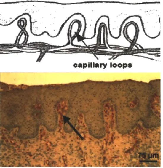
Nevertheless, induced regeneration observed to date is described as 'partial' since perfectly physiological organs have not yet been regenerated. Regenerated skin was histologically and functionally different from scar and identical to physiological skin in almost all respects, including a physiological epidermis, well-formed basement membrane, well-formed capillary loops at the rete ridges of the dermal-epidermal junction, nerve endings with confirmed tactile and heat-cold feeling, and a physiological dermis; however, the regenerate lacked certain organelles (hair follicles, sweat glands, etc.). Evidence for the induced regeneration of partial skin is presented in Fig. 1.2. The supportive data for induced regeneration of peripheral nerves is
opillory loops
FIG. 1.2. EVIDENCE FOR INDUCED REGENERATION OF SKIN. The dermal-epidermal junction for normal skin is represented schematically (top) and contrasted with that of partially regenerated skin in the swine, following grafting with the keratinocyte-seeded dermal regeneration template scaffold (bottom). The new skin is not scar, as evidenced by the presence of rete ridges and capillary loops inside the ridges. (Top, from Burkitt HG, Young B, Heath JW. Wheater's Functional Histology. Edinburgh, Scotland: Churchill Livingstone; 1993. Bottom, from Compton, Butler et al, J Invest Dermatol. 1998).
99
06
0 .9 d"0
01 8 5 A A- ber ask IgkuurMse i Naem A, Peak -IthurFIG. 1.3. EVIDENCE FOR INDUCED REGENERATION OF PERIPHERAL NERVES. Histological micrographs of nerve tissue postfixed with osmium tetroxide and stained with toluidine blue, scale bars, 10 pim. (a) Tissue regenerated through the midportion of a matrix-filled large-pore collagen (LC /M) implant at 30 weeks. Note the large number of axons in this cross-section with the majority of axons being small in diameter. The largest axons have diameters of approximately 7 pim. Many Schwann cells are visible with some actively participating in myelination. The blood vessel that is visible in this micrograph was characteristic of the caliber of most vessels present in the regenerated tissue. (b) Tissue regenerated through the midportion of a LC /M implant at 60 weeks. Compared to 30 weeks, the axons are much larger (diameters up to 12 pim) and have thicker myelin sheaths. Also, fewer small diameter axons are visible. Few nonmyelinating Schwann cells are visible at 60 weeks. (c) Normal nerve tissue from the level of the lesion is shown as a control. Note the number of large diameter fibers and the thickness of the myelin sheaths compared to the regenerated nerves. (d) Typical oscilloscope tracings of A-fiber and B-fiber com- pound nerve action potentials for normal sciatic nerve and nerve regenerated through a LC /M implant at 60 weeks postimplantation. The A-fiber peak for the regenerated nerve has a significantly smaller amplitude than the normal nerve control. This was typical of all regenerated groups. In contrast, the conduction velocity of the regenerated nerve, although significantly slower than normal, was approaching normal values. The latency is measured along the x-axis from the stimulus to the peak and then combined with the constant distance between electrodes to determine conduction velocity. The dashed line indicating the B-fiber peak has been added on to the tracing for reference. Note that the normal nerve tracing has no visible B-fiber peak. In the regenerated nerves, the B-fiber peak was similar and visible in all groups. Adapted from Chamberlain et al, 2000.
1.4.2. The Defect Closure Rule
Careful review of the literature suggests that not more than three distinct processes are used to close an anatomically well-defined defect (dermis-free defect) in skin wounds, contraction originating from the edges of the defect, scar formation by stromal fibroblasts (followed by epithelialization of scar), and regeneration.
Continuous kinetic data are rarely available in regeneration experiments and often difficult to compare from one study to another. One approach to studying the regenerative activity of exogenous agents on the healing process is to establish two standardized configuration states and to evaluate the total change that is caused during this fixed period in the healing process. In the absence of kinetic data, the defect closure rule bridges the gap by presenting a quantitative
description of the healing process through comparison of snapshots of the initial and final stages of wounding. The initial state of configuration is the anatomical description of the recently generated defect, characterized by the loss of structural continuity in one or more tissues, the beginning of exudate flow, and the loss of physiological homeostatic control of the organ. As defect healing progresses, the original area, AO, eventually diminishes spontaneously because of one or more of the three processes mentioned above. The area of the closed defect (the closed wound) comprises tissues that result either from contraction (fractional amount, %C), scar formation (%S), or regeneration (%R). The configuration of the final state can be described by the following simple relation:
C + S + R =100 (1)
Eq. 1 states that the defect closure in any organ can be described by only three
outcomes: contraction, scar formation (neuroma or fibrosis), and regeneration (partial or total).
For the idealized case of early fetal wound healing (spontaneous regeneration), con- traction and scarring is absent (C, S = 0) and
R = 100 (regeneration)
(repair), regeneration is absent (R = 0) and
C + S = 100 (repair)
The literature describes several assays to determine the configuration of the final state (recently closed defect) (1). Functional assays can be used to qualitatively identify the physiological nature of the tissue and assist in providing a quantitative measure of its incidence in the final state in terms of the numerical values of these three quantities
(C, S, or R?). The defect closure rule may be interpreted as a conservation principle:
provided that the magnitude of two individual terms (C and S for example) has been determined, the magnitude of the remaining process may be calculated. Defect closure data are expressed using the following convention: [%C, %S, %R].
The defect closure rule is useful in evaluating the activity of unknown reactants as inductive agents of regeneration. This quantitative description of the structure and function of the injured organ at its final state has shed interesting light on the relationship between the characteristic elements of the adult healing response
(contraction or scar synthesis, or both) and regeneration.
1.4.3 Prevalence of Contraction During Spontaneous Healing
In the skin, the defect closure rule has been used to approximate data on the configuration of the final state following spontaneous healing of the anatomically well-defined defect (dermis-free defect) in several species. In all cases, it was ensured that the contribution of regeneration to defect closure was negligible (R = 0). Skin contraction
was measured directly as the reduction in initial wound surface area by inward (centripetal) movement of skin from the margins of the wound. Scar formation was studied qualitatively by histology or quantitatively by use of laser light scattering (to measure the average degree of collagen fiber orientation). Values for the percentage of initial defect area closed by epithelialized scar (S) were determined using the simplified
defect closure rule for repair (S = 100 - C).
The contribution of the various methods of defect closure in anatomically well-defined defects is species dependent. In rodents, in which the integument is mobile, contraction is by far the main engine of closure of skin wounds, whereas scar formation has been shown to be quantitatively much less important.
charac- teristic of several rodents and lagomorphs (rabbits), and results in the following final state configuration: [91, 9, 0] (Yannas, Burke et al 1982). In general, C >> S and defect closure for adult rodents and rabbits reduces to C approx 100. Alternatively, the approximation of the final state is expressed as [100, 0, 0].
Human integument is tethered more securely onto subcutaneous tissues and contraction and scar formation contribute approximately equally to wound closure. Experimentally, the spontaneous healing of full-thickness skin defects in the humans (R
0) results in a final state represented by [37, 63, 0] (Ramirez A.T. 1969).
In the absence of direct quantitative observations, histological analysis was used to describe the closure of the fully transected peripheral nerve in the adult rat. Spontaneous healing results in reduction of the initial area of cross-sections of nerve trunks by 95% with neuroma formation (neural scar) accounting for the remaining 5%. The resulting estimation of the final state configuration is [95, 5, 0] (Chamberlain, Yannas et al, 2000).
The contraction of a wide array of organs in response to trauma is well documented in both animals and humans, yet these reports are almost exclusively of a qualitative nature (Holmes and Z. 1942; Weiss 1944; Weiss 1944; Kiviat 1973; Peacock Jr 1984; Dahners 1986; Wilson 1988; Sunderland 1990; Rudolph 1992; Krishnan K.G. 2000; Levine D. 2000; Cornelissen A.M. 2000 ; Delaere P.R. 2001; Zeinoun T. 2001 ; Ivarsen A.
2003; Bulut T. 2004; Wong T.T.L. 2004; Chou T.D. 2004 ; Oppenheimer and Hinman 1955; Unterhauser FN 2004; Schmidt M.R. (2004) ). With very few exceptions the sole
organ in which contraction has been studied systematically to date is the skin. Despite the dearth of widespread quantitative data, the prevalence of contraction must not be overlooked; it appears to be a critical outcome of the spontaneous healing response throughout the adult organism.
1.4.4 Antagonistic Relation Between Contraction and Regeneration
The characteristic elements of the adult healing response (contraction or scar synthesis, or both) must be controlled in order for induced regeneration to occur. Extensive data, including empirical data on the final state of the defect in response to various reactants, suggest that during healing of a severe injury, contraction antagonizes regeneration (Yannas 2001).
stroma was accompanied in each case by direct observation of a significant reduction in contraction as a mode of defect closure. Conjunctival and peripheral nerve regeneration studies were guided by earlier studies of skin regeneration. Partial skin was first induced to regenerate in the adult guinea pig. The spontaneous healing behavior of the untreated dermis-free defect in this organism resulted in a final configuration of [91, 9,
0]. Grafting an identical well-defined skin defect with a highly porous polymer of type-I
collagen and chondroitin 6-sulfate (referred to as a DRT) (Fig. 1.4A), abolished scar synthesis and led to the regeneration of a small mass of dermis and subsequent synthesis of an overlying epidermis within the defect. In the context of the defect closure rule, the regenerative activity of the DRT on the configuration of the final state was as follows:
[92, 8, 0] -- [89, 0, 11] (DRT).
In addition, the DRT led to a significant delay in wound contraction over 25 days (40,41).
When a DRT seeded with KC was grafted into an identical defect, the result was more pronounced (Fig 1.4B-D):
[91, 9, 0] -- [28, 0, 72] (DRT + KC).
KC-seeded DRTs accomplished rapid wound closure through partial regeneration of skin (simultaneous synthesis of a physiological dermis and epidermis, described earlier), and completely arrested contraction at 35-40 days (Yannas, Burke et al. 1982).
The cell-free DRT that induces partial skin regeneration comprises the regenerative component of the two-layer device (Integra DRT@) approved by the Food and Drug Administration (FDA) for restoration of a physiological epidermis and dermis in patients suffering from severe burns as well as those undergoing plastic and reconstructive surgery of the skin, as described in an early study (Burkey, Yannas et al,
1981) and reviewed recently (Yannas, Orgill et al. 2011). Growth factors (Greenhalgh,
H. et al. 1990; Puolakkainen, D.R. et al. 1995), epidermal cell suspensions, and cell sheets (Billingham and
J.
1952) exhibited negligible regenerative activity when added tofull-thickness skin wounds in other rodent models. These reactants did not significantly alter the configuration of the final state or the extent of contraction delay. Similarly, a
number of synthetic polymer scaffolds (Cooper, F. et al. 1991 ; Hansbrough, L. et al.
1993) failed to induce physiological dermis (or skin) regeneration. These observations
focus attention on the mechanism of scaffold regenerative activity, to be discussed later.
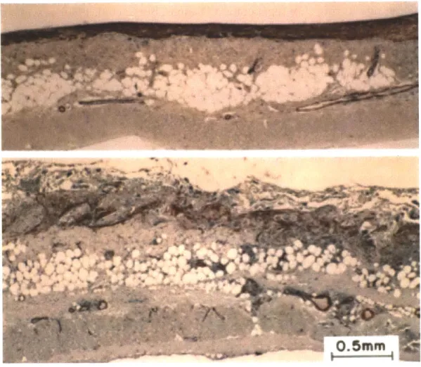
FIG. 1.4. INDUCED REGENERATION OF SKIN COINCIDES WITH SIGNIFICANT REDUCTION OF CONTRACTION AS A MODE OF WOUND CLOSURE IN THE ADULT GUINEA PIG. a) SEM micrograph of an active
collagen-glycosaminoglycan (CG) scaffold (also known as dermal regeneration template or DRT). b) A full-thickness (dermis-free) skin wound is grafted with CG scaffold and seeded with keratinocytes (KC). c) An inactive KC-seeded scaffold leads to contraction and scar formation. d) An active scaffold (dermal regeneration template or DRT) seeded with KC alters the defect closure by blocking macroscopic contraction dramatically and leads to simultaneous synthesis of epidermis and dermis. The new tissue formed is skin, not scar. SEM micrograph: E. Soller, 2008. B-D adapted from Yannas et al, 1989 PNAS.
in the adult rat. The spontaneous healing behavior of the untreated transected peripheral nerve in this organism resulted in a final configuration that was estimated using histological analysis to be [95, 5, 0]. Insertion of the fully transected nerve stumps into a silicone tube filled with a collagen-based regeneration template (referred to as a Nerve Regeneration Template or NRT) resulted in reduced contraction (as determined
by histological analysis of cross-sectional areas of regenerates) and partial regeneration
over an unprecedented gap length (Chamberlain, Yannas et al, 2000; Chamberlain, Yannas et al, 2000). Contraction was abolished and the quality of regeneration improved significantly when the NRT was used in con- junction with a degradable collagen tube. In the context of the defect closure rule, the regenerative activity of the NRT in each experimental configuration can be evaluated by inspecting the characteristics of the final state, as follows (the arrow indicates the change observed following use of the scaffold):
[95, 5, 0] - [53, 0, 47] (NRT in silicone tube),
[95, 5, 0] - [0, 0, 100] (NRT in collagen tube).
The relative importance of each method of defect closure (C, S, and R) changes during animal development. A sharp change occurs during the fetal-adult transition in mammals (roughly during the third trimester of gestation), in which contraction replaces regeneration as the dominant method of closure (Mast, J.M. et al. 1992; Lorenz and Adzick 1993 ; Martin 1996). Similarly, as amphibian development progresses, contraction becomes a more prominent method of wound closure, as regeneration recedes and scar formation becomes more evident (Stocum 1995; Yannas,
J.
et al. 1996).Although scar has been widely considered the key barrier to regeneration in adults, quantitative study reveals that contraction is the dominant mode of spontaneous closure in skin and peripheral nerve defects. Studies of induced regeneration in skin, peripheral nerves using analogs of the ECM indicate that scar formation is a process that is secondary to contraction: in studies of induced regeneration in these organs, when contraction was even slightly inhibited, scar formation was totally abolished (Yannas 2001).
[92, 8, 0] -> [89, 0, 11] (DRT)
[91, 9, 0] -+ [28, 0, 72] (skin, DRT + KC)
[95, 5, 0] -> [53, 0, 47] (peripheral nerve, NRT in silicone tube),
[95, 5, 0] -> [0, 0, 100] (peripheral nerve, NRT in collagen tube).
Suppression of contraction in certain cases of impaired healing, for example, following use of pharmacological agents, such as steroids, or application of devices, for example, use of mechanical splints or full-thickness skin grafts, was not accompanied
by regeneration, indicating that suppression of contraction alone did not suffice to
induce regeneration (Yannas 2001).
The available evidence supports the theory that selectively suppressed contraction in adult defects is required, but not sufficient to induce regeneration of skin and peripheral nerves. This can be expressed in the context of the defect closure (Eq. 1) rule
as follows:
AR>0 and S-*0 if AC<0 (2)
This condition describes an antagonistic relationship between contraction and regeneration in the closure of a defect. It suggests that successful induced regeneration strategies consist of reactants that block contraction without blocking other aspects of the healing process.
1.4.5. Repair: Mechanism of Contraction
Similarities in the mechanistic hypotheses for inducing regeneration of skin and peripheral nerves originate in their common response to irreversible injury. Both organs spontaneously respond to injury by recruiting contractile cells that, if not properly sup-pressed, drive closure of the defect by contraction and scar synthesis rather than by regeneration. Contraction of skin defects starts from a cell cluster at the edge of the defect and later extends across the entire defect area. In peripheral nerves, contraction primarily results from the activity of a circumstantial sheath of contractile cells.
The well-documented, macroscopic contraction that drives the closure of skin defects finds its origin at the cellular scale, arising from the individual contribution of contractile forces generated by differentiated myofibroblasts (MFB) (Rudolph, Abraham et al. 1978; Frangos 1993; Racine-Samson, Rockey et al. 1997; Freyman, I.V. et al. 2001; Freyman, Yannas et al. 2001; Desmouliere, Chaponnier et al. 2005) (75,77-83). The
current consensus is that MFBs present in the granulation tissue following skin wounding derive directly from fibroblasts and comprise an intermediate, contractile, cellular phenotype between the fibroblast and the smooth muscle cell (Zeinoun, Nammour et al, 2001). There is also evidence that undifferentiated fibroblasts may contribute to macroscopic contraction by applying traction to the ECM very soon after coming into contact with it (Dahners, Banes et al 1986; (Davison, McCaffrey et al. 1999; Ehrlich, Keefer et al. 1999; Ehrlich, Gabbiani et al. 2002) In response to external tension, fibroblasts exert sustained isometric force on their surrounding environment via a rho/ rho-kinase-mediated, actomyosin contractile apparatus (Kimura, Ito et al.
1996; Amano, Chihara et al. 1997; Hall 1998; Tomasek, Vaughan et al. 2006). This
three-dimensional, transcellular structure consists of bundles of actin and nonmuscle myosin microfilaments called "stress fibers." Of the many ultrastructural and biochemical
factors that distinguish MFBs from their fibroblast precursors, the most useful operational distinction of MFB differentiation is expression of the a-smooth muscle actin (SMA) phenotype (Desmouliere, Chaponnier et al, 2005). Stress fibers of immature MFBs (called proto-MFBs) contain only
p-
and y-cytoplasmic actins. Additionally, differentiated MFBs exhibit stress fibers typically arranged parallel to the long axis of the cell, nuclei which consistently show multiple indentations or deep folds, and two cell-matrix adhesion macromolecules (vinculin and fibronectin) (Serini, Bochaton-Piallat et al. 1998; Dugina, Fontao et al. 2001)Simplistically, the myofibroblast differentiation process can be described as a positive feedback loop that requires the concurrent action of at least three factors: the cytokine transforming growth factor (TGF)-
P1,
the presence of mechanical tension, and the ED-A splice variant of cellular fibronectin (an ECM component) (Fig. 1.5). Fibroblasts respond to the development of mechanical tension by upregulating TGF-P1
production and expressing the a-SMA isoform; in turn, a-SMA expression strengthens the contractile apparatus and increases tension development (Tomasek, Gabbiani et al. 2002)Desmouliere, Chaponnier et al, 2005). Recent work suggests that mature myofibroblasts in skin granulation tissue link their cytoskeletons together using cadherin proteins, which allow them to generate even higher levels of force to drive wound closure (Hinz, Pittet et al. 2004).Foci adhesn sio e
M)A8 Cortica cyopasmic actins
Cytopbsmoc actins
- Smxooh muscle ac
M-a ED-A ntrsec
TG 4
ED-A fi
Mechanical tension
Pfot-ny ft%@mbmla0 Stk
Nuw Reviews IMoeculr Cei Bology
FIG. 1.5. THE TWO-STAGE MODEL OF MYOFIBROBLAST DIFFERENTIATION. Under normal conditions in vivo
fibroblasts have a quiescent phenotype and do not express stress fibers or focal adhesions with the extracellular matrix (ECM). Mechanical tension prompts differentiation into proto-myofibroblasts, which form cytoplasmic actin-containing stress fibers terminating in fibronexus adhesion complexes. Proto-myofibroblasts express the ED-A splice variant of cellular fibronectin and are capable of generating contractile force.Transforming growth factor beta-1(TGF-p1)increases the expression of ED-A fibronectin. Both factors, in the presence of mechanical stress, promote the modulation of proto-myofibroblasts into differentiated myofibroblasts that are characterized by de novo expression of the contractile filament protein a-smooth muscle actin (a-SMA) in more extensively developed stress fibers and by large fibronexus adhesion complexes in vivo or supermature focal adhesions in vitro. Functionally, differentiated myofibroblasts generate greater contractile force than proto-myofibroblasts and exhibit a higher level of organization of extracellular fibronectin into fibrils. Reproduced from Tomasek, Gabbiani et al. 2002.
1.4.6. Structural Determinants of Scaffold Regenerative Activity
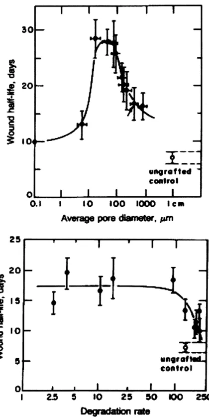
Scaffolds that induce regeneration of partial skin possess a highly specific structure that is distinctly different in pore structure and degradation rate from scaffolds that regenerate peripheral nerves (Yannas, 2001). The nature and duration of
the contractile response as well as the structure of the two organs differ greatly as do the values for several of the structural parameters of the early scaffolds that were used to control contraction and induce regeneration in each organ. The scaffolds have a common ligand identity due to an identical chemical composition (type I collagen
/
GAG, 98 / 2 w / w ratio) yet they differ in average pore diameter (higher in the case of the DRT), the pore channel orientation (axial for the nerve guide, random for the DRT), and degradation rate (a higher average molecular weight between cross-links, Mc[kDa] in the nerve guide leads to faster degradation).
In skin wounds, the mechanism of induced regeneration has been elucidated through careful modulation of the DRT's structural properties that impart contraction blocking activity. DRTs that actively block contraction in skin wounds (and induce
regeneration) have structural properties that seem to accomplish three main processes:
1) reduction in MFB number present in the wound, possibly due to inhibition of TGF-p
synthesis, leading to downregulation of myofibroblast recruitment; 2) blocking orientation of myofibroblast (MFB) axes in the plane of the defect where macroscopic contraction is observed and 3) ensuring that DRT degradation time is sufficiently long to ensure that contraction blocking persists for the duration of the interim myofibroblast contractile response but not so long as to interfere with key regenerative processes.
1) Apparent downregulation of TGF-3 synthesis. The quaternary structure of
collagen fibers is a requirement for the aggregation of platelets, an early component of the wound response. Platelet aggregation initiates a cascade of events that include the release of the cytokine TGF-p1, one of the main inductors of the myofibroblast phenotype. Collagen fibers in the DRT maintain their tertiary (triple helical) structure but are practically free of banding (due to treatment with acetic acid during scaffold preparation). Collagen that has been treated in this manner during scaffold preparation loses its quaternary structure, although it maintains its triple helical structure, and has been shown not to aggregate plaetelets in vitro (Sylvester et a., 1989). DRT apparently, therefore, disrupts platelet aggregation that normally takes place in contact with native collagen fibers within the defect, reducing production of TGF-s1, and the recruitment of contractile myofibroblasts to the wound site (Yannas 1997).
2) Blocking orientation of MFB axes in the plane of the wound as well as MFB-MFB binding. Contraction of wound edges appears to require orientation of MFB axes in the
plane of the wound as well as MFB-MFB binding and MFB-ECM binding. MFB binding on the extensive surface of the highly porous 3D scaffold inhibits such orientation as well as inhibiting MFB-MFB and MFB-ECM binding. It is suggested that these mechanisms are additionally responsible for contraction blocking by the scaffold. According to this suggested mechanism contraction blocking requires extensive MFB binding onto a sufficiently large scaffold surface, which must take place via specific integrin-ligand interactions. Fibroblasts normally bind onto a specific GFOGER ligand on a collagen surface via the clpl and a2p1 integrins (Knight 2000 ). When other structural properties are held constant, the ligand density of a scaffold increases with decreasing average pore size (since the specific surface area of the scaffold available for attachment is thereby increased). An appropriately high ligand density appears to be necessary to disrupt extensive MFB-ECM binding responsible for the onset of macroscopic contraction in skin wounds (Fig. 1.6).
When myofibroblasts bind to specific DRT integrins that are distributed evenly in a three-dimensional, interconnecting porous network, the axes of the MFB contractile apparatus becomes disoriented. At the cellular level, the randomized configuration of the preferential contractile axes that individual myofibroblasts adopt in the presence of DRT leads to approximate cancellation of the macroscopic mechanical forces that lead to two-dimensional contraction and scar synthesis in ungrafted skin wounds. When the pore diameter of DRT is increased much beyond the level of 120 ym, the contraction-blocking activity of the scaffold is lost, probably because the effective DRT ligand density drops to a value that does not provide sufficient binding of myofibroblasts. (Yannas 2001; Yannas, Lee et al, 1989, Fig. 1.7). Similarly, a minimal average pore size exists that is necessary to ensure MFB migration inside the scaffold in order to enable MFB binding on the scaffold surface. According to this interpretation if the pore size is too small, MFB does not infiltrate the scaffold, MFB-DRT ligand bonds do not form, and MFB contractile activity is not cancelled (Yannas, Lee et al, 1989). Experimentally, the
highly planar orientation of myofibroblast axes that is characteristic of the spontaneous
contractile response in ungrafted skin wounds is negligible in the presence of DRT (Troxel 1994).
3) Duration of DRT in an undegraded state over the entire contraction process. It is