HAL Id: inserm-00590412
https://www.hal.inserm.fr/inserm-00590412
Submitted on 3 May 2011
HAL is a multi-disciplinary open access
archive for the deposit and dissemination of
sci-entific research documents, whether they are
pub-lished or not. The documents may come from
L’archive ouverte pluridisciplinaire HAL, est
destinée au dépôt et à la diffusion de documents
scientifiques de niveau recherche, publiés ou non,
émanant des établissements d’enseignement et de
Automated DTI analysis of MS lesions and their
contralateral regions of interest using the mid-sagittal
plane as a reference
Nicolas Wiest-Daesslé, Sylvain Prima, Sean Patrick Morrissey, Christian
Barillot
To cite this version:
Nicolas Wiest-Daesslé, Sylvain Prima, Sean Patrick Morrissey, Christian Barillot. Automated DTI
analysis of MS lesions and their contralateral regions of interest using the mid-sagittal plane as a
ref-erence. MICCAI workshop on Medical Image Analysis on Multiple Sclerosis (validation and
method-ological issues) (MIAMS’2008), Sep 2008, New York, United States. pp.51-59. �inserm-00590412�
Automated DTI analysis of MS lesions and their
contralateral regions of interest using the
mid-sagittal plane as a reference
Nicolas Wiest-Daessl´e, Sylvain Prima, Sean Patrick Morrissey, Christian Barillot
Unit/Project VisAGeS U746, INSERM - INRIA - CNRS - Univ-Rennes 1, IRISA campus Beaulieu 35042 Rennes Cedex, France
{nwiestda,sprima,pcoupe,spmorris,cbarillo}@irisa.fr, http://www.irisa.fr/visages
Abstract. Diffusion tensor MRI (DT-MRI) allows the in vivo assess-ment of the abnormalities of white matter in multiple sclerosis (MS). DT-MRI is complementary to conventional MRI sequences where such abnormalities are often not visible. Most studies have shown differences of mean diffusivity (MD) and fractional anisotropy (FA) between patients and controls in MS lesions (MSL) and normal appearing white matter (NAWM) based on histogram analyses. However, the majority of these studies are based on histogram analysis, i.e. local information of DT-MRI is lost, and moreover a number of those studies were not conclusive, partly explained by methodological issues, because these tensor indices vary within the brain, which is likely to make such global, histogram-based analyses, fail. Here we propose a new framework to compare these indices between MSL and NAWM and between two populations (patients and controls). First, MSL are manually delineated in MS patients. The mid-sagittal plane is then automatically computed, allowing to define a contralateral region of interest (ROI) in NAWM for each MSL. This al-lows the local comparison of DTI indices in anatomically similar regions in each MS patient. Second, each MS patient is linearly registered to each control subject, and the same left-right comparison between MSL and contralateral NAWM is then performed in controls. The results (ANOVA with multiple comparisons procedure) show that 1) FA values are lower in MSL than in contralateral NAWM in MS patients (p < 0.05) but not in controls, 2) FA values are lower in MS patients (MSL and contralateral NAWM) compared to controls (p < 0.05), 3) MD values are not differ-ent between MSL/contralateral NAWM in MS patidiffer-ents and controls. We also show that combining different preprocessing methods (3 estimation methods and 3 distortion correction methods) has little impact on such results. Nevertheless, our fully automated approach is superior to man-ual or semi-automated DT-MRI analyses regarding the robustness of the results (reproducibility and accuracy).
1
Introduction
Since its description in 1986 by Le Bihan et al. [1], diffusion-weighted MRI has gained increasing attention in the neuroimaging community. DW-MRI allows to measure non-invasively the water molecular self-diffusion in biologic tissues. The movement of water molecules is strongly related to the underlying anatom-ical structure and allows a biophysanatom-ical characterisation of the tissue organisa-tion. This information is especially relevant for fibres where the water molecular movement is orientation-dependent due to microscopic barriers, such as muscles, ligaments, tendons, or fibre bundles composing the white matter of the central nervous system (CNS). DW-MRI allows the study of the normal and pathological brain, as it provides a unique insight into the microscopic physiological phenom-ena occurring in living tissues. A particularly simple way to exploit DW-MRI data has been introduced by Basser et al. [2] in 1994, termed diffusion-tensor magnetic resonance imaging (DT-MRI). In MS, DT-MRI findings correlated with qualitative characteristics of MSL using conventional MRI sequences, but in contrast to conventional MRI, DT-MRI conveys at the same time biophysi-cal and quantitative properties. In patients with MS, important and significant DW-MRI findings were reported with regard to focal (MSL) and diffuse (normal appearing white matter (NAWM) and normal appearing grey matter (NAGM)) pathology both in the brain and the spinal cord. Studies were performed with histogram analysis of the diffusion characteristics (ADC/FA/MD) in the whole brain or large parts of it [3–6]. Analysis of semi-automatically delineated re-gions of interest (ROI) were performed both in MSL and the NAWM [7–16]. Overall, in MSL, but also in the NAWM and NAGM, increased values of MD and decreased values of FA/RA were reported. Even if DT-MRI in MS conveys more detailed information about tissue damage than conventional MRI studies it should be kept in mind that DT-MRI shows a good sensitivity to detect diffu-sion abnormalities and has the potential to exhibit de- or re- myelinisation effect . On the other hand DW-MRI lacks specificity to distinguish between changes in membrane permeability, tissue integrity, gliosis, inflammation or axonal loss. Heterogeneous results of diffusion imaging in MS lesions have been explained by lesion heterogeneity, basically in terms of lesion age, degree of tissue loss and presence or absence of active inflammation on conventional MRI (i.e. Gd-enhancement) [7–13, 17]. Correlations between diffusion measures and clinical scales have been rather disappointing, and their correlation is at best moder-ate [5, 6, 13, 17–21]. Furthermore, DW-MRI studies in MS suggest that focal MD or FA changes do not correlate with brain atrophy measurements [22, 23], and moderate correlations were found with ROI histogram analysis [4].
In this paper, we propose to compare three automated methods of diffusion tensor estimation, and also three automated image distortion corrections for the processing of MR Diffusion Tensor Images (DTI) in patients with multiple sclerosis (MS). Here we propose automated tools for the exact comparison of DTI invariants (fractional anisotropy (FA) and mean diffusivity (MD)) between lesions and their contralateral regions of interest (ROI) in the normal appearing white matter (NAWM).
2
Methods
A lot of different methods for the estimation of tensors have been proposed in the past few years, but none has been evaluated in a pathological context. As of today most of the studies involving MS and DTI were conducted using standard least squares (LS) estimation of the tensors. This classical method has been shown to have more variation in both trace and orientation of the tensors than the weighted least squares (wLS) or constrained non linear least squares (CNLS) methods [24]. Another very important pre-processing is the correction of eddy current distortion [25], and is often either not performed or done using a very simple model. In this paper we compare the effects of distortion correction using linear, polynomial second order and polynomial third order transformations, and no distortion correction on DTI invariants of MSL and their contralateral counterparts. The contralateral ROIs are automatically computed using the mid-sagittal plane as a reference [26]. The comparison was performed using ANOVA and multiple comparison procedure in two main groups : 1) MSL and 2) contralateral ROI as well as for each of the combination of pre-processing (12 groups), resulting in 24 entries in our multiple comparisons procedure.
2.1 Diffusion tensor estimation
In order to exploit the information included in diffusion-weighted MRI (DW-MRI) a model of the diffusion is required. The first proposed model that can provide information on the fiber orientation is the tensor model of the diffusion. This model is the simplest available one for the diffusion in order to include fiber orientation and is the most often used model in the clinical context. A tensor is mathematically represented by a 3 × 3 matrix, which is symmetric and definite positive (SDP), this reflects the physical meaning that the diffusion in a direction can neither be negative nor null. The computation of a tensor is based on the Stejskal-Tanner equation, Si = S0e−bDi, which links each image point,
voxel, of a diffusion unweighted image S0to the spatially corresponding voxels in
the diffusion-weighted images Si with the diffusion coefficient Di dependant on
the acquisition parameter b. The previous equation is written for each diffusion-weighted image with gradient giusing tensors as Si = S0e−bg
T iDg
T
i where D is a
tensor. The diffusion tensor being a symmetric matrix only six coefficients need to be computed. This is the reason for the acquisition of a number of diffusion weighted images equal at least to six.
In a paper reviewing the tensor estimation techniques Koay et al. [27] showed that this estimation can result in quite different tensors in terms of orientation and shape. The most interesting result of this paper is that the constrained non linear least square has been shown to have the lower relative error in estimating the MD and FA than other methods [24,27]. In most DTI and MS related papers the estimation if performed using simple linear least squares (LS) estimation or weighted least squares (wLS). We propose to compare these two methods
(LS,wLS) and CNLS (the best one according to Koay et al.) in order to measure the impact of such techniques.
2.2 Correction of distortions
DT-MRI consists in acquiring one unweighted image and several diffusion-weighted images with non-collinear direction-encoding gradients. The tensor summarising the diffusion information is then computed on a voxel-by-voxel ba-sis. Echo Planar Imaging (EPI) is generally used for the acquisition of DW-MRI data. This fast technique reduces the effects due to the subject’s motion, but is especially sensitive to eddy currents. These induce geometrical distortions that cause misregistration of the MRI data and thus inaccuracy of the reconstructed tensor. We use different models for the transformation due to the distortions:
– An Affine model (12 parameters)
– Two global polynomial models with polynomials of order 2 and 3 (30 and 60 parameters).
2.3 Mid-Sagittal Plane (MSP) computation
We propose a method for the automated computation of the mid-sagittal plane (MSP) of the brain in diffusion tensor MR images. We estimate this plane as the one that best superposes the two hemispheres of the brain by reflection symmetry [28]. This is done via the automated minimization of a correlation-type global criterion over the tensor image which computes the plane parameters. The MSL are then flipped with respect to the MSP, giving contralateral ROI located in the NAWM.
3
Experiments
The data are acquired using axial (2 mm slice thickness) DW-MRIs on a 3T (Philips) with 15 directions. The database was constituted with five patients with MS and five control subjects (sex- and age-matched).
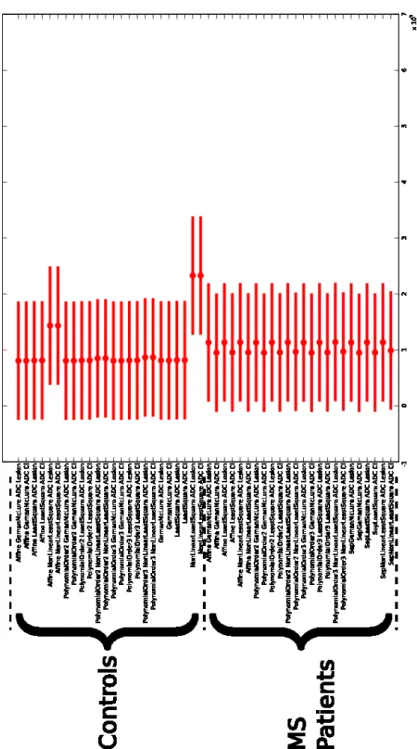
Conventional DT-MRI tools were applied for the computation of DTI in-variants (FA and MD maps). The diffusion tensors were calculated using the LS, wLS and CNLS methods and four methods for the corrections of distortions were applied to the DW-MRI (no correction, affine and the two polynomial mod-els). For controls, the mask of lesions from the MS patients were automatically aligned with the images of the controls, using a linear registration method [25]. The lesion mask and contralateral mask of lesion were then used to extract paired-data on the FA and MD maps for each MS patient and control. An analysis of variance (ANOVA) combined with a multiple comparison procedure is then applied to the FA and MD data from each ROI. The ANOVA is fed with voxel data intensity, grouped by ROI: 1) MSL, 2) contralateral MSL ROI, 3) control ROI, 4) control contralateral ROI and by preprocessing. The Figure
1 and 2 show the multiple comparisons procedure output. These Figures has the controls and MS patients displayed as two groups, for this two groups the pre-processing are ordered as follow:
– correction of distortion with affine model
• tensor estimation with WLS for the lesion and contralateral ROI • tensor estimation with LS for the lesion and contralateral ROI • tensor estimation with CNLS for the lesion and contralateral ROI – correction of distortion with polynomial second order model
• tensor estimation with WLS for the lesion and contralateral ROI • tensor estimation with LS for the lesion and contralateral ROI • tensor estimation with CNLS for the lesion and contralateral ROI – correction of distortion with polynomial third order model
• tensor estimation with WLS for the lesion and contralateral ROI • tensor estimation with LS for the lesion and contralateral ROI • tensor estimation with CNLS for the lesion and contralateral ROI – no correction of distortions
• tensor estimation with WLS for the lesion and contralateral ROI • tensor estimation with LS for the lesion and contralateral ROI • tensor estimation with CNLS for the lesion and contralateral ROI In our experiments, whatever processing was applied, three statistically dif-ferent groups appear on the FA (Fig. 1):
– MSL in MS patients
– Contralateral of MSL in MS patients
– Controls (both ”lesion” and contralateral ROI).
On the FA maps of controls, a slight difference appears depending on the applied processing. On average, this is mainly due to the higher anisotropy in the controls data. The differences in the tensor estimation techniques are clearer in regions of high anisotropy, which is reflected by our experiments. The correction of distortions does not seem to yield specifically different results. For the MD, no statistically different results appear (Fig. 1) but a small variation still exists on two of the preprocessings.
4
Results
In MS patients a statistical difference between lesions and their contralateral counterparts was found for the FA (but not for the MD), irrespective of the automated image processing methods used. In controls, no statistical difference between ROI associated with MS patient lesions and contralateral ROI was found.
5
Conclusion
In comparison with widely used manual or semi-automated DTI analysis method-ology, in this pilot study with MS patients and age- and sex-matched controls, we show with our automated approach using the mid-sagittal plane as a refer-ence that we were able to replicate results from the literature. Automated image analysis approaches, however, have the advantage being more accurate, repro-ducible and robust. A statistical difference between MSL and their contra lateral ROI is confirmed as shown in the literature, which does not exists for controls. A statistical difference is present when comparing the three tissues classes from MS patients and controls: 1) MSL ROI, 2) contralateral ROI MSL and 3) controls ROI. Even if the pre processing seems to impact little on statistical differences of the DTI measures in healthy volunteers and MS patients our fully automated approach is superior to manual or semi-automated DT-MRI analyses regarding the robustness of the results (reproducibility and accuracy).
References
1. Le Bihan, D. et al.: MR imaging of intravoxel incoherent motions: application to diffusion and perfusion in neurologic disorders. Radiology 161(2) (November 1986) 401–7
2. Basser, P. et al.: Estimation of the effective self-diffusion tensor from the NMR spin echo. Journal of Magnetic Resonance B(103) (1994) 247–254
3. Rovaris, M. et al.: Diffusion MRI in multiple sclerosis. Neurology 65(10) (Novem-ber 2005) 1526–1532
4. Wilson, M. et al.: Quantitative diffusion weighted magnetic resonance imaging, cerebral atrophy, and disability in multiple sclerosis. J Neurol Neurosurg Psychiatry 70 (march 2001) 318–322
5. Ciccarelli, O. et al.: Investigation of MS normal-appearing brain using diffusion tensor MRI with clinical correlations. Neurology 56(7) (April 2001) 926–933 6. Nusbaum, A.O. et al.: Whole-brain diffusion MR histograms differ between MS
subtypes. Neurology 54(7) (April 2000) 1421–1427
7. Bammer, R. et al.: Magnetic resonance diffusion tensor imaging for characteriz-ing diffuse and focal white matter abnormalities in multiple sclerosis. Magnetic Resonance in Medicine 44 (2000) 583–591
8. Droogan, A.G. et al.: Comparison of multiple sclerosis clinical subgroups using navigated spin echo diffusion-weighted imaging. Magn Reson Imaging 17 (June 1999) 653–661
9. Filippi, M. et al.: A quantitative study of water diffusion in multiple sclerosis lesions and normal-appearing white matter using echo-planar imaging. Arch Neurol 57(7) (July 2000) 1017–1021
10. Roychowdhury, S. et al.: Multiple Sclerosis: Comparison of Trace Apparent Diffu-sion Coefficients with MR Enhancement Pattern of LeDiffu-sions. AJNR Am J Neuro-radiol 21(5) (May 2000) 869–874
11. Werring, D.J. et al.: Diffusion tensor imaging of lesions and normal-appearing white matter in multiple sclerosis. Neurology 52 (May 1999) 1626–1632
12. Castriota-Scanderbeg, A. et al.: Coefficient Dav Is More Sensitive Than Fractional Anisotropy in Monitoring Progression of Irreversible Tissue Damage in Focal Non-active Multiple Sclerosis Lesions. AJNR Am J Neuroradiol 24(4) (April 2003) 663–670
13. Filippi, M. et al.: Diffusion tensor magnetic resonance imaging in multiple sclerosis. Neurology 56(3) (February 2001) 304–311
14. Nusbaum, A.O. et al.: Regional and Global Changes in Cerebral Diffusion with Normal Aging. AJNR Am J Neuroradiol 22 (January 2001) 136–142
15. Ranjeva, J.P. et al.: MRI/MRS of corpus callosum in patients with clinically isolated syndrome suggestive of multiple sclerosis. Mult Scler 9(6) (December 2003) 554–565
16. Cassol, E. et al.: Diffusion tensor imaging in multiple sclerosis: a tool for monitoring changes in normal-appearing white matter. Mult Scler 10(2) (April 2004) 188–196 17. Scanderbeg, A.C. et al.: Demyelinating Plaques in Relapsing-remitting and Secondary-progressive Multiple Sclerosis: Assessment with Diffusion MR Imaging. AJNR Am J Neuroradiol 21(5) (May 2000) 862–868
18. Cercignani, M. et al.: Intra-voxel and inter-voxel coherence in patients with mul-tiple sclerosis assessed using diffusion tensor MRI. Journal of Neurology 249(7) (July 2002) 875–883
19. Cercignani, M. et al.: Mean Diffusivity and Fractional Anisotropy Histograms of Patients with Multiple Sclerosis. AJNR Am J Neuroradiol 22(5) (May 2001) 952–958
20. Bozzali, M. et al.: Quantification of Brain Gray Matter Damage in Different MS Phenotypes by Use of Diffusion Tensor MR Imaging. AJNR Am J Neuroradiol 23(6) (June 2002) 985–988
21. Rovaris, M. et al.: Assessment of normal-appearing white and gray matter in patients with primary progressive multiple sclerosis: a diffusion-tensor magnetic resonance imaging study. Arch Neurol 59(9) (September 2002) 1406–1412 22. Iannucci, G. et al.: Correlation of Multiple Sclerosis Measures Derived from
T2-Weighted, T1-T2-Weighted, Magnetization Transfer, and Diffusion Tensor MR Imag-ing. AJNR Am J Neuroradiol 22(8) (September 2001) 1462–1467
23. Stefano, N.D. et al.: MR correlates of cerebral atrophy in patients with multiple sclerosis. J Neurol 249 (August 2002) 1072–1077
24. Koay, C.G., Basser, P.J.: Analytically exact correction scheme for signal extraction from noisy magnitude MR signals. J Magn Reson 179(2) (April 2006) 317–322 25. Wiest-Daessl´e, N. et al.: Evaluation of a new optimisation algorithm for rigid
registration of MRI data. In: SPIE Medical Imaging 2007: Image Processing. (2007) to be published.
26. Prima, S., Wiest-Daessl´e, N.: Computation of the Mid-Sagittal Plane
in Diffusion Tensor MR Brain Images. In: SPIE Medical Imaging 2007: Image Processing. (2007) to be published.
27. Koay, C.G. et al.: A unifying theoretical and algorithmic framework for least squares methods of estimation in diffusion tensor imaging. J Magn Reson 182(1) (September 2006) 115–125
28. Wiest-Daessl´e, N. et al.: Validation of a new optimisation algorithm for registra-tion tasks in medical imaging. In: IEEE Internaregistra-tional Symposium on Biomedical Imaging 2007, Washington DC, USA (April 2007)