HAL Id: hal-00838540
https://hal.archives-ouvertes.fr/hal-00838540
Submitted on 25 Jun 2013
HAL is a multi-disciplinary open access
archive for the deposit and dissemination of
sci-entific research documents, whether they are
pub-lished or not. The documents may come from
teaching and research institutions in France or
abroad, or from public or private research centers.
L’archive ouverte pluridisciplinaire HAL, est
destinée au dépôt et à la diffusion de documents
scientifiques de niveau recherche, publiés ou non,
émanant des établissements d’enseignement et de
recherche français ou étrangers, des laboratoires
publics ou privés.
of the fluoroprotein asFP595.
Lars Schäfer, Gerrit Groenhof, Martial Boggio-Pasqua, Michael Robb, Helmut
Grubmüller
To cite this version:
Lars Schäfer, Gerrit Groenhof, Martial Boggio-Pasqua, Michael Robb, Helmut Grubmüller.
Chro-mophore protonation state controls photoswitching of the fluoroprotein asFP595.. PLoS
Computa-tional Biology, Public Library of Science, 2008, 4 (3), pp.e1000034. �10.1371/journal.pcbi.1000034�.
�hal-00838540�
Photoswitching of the Fluoroprotein asFP595
Lars V. Scha¨fer1, Gerrit Groenhof1, Martial Boggio-Pasqua2, Michael A. Robb3, Helmut Grubmu¨ller1*
1 Department of Theoretical and Computational Biophysics, Max-Planck-Institute for Biophysical Chemistry, Go¨ttingen, Germany, 2 Laboratoire de Chimie et Physique Quantiques, IRSAMC, Universite´ Paul Sabatier, Toulouse, France,3 Department of Chemistry, Imperial College London, London, United Kingdom
Abstract
Fluorescent proteins have been widely used as genetically encodable fusion tags for biological imaging. Recently, a new class of fluorescent proteins was discovered that can be reversibly light-switched between a fluorescent and a non-fluorescent state. Such proteins can not only provide nanoscale resolution in far-field fluorescence optical microscopy much below the diffraction limit, but also hold promise for other nanotechnological applications, such as optical data storage. To systematically exploit the potential of such photoswitchable proteins and to enable rational improvements to their properties requires a detailed understanding of the molecular switching mechanism, which is currently unknown. Here, we have studied the photoswitching mechanism of the reversibly switchable fluoroprotein asFP595 at the atomic level by multiconfigurational ab initio (CASSCF) calculations and QM/MM excited state molecular dynamics simulations with explicit surface hopping. Our simulations explain measured quantum yields and excited state lifetimes, and also predict the structures of the hitherto unknown intermediates and of the irreversibly fluorescent state. Further, we find that the proton distribution in the active site of the asFP595 controls the photochemical conversion pathways of the chromophore in the protein matrix. Accordingly, changes in the protonation state of the chromophore and some proximal amino acids lead to different photochemical states, which all turn out to be essential for the photoswitching mechanism. These photochemical states are (i) a neutral chromophore, which can trans-cis photoisomerize, (ii) an anionic chromophore, which rapidly undergoes radiationless decay after excitation, and (iii) a putative fluorescent zwitterionic chromophore. The overall stability of the different protonation states is controlled by the isomeric state of the chromophore. We finally propose that radiation-induced decarboxylation of the glutamic acid Glu215 blocks the proton transfer pathways that enable the deactivation of the zwitterionic chromophore and thus leads to irreversible fluorescence. We have identified the tight coupling of trans-cis isomerization and proton transfers in photoswitchable proteins to be essential for their function and propose a detailed underlying mechanism, which provides a comprehensive picture that explains the available experimental data. The structural similarity between asFP595 and other fluoroproteins of interest for imaging suggests that this coupling is a quite general mechanism for photoswitchable proteins. These insights can guide the rational design and optimization of photoswitchable proteins.
Citation: Scha¨fer LV, Groenhof G, Boggio-Pasqua M, Robb MA, Grubmu¨ller H (2008) Chromophore Protonation State Controls Photoswitching of the Fluoroprotein asFP595. PLoS Comput Biol 4(3): e1000034. doi:10.1371/journal.pcbi.1000034
Editor: Arieh Warshel, University of Southern California, United States of America Received September 10, 2007; Accepted February 12, 2008; Published March 28, 2008
Copyright: ß 2008 Scha¨fer et al. This is an open-access article distributed under the terms of the Creative Commons Attribution License, which permits unrestricted use, distribution, and reproduction in any medium, provided the original author and source are credited.
Funding: Support from the EU Nanomot project (grant 29084) is thankfully acknowledged. This work has been supported by EPSRC UK (grant GR/S94704/01). LVS thanks the Boehringer Ingelheim Fonds for a PhD fellowship.
Competing Interests: The authors have declared that no competing interests exist. * E-mail: hgrubmu@gwdg.de
Introduction
Fluorescent proteins have been widely used as genetically encodable fusion tags to monitor protein localizations and dynamics in live cells [1–3]. Recently, a new class of green fluorescent protein (GFP)-like proteins has been discovered, which can be reversibly photoswitched between a fluorescent (on) and a non-fluorescent (off) state [4–10]. As the reversible photoswitching of photochromic organic molecules such as fulgides or diarylethenes is usually not accompanied by fluorescence [11], this switching reversibility is a very remarkable and unique feature that may allow fundamentally new applications. For example, the reversible photoswitching, also known as kindling, may provide nanoscale resolution in far field fluorescence optical microscopy much below the diffraction limit [12–15]. Likewise, reversibly switchable fluorescent proteins will enable the repeated tracking of protein location and movement in single cells [16]. Since fluorescence can be sensitively read out from
a bulky crystal, the prospect of erasable three-dimensional data storage is equally intriguing [17].
The GFP-like protein asFP595, isolated from the sea anemone Anemonia sulcata, is a prototype for a reversibly switchable fluorescent protein. The protein can be switched from its non-fluorescent off state to the non-fluorescent on state by green light of 568 nm wavelength [5,6,18,19]. From this so-called kindled on state, the same green light elicits a red fluorescence emission at 595 nm. Upon kindling, the intensity of the absorption maximum at 568 nm diminishes, and an absorption peak at 445 nm appears. The kindled on state can be promptly switched back to the initial off state by this blue light of 445 nm. Alternatively, the off state is repopulated through thermal relaxation within seconds. In addition, if irradiated with intense green light over a long period of time, asFP595 can also be irreversibly converted into a fluorescent state that cannot be quenched by light any more [5]. The nature of this state is hitherto unknown.
The switching cycle of asFP595 is reversible and can be repeated many times without significant photobleaching. These properties render asFP595 a promising fluorescence marker for high-resolution optical far-field microscopy, as recently demon-strated by Hofmann and coworkers [20]. Currently, however, with its low fluorescence quantum yield (,0.1% and 7% before and after activation, respectively [6,16]) and rather slow switching kinetics, the photochromic properties of asFP595 need to be improved. To systematically exploit the potential of such switchable proteins and to enable rational improvements to the properties of asFP595, a detailed molecular understanding of the photoswitching mechanism is mandatory.
The aim of this study is to obtain a detailed mechanistic picture of the photoswitching mechanism of asFP595 at the atomic level, i.e., to understand the dynamics of both the activation process (off-to-on switching) and the de-activation process (on-to-off switching). High-resolution crystal structures of the wild-type (wt) asFP595 in its off state [19,21,22], of the Ser158Val mutant in its on state [19], and of the Ala143Ser mutant in its on and off states [19] were recently determined. Similar to GFP, asFP595 adopts a b-barrel fold enclosing the chromophore, a 2-acetyl-5-(p-hydroxybenzyli-dene)imidazolinone (Figure 1). The chromophore is post-transla-tionally formed in an autocatalytic cyclization-oxidation reaction of the Met63-Tyr64-Gly65 (MYG) triad. As compared to the GFP chromophore, the p-system of MYG is elongated by an additional carbonyl group [23].
Reversible photoswitching of asFP595 was possible even within protein crystals, and x-ray analysis showed that the off-on switching of the fluorescence is accompanied by a conformational trans-cis isomerization of the chromophore [19]. In a recent study [24], we have shown that the isomerization induces changes of the protonation pattern of the chromophore and some of the surrounding amino acids, and that these changes account for the observed shifts in the absorption spectrum upon kindling. Based on the comparison between measured and calculated absorption spectra, the major protonation states in the ground state have been assigned to the zwitterion (Z) and the anion (A) for the trans conformer, whereas the neutral (N) chromophore is dominant for the cis conformation (Figure 1B).
Here, we study the photochemical behavior of each of the previously identified protonation states. We have addressed the following questions: How does light absorption induce the isomerization of the chromophore within the protein matrix, and how do the different protonation states affect the internal conversion mechanism? Which is the fluorescent species, and how can the fluorescence quantum yield be increased? To address these questions, we have carried out nonadiabatic molecular dynamics (MD) simulations using a hybrid quantum-classical QM/MM approach. This approach includes diabatic surface hopping between the excited state and the ground state. The forces acting on the chromophore were calculated using the CASSCF [25,26] multi-reference method, which, although not always yielding highly accurate excitation and fluorescence energies, has shown to be a reliable method for mechanistic studies of photochemical reactions involving conical intersections [27].
A number of approaches for modeling nonadiabatic dynamics have been described in the literature, such as Tully’s fewest switches surface hopping [28], and multiple spawning [29]. For recent reviews, see [30,31]. In the context of QM/MM simulations, the surface hopping approach to photobiological problems has been pioneered by Warshel and coworkers [32,33].
zwitterion “Z”
anion “A”
A
B
His197
Ser158
Glu215
MYG
W233
neutral “N”
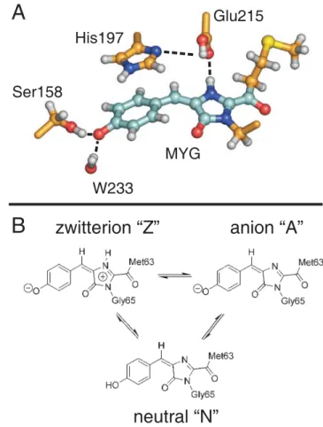
Figure 1. (A) Chromophore (MYG) in the trans conformation and adjacent amino acid side chains. Ser158, Glu215, and the crystallo-graphic water molecule W233 are hydrogen-bonded to MYG (dashed lines). His197 is p-stacked to the MYG phenoxy-moiety and forms a hydrogen bond to Glu215 (dashed line). The carbon skeleton of the quantum mechanical (QM) subsystem is shown in cyan, and the carbon atoms modelled by molecular mechanics (MM) are shown in orange. (B) Schematic drawings of the different MYG protonation states considered in this work.
doi:10.1371/journal.pcbi.1000034.g001 Author Summary
Proteins whose fluorescence can be reversibly switched on and off hold great promise for applications in high-resolution optical microscopy and nanotechnology. To systematically exploit the potential of such photoswitch-able proteins and to enphotoswitch-able rational improvements of their properties requires a detailed understanding of the molecular switching mechanism. Here, we have studied the photoswitching mechanism of the reversibly switch-able fluoroprotein asFP595 by atomistic molecular dynam-ics simulations. Our simulations explain measured quan-tum yields and excited state lifetimes, and also predict the structures of the hitherto unknown intermediates and of the irreversibly fluorescent state. Further, we find that the proton distribution in the active site of the asFP595 controls the photochemical conversion pathways of the chromophore in the protein matrix. Our results show that a tight coupling between trans-cis isomerization of the chromophore and proton transfer is essential for the function of asFP595. The structural similarity between asFP595 and other fluoroproteins suggests that this coupling is a quite general mechanism for photoswitch-able proteins. These insights can guide the rational design and optimization of photoswitchable proteins.
The diabatic surface hopping approach used in this work differs from the other approaches in two main respects. First, in our approach a binary decision (hop or no hop) is made at each integration time step of the trajectory, based only on the current wavefunctions of the ground and excited states. Second, hopping is only allowed at the conical intersection (CI) seam, where hopping probability approaches unity. This could in principle underesti-mate the crossing probabilty, because we do not allow for transitions in regions of strong coupling but no real crossing. However, for ultra-fast photochemical reactions in large poly-atomic systems, decay predominantly takes place at the CI seam, as also shown by others [31]. Thus, most surface hops are essentially diabatic, justifying our approach. In addition, both energy and momentum are conserved upon a transition, as the trajectory never leaves the diabatic energy surface. The theoretical background and algorithmic implementation of the diabatic surface hopping are detailed in the Supporting Information (Text S4).
Several theoretical studies on the photochemistry of the GFP chromophore have been conducted, applying both static ab initio [34–36] and DFT calculations [37], and dynamics simulations based on a semi-empirical Hamiltonian [38]. In addition, vertical excitation energies of asFP595 model chromophores in the gas phase and in a continuum dielectric were calculated by DFT and ab initio methods [39,40], as well as in a minimal protein environment by means of DFT and CASSCF calculations within a QM/MM approach [41].
By identifying key residues in the cavity of the asFP595 chromophore, our nonadiabatic QM/MM molecular dynamics simulations elucidate how the protein surrounding governs the photoreactivity of this photoswitchable protein. Based on the simulations, we provide a new mechanism that qualitatively explains measured decay times and quantum yields, and that predicts the structures and protonation states of the photochemical intermediates and of the irreversibly fluorescent state. We also suggest excited state proton transfer (ESPT) to play an important mechanistic role. However, the detailed study of such ESPT processes is beyond the scope of this paper. Our predictions can be probed by, e.g., time-resolved Fourier transform infrared (FTIR) spectroscopy and x-ray crystallography.
Results/Discussion
Our results reveal that the excited state behavior of asFP595 is determined by the protonation pattern of the chromophore and some amino acids in the surrounding protein matrix rather than by the chromophore conformation (trans or cis). The latter, however, modulates the excited state properties by changing the hydrogen-bonded network in the chromophore cavity.
For both conformers, we identified three possible species that differ in protonation state, explaining the complex photochemical behavior of asFP595. First, the neutral chromophores Ntransand
Ncis undergo reversible trans-cis photoisomerization and thus
account for the photoswitching between the dark off and fluorescent on states. Second, anionic chromophores Atrans and
Acis lead to the observed ultra-fast radiationless deactivation.
Third, fluorescence emission can in principle originate from both zwitterions Ztransand Zcis. The protonation states are
interchange-able via proton transfers. In the following we will describe the excited state behavior of all three protonation states.
trans-cis Isomerization of the Neutral Chromophore
The five excited state simulations that were initiated from the ground state trajectory of the trans neutral chromophore Ntransare
listed in Table 1. Trans-to-cis photoisomerization of the chromo-phore was observed in one of these simulations (run b, Table 1; Video S2 in Supporting Information). Figure 2 shows a schematic representation of the S0 (green) and S1 (red) potential energy
surfaces of the neutral chromophore, along with a photoisomer-ization MD trajectory (yellow dashed line). Two coordinates are shown, the isomerization coordinate and a skeletal deformation coordinate of the chromophore (see below). The dynamics can be separated into three distinct phases: (i) evolution on the electronic ground state S0, (ii) excitation and evolution on the excited state
S1, and (iii) decay back to S0at the surface crossing seam followed
Table 1. Excited state lifetimes and final conformations from the MD simulations initiated in the neutral trans chromophore conformation.
Starting in Ntrans
Run S1lifetime (ps) Final conformation
a 0.516 trans b 0.475 cis c 0.309 trans d 0.224 trans e 0.718 trans doi:10.1371/journal.pcbi.1000034.t001
Figure 2. Schematic representation of potential energy surfac-es. The excited (S1, red) and ground (S0, green) states of the neutral
chromophore are shown along the trans-cis isomerization coordinate (torsion A) and a skeletal deformation coordinate of the chromophore. Radiationless decay occurs at the S1/S0conical intersection (CI, dashed
white line). In this representation, the CI occurs as an extended seam, because the torsion coordinate is from the (N-2)-dimensional intersec-tion space, and the skeletal deformaintersec-tion coordinate is from the 2-dimensional branching pace. The dashed yellow line represents the path sampled in a QM/MM photoisomerization trajectory.
by subsequent relaxation on the ground state surface. The position of the surface crossing seam controls the passage of the trajectory from S1 to S0. The seam is accessed from a global twisted
minimum on S1, which is separated by a small S1barrier from a
local planar minimum near the Franck-Condon (FC) region. In our simulations, individual excited state (S1) lifetimes between
0.224 ps and 0.718 ps were observed (Table 1). A simple exponential fit to the observed lifetimes yielded a decay time of t = 0.34 ps (s+= 0.21 ps, s2= 0.13 ps, with s being the statistical
error). Given the low number of trajectories, the statistical error of our estimated lifetime may seem unexpectedly low, but results from a rigorous analysis assuming an underlying single exponential decay [42]. Recent femtosecond time-resolved pump/probe experiments by Schu¨ttrigkeit and coworkers have yielded excited state decay time constants of 0.32 ps (78%), 2.6 ps (19%), and 12.1 ps (3%) as well as a fluorescence lifetime of 2.2 ns for asFP595 [43]. However, although the simulated decay times seem to be in good agreement to the experimental results, we believe that the results should not be directly compared. In previous work [24], we demonstrated that the Ntransprotonation state is hardly
populated in asFP595 and therefore is unlikely to contribute to the observed excited state decay. Instead, the species that is predominantly responsible for the ultra-fast radiationless decay observed in the experiments is the anionic trans chromophore Atrans, as shown in detail below.
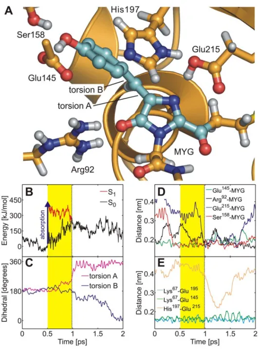
Figure 3A shows the snapshot from the isomerization trajectory (run b, Table 1) shortly before the surface crossing seam was encountered.
After photon absorption (blue arrow in Figures 2 and 3B), the chromophore spontaneously rotated around torsion A (imidazo-linone-twist), and the ring-bridging CH group pointed downwards (away from His197) by almost 90u. The time-evolution of the S0
and S1 potential energies and of the two ring-bridging torsion
angles during trans-cis photoisomerization are shown in Figures 3B and 3C. After excitation to S1, the chromophore rapidly relaxed
from the FC region into a nearby planar S1 minimum, as is
evident from the decreasing S1energy in panel b (red curve). The
system stayed in this planar minimum for about 0.2 ps; subsequently the global twisted S1minimum was reached through
rotation around torsion A (Figures 2, 3B, and 3C). The system then oscillated around this minimum until the conical intersection seam was encountered. After the surface hop to S0, torsion B
(hydroxyphenyl-twist) rotated after a short delay of about 0.5 ps. Previously, an ideal ‘‘hula-twist’’ isomerization mechanism of the zwitterionic chromophore was proposed based on a force field model [19]. This hula-twist involves a simultaneous rotation around both torsion angles A and B. In the QM/MM simulations of the neutral chromophore presented here, rotation around both torsions was also observed. However, twisting around torsion B was slower than around torsion A (Figure 3C), and the isomerization hence proceeded via the twisted conformer shown in Figure 3A, with perpendicular imidazolinone and hydroxyphe-nyl moieties. The hula-twist isomerization mechanism of the zwitterion at the QM/MM level will be discussed below.
During the initial equilibration of Ntrans, the hydrogen bonding
network of the x-ray crystal structures of the anionic and zwitterionic chromophores (see above) changed to accommodate the non-native neutral chromophore. First, a stable hydrogen bond formed between the hydroxyphenyl OH group of MYG and Glu145 (Figure 3A). Second, the hydrogen bonds between the imidazolinone nitrogen and Glu215 as well as between His197 and Glu215 ruptured. Interestingly, these two hydrogen bonds were transiently re-established during the end of the second isomerization phase (blue and orange curves in Figures 3D and
3E, respectively) in which torsion B followed torsion A. In the twisted intermediate structure, the imidazolinone nitrogen atom was sterically more exposed as compared to the planar conformation, which facilitated the formation of the hydrogen bonds. During an extended 10 ns force field simulation, the His197-Glu215 and MYG-Glu215 hydrogen bonds repeatedly broke and re-formed at a timescale of several hundred picoseconds (data not shown), further underlining the flexibility of these two hydrogen bonds. Figures 3D and 3E furthermore show that during the isomerization, the hydrogen bonding network in the chromophore cavity was stable, as none of the hydrogen bonds that were established at the instant of photoexcitation ruptured. A similar stability was found in all simulations, irrespective of the chromophore conformation or protonation state. Thus, the hydrogen bonding network around the chromophore is flexible enough to allow for photoexcitation and even photoisomerization without being ruptured.
For the neutral cis chromophore Ncis, five excited state
simulations were initiated from the ground state trajectory (Table 2). Only two of these trajectories returned to the ground state within 10 ps (Table 2, runs a and b), which was the maximum affordable trajectory length in terms of computation time. In one of these two simulations, a spontaneous cis-to-trans photoisomerization was observed (run a; Video S1 in Supporting Information). As expected, the isomerization pathway was similar to the reverse trans-to-cis pathway in that the conical intersection seam was accessed via rotation around torsion A, followed by a slightly delayed rotation around torsion B in S0(see Figure S4 in
Supporting Information). However, in contrast to the activation pathway, the ring-bridging CH group rotated upwards (i.e., towards His197). Thus, despite the anisotropic protein surround-ing, both rotational orientations of the chromophore CH bridge are feasible. In the second simulation, the CI seam was also encountered after rotation around torsion A, but the chromophore returned to the initial cis conformation.
For the other three trajectories, the chromophore remained trapped in a planar S1minimum conformation near the FC region
throughout the simulation (data not shown in Table 2). The starting structures of these trajectories were used for three additional simulations in which the escape from the planar S1
minimum was accelerated by means of conformational flooding [42,44] (Table 2, runs c–e). In these accelerated simulations, the flooding potential successfully induced the escape from the local S1
minimum, and the surface crossing seam was encountered in all cases. Isomerization was observed in two of these simulations. In total, cis-to-trans photoisomerization was seen in three out of five simulations initiated in the Ncis state. Although the number of
trajectories is small, our simulations suggest that the probability for cis-to-trans photoisomerization is larger than for the reverse trans-to-cis process discussed before.
S1/S0Conical Intersection Topology and S1Minima
To further characterize the potential energy surfaces underlying the photochemical conversion processes of the neutral chromo-phore (Figure 2), we have optimized three S1 minima and a
minimum energy S1/S0 conical intersection (MECI) in the gas
phase (see Supporting Information, Figure S1, Tables S1 and S5). We found a local planar S1minimum for the trans isomer 76 kJ/
mol below the FC region. The structure corresponding to the global S1 minimum is twisted around torsion A by 85u and lies
2131 kJ/mol relative to the FC region. The structure of the nearby MECI is twisted around torsion A by 81u. The MECI is energetically lower than the FC region by 62 kJ/mol, and the CI seam is therefore readily accessible. Twisting around torsion B
instead of torsion A also leads to a local minimum on S1, whose
energy is 28 kJ/mol below the FC energy.
The MD simulations reflect this surface topology. Immediately after excitation, the system relaxed from the FC region to the global S1minimum by rotation around torsion A (Figure 3C). The
system oscillated around this minimum until the conical intersection seam was encountered, with a subsequent surface hop back to S0. The gradients on S0and S1are almost parallel at
the CI, which indicates that the CI is sloped. The gradient difference vector and the derivative coupling vector that span the
branching space largely correspond to skeletal deformations of the imidazolinone moiety (see Supporting Information, Figure S1). Thus, as shown in Figure 2, the rotation coordinate around torsion A is parallel to the seam and does not lift the S1/S0degeneracy.
The seam is accessible anywhere along this torsional rotation coordinate, and therefore such torsional rotation is in principle not essential for the radiationless decay. The extended surface crossing seam parallel to the isomerization coordinate accounts for the low isomerization quantum yield seen in our simulations. In most of our MD simulations, the seam was encountered rather ‘‘early’’
Figure 3.Trans-cisisomerization of the neutral chromophore. (A) Chromophore (MYG) conical intersection geometry adopted during the MD simulation. MYG forms hydrogen bonds to Arg92, Glu145, Ser158, and Glu215. Color code as in Figure 1. (B) Ground (S0, black) and excited (S1, red)
potential energy traces along the QM/MM molecular dynamics trajectory. Photon absorption (blue arrow) excites the chromophore into S1(yellow
area) until it decays back to S0at the conical intersection seam (dashed line). (C) Time-evolution of the ring-bridging torsion angles A (magenta) and B
(blue). (D, E) Change of the hydrogen bonding network in the chromophore cavity during trans-cis isomerization. The Arg92 (black), MYG-Glu145 (green), MYG-Ser158 (red), Lys67-Glu195 (cyan, residues not shown in (A)), and Lys67-MYG-Glu145 (green) hydrogen bonds were stable during isomerization. Additional hydrogen bonds between MYG and Glu215 (blue) as well as between His197 and Glu215 (orange) were transiently formed. doi:10.1371/journal.pcbi.1000034.g003
along the torsional rotation coordinate (Figure 2), and the system thus returned to the ground state before overcoming the S0barrier
maximum. In these cases, relaxation on S0after the surface hop
led back to the starting conformation.
Role of the Protein Environment
To elucidate the influence of the protein environment on the photoisomerization process of the chromophore, we have re-calculated the S1 and S0 energies along two excited state
trajectories (run b, Table 1 and run a, Table 2) in the gas phase. In these simulations, the chromophore followed the same trajectory as before, but did not interact with the rest of the system (protein and solvent surrounding). We have not attempted to further characterize the electrostatic influence of the surround-ing by, e.g., pKacalculations.
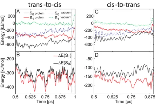
Figure 4A and 4C show the obtained energy traces. In the protein, both S1and S0are stabilized with respect to the gas phase.
For the trans-to-cis isomerization process, the protein stabilized the energies of the S1and S0states on average by 2339 kJ/mol and
2307 kJ/mol, respectively. For the cis-to-trans process, the average stabilization energies were 2173 kJ/mol and 2126 kJ/mol, respectively. Thus, the protein (and solvent) environment favors S1over S0by about 30–50 kJ/mol. Figure 4B and 4D show the
energy differences between the protein and the gas phase, DE = E(protein)2E(gas phase). The S1 stabilization was rather
strong at the surface crossing seam (Figure 4). We found S1to be
stabilized stronger than S0 by 78 kJ/mol and 93 kJ/mol at the
conical intersection in both MD simulations. In summary, the protein environment energetically stabilizes S1 more than S0,
thereby enhancing fast radiationless decay.
Ultra-Fast Radiationless Deactivation of the Anionic Chromophore
In total, 20 simulations of the anionic chromophore protonation state were carried out, 10 of which were initiated in the trans conformation and the other 10 were initiated in the cis conformation. Ultra-fast radiationless deactivation was observed in all 20 trajectories (Table S6 in Supporting Information). However, trans-cis photoisomerization never occurred. A simple exponential fit to the S1lifetimes of the trans anion yielded a decay
time of t = 0.45 ps (s+= 0.19 ps, s2= 0.12 ps). Since Atransis one
of the two dominant protonation states in the off state besides Ztrans
[24], we expect Atrans to significantly contribute to the
experi-mentally observed decay. The measured decay time of 0.32 ps [43] agrees well with the decay time derived from the simulations. For Acis, an excited state decay time of t = 1.81 ps (s+= 0.77 ps,
s2= 0.48 ps) was obtained, which is about four times longer as
compared to the decay time of Atrans.
Figure 4. Influence of the protein environment on the photoisomerization process of the neutral asFP595 chromophore. (A, C) Ground and excited state energies along trans-to-cis (A) and cis-to-trans (C) isomerization trajectories (run b, Table 1 and run a, Table 2). The protein environment stabilizes S0and S1(black and red lines, respectively) relative to the gas phase (dashed blue and green lines, respectively). (B, D) Energy
difference between the protein and the gas phase. DE(S0) = E(S0, protein)2E(S0, gas phase) is plotted in black, DE(S1) = E(S1, protein)2E(S1, gas phase)
in red. The protein environment energetically stabilizes S1more strongly than S0. The vertical dashed black line represents the surface crossing. The
energy offset in (A) and (C) is 1.96996106kJ/mol. doi:10.1371/journal.pcbi.1000034.g004
Table 2. Excited state lifetimes and final conformations from the MD simulations initiated in the neutral cis chromophore conformation.
Starting in Ncis
Run S1lifetime (ps) Final conformation
a 0.374 trans b 3.561 cis c* 1.573 trans d* 0.867 cis e* 1.206 trans *
In runs c, d, and e, the escape from the S1minimum was accelerated by
conformational flooding.
Figure 5A shows the conical intersection geometry adopted during a typical trajectory. In contrast to the neutral chromo-phore, the CI seam was accessed through a phenoxy-twist (rotation around torsion B, see Figure 5C), and the CH bridge remained in the imidazolinone plane. Shortly after excitation, rotation around torsion B drove the system towards the surface crossing seam (Figure 5B and 5C). Back on S0, the system returned
to the initial configuration. The hydrogen bonding network in the
chromophore cavity was very similar to the network observed in the x-ray crystal structures and remained stable during the excited state MD simulations.
Since rotation around torsion B does not lead to trans-cis isomerization and rotation around torsion A did not occur, the quantum yield for the isomerization of the anion was zero in our simulations. However, due to the limited number of trajectories (20), we cannot rule out the trans-cis photoisomerization of the
Figure 5. Ultra-fast internal conversion mechanism of thetransanion. (A) Snapshot at the conical intersection: the chromophore is twisted around torsion B, yet the hydrogen bonded network in the chromophore cavity remains intact. (B) Ground (S0, black) and excited (S1, red) potential
energy traces along the QM/MM molecular dynamics trajectory. Photon absorption (green arrow) brings the chromophore into S1(yellow area) until
it decays back to S0at the conical intersection seam (dashed line). (C) Time-evolution of the torsion angles A (magenta) and B (blue). (D) S0and S1
energies along a representative excited state trajectory of Atrans. The protein environment strongly stabilizes S0and S1(black and red lines,
respectively) relative to the gas phase (dashed blue and green lines, respectively). The energy offset is 1.96866106kJ/mol. (E) Energy difference DE
between the protein and the gas phase for S0(black) and S1(red).
anion. Our results agree with recent MRPT2 computations of Olsen and coworkers on an anionic DsRed-like model chromo-phore in the gas phase [45], who have shown that the imidazolinone-twisted S1/S0 CI (i.e., twisted around torsion A),
which leads to cis-trans isomerization, is disfavored by more than 150 kJ/mol as compared to the phenoxy-twisted CI. The latter CI is lower in energy than the planar S1 minimum by 9.2 and
48.1 kJ/mol at the MRPT2 and CASSCF levels, respectively [45]. In our calculations, this energy difference is about 30 kJ/mol (see Supporting Information, Table S2), in qualitative agreement with Olsen and coworkers. Due to the slightly different chromophores in DsRed and asFP595, a quantitative agreement cannot be expected.
The deviation from planarity of the trans chromophore observed in the crystal structures was speculated to enhance ultra-fast deactivation [24,43]. Indeed, the difference between the S1
lifetimes of the cis and trans conformers seen in our simulations can be attributed to steric constraints imposed by the protein matrix. In the trans conformation the phenoxy-ring deviates from planarity by about 20u, whereas the cis chromophore is essentially planar [19,24]. We observed in our simulations that only a slight additional twisting was required for the trans conformer to reach the surface crossing seam. Thus, the pre-twisting of the phenoxy-moiety due to the protein matrix facilitated fast internal conversion of the trans conformer.
S1/S0Conical Intersection Topology and S1Minima
As shown in Supporting Information (Figure S2, Table S2), we have optimized the S1/S0 MECI, a planar and two twisted S1
minima (imidazolinone-twist and phenoxy-twist) for an isolated anionic chromophore. The planar minimum lies 31 kJ/mol below the FC point. The structure of the global S1minimum is twisted
around torsion B by 269u and its energy lies 282 kJ/mol relative to the FC energy. The MECI structure is also twisted about torsion angle B by 269u and is energetically lower than the FC point by 61 kJ/mol, thus explaining the ultra-fast decay seen in our MD simulations. Twisting around torsion A leads to a local S1
minimum that is 46 kJ/mol below the FC point. The CI of the anion is sloped, and the gradient difference vector corresponds to a skeletal deformation of the imidazolinone ring, analogous to the neutral chromophore (see above). In contrast to the neutral chromophore, the derivative coupling vector involves rotation around torsion B. However, the amplitude of this vector is small. Thus, the two electronic states may remain close in energy along torsion B, allowing the system to decay at various phenoxy-twist angles.
Role of the Protein Environment
To study the influence of the protein matrix on the deactivation process of the anionic chromophore, we have re-evaluated the S0
and S1energies along two representative excited state trajectories
(trans and cis) with all interactions between the QM atoms of the chromophore and the MM surrounding switched off, as was done for the neutral chromophores (see above). As Figure 5D and 5E show, the protein (and solvent) environment stabilizes the chromophore with respect to the gas phase. The S0and S1states
of the trans chromophore are strongly stabilized by 2840 kJ/mol and 2832 kJ/mol, respectively. The S0and S1 states of the cis
chromophore were stabilized relative to the gas phase by 2407 kJ/mol and 2433 kJ/mol, respectively, during a represen-tative Acis trajectory (Figure S5 in Supporting Information).
Similar to the neutral chromophore, the protein environment favors the trans conformation over cis. Before reaching the CI seam, the S0and S1states of the chromophore were stabilized to
the same extent. At the CI, however, the protein environment lowered the energy of the S1state more strongly than the energy of
the S0 state by 26 kJ/mol and 20 kJ/mol for Atrans and Acis,
respectively. This preferential stabilization of S1 enhanced the
ultra-fast radiationless deactivation seen in our MD simulations.
Fluorescence Emission of the Zwitterionic Chromophores
Ten simulations were carried out for the zwitterion. Five simulations were started in the Ztransconformation, and the other
five simulations were initiated in the Zcisconformation. No decay
back to the ground state was observed within a maximum trajectory length of 10 ps, neither for Ztrans nor for Zcis. The
chromophore did not escape from a planar S1 minimum in the
vicinity of the FC region in any of the excited state simulations. This suggests that Ztransand Zciscould be the fluorescent species in
asFP595, although the measured fluorescence lifetime of 2.2 ns [43] is still much longer than our maximal trajectory length (10 ps).
For Ztrans and for Zcis, we have carried out three additional
simulations each, in which we applied the conformational flooding technique [42,44,46] to accelerate the escape from the S1
minimum in an unbiased manner (see Materials and Methods). The results of these flooding simulations are shown in the Supporting Information (Figure S6, Text S3). The flooding potential induced isomerization of the chromophore, which followed a hula-twist pathway, in agreement with our previous work [19]. From the flooding simulations, we obtained a lower bound for the excited state lifetime of the order of 1 ns. The qualitative agreement with the measured fluorescence decay time of 2.2 ns provides further support for the assignment of the zwitterion as the fluorescent species. The results thus obtained for the zwitterionic chromophore suggest that a hula-twist CI may be spontaneously accessed if the trajectories were extended to (significantly) longer times, i.e., nanoseconds. However, at the nanosecond timescale, fluorescence (and not isomerization) will be the predominant decay process. Note that in Ref. [19], the energy barrier for hula-twist isomerization of the zwitterion was underestimated, thus favoring this isomerization over fluorescence. In the next paragraph, we characterize the CI and show that the minimum energy crossing point for the zwitterionic chromophore has a high energy, thus hampering radiationless decay through hula-twist isomerization.
S1/S0Conical Intersection Topology and Influence of the
Protein Environment
To characterize the topology of the S1and S0potential energy
surfaces and of the conical intersection that occurs between them, multiconfigurational calculations were carried out for the isolated zwitterionic chromophore, as was also done for the other protonation states (see above). We optimized a planar S1minimum
and a hula-twist S1/S0MECI (see Supporting Information, Figure
S3, Tables S3 and S4). In contrast to the anion and the neutral chromophore, no twisted S1 minima were found. The gradient
difference vector and the derivative coupling vector at the MECI do not involve torsional rotation of either torsion A or torsion B, indicating that the CI seam lies parallel to the isomerization coordinate. The MECI lies 70 kJ/mol above the planar S1
minimum and 23.4 kJ/mol above the FC energy. Hence, in contrast to the anion and the neutral chromophore, no low-lying CI is present for the zwitterion, demonstrating that radiationless decay in the gas phase cannot occur in an unactivated manner. For the CI seam to become accessible, a significant stabilization of S1relative to S0by the protein environment would be required.
protein surrounding does not reduce the S1/S0 energy gap
anywhere along the isomerization coordinate.
Deactivation of Ztransthrough Proton Transfer
Our results suggest that the zwitterionic chromophore is potentially fluorescent, irrespective of the conformation. However, the x-ray analysis of the emitting species has shown that only the cis chromophore fluoresces, whereas the trans chromophore is dark [19]. A possible explanation for this discrepancy is the presence of an alternative deactivation channel that does not involve isomerization. This deactivation pathway would have to be more easily accessible for Ztrans than for Zcis. Only the latter would
therefore be trapped in S1and fluoresce.
The hydrogen bond between the NH group of the imidazoli-none ring and Glu215 strongly suggests that the alternative decay involves an excited state proton transfer (ESPT). Such ESPT would quench the fluorescence, because the resulting anion rapidly deactivates, as shown above. However, by including only the chromophore into the QM subsystem, we have excluded the possibility of observing such ESPT in our QM/MM simulations. To identify possible ESPT pathways, we have carried out extended force field MD simulations of both Ztrans and Zcisand
analyzed the relevant hydrogen bonds. Figure 6A shows that, during the simulation of Ztrans, two stable hydrogen bonds were
formed between the protonated OH group of Glu215 and His197 as well as between the NH proton of MYG and Glu215. These two hydrogen bonds allow for a proton transfer from Ztransto the
rapidly deactivating Atrans. The OH proton of Glu215 could
transfer to the Nd atom of His197, with a simultaneous or
subsequent transfer of the NH proton of the imidazolinone moiety to Glu215. In contrast, during the force field simulation of Zcis, the
MYG-Glu215 hydrogen bond remained intact, whereas the Glu215-His197 hydrogen bond broke after about 1 ns (Figure 6B). This differential behavior of Ztrans and Zcis was
confirmed by two additional independent MD simulations (data not shown). Based on these results, we assume that only the trans
zwitterion can be converted to the anion through a short proton wire. Therefore, an ultra-fast deactivation channel is available only for the trans zwitterion, and not for the fluorescent cis zwitterion. From the presence of the hydrogen bonding network in our force field trajectories, we do not obtain insights into the energetics of proton transfer. Studying these transfers along the identified pathways in asFP595, both in the ground and the excited state, is beyond the scope of the present work.
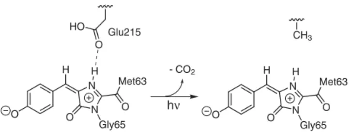
Having established that fluorescence can only originate from the zwitterionic chromophores, the structure of the irreversibly fluorescent state of asFP595 can now be predicted. We expect that intense irradiation over a prolonged period of time leads to a decarboxylation of the Glu215 side chain (Figure 7). Such process is also known to occur in GFP [47,48] and DsRed [49]. A decarboxylated Glu215 can no longer take up the NH proton from the zwitterionic chromophores. The absence of an S1ESPT
deactivation channel leads to fluorescence. The finding that the irreversibly fluorescent state cannot be switched off by light (see Introduction) is corroborated by our observation that even in the flooding-induced isomerization trajectories, no radiationless decay back to S0 occurred (see Figure S6 in Supporting
Information).
Figure 6. Hydrogen bonding network in the chromophore cavity during force field simulations of zwitterionic chromophores. (A, C) Snapshots from MD simulations of Ztransand Zcis, respectively. The blue dashed lines indicates the distance between the NH proton of MYG and
Glu215, and the red dashed line that between the OH-group of Glu215 and the Ndatom of His197. (B, D) Time-evolution of the two hydrogen bonds
shown in (A) and (C) during representative force field MD simulations. doi:10.1371/journal.pcbi.1000034.g006 O N N O Gly65 O Met63 H H O N N O Gly65 O Met63 H H O HO CH3 - CO2 Glu215 hν
Figure 7. Scheme of the proposed decarboxylation of Glu215, which yields an irreversibly fluorescent zwitterion.
Switching Efficiency of asFP595
Figure 8 summarizes our proposed photoswitching mechanism. The proton distribution at the active site of asFP595 governs the photochemical conversion pathways of the chromophore in the protein matrix. Changes in the protonation state of the chromophore and several proximal amino acids lead to different photochemical states, which are all involved in the photoswitching process. These photochemical states are (i) the neutral chromo-phores Ntrans and Ncis, which can undergo trans-cis
photoisomer-ization, (ii) the anionic chromophores Atrans and Acis, which
rapidly undergo radiationless decay after excitation, and (iii) the potentially fluorescent zwitterions Ztrans and Zcis. The overall
stability of the different protonation states is controlled by the isomeric state of the chromophore.
To switch from the non-fluorescent off to the fluorescent on state, the chromophore has to isomerize from trans to cis. As shown in Figure 8, this photoisomerization was only observed for the neutral form of the chromophore (NtransR Ncis). However, the Ntransstate
is only marginally populated [24], thus explaining the low quantum yield for switching asPF595 to the on state. Moreover, green light is used to switch on asFP595, whereas the absorption maximum of Ntrans is significantly blue-shifted. The use of blue
light, however, would lead to an unfavorable NcisR Ntrans
back-reaction due to the absorption of the blue light by Ncis.
The reverse cis-to-trans isomerization, i.e., on-to-off switching, requires the excitation of the neutral cis chromophore. Since Ncisis
the predominant protonation state in the cis conformation, the efficiency for the on-to-off switching is high, as observed experimentally [19]. Fluorescence originates from Zcis, which is
also hardly populated, like Ntrans [24]. Taken together, the low
populations of the involved states give rise to the low overall fluorescence quantum yield of asFP595.
The insight obtained from our simulations can be exploited for a targeted improvement of asFP595 for applications as a fluorescence marker in optical microscopy. In particular, to improve the signal-to-noise ratio, a higher fluorescence quantum yield is desired. Our results suggest that one way to enhance fluorescence would be to increase the stability of Zcis, e.g., by
introducing additional hydrogen bond donors near the phenoxy-group of the chromophore. Another possibility would be to implement an internal proton relay, similar to that in GFP. In GFP, a hydroxyphenyl-bound serine residue, a water molecule, and a glutamic acid form an internal proton wire that enhances the formation of the fluorescent Acis chromophore from the
neutral chromophore via ESPT. GFP has a significantly higher fluorescence quantum yield as compared to asFP595 [50–54]. Although the fluorescent species in asFP595 and GFP are different, the similarity between the chromophores suggests that implementing a similar internal proton relay in asFP595 might increase its fluorescence quantum yield.
Note, however, that due to the competition between different reaction channels in asFP595, shifting the relative populations of the protonation states will also affect the photoswitchability. For example, increasing the population of the fluorescent species at the cost of the neutral species will decrease the back-isomerization efficiency. Thus, a compromise has to be found between increasing the fluorescence quantum yield on the one hand while maintaining the photoswitchability of asFP595 on the other hand.
Conclusions
Understanding the excited state dynamics of ultra-fast photo-activated processes in biomolecular systems such as the reversible photoswitching of the fluorescent protein asFP595 represents a major challenge, but is essential to unveil the underlying molecular mechanisms. In the present work we have demonstrated that by using an ab initio QM/MM excited state molecular dynamics strategy together with explicit surface hopping, it is not only possible to explain at the atomic level experimentally accessible quantities of asFP595, such as quantum yields and excited state lifetimes, but also to make predictions that are rigorously testable by experiment, such as the nature of the irreversibly fluorescent state or possible improvement of the fluorescence quantum of this protein.
We have revealed that the protonation pattern of the chromophore cavity determines the photochemical behavior of asFP595, and that photon absorption can lead to trans-cis
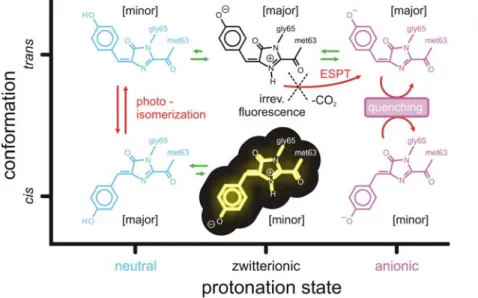
Figure 8. Scheme of the reversible photoswitching mechanism of asFP595 proposed in this work. The fluorescent state Zcis is
highlighted. The green arrows indicate ground state equilibria, whereas the red arrows indicate excited state processes. The major protonation states are the zwitterionic and the anionic chromophores in the trans conformation, and the neutral chromophore in the cis conformation, as indicated in the square brackets.
isomerization of the neutral chromophore. Based on our results, we suggest that a reversibly switchable protein must fulfill three criteria. First, to enable switching, trans-cis photoisomerization is necessary. Second, this photoisomerization has to be coupled to proton transfer events, that is, the preferred protonation state is different for the two conformers. Third, only one of the two isomers fluoresces, while the other can undergo rapid radiationless decay.
Interestingly, other fluoroproteins contain a chromophoric moiety similar to asFP595, like, e.g., Dronpa [8,55,56], DsRed [49,57], Kaede or KiKG [58,59], eqFP611 [60–63], Rtms5 [64], and HcRed [65]. In these structures cis or trans conformations of the chromophore have been observed. Our simulations on asFP595 suggest that chromophore photoisomerization could also be possible in these fluoroproteins. In particular, the high structural similarity between asFP595 and the reversibly photo-switchable Dronpa protein suggests that the Dronpa chromophore can undergo trans-cis photoisomerization as well. Indeed, the crystal structure of the isomerized state of Dronpa was solved very recently [66] and confirms our prediction. Furthermore, recent experiments on Dronpa [67,68] also provide strong support for our proposed protonation/deprotonation mechanism. The simi-larity between the chromophores in a variety of fluoroproteins suggests that during molecular evolution, the (p-hydroxybenzyli-dene)imidazolinone chromophoric moiety served as a template and that the photochromic properties - and thus the function - was fine-tuned by the protein environment.
Materials and Methods
To model the dynamics of the photoactivated asFP595 chromophore, we carried out excited state QM/MM [69] molecular dynamics (MD) simulations. Energies and forces of the excited (S1) and ground (S0) states were calculated on-the-fly at
the CASSCF/3-21G level of theory with a reduced active space of 6 electrons in 6 orbitals. The studied processes start in S1and end
up in S0. The transitions (hops) between the two energy surfaces
were modeled by surface selection at the conical intersection (CI) seam (see Supporting Information, Text S4 for details). This diabatic hopping procedure is known to potentially underestimate the surface hopping probability [70,71]. However, our experience has shown that in large polyatomic systems, low-lying CI hyperlines are easily accessed due to their high dimensionality (N22, where N is the number of internal degrees of freedom). We therefore expect that, for the case at hand, diabatic surface hopping is sufficiently accurate. For all MD simulations, Gromacs 3.3 [72] with an interface [70] to Gaussian03 [73] was used. Modifications were made to the one-electron integral routines of Gaussian03 to account for the polarization of the CASSCF wavefunctions due to the pointcharges on the MM atoms.
The reduced active space used in the MD simulations was validated using geometry optimizations of relevant excited state minima and minimum energy crossing points for the isolated chromophores. The full CASSCF active space for the p-system of the asFP595 chromophore would require 18 p electrons in 16 p orbitals, rendering geometry optimizations prohibitively expen-sive. To make the respective calculations feasible, we have restricted the number of excitations in the wavefunction by employing the RASSCF method (see Supporting Information, Text S1) [74–76]. We have characterized minima and conical intersections at the RASSCF(18,7+4+5)[2,2]/6-31G* level of theory. The final 6 electron, 6 orbital active space used in the CASSCF QM/MM MD simulations was selected from the RASSCF calculations such as to enable the simultaneous
description of the electronic ground and first excited states. This approach allowed us to compute on-the-fly QM/MM trajectories using a CASSCF/3-21G wavefunction with an accuracy that is comparable to RASSCF/6-31G* at a drastically reduced computational cost.
All MD simulations were based on the crystal structure of a mutant of asFP595 [24]. The mutant has similar photochromic properties as the wild-type, but high-resolution crystal structures are available for both the trans and the cis conformations. For both trans and cis isomers, MD simulations were initiated for three different chromophore protonation states; anionic (A), neutral (N), and zwitterionic (Z). The simulations were performed in a rectangular periodic box of about 730 nm3. Each system contained about 21,500 TIP4P water molecules, including 340 crystallographic water molecules. After assigning the protonation pattern of the chromophore pocket, all other polar, aromatic, and aliphatic hydrogen atoms were added to the protein with the HB2MAK [77] routine of WHATIF [78]. To each of the systems, sodium and chloride ions were added at physiological concentra-tion to compensate for the net positive charge of the protein. The actual number of ions varied with the total charge of the protein, which differed for the chosen protonation patterns of the chromophore cavity. The final systems comprised about 90,000 atoms.
Prior to the MD simulations, the systems were energy minimized (1000 steps steepest descent). Subsequently, force field based MD simulations were carried out. First, 500 ps MD simulations with harmonic position restraints on all protein heavy atoms (force constant 1000 kJ mol21nm22) were carried out to equilibrate the solvent and the ions. Then, 500 ps free MD simulation were run at 300 K. All simulations were carried out using the OPLS all-atom force field [79]. Parameters for the chromophore were taken from [19].
The simulations were run at constant temperature and pressure by coupling to an external heat bath (tT= 0.1 ps, tp= 1 ps) [80].
In the force field simulations, LINCS [81] was used to constrain bond lengths, thus allowing a time step of 2 fs. SETTLE [82] was applied to constrain the internal degrees of freedom of the water molecules. A twin-range cut-off was used for the Lennard-Jones interactions. Interactions within 1.0 nm were updated at every step, whereas interactions between 1.0 nm and 1.6 nm were updated every ten steps. Coulomb interactions within 1.0 nm were computed at each step as well. Beyond this cut-off, the particle-mesh Ewald (PME) method [83] with a grid spacing of 0.12 nm was used.
The QM subsystem in the excited state QM/MM simulations comprised the chromophore; the rest of the system was described by the OPLS all-atom force field (Figure 1). The N2Cabond of
Gly65 and the Ca2Cbbond of Met63, respectively, were replaced
by a constraint, and the QM part was capped with hydrogen link atoms [84]. The forces on the link atoms were distributed over the two heavy atoms at the boundary according to the lever rule. The QM system experienced the Coulomb field of all MM atoms within a sphere of 1.6 nm, and Lennard-Jones interactions between QM and MM atoms were included. For the QM/MM simulations, a time step of 1 fs was used, and no constraints were applied in the QM subsystem. Prior to the excited state simulations, the systems were simulated in the ground state, first for 1 ps at the RHF/3-21G//OPLS level of theory and then for an additional 2.5 ps at the CASSCF(6,6)/3-21G//OPLS level. From the latter trajectory, frames at equal time intervals (Dt = 0.5 ps) were used as starting configurations for the excited state MD simulations.
To accelerate the escape from the S1minima, additional excited
state conformational flooding [42,44] simulations were carried out for the Ncis, Zcis, and Ztrans systems, respectively. A
Gaussian-shaped flooding potential Vfl was constructed from a principal
component analysis (PCA) [85–87] of free QM/MM S1
simulations. For all systems, the covariance matrix of the motion of all QM atoms, except the exocyclic carbonyl group of the chromophore and the hydroxyphenyl-OH proton (for Ncis), was
computed from two independent 10 ps S1trajectories. All internal
degrees of freedom of the chromophore were affected by the flooding potential to ensure that the escape from the initial minimum was accelerated in an unbiased manner. Unless stated differently, adaptive flooding [42,46] with target destabilization free energies of 300 kJ/mol (Ztrans) or 100 kJ/mol (Ncis) and time
constants of t = 0.1 ps was applied. In all flooding simulations, Vfl
was switched off as soon as the conical intersection seam was encountered to allow for an unperturbed relaxation on the ground state potential energy surface.
In all our simulations of the anionic and zwitterionic chromo-phores, we considered both the cationic and neutral protonation states of the imidazole side chain of His197, which lies coplanar to the chromophore (Figure 1). Recent Poisson-Boltzmann electrostat-ics calculations have shown that slight structural fluctuations in the local environment change the preferred protonation of the His197 imidazole ring between cationic and neutral and that both protonation states are populated at room temperature [24]. Thus, we ran the same number of trajectories with a cationic and with a neutral His197. As shown in Supporting Information (Text S2), the His197 protonation state had no major influence on the decay mechanisms and lifetimes.
Supporting Information
Figure S1 Optimized ab initio geometries of Ntrans.
Found at: doi:10.1371/journal.pcbi.1000034.s001 (0.53 MB MPG)
Figure S2 Optimized ab initio geometries of Atrans.
Found at: doi:10.1371/journal.pcbi.1000034.s002 (0.48 MB DOC)
Figure S3 Optimized ab initio geometries of Ztrans.
Found at: doi:10.1371/journal.pcbi.1000034.s003 (0.42 MB DOC)
Figure S4 Cis-to-trans isomerization of the neutral chromophore. (A) Chromophore (MYG) conical intersection geometry adopted during excited state QM/MM MD simulation. The CH bridge rotated upwards (i.e., towards His197). (B) Time-evolution of the ring-bridging dihedral angles A (magenta) and B (blue). (C) S0
(black) and S1(red) potential energy traces along the QM/MM
trajectory. Photon absorption brings the system into S1 (yellow
area), until decay back to S0occurs (dashed line) at the CI seam.
(D,E) The hydrogen-bonding network in the chromophore cavity stayed stable during the isomerization of MYG.
Found at: doi:10.1371/journal.pcbi.1000034.s004 (8.79 MB DOC)
Figure S5 Influence of the protein environment on the deactivation of the anionic cis chromophore Acis. (A) S0and S1
energies along a representative trajectory (run h, Table S6). The protein environment stabilized S0 and S1 (black and red lines,
respectively) relative to the gas phase (dashed blue and green lines, respectively). (B) Energy difference DE between the protein and the gas phase for S0 (black) and for S1 (red). The dashed line
indicates the decay at the CI seam.
Found at: doi:10.1371/journal.pcbi.1000034.s005 (1.30 MB TIF)
Figure S6 Isomerization of the cis zwitterion induced by conformational flooding. (A) Hula-twist structure adopted during isomerization trajectory (B) Ground (S0, black) and excited state
(S1, red) potential energy traces along the trajectory. S0 and S1
come energetically close, but the surface crossing seam was not encountered. The surface hop was therefore imposed at the structure with the minimum energy gap (dashed black line). The time evolution of the flooding potential Vflis shown in the inset.
(C) Time evolution of the torsion angles A (magenta) and B (blue). (D) S0and S1energies (black and red lines, respectively) along the
isomerization trajectory. The protein environment stabilizes S0
and S1 relative to the gas phase (dashed blue and green lines,
respectively). The energy offset is 1.97?106kJ/mol. (E) Energy difference DE between the protein and the gas phase for S0(black)
and S1 (red). S0 is stabilized slightly stronger than S1 along the
whole isomerization pathway.
Found at: doi:10.1371/journal.pcbi.1000034.s006 (7.52 MB TIF) Table S1 RASSCF(18,7+4+5)2,2/6-31G* results on Ntrans.
Found at: doi:10.1371/journal.pcbi.1000034.s007 (0.03 MB TIF) Table S2 RASSCF(18,7+4+5)2,2/6-31G* results on Atrans.
Found at: doi:10.1371/journal.pcbi.1000034.s008 (0.03 MB DOC)
Table S3 RASSCF(18,7+4+5)2,2/6-31G* results on Ztrans.
Found at: doi:10.1371/journal.pcbi.1000034.s009 (0.03 MB DOC)
Table S4 CASSCF(6,6)/3-21G results on Ztrans.
Found at: doi:10.1371/journal.pcbi.1000034.s010 (0.03 MB DOC)
Table S5 Cartesian coordinates of optimized structures at RASSCF(18,7+4+5)2,2/6-31G* level.
Found at: doi:10.1371/journal.pcbi.1000034.s011 (0.05 MB DOC)
Table S6 Excited state lifetimes and conformations from the MD simulations of the anionic chromophores Atransand Acis. In
runs A–J, His197 was modelled as cationic, whereas in runs K-T, His197 was modelled as neutral (singly protonated at Nd).
Found at: doi:10.1371/journal.pcbi.1000034.s012 (0.04 MB DOC)
Text S1 Ab initio calculations in the gas phase.
Found at: doi:10.1371/journal.pcbi.1000034.s013 (0.03 MB DOC)
Text S2 Influence of the p-stacked Histidine 197.
Found at: doi:10.1371/journal.pcbi.1000034.s014 (0.02 MB DOC)
Text S3 Conformational flooding simulations of the zwitterionic chromophores.
Found at: doi:10.1371/journal.pcbi.1000034.s015 (0.02 MB DOC)
Text S4 Diabatic Surface Hopping: Theory and Implementa-tion.
Found at: doi:10.1371/journal.pcbi.1000034.s016 (0.06 MB DOC)
Video S1 cis-to-trans isomerization of neutral chromophore. Found at: doi:10.1371/journal.pcbi.1000034.s017 (24.36 MB DOC)
Video S2 trans-to-cis isomerization of neutral chromophore. Found at: doi:10.1371/journal.pcbi.1000034.s018 (51.31 MB MPG)
Acknowledgments
We thank Ira Tremmel for carefully reading the manuscript and for valuable suggestions, and M. Andresen, C. Eggeling, S. Hell, S. Jakobs, A. Stiel, and M. Wahl for the crystal structures and stimulating discussions.
Author Contributions
Conceived and designed the experiments: LS GG MB-P MR HG. Performed the experiments: LS MB-P. Analyzed the data: LS GG MB-P. Wrote the paper: LS GG MB-P MR HG.
References
1. Lippincott-Schwartz J, Altan-Bonnet N, Patterson GH (2003) Photobleaching and photoactivation: following protein dynamics in living cells. Nat Cell Biol. pp S7–S14.
2. Miyawaki A, Sawano A, Kogure T (2003) Lighting up cells: labelling proteins with fluorophores. Nat Cell Biol. pp S1–S7.
3. Tsien RY (1998) The green fluorescent protein. Annu Rev Biochem 67: 509–544.
4. Ando R, Mizuno H, Miyawaki A (2004) Regulated fast nucleocytoplasmic shuttling observed by reversible protein highlighting. Science 306: 1370–1373. 5. Chudakov DM, Belousov VV, Zarisky AG, Novoselov VV, Staroverov DB, et al. (2003) Kindling fluorescent proteins for precise in vivo photolabeling. Nature Biotechnol 21: 191–194.
6. Lukyanov KA, Fradkov AF, Gurskaya NG, Matz MV, Labas YA, et al. (2000) Natural animal coloration can be determined by a nonfluorescent green fluorescent protein homolog. J Biol Chem 275: 25879–25882.
7. Habuchi S, Ando R, Dedecker P, Verheijen W, Mizuno H, et al. (2005) Reversible single-molecule photoswitching in the GFP-like fluorescent protein Dronpa. Proc Natl Acad Sci USA 102: 9511–9516.
8. Wilmann PG, Turcic K, Battad JM, Wilce MCJ, Devenish RJ, et al. (2006) The 1.7 A˚ crystal structure of Dronpa: A photoswitchable green fluorescent protein. J Mol Biol 364: 213–224.
9. Dedecker P, Hotta J, Ando R, Miyawaki A, Engelborghs Y, et al. (2006) Fast and reversible photoswitching of the fluorescent protein Dronpa as evidenced by fluorescence correlation spectroscopy. Biophys J 91: L45–L47.
10. Remington SJ (2006) Fluorescent proteins: maturation, photochemistry and photophysics. Curr Opin Struct Biol 16: 714–721.
11. Crano JC, Guglielmetti RJ (1999) Organic photochromic and thermochromic compounds: Main photochromic families, Vol. 1. New York: Plenum. 12. Hell SW, Jakobs S, Kastrup L (2003) Imaging and writing at the nanoscale with
focused visible light through saturable optical transitions. Appl Phys A-Mater Sci Process 77: 859–860.
13. Hell SW (2003) Toward fluorescence nanoscopy. Nature Biotechnol 21: 1347–1355.
14. Hell SW, Dyba M, Jakobs S (2004) Concepts for nanoscale resolution in fluorescence microscopy. Curr Opin Neurobiol 14: 599–609.
15. Geisler C, Schonle A, von Middendorff C, Bock H, Eggeling C, et al. (2007) Resolution of lambda/10 in fluorescence microscopy using fast single molecule photo-switching. Appl Phys A-Mater Sci Process 88: 223–226.
16. Lukyanov KA, Chudakov DM, Lukyanov S, Verkhusha VV (2005) Photo-activatable fluorescent proteins. Nat Rev Mol Cell Biol 6: 885–891. 17. Sauer M (2005) Reversible molecular photoswitches: A key technology for
nanoscience and fluorescence imaging. Proc Natl Acad Sci USA 102: 9433–9434.
18. Chudakov DM, Feofanov AV, Mudrik NN, Lukyanov S, Lukyanov KA (2003) Chromophore environment provides clue to ‘‘kindling fluorescent protein’’ riddle. J Biol Chem 278: 7215–7219.
19. Andresen M, Wahl MC, Stiel AC, Gra¨ter F, Scha¨fer LV, et al. (2005) Structure and mechanism of the reversible photoswitch of a fluorescent protein. Proc Natl Acad Sci USA 102: 13070–13074.
20. Hofmann M, Eggeling C, Jakobs S, Hell SW (2005) Breaking the diffraction barrier in fluorescence microscopy at low light intensities by using reversibly photoswitchable proteins. Proc Natl Acad Sci USA 102: 17565–17569. 21. Wilmann PG, Petersen J, Devenish RJ, Prescott M, Rossjohn J (2005) Variations
on the GFP chromophore. J Biol Chem 280: 2401–2404.
22. Quillin ML, Anstrom DA, Shu XK, O’Leary S, Kallio K, et al. (2005) Kindling fluorescent protein from Anemonia sulcata: Dark-state structure at 1.38 A˚ resolution. Biochemistry 44: 5774–5787.
23. Yampolsky IV, Remington SJ, Martynov VI, Potapov VK, Lukyanov S, et al. (2005) Synthesis and properties of the chromophore of the asFP595 chromoprotein from Anemonia sulcata. Biochemistry 44: 5788–5793. 24. Scha¨fer LV, Groenhof G, Klingen AR, Ullmann GM, Boggio-Pasqua M, et al.
(2007) Photoswitching of the fluorescent protein asFP595: Mechanism, proton pathways, and absorption spectra. Angew Chemie Int Ed 46: 530–536. 25. Siegbahn P, Heiberg A, Roos B, Levy B (1980) Comparison of the super-CL and
the Newton-Raphson scheme in the complete active space SCF method. Phys Scr 21: 323–327.
26. Siegbahn PEM, Almlof J, Heiberg A, Roos BO (1981) The complete active space SCF (CASSCF) method in a Newton-Raphson formulation with application to the HNO molecule. J Chem Phys 74: 2384–2396.
27. Lipkowitz KB, Boyd DB, Robb MA, Garavelli M, Olivucci M, et al. (2000) Reviews in computational chemistry. New York: Wiley.
28. Tully JC (1990) Molecular dynamics with electronic transitions. J Chem Phys 93: 1061–1071.
29. Ben-Nun M, Quenneville J, Martinez TJ (2000) Ab initio multiple spawning: Photochemistry from first principles molecular dynamics. J Phys Chem A 104: 5161–5175.
30. Martinez TJ (2006) Insights for light-driven molecular devices from ab initio multiple spawning excited-state dynamics of organic and biological chromo-phores. Acc Chem Res 39: 119–126.
31. Levine BG, Martinez TJ (2007) Isomerization through conical intersections. Annu Rev Phys Chem 58: 613–634.
32. Warshel A (1976) Bicycle-pedal model for 1st step in vision process. Nature 260: 679–683.
33. Warshel A, Chu ZT (2001) Nature of the surface crossing process in bacteriorhodopsin: Computer simulations of the quantum dynamics of the primary photochemical event. J Phys Chem B 105: 9857–9871.
34. Martin ME, Negri F, Olivucci M (2004) Origin, nature, and fate of the fluorescent state of the green fluorescent protein chromophore at the CASPT2// CASSCF resolution. J Am Chem Soc 126: 5452–5264.
35. Altoe P, Bernardi F, Garavelli M, Orlandi G, Negri F (2005) Solvent effects on the vibrational activity and photodynamics of the green fluorescent protein chromophore: A quantum-chemical study. J Am Chem Soc 127: 11534–11535. 36. Sinicropi A, Andruniow T, Ferre N, Basosi R, Olivucci M (2005) Properties of the emitting state of the green fluorescent protein resolved at the CASPT2// CASSCF/CHARMM level. J Am Chem Soc 127: 11534–11535.
37. Laino T, Nifosi R, Tozzini V (2004) Relationship between structure and optical properties in green fluorescent proteins: a quantum mechanical study of the chromophore environment. Chem Phys 298: 17–28.
38. Toniolo A, Olsen S, Manohar L, Martinez TJ (2004) Conical intersection dynamics in solution: the chromophore of green fluorescent protein. Faraday Discuss 127: 149–163.
39. Amat P, Granucci G, Buda F, Persico M, Tozzini V (2006) The chromophore of asFP595: a theorectical study. J Phys Chem B 110: 9348–9353.
40. Nemukhin AV, Topol IA, Burt SK (2006) Electronic excitations of the chromophore from the fluorescent protein asFP595 in solutions. J Chem Theory Comput 2: 292–299.
41. Grigorenko B, Savitsky A, Topol I, Burt S, Nemukhin A (2006) Ground-state structures and vertical excitations for the kindling fluorescent protein asFP595. J Phys Chem B 110: 18635–18640.
42. Lange OF, Scha¨fer LV, Grubmu¨ller H (2006) Flooding in GROMACS: Accelerated barrier crossings in molecular dynamics. J Comp Chem 27: 1693–1702.
43. Schu¨ttrigkeit TA, von Feilitzsch T, Kompa CK, Lukyanov KA, Savitsky AP, et al. (2006) Femtosecond study of light-induced fluorescence increase of the dark chromoprotein asFP595. Chem Phys 323: 149–160.
44. Grubmu¨ller H (1995) Predicting slow structural transitions in macromolecular systems: Conformational flooding. Phys Rev E 52: 2893–2906.
45. Olsen S, Smith SC (2007) Radiationless decay of red fluorescent protein chromophore models via twisted intermolecular charge transfer states. J Am Chem Soc 129: 2054–2065.
46. Mu¨ller EM, de Meijere A, Grubmu¨ller H (2002) Predicting unimolecular chemical reactions: Chemical flooding. J Chem Phys 116: 897–905. 47. van Thor JJ, Gensch T, Hellingwerf KJ, Johnson LN (2002)
Phototransforma-tion of green fluorescent protein with UV and visible light leads to decarboxylation of glutamate 222. Nat Struct Biol 9: 37–41.
48. Bell AF, Stoner-Ma D, Wachter RM, Tonge PJ (2003) Light-driven decarboxylation of wild-type green fluorescent protein. J Am Chem Soc 125: 6919–6926.
49. Habuchi S, Cotlet M, Gensch T, Bednarz T, Haber-Pohlmeier S, et al. (2005) Evidence for the isomerization and decarboxylation in the photoconversion of the red fluorescent protein DsRed. J Am Chem Soc 127: 8977–8984. 50. Helms V (2002) Electronic excitations of biomolecules studied by quantum
chemistry. Curr Opin Struct Biol 12: 169–175.
51. Stoner-Ma D, Jaye AA, Matousek P, Towrie M, Meech SR, et al. (2005) Observation of excited-state proton transfer in green fluorescent protein using ultrafast vibrational spectroscopy. J Am Chem Soc 127: 2864–2865. 52. Agmon N (2005) Proton pathways in green fluorescence protein. Biophys J 88:
2452–2461.
53. Leiderman P, Huppert D, Agmon N (2006) Transition in the temperature-dependence of GFP fluorescence: From proton wires to proton exit. Biophys J 90: 1009–1018.
54. Vendrell O, Gelabert R, Moreno M, Lluch JM (2006) Potential energy landscape of the photoinduced multiple proton-transfer process in the green fluorescent protein: classical molecular dynamics and multiconfigurational electronic structure calculations. J Am Chem Soc 128: 3564–3574.
55. Stiel AC, Trowitzsch S, Weber G, Andresen M, Eggeling C, et al. (2006) 1.8 A˚ bright-state structure of the reversibly switchable fluorescent protein Dronpa guides the generation of fast switching variants. Biochem J 402: 35–42.