HAL Id: hal-01764683
https://hal-amu.archives-ouvertes.fr/hal-01764683
Submitted on 13 Apr 2018
HAL is a multi-disciplinary open access archive for the deposit and dissemination of sci-entific research documents, whether they are pub-lished or not. The documents may come from teaching and research institutions in France or abroad, or from public or private research centers.
L’archive ouverte pluridisciplinaire HAL, est destinée au dépôt et à la diffusion de documents scientifiques de niveau recherche, publiés ou non, émanant des établissements d’enseignement et de recherche français ou étrangers, des laboratoires publics ou privés.
Host resistance to endotoxic shock requires the
neuroendocrine regulation of group 1 innate lymphoid
cells
Linda Quatrini, Elisabeth Wieduwild, Sophie Guia, Claire Bernat, Nicolas
Glaichenhaus, Eric Vivier, Sophie Ugolini
To cite this version:
Linda Quatrini, Elisabeth Wieduwild, Sophie Guia, Claire Bernat, Nicolas Glaichenhaus, et al.. Host resistance to endotoxic shock requires the neuroendocrine regulation of group 1 innate lymphoid cells. Journal of Experimental Medicine, Rockefeller University Press, 2017, 214 (12), pp.3531-3541. �10.1084/jem.20171048�. �hal-01764683�
Br ief Definitive Repor t
The J
our
nal of Exper
imental Medicine
IntroductIonBoth the immune and nervous systems are involved in the maintenance of homeostasis and host integrity. Upon in-fection, a delicate balance is established between resistance mechanisms, which induce pathogen elimination, and tol-erance mechanisms, which prevent excessive tissue damage (Medzhitov et al., 2012). It is becoming increasingly clear that neuroendocrine–immune interactions play an important role in these regulatory processes (Irwin and Cole, 2011), but the mechanisms involved are still not well understood.
IFN-γ is an important endogenous regulator of the im-mune response (Schroder et al., 2004). In bacterial infections, IFN-γ primes mononuclear phagocytes for phagocytosis and production of inflammatory cytokines promoting pathogen clearance (Schroder et al., 2004). These inflammatory pro-cesses are tightly regulated, and uncontrolled inflammation can induce clinical complications, such as septic shock. In par-ticular, a process of tolerance to endotoxins has an important role for protecting the host against bacteria-induced shock (López-Collazo and del Fresno, 2013). This phenomenon is observed when exposure to low doses of endotoxins such as LPS, a major component of inflammation produced by gram-negative bacteria, reprograms the innate immune sys-tem, which becomes transiently more tolerant to subsequent high-dose endotoxin challenges. In experimental models of endotoxin tolerance, myeloid cells have been shown to switch to an antiinflammatory phenotype (Adib-Conquy et al., 2006; Foster et al., 2007; Porta et al., 2009; Yoza and McCall, 2011).
This reprogramming of myeloid functions can be prevented by IFN-γ treatment both in vitro and in vivo (Mengozzi et al., 1991; Bundschuh et al., 1997; Chen and Ivashkiv, 2010). Interestingly, reduced IFN-γ production following the induc-tion of endotoxin tolerance has been described in both hu-mans and mice (Balkhy and Heinzel, 1999; Lauw et al., 2000; Varma et al., 2001). However, the mechanisms involved in the down-regulation of IFN-γ production and its consequences for host resistance to disease remain to be addressed.
The main cellular sources of IFN-γ immediately after pathogen invasion are group 1 innate lymphoid cells (ILCs; Chiche et al., 2011; Sonnenberg and Artis, 2015). These in-nate lymphocytes produce IFN-γ in response to various stim-uli, including the inflammatory cytokines IL-12 and IL-18 released by myeloid cells (Varma et al., 2001). Group 1 ILCs comprise conventional natural killer (NK) cells, which are present in many organs, including the spleen, and circulate between the blood and tissues (Vivier et al., 2008). In addition, tissue-resident ILC1s that produce IFN-γ have been identi-fied. In the liver, for instance, these cells share several mark-ers with NK cells (such as NKp46 and NK1.1) and can be distinguished from conventional NK cells on the basis of the mutually exclusive expression of CD49a and CD49b (Peng et al., 2013; Yokoyama et al., 2013; Cortez and Colonna, 2016).
In addition to inducing an inflammatory response, LPS also indirectly activates the central nervous system. Indeed, LPS-induced IL-6, IL-1β, and TNF-α stimulate the
hypotha-upon infection, the immune system produces inflammatory mediators important for pathogen clearance. However, inflamma-tion can also have deleterious effect on the host and is tightly regulated. Immune system–derived cytokines stimulate the hypothalamic–pituitary–adrenal (HPA) axis, triggering endogenous glucocorticoid production. through interaction with ubiq-uitously expressed glucocorticoid receptors (Grs), this steroid hormone has pleiotropic effects on many cell types. using a genetic mouse model in which the gene encoding the Gr is selectively deleted in nKp46+ innate lymphoid cells (ILcs), we
demonstrated a major role for the HPA pathway in host resistance to endotoxin-induced septic shock. Gr expression in group 1 ILcs is required to limit their IFn-γ production, thereby allowing the development of IL-10–dependent tolerance to endo-toxin. these findings suggest that neuroendocrine axes are crucial for tolerization of the innate immune system to microbial endotoxin exposure through direct corticosterone-mediated effects on nKp46-expressing innate cells, revealing a novel strat-egy of host protection from immunopathology.
Host resistance to endotoxic shock requires the
neuroendocrine regulation of group 1 innate lymphoid cells
Linda Quatrini,
1Elisabeth Wieduwild,
1Sophie Guia,
1Claire Bernat,
1Nicolas Glaichenhaus,
2Eric Vivier,
1,3and Sophie Ugolini
11Aix-Marseille Université, CNRS, INS ERM, CIML, Centre d'Immunologie de Marseille-Luminy, Marseille, France 2Université Côte d’Azur, CNRS, INS ERM, Institut de Pharmacologie Moléculaire et Cellulaire, Valbonne, France 3Service d’Immunologie, Hôpital de la Timone, Assistance Publique-Hôpitaux de Marseille, Marseille, France
© 2017 Quatrini et al. This article is available under a Creative Commons License (Attribution 4.0 International, as described at https ://creativecommons .org /licenses /by /4 .0 /).
Correspondence to Sophie Ugolini: ugolini@ciml.univ-mrs.fr on April 13, 2018 jem.rupress.org
Downloaded from
http://doi.org/10.1084/jem.20171048 Published Online: 15 November, 2017 | Supp Info:
lamic–pituitary–adrenal (HPA) axis, which in turn induces the production of glucocorticoids (GCs; Rivier et al., 1989; Perlstein et al., 1993). In steady-state conditions, GCs (cortisol in humans and corticosterone in rodents) are released into the bloodstream by the adrenal gland according to a circadian rhythm regulated by the HPA axis. These steroid hormones optimize the synchronization of physiological and behav-ioral processes with the external environment (Dumbell et al., 2016; Oster et al., 2016). In conditions of inflammation, such as infection, that induce systemic cytokine production, GCs are among the principal effectors of the “stress response,” which results from an interaction between the neuroendo-crine and immune systems responsible for maintaining phys-iological homeostasis (Cain and Cidlowski, 2017). GCs have long been recognized to have salient immunosuppressive and antiinflammatory functions and these properties have been widely exploited in clinical practice (Kadmiel and Cidlowski, 2013). However, understanding the role and mode of action of endogenously produced GCs is much more challenging (Cain and Cidlowski, 2017). Importantly, the disruption of the HPA axis through surgery (adrenalectomy) or pharma-cological modulation (with GC receptor antagonists) renders mice more susceptible to septic shock because of the del-eterious effects of hyperinflammation (Bertini et al., 1988; Edwards et al., 1991; Ramachandra et al., 1992). In addition, adrenalectomized mice cannot develop LPS tolerance, sug-gesting that endogenous GCs may regulate this process (Evans and Zuckerman, 1991). However, the cellular targets of GCs and the mechanisms involved remain elusive.
We used here a conditional knockout mouse model in which the gene encoding the glucocorticoid receptor is se-lectively deleted in NKp46+ innate lymphoid cells. This
ap-proach allowed us to investigate precisely how group 1 ILC functions are regulated by endogenous glucocorticoids and revealed the importance of this pathway for host resistance to endotoxin-induced systemic inflammatory disease.
resuLts And dIscussIon
tolerance to endotoxic shock is associated with high systemic Gc levels
Upon bacterial infections, the cytokines produced in the in-flammatory cascade (including TNF-α, IL-1, IL-6, IL-12, and IFN-γ) are important for pathogen elimination, but they also have deleterious effects on the host. We studied the molecu-lar and cellumolecu-lar mechanisms involved in the establishment of tolerance to endotoxin, by challenging WT C57BL/6 mice i.p. with LPS to mimic inflammation induced by a bacterial infection without the complexity associated with pathogen replication. In our model of endotoxin tolerance, the injec-tion of a low dose of LPS into the mouse footpad to mimic peripheral infection is followed, 24 h later, by a systemic in-jection (i.p.) of a high dose of LPS (Fig. 1 A). We compared these primed mice with control mice receiving only a sin-gle high dose of LPS (model of septic shock). LPS priming significantly improved survival upon rechallenge (Fig. 1 B),
and these tolerant mice had lower serum concentrations of the proinflammatory cytokines TNF-α, IL-6, and IFN-γ (Fig. 1 C), validating our in vivo model of endotoxin toler-ance and consistent with previous studies.
We then investigated whether exposure to endotoxin differentially affected HPA axis activation by measuring GC production. The mechanisms underlying HPA axis activation in response to bacterial products have been described else-where (Rivier et al., 1989; Perlstein et al., 1993); IL-6, TNF-α, and IL-1 stimulate the hypothalamus to induce GC secretion (Dunn, 2000). A single injection of a low dose of LPS (used for priming) was sufficient to trigger the same level of cor-ticosterone in the blood as a single injection of a high dose of LPS (challenge; Fig. 1 D). Thus, low doses of LPS used for priming in the tolerance model induced a systemic response stimulating the HPA axis comparably to a large dose of LPS used for the challenge in the sepsis model (Fig. 1 C). In addi-tion, priming with a low dose of LPS significantly increased the amount of corticosterone produced after the subsequent injection of a high dose of LPS (Fig. 1 D). These data thus show that endotoxin tolerance is associated with high levels of GC production after both LPS priming and challenge, and they led us to dissect further the role of the HPA axis in the development of endotoxin tolerance.
Gr expression and function on nKp46+ ILcs
The GC receptor (GR; encoded by the nuclear receptor subfamily 3 group C member 1 gene Nr3c1) is an ubiqui-tously expressed transcription factor. It can regulate many different gene networks, depending on particular cellular and physiological contexts, making it difficult to precisely dissect the mechanisms involved in a given physiological process (Weikum et al., 2017). This complexity can be circumvented by addressing its role in specific cell types. In the LPS-induced septic shock model, previous studies have shown that GCs can inhibit the expression of inflam-matory cytokines by specifically acting on their cellular sources; such mechanisms have been shown to operate for the production of IL-1β, TNF-α, and IL-6 by monocytes and macrophages (Bhattacharyya et al., 2007; Kleiman et al., 2012) and of IL-12 by dendritic cells (DCs; Li et al., 2015). Because IFN-γ is a central mediator of inflamma-tion that is down-regulated in endotoxin tolerance models (Varma et al., 2001), we focused our study on group 1 ILCs, which are characterized by their capacity to pro-duce IFN-γ. We investigated the regulatory mechanisms involved and the role of IFN-γ production in these inflam-matory conditions by generating a conditional knockout mutant of the Nr3c1 gene to eliminate the GR specifically in NKp46+ cells. We crossed Ncr1iCre mice (expressing an
“improved” Cre [iCre] recombinase from the locus en-coding NKp46; Narni-Mancinelli et al., 2011) with mice in which the third exon of the Nr3c1 gene encoding GR was flanked by loxP recombination sites (Nr3c1LoxP/LoxP;
mice (referred to hereafter as GRNcr1-iCre mice). In this
study, Ncr1iCre/+ littermates were used as controls (referred
to hereafter as control mice).
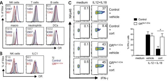
We observed that splenic NKp46+ ILCs, most of which
are conventional NK cells, expressed higher levels of GR than T and B lymphocytes (Fig. 2 A). In the liver, both NK cells and ILC1s also expressed GR (Fig. 2 B). An analysis of NKp46+
ILCs subsets in the small intestine showed that GR was ex-pressed in NK cells, ILC1s, and the NKp46+ subset of ILC3s
(not depicted). As expected, flow cytometry analysis revealed that the GR protein was selectively depleted in all NKp46+
ILCs subsets from the spleen (Fig. 2 A), liver (Fig. 2 B), and small intestine (not depicted) of GRNcr1-iCre mice, whereas it
was retained in other hematopoietic cells, including T cells, B cells, macrophages, neutrophils and DCs (Fig. 2 A).
We then analyzed the effect of the Nr3c1 gene deletion in NKp46+ cells at steady-state. The numbers and percentages
of all NKp46+ ILC subsets in the spleen, the liver and the
small intestine of GRNcr1-iCre mice were normal (Fig. S1 A and
not depicted). In addition, the expression of the inhibitory and activating receptors Ly49G2, NKG2A, KLRG1, Ly49D, Ly49H and the maturation markers CD11b, CD27, CD43 re-vealed no change in NK cell maturation and phenotype (Fig. S1, B–D). These results demonstrate the lack of requirement of the GC–GR pathway for the development, maturation,
and homeostasis of NK cells, ILC1s, and NKp46+ ILC3s at
steady state, contrary to T cells, in which such signaling is re-quired for thymocyte selection (Mittelstadt et al., 2012).
We then investigated the modulation of NK cell func-tions by GCs in an in vitro activation assay in the pres-ence or abspres-ence of corticosterone. Corticosterone treatment inhibited IFN-γ production by spleen NK cells stimulated with IL-12 and IL-18 (Fig. 2 C). Importantly, this regulation was NK cell intrinsic, as it was not observed with spleno-cytes from GRNcr1-iCre mice (Fig. 2 C).
These in vitro results highlighted the potential role of GR in NK cells in inflammatory conditions, prompting us to inves-tigate whether the HPA axis, via the production of corticoste-rone, could modulate IFN-γ–producing ILC functions in vivo.
the regulation of IFn-γ production associated with
endotoxin tolerance requires Gr expression in group 1 ILcs
The mechanisms involved in the reduced IFN-γ produc-tion after the inducproduc-tion of endotoxin tolerance are poorly understood. We assessed the in vivo consequences of the modulation of NKp46+ ILC functions by endogenous GCs
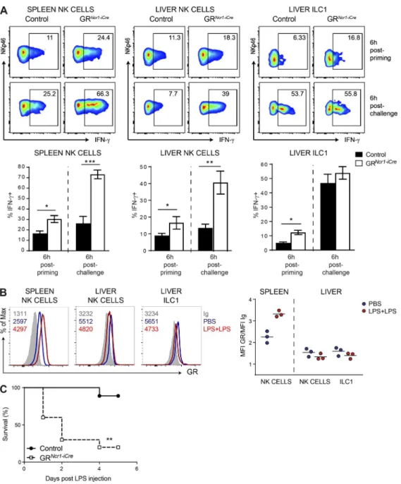
by challenging GRNcr1-iCre mice and their control littermates
using the endotoxin tolerance protocol. Six hours after LPS priming, we found that the frequency of splenic and liver NK cells and liver-resident ILC1s producing IFN-γ
Figure 1. repeated LPs injections pro-mote resistance to endotoxic shock and Gc production. (A) Protocol to induce
endotox-in-induced sepsis or endotoxin tolerance. For the sepsis model, mice not injected or injected with PBS at day 1 (D-1) behave equally (not depicted). (B) Survival curve for mice receiving LPS injections according to the protocol in A. n = 11–12 mice pooled from two indepen-dent experiments; ***, P < 0.001 (Mantel-Cox test). (C and D) Cytokines and corticoste-rone concentrations in the serum. Data are presented as mean ± SEM. Each symbol in D represents a single mouse. n = 6–10 mice pooled from two independent experiments; *, P < 0.05; **, P < 0.01; ***, P < 0.001; ****, P < 0.0001 (one-way ANO VA).
in GRNcr1-iCre mice was much higher than that in control
mice (Fig. 3 A). Moreover, 6 h after the secondary LPS in-jection (LPS challenge) the frequency of IFN-γ+ NK cells
in both the spleen and liver of GRNcr1-iCre mice was higher
than that in the controls, whereas the production of IFN-γ by liver-resident ILC1s was unaffected at this time point (Fig. 3 A). Thus, during the priming phase, endogenous cor-ticosterone inhibited IFN-γ production by NK cells and ILC1s. In contrast, upon secondary challenge, only spleen and liver NK cells had higher levels of IFN-γ production in the absence of GR signaling.
We then investigated the roles of the GR on group 1 ILC functions during the primary and secondary phases by determining whether GR expression was modulated during these inflammatory challenges. We found that multiple LPS challenge increased GR expression in spleen NK cells but had no effect or slightly decreased GR expression in liver NK cells and resident ILC1s (Fig. 3 B). The regulation of GR ex-pression in NK cells in this model is, therefore, organ specific. Our results also suggest that the level of GR expression in ILC1s is not sufficient to account for the lack of regulation of IFN-γ production by the GC–GR pathway upon the second-ary LPS challenge (although it may contribute to this effect), as liver NK cells, which have a similar phenotype, are sensitive to this regulation. This difference in ILC1 sensitivity to GR regulation between the two phases indicates that the mech-anisms of control of IFN-γ production in these cells differs from that in NK cells in conditions of endotoxin tolerance.
As mentioned in the previous section, the NKp46+
innate cells targeted with the Ncr1iCre model include NK
cells, ILC1s, and NKp46+ ILC3s (also called NCR+ ILC3s),
which are undetectable in spleen and liver at steady state but present in the gut (Narni-Mancinelli et al., 2011). Upon LPS challenge, no IFN-γ production by small in-testine RORγt+ ILC3s, NK1.1+RORγt− Eomes− ILC1s,
and NK1.1+RORγt− Eomes+ NK cells was detected ex
vivo (not depicted), suggesting that these intestinal cell subsets do not play a major role in IFN-γ production in this model. As a control, we induced IFN-γ production in small-intestine NK cells and ILC1s (but not NCR+
ILC3s) upon in vitro restimulation with cytokines (IL-12 and IL-18), PMA, and ionomycin, showing that these cells can be responsive (Fig. S2 A). However, under these in vitro experimental conditions, the frequency of IFN-γ+
cells observed after the stimulation of cells from mice treated with PBS or LPS was similar, demonstrating that the responsiveness of these cells was not affected by the inflammatory or endogenous GC responses induced by the endotoxin (Fig. S2 A). We can thus conclude that NKp46+
cells in the small intestine are not a significant source of IFN-γ in our system.
We then checked whether parameters other than IFN-γ production were regulated by GR in group 1 ILCs by ana-lyzing the TNF-α production of these cells, their prolifera-tion, and their expression of other functional markers. The frequency of TNF-α-producing NK cells and ILC1s after LPS challenge was unchanged in the spleen and liver of GRNcr1-iCre
mice, as shown by comparison with control animals (Fig. S2 B). In addition, the absolute number of NK cells and ILC1s and their proliferative status, analyzed by evaluating the levels
Figure 2. corticosterone inhibits IFn-γ production by nK cells through Gr signaling. (A and B) GR expression and isotype control (Ig) in NK cells (NK-p46+NK1.1+CD3−CD19−), T cells (CD3+NK1.1−CD19−), B cells (CD19+CD3−), macrophages (CD11b+F4/80+), neutrophils (CD11b+Ly6G+), and DCs (CD11c+MHC
II+) from the spleen (A) and NK cells (NKp46+NK1.1+CD3−CD19−DX5+CD49a−) and resident ILC1s (NKp46+NK1.1+CD3−CD19−DX5−CD49a+) in the liver (B).
The numbers on the plots represent the mean fluorescence intensity. The data shown are representative of two independent experiments with five mice per group. (C) Splenocytes were left untreated (medium) or stimulated for 4 h with IL-12 and IL-18 in the presence of 500 nM corticosterone (cort) or vehicle alone. The percentage of IFN-γ+ NKp46+ cells is indicated for each FACS plot. Histograms represent these percentages as means + SEM (n = 7–8 mice pooled
of the Ki67 marker, were similar in GRNcr1-iCre and control
mice (Fig. S2 C). Finally, levels of the activation markers CD69 and granzyme B were also similar (Fig. S2 D), demonstrating that, in the context of endotoxin tolerance, GR expression has no effect on these other parameters linked to cell activation and functional competence. Thus IFN-γ production is the only one of the “canonical” NK cell and ILC1 activation path-ways analyzed to display selective sensitivity to GR regulation.
nKp46+ cell responsiveness to Gcs is required for the
development of endotoxin tolerance
We evaluated the physiological relevance of the regulation of IFN-γ production in group 1 ILCs by the GR pathway by comparing the survival of GRNcr1-iCre mice and their control
littermates after LPS challenge. We found that priming with a low dose of LPS did not rescue survival in GRNcr1-iCre mice, in
marked contrast with control animals (Fig. 3 C), showing that
Figure 3. spleen and liver group 1 ILc responsiveness to Gcs is required for the development of endotoxin tolerance. (A) Percentage of IFN-γ+
NK cells (NKp46+NK1.1+CD3−CD19−DX5+CD49a−) and ILC1s (NKp46+NK1.1+CD3−CD19−DX5−CD49a+) in the spleen and liver of control and GRNcr1-iCre mice,
as determined 6 h after LPS priming and challenge. Data are presented as representative FACS plots and as mean ± SEM (n = 5–7 mice pooled from two independent experiments; *, P < 0.05; **, P < 0.01; ***, P < 0.001; Mann–Whitney U test). (B) GR expression and isotype control (Ig) measured 6 h after PBS injection or LPS challenge. FACS histograms and the mean value of the GR/Ig ratio from one representative experiment with three mice per group are shown. Each symbol represents a single mouse. (C) Survival curve for control and GRNcr1-iCre mice (n = 7–11 mice pooled from two experiments; **, P <
the responsiveness of NKp46-expressing cells to corticoste-rone was required for the development of endotoxin tolerance. In vivo studies in animal models (Delano et al., 2007) and ex vivo experiments on human peripheral blood mono-nuclear cells (Pena et al., 2011) have shown that multiple challenges with LPS induce myeloid cells to switch to an an-ti-inflammatory phenotype. Several tolerization mechanisms have been reported to operate at the signal transduction, transcriptional, and epigenetic levels (Mengozzi et al., 1991; Frankenberger et al., 1995; Foster et al., 2007). Interestingly, some of these mechanisms can be abolished by IFN-γ (Fran-kenberger and Ziegler-Heitbrock, 1997; Chen and Ivashkiv, 2010; Turrel-Davin et al., 2011). In particular, pretreatment with IFN-γ in vitro can prevent the tolerization of primary human monocytes and restore the TLR4-mediated induction of various proinflammatory cytokines, including IL-6 and TNF-α (Chen and Ivashkiv, 2010).
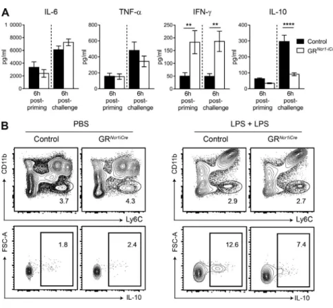
We investigated the mechanisms by which this HPA-driven regulation of NKp46+ cells promotes
endo-toxin tolerance by determining the levels of inflammatory cytokines in the serum 6 h after the first (priming) and the second (challenge) LPS injections. In this in vivo model, we found that serum IL-6 and TNF-α concentrations were not significantly affected in GRNcr1-iCre mice, despite the higher
serum levels of IFN-γ in GRNcr1-iCre mice both after
prim-ing and after rechallenge (Fig. 4 A). In contrast, IL-10 serum levels were similar after priming but significantly lower in these GRNcr1-iCre animals after LPS rechallenge (Fig. 4 A).
These results suggest that the down-regulation of IFN-γ
production by corticosterone in group 1 ILCs has a major impact on systemic IFN-γ levels during both the priming and challenge phases. Moreover, a GR-dependent pathway in NKp46-expressing cells allows the development of an anti-inflammatory status associated with the production of IL-10. The reduction of systemic IL-10 levels in GR condi-tional KO mice was not caused by an intrinsic effect of GCs on IL-10 production by NKp46+ cells because these cells do
not produce this cytokine (Fig. S3 A). Similarly, we did not detect IL-10 production in T cells, B cells, or DCs upon LPS challenge (not depicted). In contrast, we identified a popula-tion of Ly6Chigh CD11blow myeloid cells in the spleen as the
main source of IL-10 in this model (Fig. 4 B). These data therefore suggest that microbial LPS exposure induces a direct corticosterone-mediated effect on NKp46-expressing group 1 ILCs, inhibiting their IFN−γ production. In addition, after a second encounter with endotoxin an indirect effect on the production of IL-10 by myeloid cells is observed.
corticosterone allows the development of IL-10–dependent endotoxin tolerance by preventing IFn-γ production
by group 1 ILcs
The regulation by GCs of IFN-γ production by group 1 ILCs may therefore be crucial to maintain the correct balance between resistance to pathogens and resistance to inflammatory disease and immunopathology. We directly addressed the question of the role of the regulation of IFN-γ production by group 1 ILCs by the HPA axis in mouse resistance to disease, by challenging GRNcr1-iCre and control Figure 4. Higher levels of IFn-γ in GrNcr1-iCre
mice are associated with lower systemic IL-10 concentrations. (A) Cytokines in the serum, as
determined 6 h after priming and after challenge with LPS. Data are presented as mean ± SEM (n = 14–16 mice pooled from three experiments; **, P < 0.01; ****, P < 0.0001; Student’s t test). (B) FACS plots showing IL-10 intracellular staining in Ly6C+CD11blow cells 6 h after challenge with LPS,
after gating out CD19+, CD3+, and CD11c+ cells.
The data shown are representative of two inde-pendent experiments with seven mice per group.
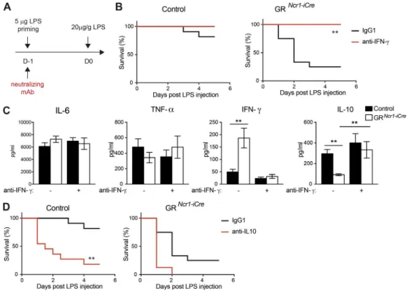
animals with LPS in the presence or absence of monoclo-nal antibodies (mAbs) neutralizing IFN-γ during the prim-ing phase (Fig. 5 A). In the presence of IFN-γ blockade, tolerance to endotoxin in GRNcr1-iCre mice was rescued to
levels similar to those observed in control mice (Fig. 5 B). Importantly, serum IL-10 concentration in GRNcr1-iCre mice
was also restored to control levels, confirming that the down-regulation of this cytokine was not intrinsically regu-lated in NKp46+ cells but extrinsically linked to the higher
levels of IFN-γ (Fig. 5 C). In contrast, in accordance with the lack of change in TNF-α and IL-6 production levels in GRNcr1-iCre mice upon endotoxin tolerance (Fig. 4 A), IFN-γ
neutralization did not affect the production of these cyto-kines after LPS challenge (Fig. 5 C). Endogenous GCs pro-duced after a first low-level microbial exposure thus regulate an IFN-γ–IL-10 axis, which is crucial for the establishment of endotoxin tolerance and host survival upon a secondary pathogen encounter. GR signaling in NKp46-expressing ILCs is essential for the regulation of this pathway. Inter-estingly, in the sepsis model (only one high dose of LPS injected), despite an increase in the systemic levels of IFN-γ in GRNcr1-iCre mice, the amount of IL-10 was similar to that
in the control (Fig. S3 B), suggesting that the
reprogram-ing of myeloid cell functions by IFN-γ is dependent on the inflammatory context.
Finally, the importance of the corticosterone regulation of group 1 ILC IFN-γ production and the consequent in-crease in systemic IL-10 levels was further demonstrated by IL-10 neutralization (Fig. S3 C), which abolished protection in control mice, reducing survival to a level similar to that in GRNcr1-iCre mice (Fig. 5 D). The role of IL-10 in endotoxin
tol-erance has been a matter of debate. Some studies have reported a weak effect of this cytokine (Berg et al., 1995; Murphey and Sherwood, 2006), whereas others have reported a requirement for systemic IL-10 in endotoxin tolerance (Randow et al., 1995; Sfeir et al., 2001; Muehlstedt et al., 2002; Muenzer et al., 2010). It is possible that, in some specific experimental set-tings, the expression of other antiinflammatory factors (such as TGF-β) contributes to endotoxin tolerance. However, our data are consistent with a major role of IL-10 in this process, as we demonstrated that IL-10 neutralization completely abolished endotoxin tolerance. We also found that IFN-γ production by group 1 ILCs has to be intrinsically regulated by GR to allow this IL-10–dependent host resistance to inflammatory disease.
Altogether, these data support a model in which HPA axis activation leads to GC production during an initial phase
Figure 5. IFn-γ neutralization restores tolerance to endotoxin and IL-10 production in GrNcr1-iCre mice. (A, B, and D) Mice received LPS injections
and were treated with anti–IFN-γ (B) or anti–IL-10 (D) neutralizing antibodies or isotype control (IgG1) antibodies at the priming phase. (B) Survival curves for control and GRNcr1-iCre mice treated with anti–IFN-γ neutralizing antibodies (n = 8–11 mice from two independent experiments, plotted in separated
graphs; **, P < 0.01; Mantel–Cox test). (C) Cytokines in the serum as determined 6 h after challenge with LPS. Data are presented as mean ± SEM (n = 8–14 mice pooled from three independent experiments; **, P < 0.01; one-way ANO VA). (D) Survival curves for control and GRNcr1-iCre mice treated with anti–IL-10
of inflammation induced by bacterial infection, which in turn controls IFN-γ production by group 1 ILCs in spleen and liver via GR. The respective roles of NK cells and ILC1s in this process are difficult to differentiate in the absence of an available model for selectively depleting one of these sub-populations. However, based on the frequency of each cell type and the fact that ILC1s are susceptible to this regulation by GR only during the priming phase, we favor a model in which NK cells have a more prominent role. Nevertheless, this regulatory mechanism is required for the establishment of a state of immunosuppression characterized by high serum concentrations of the anti-inflammatory cytokine IL-10 and resistance to septic shock.
concluding remarks
The ability of host organisms to tolerate, at least to some ex-tent, the presence of a pathogen can be seen as part of a global defense strategy enabling the host organism to protect itself from infectious diseases not only by fighting against patho-gens but also by reducing the negative impact of infection on host fitness (Medzhitov et al., 2012). The HPA-dependent pathway controlling IFN-γ release by spleen and liver group 1 ILCs described here reveals an important mechanism of the fine balance facilitating the activation of the immune re-sponse by an infectious agent while preserving host integrity, which could be integrated into the broader concept of “dis-ease tolerance.” This pathway plays a role upstream from my-eloid cell reprogramming, the principal process investigated to date, and is essential for the development of an immune suppressive state, in which IL-10 is a major player.
In some clinical circumstances, the refractory state that allows resistance to endotoxin may also be associated with an increase in susceptibility to secondary nosocomial infec-tions (Cavaillon et al., 2003). A loss of LPS reactivity similar to that reported in patients with sepsis has been found in patients with noninfectious systemic inflammation response syndrome (SIRS) caused by trauma, surgery, or cardiac arrest, who have a higher risk of succumbing to infections than other patients. Interestingly, in blood samples from patients with bacterial sepsis or SIRS, IFN-γ production in response to TLR agonists is abolished (Souza-Fonseca-Guimaraes et al., 2012a,b). In addition, NK cell immunosuppression has been observed in blood samples from trauma patients with brain injury, where poor NK cell recruitment into a BCG-induced granuloma model was observed (Deknuydt et al., 2013). Because the HPA axis can also be activated in such stressful situations, a knowledge of the molecular and cellular mechanisms involved in the induction of this state of tolerance may be important for the development of strat-egies for restoring a functional immune response in these patients. Our present findings thus shed new light on the critical role of group 1 ILCs in inflammation and the reg-ulatory mechanisms promoting disease tolerance, paving new avenues of translational research for the treatment of inflammatory disorders.
MAterIALs And MetHods Mice
C57BL/6J mice were purchased from Janvier Labs; Ncr1iCre
mice were generated as described previously (Narni-Manci-nelli et al., 2011), and Nr3c1LoxP/LoxP mice were provided
by F. Tronche (Sorbonne Universités, Université Pierre et Marie Curie, UMR_CR18, Neuroscience, Paris-Seine, Paris, France; Tronche et al., 1999). All mice were bred and maintained under specific-pathogen–free conditions at the Centre d’Immunophenomique de Marseille and in the Cen-tre d’Immunologie de Marseille Luminy. Mice were housed under a standard 12 h/12 h light–dark cycle with food and water ad libitum. Age-matched (7–12 wk old) and sex-matched littermate mice were used. All experiments were conducted in accordance with institutional committees (Comité d’éthique de Marseille 14) and French and Euro-pean guidelines for animal care.
Flow cytometry
Single-cell suspensions from the spleen or liver (after lym-phocyte isolation on a 37.5%-67.5% Percoll gradient) or small intestine (after digestion with collagenase VIII from Sigma and lymphocyte isolation on a 40–100% Percoll gra-dient) were incubated with Fc blocking antibody (2.4G2) and the fixable blue dead cell stain kit (Invitrogen). Surface molecules were stained using antibodies against CD45.2 (104), CD3 (145-2C11), CD19 (1D3), NK1.1 (PK136), CD49a (Ha31/8), CD11b (M1/70), CD27 (LG.3A10), CD43 (S7), KLRG1 (2F1), Ly49G2 (4D11), CD69 (H1.2F3), CD11b (M1/70), MHC II (M5/114.15.2), Ly6C (AL-21), CD3 (145-2C11), CD19 (1D3) from BD Biosci-ences; NKp46 (29A1.4), CD49b (DX5), NKG2A (16a11), Ly49D (4E5), Ly49H (3D10), and F4/80 (BM8) from eBioscience and CD11c (N418) and Ly6G (1A8) from BioLegend. For intracellular staining, cells were fixed and permeabilized with an intracellular staining kit (eBiosci-ence) and the following antibodies were used: anti–GR XP rabbit mAb (D8H2) and rabbit mAb IgG XP (DA1E) from Cell Signaling Technology; anti–IFN-γ (XMG1.2) from BioLegend; anti–Rorγt (Q31-378), anti-TNF (MP6-XT22), anti–granzyme B (GB11), anti-Ki67 (B56), and anti–IL-10 (JE5-16E3) from BD Biosciences; and an-ti-Eomes (Dan11mag) from eBioscience.
In vitro splenocyte stimulation
Splenocytes from control and GRNcr1-iCre mice were
stim-ulated in vitro with 25 ng/ml IL-12 and 20 ng/ml IL-18 in complete culture medium (RPMI 10% FCS, 100 µg/ml penicillin/streptomycin, 2 mM l-glutamine, 1 mM sodium pyruvate, and 0.01 M Hepes) with the addition of 500 nM corticosterone (Sigma; dissolved in ethanol) or the same vol-ume of vehicle alone. Cells were stimulated at 37°C in the presence of Golgi Stop and Golgi Plug from BD Biosci-ences. After 4 h of stimulation, the cells were washed and stained for FACS analysis.
ex vivo cell stimulation
For IL-10 detection, splenocytes were stimulated with 500 ng/ml PMA and 500 ng/ml ionomycin (both from Sigma) for 2 h at 37°C in the presence of Golgi Plug. For IFN-γ detection in small intestine lymphocytes, cells were stimu-lated with 200 ng/ml PMA, 1 µg/ml ionomycin, 25 ng/ml IL-12, and 20 ng/ml IL-18 for 2 h at 37°C in the presence of Golgi Stop and Golgi Plug.
serum analysis
Blood was collected from the retro-orbital sinus of mice receiving LPS injections under low-stress condi-tions (i.e., within 2 min of handling). Sera were tested with the Corticosterone ELI SA kit (Enzo) according to the manufacturer’s instructions for determining cor-ticosterone concentration. The concentrations of IL-6, TNF-α, IFN-γ, and IL-10 were assessed with cytomet-ric bead arrays according to the manufacturer’s protocol (CBA; BD Biosciences).
In vivo LPs challenge
LPS (from Escherichia coli strain 055:B5; Sigma) diluted in PBS was injected intraperitoneally (20 µg per gram mouse body weight), and in endotoxin tolerance ex-periments, 5 µg LPS was injected, in a total volume of 30 µl, into the footpads of mice under anesthesia with 3.5% isoflurane. All LPS injections were performed be-tween 9 a.m. and 10 a.m., and mice were checked every 12 h for signs of distress in survival experiments. Cyto-kine neutralization was achieved by the intraperitoneal injection of 500 µg anti–IFN-γ (XMG1.2), anti–IL-10 (JES5-2A5), or rat IgG1 (HRPN; all from BioXCell) at the time of LPS priming.
statistical analysis
Statistical analysis was performed with GraphPad Prism software. Data were considered statistically significant if the p-value obtained was lower than 0.05. Data were compared with unpaired Student’s t tests if the values followed a Gaussian distribution with similar variances or the Mann–Whitney U test otherwise. For multi-group comparisons, we applied one-way ANO VA or multiple t tests. Differences in survival were evaluated with the Mantel–Cox test.
online supplemental material
Fig. S1 shows that GRNcr1-iCre mice have normal NKp46+
cell phenotype, numbers, and maturation. Fig. S2 shows that GR expression does not affect parameters linked to NKp46+
cell activation and functional competence other than IFN-γ production by group 1 ILCs in the spleen and liver in endo-toxin tolerance. Fig. S3 shows that the low systemic IL-10 concentration in GRNcr1-iCre mice is not directly caused
by NKp46+ cell-intrinsic regulation by GR and is specific
to endotoxin tolerance.
AcKnowLedGMents
The authors thank François Tronche for providing the Nr3c1LoxP/LoxP transgenic mice, Justine Galluso and Pascaline Morganti for mice breeding and genotyping, Bruno Lucas for helpful discussions and for providing a crucial experimental protocol, and the Centre d'Immunologie de Marseille-Luminy mouse house and core cy-tometry facilities.
The S. Ugolini laboratory is supported by the European Research Council (ERC), Agence Nationale de la Recherche (ANR), Fondation ARC, and Ligue Nationale contre le Cancer. In particular, this project has received funding from the ERC under the European Union's Horizon 2020 research and innovation programme (under grant no. 648768), from the ANR (ANR-14-CE14-0009-01), and from the Fondation ARC (PGA120140200817). The E. Vivier laboratory is supported by the European Re-search Council, Agence Nationale de la Recherche, Innate Pharma, MSDAvenir, and Ligue Nationale contre le Cancer (Equipe labelisée “La Ligue”). L. Quatrini was partially supported by a fellowship from the Italian Foundation for Cancer Research (AIRC). This work was also supported by institutional grants from Institut National de la Santé et de la Recherche Médicale, Centre National de la Recherche Scientifique, Aix-Marseille University, and Marseille-Immunopole to the Centre d'Immunologie de Marseille-Luminy.
E. Vivier is the cofounder and a shareholder of Innate Pharma. The remaining authors declare no competing financial interests.
Author contributions: L. Quatrini designed and performed experiments and analyzed data. E. Wieduwild, S. Guia, and C. Bernat performed experiments. N. Glaic-henhaus and E. Vivier contributed to design the study. S. Ugolini conceived, designed, and directed the study and wrote the manuscript with L. Quatrini. All authors re-viewed and provided input on the manuscript.
Submitted: 9 June 2017 Revised: 7 September 2017 Accepted: 11 October 2017
reFerences
Adib-Conquy, M., C. Adrie, C. Fitting, O. Gattolliat, R. Beyaert, and J.M. Cavaillon. 2006. Up-regulation of MyD88s and SIG IRR, molecules inhibiting Toll-like receptor signaling, in monocytes from septic patients.
Crit. Care Med. 34:2377–2385. https ://doi .org /10 .1097 /01 .CCM
.0000233875 .93866 .88
Balkhy, H.H., and F.P. Heinzel. 1999. Endotoxin fails to induce IFN-gamma in endotoxin-tolerant mice: deficiencies in both IL-12 heterodimer pro-duction and IL-12 responsiveness. J. Immunol. 162:3633–3638. Berg, D.J., R. Kühn, K. Rajewsky, W. Müller, S. Menon, N. Davidson, G.
Grünig, and D. Rennick. 1995. Interleukin-10 is a central regulator of the response to LPS in murine models of endotoxic shock and the Shwartzman reaction but not endotoxin tolerance. J. Clin. Invest. 96:2339–2347. https ://doi .org /10 .1172 /JCI118290
Bertini, R., M. Bianchi, and P. Ghezzi. 1988. Adrenalectomy sensitizes mice to the lethal effects of interleukin 1 and tumor necrosis factor. J. Exp.
Med. 167:1708–1712. https ://doi .org /10 .1084 /jem .167 .5 .1708
Bhattacharyya, S., D.E. Brown, J.A. Brewer, S.K. Vogt, and L.J. Muglia. 2007. Macrophage glucocorticoid receptors regulate Toll-like receptor 4-mediated inflammatory responses by selective inhibition of p38 MAP kinase. Blood. 109:4313–4319. https ://doi .org /10 .1182 /blood -2006 -10 -048215
Bundschuh, D.S., J. Barsig, T. Hartung, F. Randow, W.D. Döcke, H.D. Volk, and A. Wendel. 1997. Granulocyte-macrophage colony-stimulating factor and IFN-gamma restore the systemic TNF-alpha response to endotoxin in lipopolysaccharide-desensitized mice. J. Immunol. 158:2862–2871. Cain, D.W., and J.A. Cidlowski. 2017. Immune regulation by glucocorticoids.
Nat. Rev. Immunol. 17:233–247. https ://doi .org /10 .1038 /nri .2017 .1
Cavaillon, J.M., C. Adrie, C. Fitting, and M. Adib-Conquy. 2003. Endotoxin tolerance: is there a clinical relevance? J. Endotoxin Res. 9:101–107. https ://doi .org /10 .1177 /09680519030090020501
Chen, J., and L.B. Ivashkiv. 2010. IFN-γ abrogates endotoxin tolerance by facilitating Toll-like receptor-induced chromatin remodeling. Proc.
Natl. Acad. Sci. USA. 107:19438–19443. https ://doi .org /10 .1073 /pnas
.1007816107
Chiche, L., J.M. Forel, G. Thomas, C. Farnarier, F. Vely, M. Bléry, L. Papazian, and E. Vivier. 2011. The role of natural killer cells in sepsis. J. Biomed.
Biotechnol. 2011:986491. https ://doi .org /10 .1155 /2011 /986491
Cortez, V.S., and M. Colonna. 2016. Diversity and function of group 1 innate lymphoid cells. Immunol. Lett. 179:19–24. https ://doi .org /10 .1016 /j .imlet .2016 .07 .005
Deknuydt, F., A. Roquilly, R. Cinotti, F. Altare, and K. Asehnoune. 2013. An in vitro model of mycobacterial granuloma to investigate the immune response in brain-injured patients. Crit. Care Med. 41:245–254. https :// doi .org /10 .1097 /CCM .0b013e3182676052
Delano, M.J., P.O. Scumpia, J.S. Weinstein, D. Coco, S. Nagaraj, K.M. Kelly-Scumpia, K.A. O’Malley, J.L. Wynn, S. Antonenko, S.Z. Al-Quran, et al. 2007. MyD88-dependent expansion of an immature GR-1(+) CD11b(+) population induces T cell suppression and Th2 polarization in sepsis. J. Exp. Med. 204:1463–1474. https ://doi .org /10 .1084 /jem .20062602
Dumbell, R., O. Matveeva, and H. Oster. 2016. Front. Endocrinol. (Lausanne). 7:37. https ://doi .org /10 .3389 /fendo .2016 .00037
Dunn, A.J. 2000. Cytokine activation of the HPA axis. Ann. N. Y. Acad. Sci. 917:608–617. https ://doi .org /10 .1111 /j .1749 -6632 .2000 .tb05426 .x Edwards, C.K. III, L.M. Yunger, R.M. Lorence, R. Dantzer, and K.W. Kelley.
1991. The pituitary gland is required for protection against lethal effects of Salmonella typhimurium. Proc. Natl. Acad. Sci. USA. 88:2274–2277. https ://doi .org /10 .1073 /pnas .88 .6 .2274
Evans, G.F., and S.H. Zuckerman. 1991. Glucocorticoid-dependent and -independent mechanisms involved in lipopolysaccharide tolerance. Eur.
J. Immunol. 21:1973–1979. https ://doi .org /10 .1002 /eji .1830210902
Foster, S.L., D.C. Hargreaves, and R. Medzhitov. 2007. Gene-specific control of inflammation by TLR-induced chromatin modifications. Nature. 447:972–978. https ://doi .org /10 .1038 /nature05836
Frankenberger, M., and H.W. Ziegler-Heitbrock. 1997. LPS tolerance in monocytes/macrophages: three 3′ cytosins are required in the DNA binding motif for detection of upregulated NF-kappa B p50 homodimers. Immunobiology. 198:81–90. https ://doi .org /10 .1016 / S0171 -2985(97)80029 -0
Frankenberger, M., H. Pechumer, and H.W. Ziegler-Heitbrock. 1995. Interleukin-10 is upregulated in LPS tolerance. J. Inflamm. 45:56–63. Irwin, M.R., and S.W. Cole. 2011. Reciprocal regulation of the neural and
innate immune systems. Nat. Rev. Immunol. 11:625–632. https ://doi .org /10 .1038 /nri3042
Kadmiel, M., and J.A. Cidlowski. 2013. Glucocorticoid receptor signaling in health and disease. Trends Pharmacol. Sci. 34:518–530. https ://doi .org /10 .1016 /j .tips .2013 .07 .003
Kleiman, A., S. Hübner, J.M. Rodriguez Parkitna, A. Neumann, S. Hofer, M.A. Weigand, M. Bauer, W. Schmid, G. Schütz, C. Libert, et al. 2012. Glucocorticoid receptor dimerization is required for survival in septic shock via suppression of interleukin-1 in macrophages. FAS EB J. 26:722–729. https ://doi .org /10 .1096 /fj .11 -192112
Lauw, F.N., T. ten Hove, P.E. Dekkers, E. de Jonge, S.J. van Deventer, and T. van Der Poll. 2000. Reduced Th1, but not Th2, cytokine production by lymphocytes after in vivo exposure of healthy subjects to endotoxin.
Infect. Immun. 68:1014–1018. https ://doi .org /10 .1128 /IAI .68 .3 .1014
-1018 .2000
Li, C.C., I. Munitic, P.R. Mittelstadt, E. Castro, and J.D. Ashwell. 2015. Suppression of Dendritic Cell-Derived IL-12 by Endogenous Glucocorticoids Is Protective in LPS-Induced Sepsis. PLoS Biol. 13:e1002269. https ://doi .org /10 .1371 /journal .pbio .1002269
López-Collazo, E., and C. del Fresno. 2013. Pathophysiology of endotoxin tolerance: mechanisms and clinical consequences. Crit. Care. 17:242. https ://doi .org /10 .1186 /cc13110
Medzhitov, R., D.S. Schneider, and M.P. Soares. 2012. Disease tolerance as a defense strategy. Science. 335:936–941. https ://doi .org /10 .1126 /science .1214935
Mengozzi, M., M. Sironi, M. Gadina, and P. Ghezzi. 1991. Reversal of de-fective IL-6 production in lipopolysaccharide-tolerant mice by phorbol myristate acetate. J. Immunol. 147:899–902.
Mittelstadt, P.R., J.P. Monteiro, and J.D. Ashwell. 2012. Thymocyte responsiveness to endogenous glucocorticoids is required for immunological fitness. J. Clin. Invest. 122:2384–2394. https ://doi .org /10 .1172 /JCI63067
Muehlstedt, S.G., M. Lyte, and J.L. Rodriguez. 2002. Increased IL-10 production and HLA-DR suppression in the lungs of injured patients precede the development of nosocomial pneumonia. Shock. 17:443–450. https ://doi .org /10 .1097 /00024382 -200206000 -00001
Muenzer, J.T., C.G. Davis, K. Chang, R.E. Schmidt, W.M. Dunne, C.M. Coopersmith, and R.S. Hotchkiss. 2010. Characterization and modulation of the immunosuppressive phase of sepsis. Infect. Immun. 78:1582–1592. https ://doi .org /10 .1128 /IAI .01213 -09
Murphey, E.D., and E.R. Sherwood. 2006. Bacterial clearance and mortality are not improved by a combination of IL-10 neutralization and IFN-gamma administration in a murine model of post-CLP immunosuppression. Shock. 26:417–424. https ://doi .org /10 .1097 /01 .shk .0000226343 .70904 .4f
Narni-Mancinelli, E., J. Chaix, A. Fenis, Y.M. Kerdiles, N. Yessaad, A. Reynders, C. Gregoire, H. Luche, S. Ugolini, E. Tomasello, et al. 2011. Fate mapping analysis of lymphoid cells expressing the NKp46 cell surface receptor.
Proc. Natl. Acad. Sci. USA. 108:18324–18329. https ://doi .org /10 .1073
/pnas .1112064108
Oster, H., E. Challet, V. Ott, E. Arvat, E.R. de Kloet, D.J. Dijk, S. Lightman, A. Vgontzas, and E. Van Cauter. 2016. The functional and clinical significance of the 24-h rhythm of circulating glucocorticoids. Endocr.
Rev. 38:3–45. https ://doi .org /10 .1210 /er .2015 -1080
Pena, O.M., J. Pistolic, D. Raj, C.D. Fjell, and R.E. Hancock. 2011. Endotoxin tolerance represents a distinctive state of alternative polarization (M2) in human mononuclear cells. J. Immunol. 186:7243–7254. https ://doi .org /10 .4049 /jimmunol .1001952
Peng, H., X. Jiang, Y. Chen, D.K. Sojka, H. Wei, X. Gao, R. Sun, W.M. Yokoyama, and Z. Tian. 2013. Liver-resident NK cells confer adaptive immunity in skin-contact inflammation. J. Clin. Invest. 123:1444–1456. https ://doi .org /10 .1172 /JCI66381
Perlstein, R.S., M.H. Whitnall, J.S. Abrams, E.H. Mougey, and R. Neta. 1993. Synergistic roles of interleukin-6, interleukin-1, and tumor necrosis factor in the adrenocorticotropin response to bacterial lipopolysaccharide in vivo. Endocrinology. 132:946–952. https ://doi .org /10 .1210 /endo .132 .3 .8382602
Porta, C., M. Rimoldi, G. Raes, L. Brys, P. Ghezzi, D. Di Liberto, F. Dieli, S. Ghisletti, G. Natoli, P. De Baetselier, et al. 2009. Tolerance and M2 (alternative) macrophage polarization are related processes orchestrated by p50 nuclear factor kappaB. Proc. Natl. Acad. Sci. USA. 106:14978– 14983. https ://doi .org /10 .1073 /pnas .0809784106
Ramachandra, R.N., A.H. Sehon, and I. Berczi. 1992. Neuro-hormonal host defence in endotoxin shock. Brain Behav. Immun. 6:157–169. https ://doi .org /10 .1016 /0889 -1591(92)90015 -G
Randow, F., U. Syrbe, C. Meisel, D. Krausch, H. Zuckermann, C. Platzer, and H.D. Volk. 1995. Mechanism of endotoxin desensitization: involvement of interleukin 10 and transforming growth factor beta. J. Exp. Med. 181:1887–1892. https ://doi .org /10 .1084 /jem .181 .5 .1887
Rivier, C., R. Chizzonite, and W. Vale. 1989. In the mouse, the activation of the hypothalamic-pituitary-adrenal axis by a lipopolysaccharide
(endotoxin) is mediated through interleukin-1. Endocrinology. 125:2800– 2805. https ://doi .org /10 .1210 /endo -125 -6 -2800
Schroder, K., P.J. Hertzog, T. Ravasi, and D.A. Hume. 2004. Interferon-gamma: an overview of signals, mechanisms and functions. J. Leukoc. Biol. 75:163–189. https ://doi .org /10 .1189 /jlb .0603252
Sfeir, T., D.C. Saha, M. Astiz, and E.C. Rackow. 2001. Role of interleukin-10 in monocyte hyporesponsiveness associated with septic shock. Crit.
Care Med. 29:129–133. https ://doi .org /10 .1097 /00003246 -200101000
-00026
Sonnenberg, G.F., and D. Artis. 2015. Innate lymphoid cells in the initiation, regulation and resolution of inflammation. Nat. Med. 21:698–708. https ://doi .org /10 .1038 /nm .3892
Souza-Fonseca-Guimaraes, F., M. Parlato, C. Fitting, J.M. Cavaillon, and M. Adib-Conquy. 2012a. NK cell tolerance to TLR agonists mediated by regulatory T cells after polymicrobial sepsis. J. Immunol. 188:5850–5858. https ://doi .org /10 .4049 /jimmunol .1103616
Souza-Fonseca-Guimaraes, F., M. Parlato, F. Philippart, B. Misset, J.M. Cavaillon, and M. Adib-Conquy. Captain study group. 2012b. Toll-like receptors expression and interferon-γ production by NK cells in human sepsis. Crit. Care. 16:R206. https ://doi .org /10 .1186 /cc11838
Tronche, F., C. Kellendonk, O. Kretz, P. Gass, K. Anlag, P.C. Orban, R. Bock, R. Klein, and G. Schütz. 1999. Disruption of the glucocorticoid receptor gene in the nervous system results in reduced anxiety. Nat. Genet. 23:99– 103. https ://doi .org /10 .1038 /12703
Turrel-Davin, F., F. Venet, C. Monnin, V. Barbalat, E. Cerrato, A. Pachot, A. Lepape, C. Alberti-Segui, and G. Monneret. 2011. mRNA-based approach to monitor recombinant gamma-interferon restoration of LPS-induced endotoxin tolerance. Crit. Care. 15:R252. https ://doi .org /10 .1186 /cc10513
Varma, T.K., T.E. Toliver-Kinsky, C.Y. Lin, A.P. Koutrouvelis, J.E. Nichols, and E.R. Sherwood. 2001. Cellular mechanisms that cause suppressed gamma interferon secretion in endotoxin-tolerant mice. Infect. Immun. 69:5249–5263. https ://doi .org /10 .1128 /IAI .69 .9 .5249 -5263 .2001 Vivier, E., E. Tomasello, M. Baratin, T. Walzer, and S. Ugolini. 2008. Functions
of natural killer cells. Nat. Immunol. 9:503–510. https ://doi .org /10 .1038 /ni1582
Weikum, E.R., M.T. Knuesel, E.A. Ortlund, and K.R. Yamamoto. 2017. Glucocorticoid receptor control of transcription: precision and plasticity via allostery. Nat. Rev. Mol. Cell Biol. 18:159–174. https ://doi .org /10 .1038 /nrm .2016 .152
Yokoyama, W.M., D.K. Sojka, H. Peng, and Z. Tian. 2013. Tissue-resident natural killer cells. Cold Spring Harb. Symp. Quant. Biol. 78:149–156. https ://doi .org /10 .1101 /sqb .2013 .78 .020354
Yoza, B.K., and C.E. McCall. 2011. Facultative heterochromatin formation at the IL-1 beta promoter in LPS tolerance and sepsis. Cytokine. 53:145– 152. https ://doi .org /10 .1016 /j .cyto .2010 .10 .007