HAL Id: inserm-01391784
https://www.hal.inserm.fr/inserm-01391784
Submitted on 3 Nov 2016
HAL is a multi-disciplinary open access
archive for the deposit and dissemination of
sci-entific research documents, whether they are
pub-lished or not. The documents may come from
teaching and research institutions in France or
abroad, or from public or private research centers.
L’archive ouverte pluridisciplinaire HAL, est
destinée au dépôt et à la diffusion de documents
scientifiques de niveau recherche, publiés ou non,
émanant des établissements d’enseignement et de
recherche français ou étrangers, des laboratoires
publics ou privés.
A prospective multicenter study on bladder cancer: the
COBLAnCE cohort
Simone Benhamou, Julia Bonastre, Karine Groussard, François Radvanyi,
Yves Allory, Thierry Lebret
To cite this version:
Simone Benhamou, Julia Bonastre, Karine Groussard, François Radvanyi, Yves Allory, et al.. A
prospective multicenter study on bladder cancer: the COBLAnCE cohort. BMC Cancer, BioMed
Central, 2015, 16 (1), pp.837. �10.1186/s12885-016-2877-x�. �inserm-01391784�
S T U D Y P R O T O C O L
Open Access
A prospective multicenter study on bladder
cancer: the COBLAnCE cohort
Simone Benhamou
1,2,3*, Julia Bonastre
3,4, Karine Groussard
1,2, François Radvanyi
5,6†, Yves Allory
7,8,9†and Thierry Lebret
10,11Abstract
Background: Bladder cancer is a very heterogeneous disease as regards natural history. Environmental exposures, constitutional genetic and/or epigenetic background may affect not only the likelihood of bladder tumor occurrence, but also the histologic type of cancer and its outcome. Currently, only a few data are available to study the prognostic role of genetic and environmental factors. Likewise, data on the economic burden of bladder cancer and the longitudinal impact of the disease and the treatments on patient quality of life are scarce.
Methods: COBLAnCE is a large French-based clinical cohort study on bladder cancer. Newly diagnosed patients are enrolled prospectively in 12 public hospitals and 5 private for profits hospitals. The target sample size is 2,000 patients. All patients are to be followed for 6 years. Information on patient characteristics and lifestyle is collected during a face-to-face interview at enrollment. Clinical information on disease presentation, diagnosis, and treatment is extracted from medical records for the primary tumor and for all subsequent local and distant recurrences. Quality of life and resource use is collected at recruitment and during follow-up. In parallel, 4 types of biological samples (blood, tumor tissue, urine and nail) are collected, at baseline and during follow-up. DNA, RNA and PBMLs are extracted from blood samples, DNA and RNA from stabilized urine, proteins from frozen urine, DNA, RNA and proteins from frozen tumor tissues, and DNA and RNA from formalin-fixed paraffin-embedded tumor tissues. All derived products are stored at−80 °C or in liquid nitrogen. Main endpoints are gene-environment interactions, molecular classification, biomarker discovery, therapeutic innovation, treatment patterns, healthcare resource use, bladder cancer outcomes and quality of life. Discussion: The COBLAnCE cohort will provide considerable insight into the biology of bladder cancer and the mechanisms through which genetic and environmental factors may influence the prognosis. It may allow the discovery of emerging biomarkers. Finally, economic data will be useful for future cost-effectiveness studies of immunotherapy drugs or other therapeutics in bladder cancer.
Keywords: Bladder cancer, Cohort study, Biobanks, Gene-environment interactions, Molecular classification, Biomarkers, Healthcare resource use, Quality of life, Outcomes
Background
In Europe, bladder cancer is the second most common malignancy of the urinary tract, after prostate cancer, in men [1]. In 2012, the estimated incidence of bladder cancer was 151 300, of which 118 400 were diagnosed in men and 32 900 were diagnosed in women [1]. In men,
bladder cancer was the fourth most commonly diagnosed cancer and accounted for 4.0 % of all cancer-related deaths [1]. Approximately ¾ of bladder cancers occur at the age of 65 years or more [1]. Main risk factors include tobacco smoking and some occupational exposures [2]. Genetic factors could also contribute to bladder cancer development. The risk of bladder cancer is 2-fold higher in first-degree relatives of bladder cancer patients. Common polymorphisms in two carcinogen detoxification genes, N-acetyltransferase 2 (NAT2) and glutathione-S-transferase M1 (GSTM1), have been consistently associ-ated with bladder cancer risk and several studies suggested that the NAT2 slow acetylation genotype interacts with
* Correspondence:[email protected]
†Equal contributors 1
INSERM, UMR 946, Genetic Variation and Human Diseases Unit, 27 rue Juliette Dodu, 75010 Paris, France
2Université Paris Diderot, Sorbonne Paris Cité, Institut Universitaire
d’Hématologie, Paris, France
Full list of author information is available at the end of the article
© The Author(s). 2016 Open Access This article is distributed under the terms of the Creative Commons Attribution 4.0 International License (http://creativecommons.org/licenses/by/4.0/), which permits unrestricted use, distribution, and reproduction in any medium, provided you give appropriate credit to the original author(s) and the source, provide a link to the Creative Commons license, and indicate if changes were made. The Creative Commons Public Domain Dedication waiver (http://creativecommons.org/publicdomain/zero/1.0/) applies to the data made available in this article, unless otherwise stated.
smoking intensity [3]. However, additional large investi-gations of bladder cancer risk, NAT2 genetic variation and comprehensive assessment of smoking habits, notably flue- or air-cured tobacco, are still needed to further as-sess this interaction. Recently, genome-wide association studies have identified around ten common susceptibility loci associated with bladder cancer risk [4, 5]. In contrast, little is known on the relationships between genetic markers, behavioral and environmental factors, and bladder cancer outcome.
Bladder cancer is a very heterogeneous disease with a variable natural history. Ninety to 95 % of all bladder cancers are urothelial cell carcinomas. Urothelial car-cinomas are classified into two broad categories: non-muscle-invasive bladder cancers (NMIBC) which are restricted to the urothelial cell layers (stage Ta or Tis) or only penetrate the lamina propria (stage T1), and muscle-invasive bladder tumors (MIBC). About 80 % of all newly diagnosed bladder carcinomas present as NMIBC and 20 % as MIBC. The disease is characterized by a wide range of outcomes. At one end of the spectrum, low-grade NMIBC rarely progress to aggressive or metastatic tumors but as many as half will recur [6]. At the other extreme, high-grade NMIBC have a high malignant potential associ-ated with significant progression (20 % of patients develop a MIBC bladder cancer), and 50 % of MIBC are associated with cancer death [7].
In the United States, several studies have shown that bladder cancer represents an economic burden for pa-tients and society [8]. However, few economic data based on patient level data are available in Europe especially regarding the cost-effectiveness of many recent and costly interventions. Given the major differences be-tween healthcare systems, it is important to assess the economic burden of bladder cancer in different juris-dictions [9]. Similarly, good quality data on the longitu-dinal impact of both the disease and the treatments on the patient quality of life (QoL) are scarce [10]. In par-ticular, there is limited QoL data in metastatic patients and in non-muscle invasive tumors where the rhythm of endoscopic surveillance could be adapted according to the repercussions on the various dimensions of QoL (functional dimension and anxiety).
Overall, environmental exposures, constitutional gen-etic and/or epigengen-etic background may affect not only the likelihood of bladder tumor occurrence but also the type of cancer which will occur. More specifically, they may determine the oncogenesis pathway followed by bladder tumorigenesis. Today, there are only very few data available to address these issues, probably because of a lack of samples of patients well enough clinically, epidemiologically and biologically characterized. There is therefore a need for well designed, adequately-powered cohort study to investigate gene-environment interactions,
pathological and molecular classification, biomarker discovery, therapeutic innovation, treatment patterns, healthcare resource use, bladder cancer outcomes and quality of life.
Methods/Design
Study design
COBLAnCE (a COhort to study BLAdder CancEr) is a national, non-interventional, prospective cohort of newly diagnosed bladder cancer patients. Twelve French public hospitals (including 9 university institutions) and 5 pri-vate for profits hospitals participate in the enrollment. The study protocol has been approved by the French regulatory authorities (data protection authority Com-mission Nationale de l’Informatique et des Libertés (CNIL) N° DR-2012-441 and biological collections au-thority N° DC-2011-1486) and a French ethics committee (Comité de Protection des Personnes Ile de France VII (N° CO-12-001). COBLAnCE adapted the Integrated Study on Bladder Cancer (ISBlaC) tools that were kindly provided by Dr Malats (National Cancer Research Centre (CNIO), Madrid, Spain). Each patient enrolled in the study provides a written informed consent. Enrollment started in December 2012.
The study has been designated:
1) To evaluate the association between constitutional DNA polymorphisms, environmental parameters, molecular bladder cancer subtypes and outcomes. The inclusion of features of both the tumor and the host will facilitate the discovery of tumorigenesis mechanisms, drugable targets and diagnostic and prognostic biomarkers.
We will comprehensively assess interactions between smoking habits, notably flue- or air-cured tobacco, and genetic variants on bladder cancer risk. Such analyses are possible in France where the great majority of cigarette smokers were air-cured tobacco users before the 1970’s. Changes in smoking habits are recorded during the follow-up period and will allow evaluating the impact of continuing smoking after bladder cancer diagnosis on recurrence and survival. More generally, this study will make possible the assessment of the impact of various environmental and occupational exposures on bladder cancer presentation and prognosis, and whether these relationships are modulated by genetic markers.
2) To improve the molecular subtyping of bladder cancer on a large series of tumors collected prospectively integrating transcriptome (both coding and non-coding RNA), proteome, genomic alterations and epigenetic modifications. The identification of homogeneous classes of bladder
cancers is a fundamental step towards tailored treatments and more reliable predictive markers of treatment response.
Beyond the NMIBC / MIBC dichotomy, the huge heterogeneity of bladder cancer requires large number of samples and high throughput technologies to unravel reliable molecular subtypes. The specific associations between molecular subtype occurrence and genetic and/or environmental background will be explored.
3) To provide insight into the tumor progression comprehension and to help to identify biomarkers by the sequential sampling of recurrent tumors and urine from individual patients during follow-up. Molecular comparison of primary tumor with samples at recurrence and/or progression will allow phylogenetic analysis, phenotypic stability and tumor heterogeneity assessment. The specifically molecular changes associated with clinical steps will be tested as potential driving events and/or prognosis biomarkers. 4) To describe treatment patterns and to assess QoL
and direct and indirect costs attributable to bladder cancer from the payer perspective with a long-term follow-up.
These data will help decisions by all stakeholders and policymakers. It will be of paramount importance in the context of implementation of new diagnostic or therapeutic modalities.
Patient population
Patients, regardless of age and sex, are eligible to partici-pate in the study if they are diagnosed in one of the par-ticipating centers with no history of bladder cancer, with at least one signal (symptoms, imaging results) that is sus-pected to be of bladder cancer origin and are to undergo transurethral resection or cystectomy in order to obtain a final histologic diagnosis. All histological types are eligible.
Follow-up schedule
All patients will be followed over a 6-year period as most first recurrence and cases of progression occur during this time scale. Treatment follow-up is based on the European Association of Urology recommendations, i.e. routine visits at 3 months, 6 months, 12 months for high grade tumors including cytology and cystoscopy, and annually thereafter [11]. Information on health status is recorded at each follow-up visit.
Data collection and data management
Information on patient characteristics and lifestyle is collected during a face-to-face interview at enrollment. Interviews are conducted by trained interviewers at each of the 17 recruiting centers and take approximately 50 min. Clinical information on disease presentation,
diagnosis, and treatment is extracted from patient med-ical records for the primary tumor and for all subse-quent local and distant recurrences. Quality of life is collected using self-administered questionnaires at re-cruitment and at 6 months, 12 months, and then annu-ally if no local recurrence occur, and at 3 months, 6 months 12 months and then annually in case of local recurrence. Two types of questionnaires are used: the EuroQol EQ-5D questionnaire and the EORTC QLQ-C30 core questionnaire as well as additional modules, which have been developed to measure QoL in patients with bladder cancer (BLS 24 and BLM 30). Finally, re-source use is collected during the different steps of the management of bladder cancer distinguishing between non-muscle-invasive and muscle-invasive tumors: diag-nosis, surgery, adjuvant treatments, surveillance, and treatment of local and distant recurrences. Main data collected are provided in Table 1.
Structured and secured electronic Case Report Forms (eCRFs) have been developed for the study. Subjects are assigned a study identification number which identifies their data. Names are removed from the data and the correspondence between the name and the identification number is kept in centers. eCRFs are entered online at each participating center into a central database (‘MACRO’) specifically designed to store all collected data. The database comprises automatic range and logic checks to reduce data entry errors. A data manager identifies missing or inconsistent data and returns queries to centers.
Sample collection
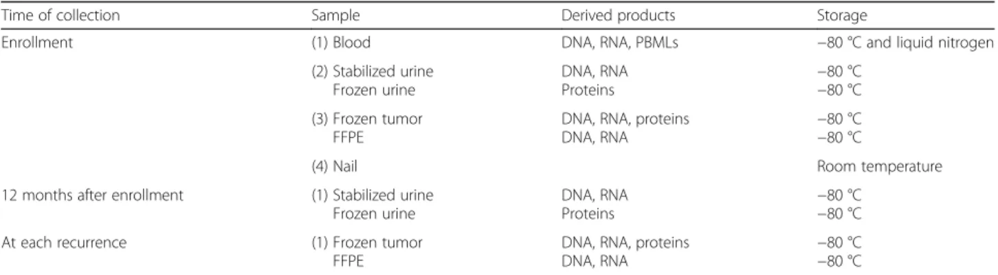
For each patient, four types of samples (blood, tumor tissue, urine and nail) are collected, at baseline and/or during follow-up, and delivered to three Biological Resources Centers (BRC) (NF S96-900 certification obtained for two BCR and being requested for one BRC) for processing and storage in cryoconservators or freezers which are alarmed and continuously moni-tored (Table 2).
Sampling kits including all tubes/containers required for the study and packaging for the shipment are prepared by the BRC of the Jean Dausset Foundation – Human Polymorphism Study Center (HPSC-BRC) in Paris and sent to participating centers. All contents are pre-labeled with a unique bar code to provide full traceability from the patient inclusion.
Blood samples
Blood samples are collected prior to surgery in three tubes (10 ml in one EDTA-containing tube for DNA ex-traction and isolation of plasma, 6 ml in one citrate-containing tube for isolation of lymphocytes, and 2.5 ml in one PAXgene tube for RNA extraction). The same
day samples are shipped by centers to the HPSC-BRC where they are processed at reception (24 to 72 h after blood sampling). Viable lymphocytes are isolated using ‘UNI-SEP Lymphocyte Separation’ tubes (Eurobio) and stored in liquid nitrogen. Aliquots of plasma and buffy-coats isolated from EDTA blood are stored at−80 °C.
DNA is extracted from buffy-coats using a salting out protocol on the Autopure LS (Qiagen). DNA concentra-tions are measured using ‘PicoGreen dsDNA reagent’ (Life Technologies). DNAs are diluted sequentially using
TE10:1 to get concentrations normalized at 100 ng/μL. Integrity of DNA is checked on agarose gel on 20 % of extracted DNAs. Gender is systematically checked on all DNAs extracted using a PCR method [12].
RNA is extracted from PAXgene tubes using PAXgene Blood miRNA Kit (Qiagen) on the QIAcube (Qiagen). RNA concentration is measured on a NanoDrop 2000C (Thermo Scientific) and quality of RNA is evaluated by calculation of the RIN (RNA Integrity Number) on a Bioanalyzer 2100 (Agilent).
Table 1 Data collection
Type of data Data collected Data Source
Epidemiology (1) Sociodemographic characteristics: age, sex, place of birth, educational level, marital status, household income, working situation, history of residences and of occupations.
(2) Lifestyle: history of tobacco consumption, dietary habits and physical activity.
(3) Medical history and medication use: weight, height, urinary tract infection, hematuria, kidney stones, skin or respiratory allergies, anti-hypercholesteremic and anti-inflammatory drugs, hormonal treatment, personal and family history of cancer
Face to face interview
Disease management (1) Disease presentation: presence of symptoms (hematuria, pollakuria, dysuria, urgency, hydronephrosis), alteration of health status
(2) Procedures before diagnostic resection: urinary cytology, urine culture, abdominal ultrasound, urinary tract fibroscopies, imaging (scanner, MRI)
(3) Diagnosis through transurethral resection of the bladder: Hervix or Narrow-Band-Imaging (NBI) fluorescence, number and location of resected tumors, size and aspect of the largest tumor (4) Treatment: for NMIBC intravesical instillations and cystectomy; for MIBC: chemotherapy,
radiotherapy, for both NMIBC and MIBC: lymphadenectomy, urethrectomy, nephro-urethrectomy, urinary diversion, blood transfusion, any hospitalization and complications.
(5) Outcomes: locoregional and distant recurrences (dates, sites, pathology, treatments, etc.) and death (date and place)
Patient medical records
Pathology (1) Histological type and subtype
(2) Prognosis factors : for NMIBC tumor stage, grade, CIS associated, lymphovascular invasion; for MIBC, TNM stage, surgical margins, lymphovascular invasion, lymph node density
(3) Pathological review for all cases at initial diagnosis and recurrence
Patient medical records
Resource use (1) All hospitalizations: dates, type of facility, service, type of care and diagnosis-related group. (2) Outpatient and community care : Cystoscopies, Imaging (CT, MRI, ultrasound, etc.), urine cytology,
urine culture and tumor biomarkers (3) Sick leaves
Patient medical records Self-administered questionnaire Quality of life (1) Generic measure: EuroQol EQ-5D-3 L questionnaire
(2) Generic measure: EORTC QLQ-C30 questionnaire
(3) Specific measures: urinary and bowel symptoms, impairment due to repeated treatments and sexual functioning.
- EORTC-BLS24 questionnaire for patients treated with transurethral resection of the bladder - EORTC-BLM30 questionnaire for those who underwent cystectomy
Self-administered questionnaires
Table 2 Sample collection
Time of collection Sample Derived products Storage
Enrollment (1) Blood DNA, RNA, PBMLs −80 °C and liquid nitrogen
(2) Stabilized urine Frozen urine DNA, RNA Proteins −80 °C−80 °C (3) Frozen tumor FFPE
DNA, RNA, proteins DNA, RNA
−80 °C −80 °C
(4) Nail Room temperature
12 months after enrollment (1) Stabilized urine Frozen urine
DNA, RNA
Proteins −80 °C−80 °C
At each recurrence (1) Frozen tumor
FFPE
DNA, RNA, proteins DNA, RNA
−80 °C −80 °C
PBMLs, peripheral blood mononuclear leukocytes; FFPE, formalin-fixed paraffin-embedded
Tumor samples
Tumor tissue is sampled at first transurethral resection of bladder tumor and transferred to the pathology de-partment for diagnosis according to routine procedures, including formalin fixation and embedding in paraffin (FFPE blocks). If the tumor is large enough (diameter > 10 mm), a sample is snap-frozen and stored at−80 °C in freezers located at each center. Frozen tumor samples are shipped every three months on dry ice to the bio-logical resources platform of the Curie Institute in Paris for DNA, RNA and proteins extraction, aliquoting and storage at−80 °C. A representative FFPE block and slides used for diagnosis are sent at room temperature to the Mondor hospital’s biological resources platform for patho-logical review, DNA and RNA extraction, and Tissue Mi-cro Arrays construction for future immunohistochemical and/or in situ hybridization analysis.
Cryosections are performed from frozen samples to check for tumor cell content by an uropathologist. Follow-ing the uropathologist report, macrodissections is per-formed on the frozen samples using a scalpel to take fragments enriched in tumor cells and the percentages of tumor cells of the microdissected fragments are recorded. When the samples are of sufficient size, a fragment of around 120 mg is taken. The remaining of the sample is stored at−80 °C. To extract RNA, DNA and protein from the same part of the tumor, each fragment is frozen in li-quid nitrogen and is ground in a mortar. If the fragment is of sufficient size (above 45 mg), the powder is divided in three equal parts, for RNA and DNA extractions and for protein extract. If the quantity of material is limiting, DNA, then RNA are prioritized. Between 20 and 45 mg, the powder will be divided in two parts for RNA and DNA extractions. Below 20 mg, only DNA will be extracted.
DNA is extracted following a phenol-chloroform proced-ure and quantified using a NanoDrop spectrophotometer and a Qubit fluorometer. The quality of the DNA is verified by electrophoresis. RNA is extracted using the TissueLyzer (Qiagen) and the Qiagen miRNeasy kit. RNA is quantified with a Nanodrop spectrophotometer. The RIN (RNA integrity number) is measured on an Agilent 2100 bioana-lyzer. For protein, the powder is dissolved in a Laemmli buffer using the TissueLyzer (Qiagen) and the proteins are quantified with the Pierce BCA kit. To spare materials, amplifications of RNA and DNA are performed as some analyses can be done on amplified materials. To obtain DNA and RNA from all tumors of the COBLAnCE cohort, DNA and RNA are being extracted from paraffin materials. For FFPE samples, slides are prepared from each block. New HES coloration, as control, is realized, the slide is scanned and archived. A GATA3/CK5/6 double-staining immunohistochemistry is performed to classify the tumor at molecular level [13, 14] and guide Tissue Micro Arrays and DNA/RNA extraction performed on selected zones.
DNA and RNA (Recover All) are extracted using Maxwel automate and specific FFPE kits (Promega). DNA and RNA concentrations are quantified using Qubit technology. Samples are stored at−80 °C.
In case of recurrence, both fresh and paraffin-embedded tumor tissue samples are collected and processed as previously described.
Urine samples
A urine sample is collected prior to surgery. 50 ml of these urine samples is stored at room temperature in Norgen® tubes (Thorold, Canada) for nucleic acid extrac-tion and the surplus is immediately frozen at−80 °C in Falcon tubes for further protein extraction.
Norgen® and frozen urine samples are shipped every three months at room temperature and on dry ice re-spectively to the Platform of Biological Resources - Henri Mondor Hospital to be processed.
DNA and RNA are extracted with the automated Maxwell 16 Instrument (Promega, Lyon, France), using the Maxwell LEV DNA Blood Kit and LEV RNA tissue Kit respectively. 25 ml of Norgen® urine is used for each DNA or RNA extraction on pellet after centrifugation. For DNA extraction, each sample is centrifuged at 2000 g for 10 min. Purified DNA and RNA samples are eluted in 50μL nuclease-free water and stored at −80 °C in the Plateform of Biological Resources (Henri Mondor Hospital) until processed. Nucleic acids concentration is determined using NanoDrop 2000C (Thermo Scientific).
To extract urine protein, thawed urine is centrifuged at 1300 xg for 10 min at room temperature. Then, protein is isolated from both the supernatant and the obtained pellet separately. Proteins from the pellet are extracted by 200μl of T-PER reagent (Tissue Protein Extraction Reagent, Thermo scientific), containing protease and phosphatase inhibitors. Proteins from the initial urine supernatant are precipitated by trichloracetic acid (TCA 30 %, Sigma-Aldrich) to a final concentration of 6 %, and the pellet is washed twice with ice-cold acetone (Sigma-Aldrich) and air-dried. Proteins are stored at−80 °C in the Platform of Biological Resources (Henri Mondor Hospital).
Protein concentration is assessed with Pierce BCA protein assay kit (Thermo Scientific).
An additional urine sample is collected at the 12 months follow-up visit for patients not treated with radical cystec-tomy and processed as previously described.
Toenail clippings
Toenail clippings are collected at the time the baseline questionnaire is completed and blood samples are collected. They are delivered to HPSC-BRC in a sealed envelope along with blood samples, and then mailed to the Mondor hospital’s biological resources platform every three months for storage at ambient temperature.
Power considerations and data analysis
We planned to enroll 2,000 patients. We assumed a recruitment period of 5 years (50 % of the patients recruited the 3 first years and 50 % the 2 last years), a loss to follow-up rate of 10 % during the 3 first years, 5 % during the 2 following years, and 5 % thereafter, and a progression rate of 40 % at 4 years and 50 % at 5 years. Based on the literature data, 30 % of bladder cancer pa-tients present with a MIBC at the time of diagnosis. We therefore expect 600 patients with MIBC to be included; the progression free survival is the primary endpoint of interest for these patients. The study will be sufficiently powered (bilateral test, α = 0.05, 1-β ≥ 80 %) to detect a hazard ratio≥ 2 considering a prevalence of marker pro-gression between 10 % and 20 %, or a hazard ratio≥ 1.5 for a prevalence marker between 30 % and 50 %.
Data analyses will be performed to complement specific objectives outlined above. Various parameters which are planned to be assessed have been summarized in Table 1. All analyses will be carried out using SAS version 9.1 (SAS Institute, Cary, NC, USA) and STATA (Version 13, Stata Corporation, Austin, Texas).
Discussion
To date (March 2016), 13 centers are actively involved in subject recruitment and 1020 patients have already been enrolled in the study. Recruitment is expected to end in the first semester of 2017. This ambitious project will allow having one of the biggest cohorts of incident bladder tumors with a longest follow-up (minimum 6 years).
This study will provide us with considerable insight into the biology of bladder cancer and the mechanisms through which various factors may alter the prognosis. It should fa-cilitate the identification and the cost-effectiveness of emer-ging biomarkers and targeted therapies in bladder cancer.
The multidisciplinary approach of this study will allow communication with other researchers and departments to share ideas and foster collaborations. In fact epidemi-ologists, clinicians, biepidemi-ologists, pathepidemi-ologists, health econo-mists, researchers coming from various French institutions joined together to build this cohort. In addition, the inter-action with many health centers will support the transla-tion of our research findings into clinical practice.
During the follow-up period, new experimental drugs for bladder cancer treatment may be recorded. For the purpose of these registrations, data prospectively collected in COBLAnCE will be useful to describe the natural history of bladder tumor evolution in France.
In summary, the establishment of this cohort study, along with the wealth of epidemiological, clinicipatholo-gical, economical and biological evidence to be collected, will lead to a much needed understanding of bladder can-cer and provide evidence to guide health professionals in the management and care of patients with this disease.
Despite the large size of this prospective cohort, larger studies are necessary for specific analyses in subgroups of patients, i.e. within stage of disease, or within groups of patients with the same treatment. Therefore, we intend to establish close collaborations with other on-going prospective studies on bladder cancer in order to combine the datasets and accordingly to increase the statistical power.
Abbreviations
BRC:Biological resources center; eCRF: Electronic case report form; FFPE: Formalin-fixed paraffin-embedded; GSTM1: Glutathione-S-transferase M1; MIBC: Muscle-invasive bladder tumors; NAT2: N-acetyltransferase 2; NMIBC: Non-muscle-invasive bladder cancers; PBML: Peripheral blood mononuclear leukocytes; QoL: Quality of life
Acknowledgements
The authors would like to thank the following hospitals for their involvement in patient recruitment: Hôpital Foch (Suresnes), Centre Hospitalier Universitaire Henri Mondor (Créteil), Centre Hospitalier Universitaire Gabriel-Montpied (Clermont-Ferrand), Centre Hospitalier Régional Universitaire de Lille, Centre Hospitalier Universitaire de Nîmes, Clinique Beau Soleil (Montpellier), Hopital Pasteur (Colmar), Centre Hospitalier René Dubos (Pontoise), Clinique du Parc Rambot (Aix en Provence), Centre Hospitalier Universitaire Grenoble Alpes (Grenoble), Clinique Urologique Atlantis (Nantes), Centre Hospitalier Européen Georges Pompidou (Paris), and Centre Hospitalier de Brabois (Nancy), and Dr Hélène Blanché at the Biological Resources Centre of Human Polymorphism Study Center, Dr Caroline Barau, Dr Nanor Sirab, Pascale Maille, Fannie Semprez and Pascale Soyeux at the Platform of Biological Resources and Research Team INSERM U955 of Henri Mondor Hospital, and Elodie Guyon and Dr Odette Mariani at the Biological Resources Centre of Curie Institute, Clémentine Krucker and Dr Isabelle Bernard-Pierrot at UMR144 CNRS, for the management of biological specimen, and Dr Marie-Claude Babron, Pascale Jan and Anthony Mangin for the data management of the COBLAnCE study.
Funding
This project is funded by the French Ministry of Research (Programme Investissements d’Avenir Cohortes), the Institute of Research in Public Health -National Institute of Health and Medical Research, and the -National League against Cancer. Sponsors were not included in the study design nor will they be in the collection, analysis, and interpretation of data, or in publications that will results from this study.
Availability of data and materials
Anonymized data and biological samples collected during the study can be obtained for conducting scientific projects. Requests will be evaluated by the Scientific Committee of the COBLAnCE cohort.
Authors’ contributions
SB, JB, YA, FR and TL contributed to the conception and design of the study. All authors are members of the study strategy group. KG provides overall study management and lead activities for monitoring evaluation. SB coordinates the project and drafted the manuscript. All authors critically read and revised the manuscript. All authors approved the final version of the manuscript. Competing interests
The authors declare that they have no competing interests. Consent for publication
Not applicable.
Ethics approval and consent to participate
This project was approved by one French ethics committee (Comité de Protection des Personnes Ile de France VII, reference number CO-12-001) for all participating centers: Hôpital Foch (Suresnes), Centre Hospitalier Universitaire Henri Mondor (Créteil), Centre Hospitalier Universitaire Gabriel-Montpied (Clermont-Ferrand), Centre Hospitalier Régional Universitaire de Lille, Centre Hospitalier Universitaire de Nîmes, Clinique Beau Soleil (Montpellier), Hopital Benhamouet al. BMC Cancer (2016) 16:837 Page 6 of 7
Pasteur (Colmar), Centre Hospitalier René Dubos (Pontoise), Clinique du Parc Rambot (Aix en Provence), Centre Hospitalier Universitaire Grenoble Alpes (Grenoble), Clinique Urologique Atlantis (Nantes), Centre Hospitalier Européen Georges Pompidou (Paris), and Centre Hospitalier de Brabois (Nancy). All patients are informed of the objectives of the study and are invited to voluntarily participate at inclusion. Patients who agree to participate provide written consent. Author details
1INSERM, UMR 946, Genetic Variation and Human Diseases Unit, 27 rue
Juliette Dodu, 75010 Paris, France.2Université Paris Diderot, Sorbonne Paris
Cité, Institut Universitaire d’Hématologie, Paris, France.3Gustave Roussy,
Service de biostatistique et d’épidémiologie, Villejuif, France.4CESP Centre for Research in Epidemiology and Population Health, INSERM U1018, University Paris-Sud, UVSQ, Université Paris-Saclay, Villejuif, France.5CNRS, UMR144,
Equipe Labellisée par la Ligue Nationale contre le Cancer, Paris, France.
6
Institut Curie, PSL Research University, Paris, France.7Université Paris-Est, Créteil, France.8INSERM, UMR 955, Créteil, France.9Département de
pathologie, APHP, Hôpital Henri-Mondor, Créteil, France.10Service d’urologie
et de transplantation rénale, Hôpital Foch, Suresnes, France.11UFR des
sciences de la santé Simone Veil, Université de Versailles-Saint-Quentin-en-Yvelines, Suresnes, France.
Received: 22 March 2016 Accepted: 24 October 2016
References
1. Ferlay J, Soerjomataram I, Ervik M, Dikshit R, Eser S, Mathers C, Rebelo M, Parkin DM, Forman D, Bray, F. GLOBOCAN 2012 v1.0, Cancer Incidence and Mortality Worldwide: IARC CancerBase No. 11 [Internet]. Lyon, France: International Agency for Research on Cancer. 2013. http://globocan.iarc.fr. Accessed 25 Feb 2016
2. World Cancer Report 2014. In: Stewart BW, Wild CP, editors. Lyon, France: International Agency for Research on Cancer; 2014. p 444–52
3. Moore LE, Baris DR, Figueroa JD, Garcia-Closas M, Karagas MR, Schwenn MR, et al. GSTM1 null and NAT2 slow acetylation genotypes, smoking intensity and bladder cancer risk: results from the New England bladder cancer study and NAT2 meta-analysis. Carcinogenesis. 2011;32:182–9.
4. Kiemeney LA, Sulem P, Besenbacher S, Vermeulen SH, Sigurdsson A, Thorleifsson G, et al. A sequence variant at 4p16.3 confers susceptibility to urinary bladder cancer. Nat Genet. 2010;42:415–9.
5. Figueroa JD, Ye Y, Siddiq A, Garcia-Closas M, Chatterjee N, Prokunina-Olsson L, et al. Genome-wide association study identifies multiple loci associated with bladder cancer risk. Hum Mol Genet. 2014;23:1387–98.
6. Bosset PO, Neuzillet Y, Paoletti X, Molinie V, Botto H, Lebret T. Long-term follow-up of TaG1 non-muscle-invasive bladder cancer. Urol Oncol. 2015;33:20.e1–7.
7. Lebret T, Neuzillet Y. Indication and timing of cystectomy in high-risk bladder cancer. Curr Opin Urol. 2012;22:427–31.
8. Botteman MF, Pashos CL, Redaelli A, Laskin B, Hauser R. The health economics of bladder cancer: a comprehensive review of the published literature. Pharmacoeconomics. 2003;21:1315–30.
9. Drummond M, Barbieri M, Cook J, Glick HA, Lis J, Malik F, et al. Transferability of economic evaluations across jurisdictions: ISPOR Good Research Practices Task Force report. Value Health. 2009;12:409–18.
10. Bartley K, DeBusk K, Duff S. Health-related quality of life in bladder cancer: a systematic literature review. Value Health. 2015;18:A474–5.
11. Witjes JA, Compérat E, Cowan NC, De Santis M, Gakis G, Lebret T, et al. EAU guidelines on muscle-invasive and metastatic bladder cancer: summary of the 2013 guidelines. Eur Urol. 2014;65:778–92.
12. Wilson JF, Erlandsson R. Sexing of human and other primate DNA. Biol Chem. 1998;379:1287–8.
13. Rebouissou S, Bernard-Pierrot I, de Reyniès A, Lepage ML, Krucker C, Chapeaublanc E, et al. EGFR as a potential therapeutic target for a subset of muscle-invasive bladder cancers presenting a basal-like phenotype. Sci Transl Med. 2014;6:244ra91.
14. Biton A, Bernard-Pierrot I, Lou Y, Krucker C, Chapeaublanc E, Rubio-Pérez C, et al. Independent component analysis uncovers the landscape of the bladder tumor transcriptome and reveals insights into luminal and basal subtypes. Cell Rep. 2014;9:1235–45.
• We accept pre-submission inquiries
• Our selector tool helps you to find the most relevant journal
• We provide round the clock customer support
• Convenient online submission
• Thorough peer review
• Inclusion in PubMed and all major indexing services
• Maximum visibility for your research Submit your manuscript at
www.biomedcentral.com/submit