409
AMS RADIOCARBON DATING OF BONES AT LSCE N Tisnérat-Laborde1,2 H Valladas1 E Kaltnecker1 M Arnold3
ABSTRACT. In this paper, we explain our routine pretreatment of bone for radiocarbon dating by accelerator mass spec-trometry (AMS), based on the specific reaction between amino acids and ninhydrin described by Nelson (1991). The values and uncertainties of the total system background are presented as a function of the carbon sample mass and the reliability of this method is discussed.
INTRODUCTION
Since the first 14C dates were obtained (Arnold et al. 1951), radiocarbon laboratories have developed many methods of bone pretreatment. Usually, these methods are based on the extraction of the bone organic matter. The extract can consist of the whole collagen (Longin 1971; Brown et al. 1988; Hedges et al. 1989; Law et al. 1989; Kretschmer et al. 1998), a mixture of collagen amino acids (Gillespie et al. 1986; Gurfinkel 1987; Long et al. 1989; Redvers-Newton et al. 1994), specific indi-vidual amino acids (van Klinken et al. 1990), or non-collagenous proteins (Ajie et al. 1992). These extracts are oxidized to CO2,then reduced to graphite and dated.
For more than 10 years at the Laboratoire des Sciences du Climat et de l’Environnement (LSCE), we have prepared bone by the method described by Nelson (1991), based on a chemical reaction that extracts CO2 from carboxylic groups of proteinaceous molecules. This chemical treatment is pre-ceded by elemental analyses (%N, %C, C/N) in order to quantify the bone collagen, and, conse-quently, to determine if the bone is datable.
In this paper, we describe the protocol of bone preparation. We present the blank values obtained on bones from 2 sites, Sclayn and Gerde. Finally, we discuss the reliability of the 14C ages obtained by this method by comparing some of them to other 14C dates available for the same archeological lay-ers. These samples are either charcoal, burnt bones, or bones treated differently.
MATERIAL AND METHOD Material
On the basis of porosity, bone may be classified as cortical bone (also known as compact bone) or trabecular bone (also called cancellous or the spongy part). The cortical bone, which is much denser and less porous than the cancellous bone, is preferred for 14C dating since it is generally less altered by diagenesis.
The fossil bones used to estimate the degree of contamination introduced by our protocol come from 2 sites. Five bones were collected in Scladina Cave (Sclayn, Belgium), under a stalagmitic floor in layer 4A, which was dated by thermoluminescence to approximately 100,000 yr ago (Debenham 1998). Another bone comes from layer 2b of Carrière cave (Gerde, France), which is below a sta-lagmitic floor and dated to 52,500 yr ago by U/Th (Clot 1987).
1Laboratoire des Sciences du Climat et de l’Environnement, UMR CEA-CNRS 1572, Avenue de la Terrasse, F-91198
Gif-sur-Yvette, France.
2Corresponding author. Email: tisnerat@lsce.cnrs-gif.fr.
Method
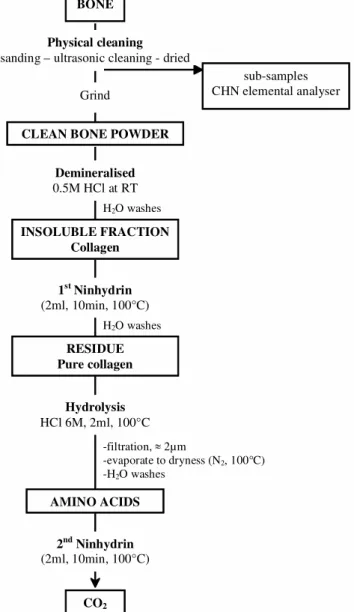
The protocol used for the bone treatment is summarized in Figure 1.
Mechanical Treatment and Elemental Analyses
A piece of bone sample ( 1–2 cm) is cleaned mechanically with an airbrasive system with 27 µg aluminum oxide to remove superficial contaminants (roots, glue) and the spongy part, which is con-sidered to be the most contaminated.
A sub-sample of approximately 5 mg is drilled out and subjected to elemental analysis. It is intro-duced in a tin capsule into a Carlo-Erba NA 1500 elemental analyzer.
Figure 1 Diagram showing pretreatment steps of bones for AMS 14C dating
H2O washes
BONE Physical cleaning
sanding – ultrasonic cleaning - dried
CLEAN BONE POWDER Demineralised 0.5M HCl at RT H2O washes INSOLUBLE FRACTION Collagen 1st Ninhydrin (2ml, 10min, 100°C) Hydrolysis HCl 6M, 2ml, 100°C AMINO ACIDS 2nd Ninhydrin (2ml, 10min, 100°C) CO2 sub-samples CHN elemental analyser RESIDUE Pure collagen -filtration, 2µm -evaporate to dryness (N2, 100°C) -H2O washes Grind
The other part of the mechanically cleaned bone (1–3 g)—if it contains enough collagen—is ultra-sonically rinsed in Milli-Q water to remove aluminum oxide and dried at 45 °C. This bone is then finely ground in a planetary micro-mill composed of bowls and balls of zirconium oxide (ZrO2). Chemical Treatment
The powdered bone is repeatedly treated with 0.5M HCl and stirred at room temperature to remove carbonates, phosphates, and fulvic acids until the residue becomes colloidal. The acid-insoluble col-lagenous residue is then rinsed with Milli-Q water until neutral pH is reached.
Next, 50 mg of ninhydrin (2,2-dihydroxy–1,3-indandione) in a 2-ml sodium citrate buffer (pH = 4.8) is added to the residue, which is heated at 100 °C for 10 min. The ninhydrin reacts specifically with the free amino acids, which come from either degraded collagen or contaminants. The ninhydrin reacts with the -NH2 of amino acids to give -imino acids, which react with water to give -ceto acids (Moore et al. 1950). These -ceto acids are unstable and release CO2 from the -carboxyl group:
The CO2 is not collected and the residue is rinsed until the solution is decolorized.
Next, this “pure” residue is hydrolyzed to free amino acids with hot acid (HCl 6M at 100 °C over-night). The solution of free amino acids is filtered on a precleaned glass filter and collected in a glass reactor. This filtrate is evaporated at 80 °C under nitrogen. The free amino acid residue is rinsed 5 times with Milli-Q water, which is then evaporated at 80 °C under nitrogen.
The reactor is connected to a vacuum line (Figure 2) and heated to 100 °C with heating coils. Once the vacuum reaches 2.10–4 mb after 2 days, 2 ml of ninhydrin solution is injected through a sep-tum. The released CO2 is dried by passing through 2 “water traps” (–78 °C, mix of dry ice and eth-anol), trapped in a liquid nitrogen trap (–196 °C), quantified into the calibrated volume, and then collected in a glass vial. The entire treatment and the CO2 transformation take more than 8 days.
R CH COOH NH2 Amino acid C C O OH OH Ninhydrin Na citrate buffer pH 4.8 100°C R C COOH NH R C COOH O H2O NH3
CO
2 R C H O Aldehyde 3H2O O C C O H OH O C C O OH OH O C C O N O C C O O Purple compound -imino acid -ceto acid Aldehyde AMS datingThe CO2 was reduced to graphite (Arnold et al. 1989) and the 14C ages were obtained by accelerator mass spectrometry (AMS) at the Gif-sur-Yvette Tandetron Facility (UMS 2004).
RESULTS AND DISCUSSION Blanks
Results of Elemental Analyses
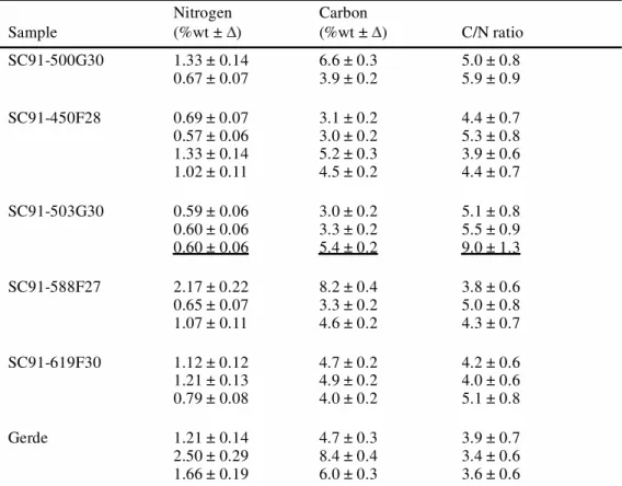
Several sub-samples were removed from different parts of the Sclayn and Gerde bones for elemental analyses (%N, %C, C/N). The results are reported in Table 1.
Nitrogen concentrations in the Sclayn bones range from 0.57–2.17%wt, in agreement with previous measurements (Bocherens et al. 1997), and in the Gerde bones from 1.21–2.5%wt. These nitrogen concentrations in the whole bone give some idea of the quantity of collagen (Hedges et al. 1992; Bocherens et al. 1997; Gillespie et al. 1984; Ambrose 1990; Hedges et al. 1995). Indeed, the quantity of nitrogen ranges from about 4% in a fresh bone (Stafford et al. 1988; Ambrose 1993) to below 0.2% in poorly preserved bone, which cannot be dated by the ninhydrin method. With nitrogen amounts ranging from 0.5–2.5%wt, the Sclayn and Gerde bones contain enough collagen for AMS dating. The scatter of the nitrogen measurements shows that the diagenesis of the organic matter is not homogeneous within any one bone. The C/N ratio of the whole bone can help to estimate the degree of diagenetic alteration. High values (i.e., >5) indicate extensive diagenesis (deamination) or a high proportion of exogenous carbon (humics). For the Sclayn bones, the C/N ratios are statistically sim-ilar with a mean value of 4.55 0.4 (n = 14; 2; P
0.05 = 9.95/22.40), excluding the value of the
Figure 2 Photograph of the vacuum line
Reactor
Septum
Liquid N2trap Water traps Glass vial
spongy part of sample SC91-503G30/3 (C/N = 9). This high C/N ratio is attributed to the addition of humic contaminants since the nitrogen concentrations are similar within the compact and the spongy parts; the high C/N ratio confirms the importance of removing this porous part of the bone. This mean C/N ratio of Sclayn bones is approximately equal to the fresh bone (5) and shows the good preservation of these bones. For the Gerde sample, the mean C/N ratio is equivalent to 3.58 0.6 (n = 3, 2; P
0.05 = 0.4/5.99), and is slightly lower than the C/N value in Sclayn. This lower value can be explained by a loss of inorganic carbon (decalcification) during burial. The Gerde bone seems less well preserved than the Sclayn bones.
The nitrogen concentrations show that the Sclayn and Gerde bones contain enough collagen for AMS datings and the C/N ratios show their degree of preservation and their non-contamination.
AMS 14C Results
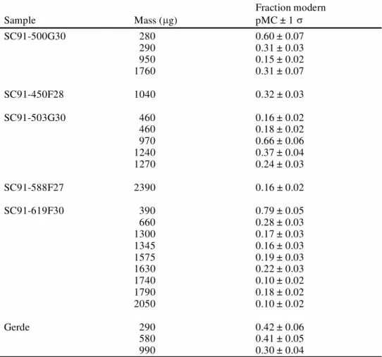
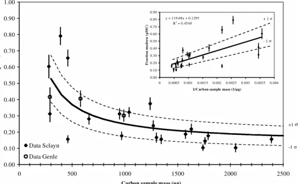
The 14C values of the Sclayn and Gerde bones (Table 2) are presented as a function of the carbon mass in Figure 3. These blank values take into account the chemical pretreatment, the conversion into CO2, the graphitization, and the machine background contaminants. They increase from 0.10 pMC to 0.80 pMC as the carbon sample size decreases from 2400 µg to less than 300 µg.
Table 1 Nitrogen and carbon concentrations in bone, expressed as % of bone weight (%wt) and the atomic C/N ratio of the Sclayn (SC91-) and Gerde blank bones. The results of the underlined line correspond to the analyses carried out on the spongy part of the bone.
Sample Nitrogen (%wt ± ) Carbon (%wt ± ) C/N ratio SC91-500G30 1.33 ± 0.14 0.67 ± 0.07 6.6 ± 0.33.9 ± 0.2 5.0 ± 0.85.9 ± 0.9 SC91-450F28 0.69 ± 0.07 0.57 ± 0.06 1.33 ± 0.14 1.02 ± 0.11 3.1 ± 0.2 3.0 ± 0.2 5.2 ± 0.3 4.5 ± 0.2 4.4 ± 0.7 5.3 ± 0.8 3.9 ± 0.6 4.4 ± 0.7 SC91-503G30 0.59 ± 0.06 0.60 ± 0.06 0.60 ± 0.06 3.0 ± 0.2 3.3 ± 0.2 5.4 ± 0.2 5.1 ± 0.8 5.5 ± 0.9 9.0 ± 1.3 SC91-588F27 2.17 ± 0.22 0.65 ± 0.07 1.07 ± 0.11 8.2 ± 0.4 3.3 ± 0.2 4.6 ± 0.2 3.8 ± 0.6 5.0 ± 0.8 4.3 ± 0.7 SC91-619F30 1.12 ± 0.12 1.21 ± 0.13 0.79 ± 0.08 4.7 ± 0.2 4.9 ± 0.2 4.0 ± 0.2 4.2 ± 0.6 4.0 ± 0.6 5.1 ± 0.8 Gerde 1.21 ± 0.14 2.50 ± 0.29 1.66 ± 0.19 4.7 ± 0.3 8.4 ± 0.4 6.0 ± 0.3 3.9 ± 0.7 3.4 ± 0.6 3.6 ± 0.6
The data indicate a statistically significant mass dependence relationship, as previously reported by several studies of 14C background (Vogel et al. 1987; Kirner et al. 1995; Brown et al. 1997; Schle-icher et al. 1998; Tisnérat-Laborde et al. 2001). By using the least-squares method, the best fit between 14C concentrations and the inverse carbon mass is obtained by:
y = 119.68 ( 28.59) / x + 0.1295 ( 0.0486)
where y = 14C concentration (pMC) and x = carbon mass (µg). This increase of the 14C background is due to the addition of 1.3 µg of modern carbon (100 pMC) per mg of sample during the whole process.
All the blank values from Sclayn and Gerde (Figure 3) are consistent, although the C/N analyses showed a lower preservation for the Gerde sample. Such agreement indicates the reliability of our protocol.
These blank values are higher than those obtained from the Carrara marble IAEA C-1, which range from 0.06–0.14 pMC as the size decreases from 2400–300 µg (Tisnérat-Laborde et al. 2001). The contamination during the chemical treatment and the conversion of bone into CO2 may be responsi-ble for the high blank values since the graphitization and the machine processing are the same for
Table 2 14C results of the Sclayn (SC91-) and Gerde bones, reported in pMC
Sample Mass ( g) Fraction modern pMC ± 1 SC91-500G30 280 290 950 1760 0.60 ± 0.07 0.31 ± 0.03 0.15 ± 0.02 0.31 ± 0.07 SC91-450F28 1040 0.32 ± 0.03 SC91-503G30 460 460 970 1240 1270 0.16 ± 0.02 0.18 ± 0.02 0.66 ± 0.06 0.37 ± 0.04 0.24 ± 0.03 SC91-588F27 2390 0.16 ± 0.02 SC91-619F30 390 660 1300 1345 1575 1630 1740 1790 2050 0.79 ± 0.05 0.28 ± 0.03 0.17 ± 0.03 0.16 ± 0.03 0.19 ± 0.03 0.22 ± 0.03 0.10 ± 0.02 0.18 ± 0.02 0.10 ± 0.02 Gerde 290 580 990 0.42 ± 0.06 0.41 ± 0.05 0.30 ± 0.04
these 2 types of sample. We also reject the intrinsic contamination because the same results were obtained for different bones and different sites. We suspect the vacuum line processes before the conversion stage of the amino acids to CO2 to be responsible forthis level of contamination for the following 2 reasons:
1. The pumping is less effective for the bone than for the carbonate (by 1 order) because the res-idue quickly becomes pasty under the vacuum;
2. Atmospheric CO2 may have been introduced during the addition of ninhydrin, either in the form of dissolved CO2 in the ninhydrin or when the septum was perforated.
The 14C ages of bone are calculated using the blank values determined from the mass dependent equation. From this equation, the age limit is about 50,000 BP (0.2 0.08 pMC) for a carbon mass of 1500 µg, and about 45,000 BP (0.37 0.1 pMC) for a carbon mass of 500 µg.
Reliability of the Method
The 14C ages of bones treated by the ninhydrin method are compared to those obtained for the same archeological layer on associated organic materials (charcoal, burnt bones) and bones treated by other methods. We use the Chi-squared test statistic to check consistency of these determinations (Ward et al. 1978).
In the first test of reliability, 3 sites (Trois-Frères Cave, Laugerie Haute, and Kozarnika) allowed the comparison of 14C dates of ninhydrin-treated bones with those of associated charcoals or burnt bones. These charcoals or burnt bones underwent the classical AAA treatment. The 14C results of the 3 archeological sites are reported in Table 3 and Figure 4. At Trois-Frères Cave and Laugerie-Figure 3 14C concentration (pMC) as a function of carbon sample mass (µg). The error bars are shown as 1 (68% of
overall confidence). The inset small figure is the relation between the inverse carbon sample weight and the 14C
concen-tration (pMC). Dashed lines correspond to the 1 error. 0.00 0.10 0.20 0.30 0.40 0.50 0.60 0.70 0.80 0.90 1.00 0 500 1000 1500 2000 2500
Carbon sample mass (µg)
F ra ct io n m od er n ( p M C ) Data Sclayn Data Gerde +1 -1 y = 119.68x + 0.1295 R2 = 0.4548 0.00 0.10 0.20 0.30 0.40 0.50 0.60 0.70 0.80 0.90 0 0.0005 0.001 0.0015 0.002 0.0025 0.003 0.0035 0.004 1/Carbon sample mass (1/µg)
F ra ct io n m od er n ( pM C ) + 1 - 1
Haute (Delpech et al. 2001; Roque et al. 2001), the burnt bones and bones have consistent 14C ages. The Kozarnika site (Fontugne et al. 2002) reveals the 14C age of the bone to be slightly older than those of the 2 charcoals, but, nevertheless, statistically in agreement.
Table 3 Comparison between 14C dates from bones (ninhydrin method) and from associated char-coals or burned bones (AAA treatment).All ages are given in 14C yr BP (before 1950). Statistical errors are given at 1 .
Site Lab nr Material
Age (yr BP)
Error
(1 ) Chi-squared test Reference
Trois-Frères Cave GifA 99552 GifA 99555 GifA 99550 GifA 99551 GifA 99553 GifA 99554 bone bone burnt bone burnt bone burnt bone burnt bone average 14210 13930 14060 13980 14210 14200 14100 110 110 110 120 110 120 50 6.21/ 2 6; 0.05 = 11.1
Laugerie Haute GifA 100634
GrN-4442 GrN-4495 Ly-1173 (OxA) GifA 100630 bone bone bone burnt bone burnt bone average 19550 19600 19740 19525 19600 19600 340 140 200 155 200 80 0.75/ 2
5; 0.05 = 9.49 Delpech & Riguad 2001
Roque et al. 2001 Kozarnika GifA 99662 Gif/LSM-10994 GifA 101050 bone charcoal charcoal average 39310 38700 37170 38000 1000 1400 700 530 3.37/ 2 3; 0.05 = 5.99 Fontugne & Tisnérat-Laborde, in press
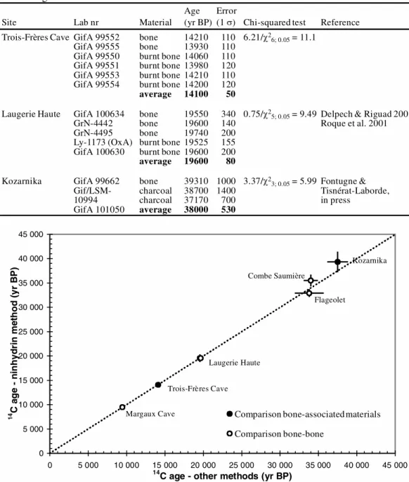
Figure 4 From a same archeological level, the average 14C dates obtained with the ninhydrin method are plotted versus
the average 14C ages obtained with the Oxford bone method (open circles) or associated materials (solid circles). The
ages are expressed as yr BP. The error bars are shown as 1 (for 5 of the data points, these error bars are smaller than the symbols). The dotted line is the 1:1 correlation line.
0 5 000 10 000 15 000 20 000 25 000 30 000 35 000 40 000 45 000 0 5 000 10 000 15 000 20 000 25 000 30 000 35 000 40 000 45 000
14C age - other methods (yr BP) 14 C a g e - ni n h yd ri n m et h o d (y r B P )
Comparison bone-associated materials Comparison bone-bone Margaux Cave Combe Saumière Flageolet Laugerie Haute Trois-Frères Cave Kozarnika
In the second test, we compared the 14C dates of bones treated by different chemical methods. The results are reported in Table 4 and Figure 4. A comparison with the Oxford method (Hedges et al. 1989; Law et al. 1989) may be made for 3 sites: Margaux Cave, Flageolet, and Combe Saumière (Delpech et al. 2001) (Table 4). A comparison with the Groningen procedure can be done for the site of Laugerie Haute (Table 3). In all cases, the 14C ages are similar whatever the chemical treatment, as previously noted by Nelson (1991).
The 2 tests show the good correlation between the 14C ages of the ninhydrin-treated bones and those of samples collected at the same archeological level (Figure 4) for time intervals ranging between 9000–45,000 yr BP. All these comparisons confirm the reliability and accuracy of the method for dates up to 45,000 yr BP.
CONCLUSION
In this paper, we described our routine protocol of bone pretreatment for AMS 14C dating. This rou-tine is applied to fossil bones containing more than 0.2% of nitrogen in whole bone.
The blank level is a function of the mass of the carbon sample. According to the equation (y = 119.68 / x + 0.1295), the blank value is equal to 0.20 0.08 pMC ( 50,000 yr BP) for a sample mass of 1500 µg and 0.37 0.10 pMC ( 45,000 yr BP) for a sample mass of 500 µg. The contamination by modern carbon is attributed either to the difficulty of degassing the sample or to the introduction of atmospheric CO2.
The validity of the method and the protocol is tested by comparing the 14C ages obtained on bones by this method and those obtained by other methods (Oxford and Groningen bone methods or asso-ciated materials). The satisfactory results of these comparisons and the good estimation of the blank level show the reliability and accuracy of the method for dates up to 45,000 yr BP.
Table 4 Comparison of 14C ages of bones pretreated by the method used by the Oxford Radiocarbon Laboratory (OxA) and by the ninhydrin method (LSCE, GifA).All ages are given in 14C yr BP (before 1950). Statistical errors are given at 1 .
Site Lab nr Material
Age (yr BP)
Error
(1 ) Chi-squared test Reference
Margaux Cave GifA 92354
GifA 92355 GifA 92362 OxA-3533 OxA-3534 bone bone bone bone bone average 9590 9530 9260 9530 9350 9460 110 110 120 120 120 50 5.76/ 2 5; 0.05 = 9.49 Flageolet GifA 95538 GifA 95559 OxA-598 bone bone bone average 32040 34300 33800 33000 850 1100 1800 630 2.87/ 2
3; 0.05 = 5.99 Delpech & Riguad 2001
Combe Saumière GifA 96768
OxA-6507 bonebone
average 35500 34000 34560 1100 850 670 1.16/ 2
ACKNOWLEDGMENTS
We gratefully acknowledge M Paterne for helpful comments and discussions. We thank M Otte for providing the Sclayn samples and H Bocherens for his help in selecting the Sclayn bones. The Gerde sample was provided by the late A Clot. We wish to thank M Fontugne for the Kozarnika results, the members of the 14C laboratory (LSCE), and the staff of Gif-sur-Yvette Tandetron Facility (UMS 2004). This work was supported by the French CNRS and the CEA, LSCE contribution nr 1001. REFERENCES
Ajie HO, Kaplan IR, Hauschka PV, Kirner D, Slota PJ, Taylor J, Taylor RE. 1992. Radiocarbon dating of bone osteocalcin: isolating and characterizing a non-collagen protein. Radiocarbon 34(3):296–305.
Ambrose SH. 1990. Preparation and characterization of bone and tooth collagen for isotopic analysis. Journal of Archaeological Science 17:431–51.
Ambrose SH. 1993. Isotopic analysis of paleodiets: methodological and interpretive considerations. In: Sandford MK, editor. Investigations of ancient human
tissue. Amsterdam: Gordon and Breach Science Pub-lishers. p 59–130.
Arnold JR, Libby WF. 1951. Radiocarbon dates. Science
113:111–20.
Arnold M, Bard E, Maurice P, Valladas H, Duplessy JC. 1989. 14C dating with the Gif-sur-Yvette Tandetron
ac-celerator: status report and study of isotopic fraction-ation in the sputter ion source. Radiocarbon 31:284– 91.
Bocherens H, Billiou D, Patou-Mathis M, Bonjean D, Otte M, Mariotti A. 1997. Paleobiological implica-tions of the isotopic signatures (13C, 15N) of fossil
mammal collagen in Scladina Cave (Sclayn, Bel-gium). Quaternary Research 48:370–80.
Bocherens H, Tresset A, Wiedmann F, Giligny F, Lafage F, Lanchon Y, Mariotti A. 1997. Diagenetic evolution of mammal bones in two French Neolithic sites.
Bul-letin de la Société géologique France 168:5. Brown TA, Nelson DE, Vogel JS, Southon JR. 1988.
Im-proved collagen extraction by modified Longin method. Radiocarbon 30(2):171–7.
Brown TA, Southon JR. 1997. Corrections for contami-nation background in AMS 14C measurements.
Nu-clear Instruments and Methods in Physics Research
B123:208–13.
Clot A. 1987. La grotte de Gerde (Hautes-Pyrénées), site préhistorique et paléontologique. Tarbes: Société Ra-mond.
Debenham NC. 1998. Thermoluminescence dating of stalagmitic calcite from la grotte Scladina at Sclayn (Namur). In: Otte M, Patou-Mathis M, Bonjean D, ed-itors. Recherches aux grottes de Sclayn, Volume 2. Liège: L’Archéologie ERAUL:39–43.
Delpech F, Rigaud J-P. 2001. Quelques exemples sur l’apport des datations en archéologie préhistorique. In: Barrandon J-N, Guibert P, Michel V, editors.
Data-tion XXIe rencontres internationales d’archéologie et
d’histoire d’Antibes. Antibes: APDCA. p 315–31. Fontugne MR, Tisnérat-Laborde N. Forthcoming. Une
séquence du Paléolithique inférieur au Paléolithique récent dans les Balkans: La grotte de Kozarnika à Ore-chets (Nord ouest de la Bulgarie). Datations
radiocar-bone.
Gillespie R, Hedges REM, Humm MJ. 1986. Routine AMS dating of bone and shell proteins. Radiocarbon
28(2A):451–6.
Gillespie R, Hedges REM, Wand JO. 1984. Radiocarbon dating of bone by accelerator mass spectrometry.
Journal of Archaeological Science 11:165–70. Gurfinkel DM. 1987. Comparative study of the
radiocar-bon dating different radiocar-bone collagen preparations. Ra-diocarbon 29(1):45–52.
Hedges REM, Law IA, Bronk CR, Housley RA. 1989. The Oxford Accelerator Mass Spectrometry Facility: technical developments in routine dating. Archaeom-etry 31(2):99–113.
Hedges REM, Millard AR. 1995. Measurements and re-lationships of diagenetic alteration of bone from three archaeological sites. Journal of Archaeological Sci-ence 22:201–9.
Hedges REM, van Klinken GJ. 1992. A review of current approaches in the treatment of bone for radiocarbon dating by AMS. Radiocarbon 34(3):279–91.
Kirner DL, Taylor RE, Southon JR. 1995. Reduction in backgrounds of microsamples for AMS 14C dating.
Radiocarbon 37(2):697–704.
Kretschmer W, Anton G, Benz M, Blasche S, Erler G, Finckh E, Fischer L, Kerscher H, Kotva A, Klein M, Leigart M, Morgenroth G. 1998. The Erlangen AMS Facility and its application in 14C sediment and bone
dating. Radiocarbon 40(1):231–8.
Law IA, Hedges REM. 1989. A semi-automated bone pretreatment system and the pretreatment of older and contaminated samples. Radiocarbon 31(3):247–53. Long A, Wilson AT, Ernst RD, Gore BH, Hare PE. 1989.
AMS radiocarbon dating of bones at Arizona.
Radio-carbon 31(3):231–8.
Longin R. 1971. New method of collagen extraction for radiocarbon dating. Nature 230:241–2.
Moore S, Stein WH. 1950. Photometric ninhydrin method for use in the chromatography of amino acids.
Journal of Biological Chemistry 176:367–88.
Nelson DE. 1991. A new method for carbon isotopic analysis of protein. Science 251:552–4.
Redvers-Newton NA, Coote GE. 1994. Bone pretreat-ments for radiocarbon dating: a study incorporating AMS dating and ion beam analysis. Nuclear Instru-ments and Methods in Physics Research B 92:270–3.
Roque C, Guibert P, Vartanian E, Bechtel F, Oberlin C, Evin J, Mercier N, Valladas H, Texier J-P, Rigaud J-P, Delpech F, Cleyet-Merle J-J, Turq A. 2001. Une ex-périence de croisement de datations TL/14C pour la
séquence solutréenne de Laugerie-Haute, Dordogne. In: Barrandon J-N, Guibert P, Michel V, editors.
Data-tion XXIe rencontres internationales d’archéologie et
d’histoire d’Antibes. Antibes: APDCA. p 217–32. Schleicher M, Grootes PM, Nadeau M-J, Schoon A.
1998. The carbonate 14C background and its
compo-nents at the Leibniz AMS Facility. Radiocarbon
40(1):85–93.
Stafford TW, JR., Brendel K, Duhamel RC. 1988.
Radio-carbon, 13C and 15N analysis of fossil bone: removal of
humates with XAD-2 resin. Geochemica at Cosmo-chimica Acta 52:2257–67.
Tisnérat-Laborde N, Poupeau J-J, Tannau J-F, Paterne M. 2001. Development of a semi-automated system for routine preparation of carbonate sample. Radiocarbon
43(2A):299–304.
van Klinken GJ, Mook WG. 1990. Preparative high-per-formance liquid chromatographic separation of indi-vidual amino acids derived from fossil bone collagen.
Radiocarbon 32(2):155–64.
Vogel JS, Nelson DE, Southon JR. 1987. 14C background
levels in an accelerator mass spectrometry system. Ra-diocarbon 29(3):323–33.
Ward GK, Wilson SR. 1978. Procedures for comparing and combining radiocarbon age determinations: a cri-tique. Archaeometry 20(1):19–31.