HAL Id: tel-02637159
https://tel.archives-ouvertes.fr/tel-02637159
Submitted on 27 May 2020
HAL is a multi-disciplinary open access archive for the deposit and dissemination of sci-entific research documents, whether they are pub-lished or not. The documents may come from teaching and research institutions in France or abroad, or from public or private research centers.
L’archive ouverte pluridisciplinaire HAL, est destinée au dépôt et à la diffusion de documents scientifiques de niveau recherche, publiés ou non, émanant des établissements d’enseignement et de recherche français ou étrangers, des laboratoires publics ou privés.
the Hungarian Conquest period (10th century) : Horse
riding and activity-related skeletal changes
William Berthon
To cite this version:
William Berthon. Bioarchaeological analysis of the mounted archers from the Hungarian Conquest pe-riod (10th century) : Horse riding and activity-related skeletal changes. Biological anthropology. Uni-versité Paris sciences et lettres; Szegedi Tudományegyetem, 2019. English. �NNT : 2019PSLEP061�. �tel-02637159�
Analyse bioarchéologique des cavaliers-archers de
l’époque de la Conquête hongroise (10
esiècle) : pratique
cavalière et modifications squelettiques liées aux activités
Bioarchaeological analysis of the mounted archers from the
Hungarian Conquest period (10
th
century): Horse riding and
activity-related skeletal changes
Soutenue parWilliam BERTHON
Le 13 décembre 2019
École doctorale n° 472
École doctorale de l’École
Pratique des Hautes Études
SpécialitéAnthropologie
Composition du jury :
M. Károly GULYA Président
Professor, University of Szeged
Mme Ildikó PAP Rapporteur
Emeritus Research Director, Hungarian Natural History Museum
M. Albert ZINK Rapporteur
Research Director, EURAC Research, Institute for Mummy Studies
Mme Anne-marie TILLIER Examinateur
Directeur de Recherche Honoraire, CNRS, UMR 5199 PACEA
M. Tamás HAJDU Examinateur
Assistant Professor, Eötvös Loránd University
M. Olivier DUTOUR Directeur de thèse
Directeur d’Études, EPHE, Université PSL
M. György PÁLFI Directeur de thèse
Associate Professor, University of Szeged
Mme Hélène COQUEUGNIOT Codirecteur de thèse
Directeur de Recherche, CNRS, UMR 5199 PACEA Directeur d’Études Cumulant, EPHE, Université PSL
0
“History, Texts and Documents”
&
UNIVERSITY OF SZEGED
Faculty of Science and Informatics
Doctoral School of Biology
Department of Biological Anthropology
International Cotutelle Doctorate
Bioarchaeological Analysis of the Mounted Archers
from the Hungarian Conquest Period (10th Century):
Horse Riding and Activity-Related Skeletal Changes
William BERTHON
PhD Dissertation
Supervisors:
Olivier DUTOUR,
MD,
PhD
École Pratique des Hautes Études, PSL University, Paris, France
György PÁLFI,
PhD
University of Szeged, Szeged, Hungary
Co-supervisors:
Hélène COQUEUGNIOT,
PhD
CNRS, UMR 5199 PACEA, Pessac, France
László RÉVÉSZ,
PhD
University of Szeged, Szeged, Hungary
2019
1
This dissertation is dedicated to my mother.
Tout ce qui est mort comme fait, est vivant comme enseignement. All that is dead as a fact is alive as a teaching.
Victor Hugo, Paris, in Œuvres complètes, vol. 10, Politique, Paris, Robert Laffont, coll. « Bouquins », 1985 [1867], p. 11
2
First of all, I would like to sincerely thank the members of the defense committee who have done me the honor of evaluating my doctoral work, in the French and the Hungarian doctoral process:
- Dr. Hélène Coqueugniot, Research Director, CNRS, UMR 5199 PACEA (Pessac, France), and, EPHE, PSL University (Paris, France)
- Pr. Olivier Dutour, Professor, EPHE, PSL University (Paris, France) - Pr. Károly Gulya, Professor, University of Szeged (Szeged, Hungary)
- Dr. Tamás Hajdu, Assistant Professor, Eötvös Loránd University (Budapest, Hungary) - Dr. Mónika Kiricsi, Associate Professor, University of Szeged (Szeged, Hungary) - Dr. György Pálfi, Associate Professor, University of Szeged (Szeged, Hungary)
- Dr. Ildikó Pap, Emeritus Research Director, Hungarian Natural History Museum (Budapest, Hungary)
- Dr. Anne-marie Tillier, Honorary Research Director, CNRS, UMR 5199 PACEA (Pessac, France)
- Dr. Albert Zink, Research Director, EURAC Research, Institute for Mummy Studies (Bolzano, Italy)
I am especially grateful to Dr. Ildikó Pap, Dr. Albert Zink, and Dr. Tamás Hajdu, who kindly accepted to be reviewers of this doctoral dissertation, in the French and the Hungarian doctoral process.
I would like to express my deepest gratitude to Pr. Olivier Dutour and Dr. Hélène Coqueugniot for their proposal to pursue a doctoral research under their supervision and co-supervision, and to Dr. György Pálfi, for confidently accepting an unknown French student under his supervision, in the frame of a French-Hungarian cotutelle doctorate. At a time of doubt and questioning, I was very lucky to be offered this generous opportunity, which turned into a life-changing experience. I am very much obliged to the three of you for your continuous support, guidance, confidence, benevolence, patience, efforts, and knowledge. It has been a great honor and a privilege to collaborate with you, and it was extremely enriching, on both academic and personal level.
I also gratefully acknowledge the collaboration of Pr. László Révész (University of Szeged), who, as the leading expert on the archaeology of the Hungarian Conquest period, accepted to be associated with this doctoral research as a co-supervisor.
3
my individual thesis follow-up committee for the EPHE, hence providing me with valuable advice for the continuation of my work.
In the frame of the cotutelle agreement, my research was conducted between research units from the two involved countries. As the host institution for the Chair of Biological Anthropology Paul Broca of the EPHE, I started my doctoral work at the research unit PACEA (UMR 5199), in Pessac, France, attached to the CNRS, University of Bordeaux, and the French Ministry of Culture and Communication, and directed by Dr. Anne Delagnes. Most of the examination of the research materials and the writing process took place then at the Department of Biological Anthropology of the University of Szeged, in Szeged, Hungary, directed by Dr. György Pálfi. I am grateful to all academic and administrative members of the two research units who made possible my presence and the successful completion of my work, and, in particular, Régine Wortmann, Viktória Kóger, and Krisztina Mócza, for their administrative support.
This research was made possible by the financial support granted by:
- the Hungarian Bilateral State Scholarships of the Tempus Public Foundation (2016-2017, 2017-2018, 2018-2019);
- the Grant for International Mobility of Doctoral Students from the Scientific Council of Region Ile-de-France (2015-2018);
- the French-Hungarian Hubert Curien Partnership Balaton, with the projects “Anthropobiologie et archéologie des anciens peuplements de Hongrie” (2015-2016), and “Anthropologie des peuplements anciens de Hongrie (2018-2019), coordinated by Pr. Olivier Dutour and Dr. György Pálfi;
- the Hungarian Institutional Doctoral Research Scholarships from the Doctoral Council of the University of Szeged (2016-2017);
- and the National Research, Development and Innovation Office, Hungary (NKFIH, project K125561, dir.: Dr. György Pálfi).
Furthermore, this doctoral research is associated with the project “Árpád-ház Program” of the Ministry of Human Capacities, Hungary.
I express my gratitude to Dr. Sébastien Reymond, Attaché de Coopération Scientifique et Universitaire (Ambassade de France en Hongrie / Institut français de Budapest) for his interest
4
Public Foundation, Higher Education Department) for her kind assistance and contribution in the granting of scholarships during my stay in Hungary.
In regards to the statistical analyses, I am greatly indebted to Frédéric Santos (PACEA) who patiently answered my numerous questions and provided insightful advice.
I would like to thank Dr. Aline Thomas (National Museum of Natural History in Paris) and Dr. Stéphane Rottier (University of Bordeaux/PACEA), for their support and for showing me the way already during my MSc at the University of Bordeaux. Similarly, thanks are also due to Dr. Aminte Thomann (INRAP) for her support since my MSc, as well as for providing skeletal materials for micro-CT analyses.
I am grateful to the National Museum of Natural History and Science in Lisbon (MUHNAC), University of Lisbon, and especially to Dr. Susana Garcia, Curator of the Anthropological Collection, and Dr. Judite Alves, Head of the Department of Zoology and Anthropology, for their assistance and for allowing the access to the Luís Lopes Skeletal Collection, from which a sample was used as a comparison group in this research.
I also thank the members of the Archaeogenetics Laboratory, Department of Genetics of the University of Szeged, Dr. Tibor Török and Dr. Endre Neparáczki, for providing results of genetic analyses performed on some individuals from the cemetery of Sárrétudvari-Hízóföld.
My thanks also go to Dr. Christèle Baillif-Ducros (INRAP) and Dr. Evan Garofalo (University of Arizona), who provided me with useful bibliographical material, as well as Patrice Courtaud (PACEA), who also allowed me the access to the Ostéothèque de Pessac, where some skeletal materials were stored in.
I am grateful to Nicolas Lenoir (CNRS) for his help with micro-CT acquisitions at PLACAMAT (UMS 3626, CNRS/University of Bordeaux), Pessac, as well as Dr. Bruno Dutailly (CNRS) and Dr. Antony Colombo (EPHE) for their kind help and insightful comments on 3D reconstructions.
I would like to express my friendly thanks to Charlotte Rittemard (EPHE) and Dany Coutinho Nogueira (EPHE), with whom everything started in Bordeaux, for the quite many shared PhD- and not PhD-related adventures. Thanks to Charlotte also for her valuable assistance with the micro-CT acquisitions and 3D reconstructions.
5
encouragements, for fruitful discussions, and for all the fun: Dr. Erika Molnár, Dr. Zsolt Bereczki, Dr. László Paja, as well as János Rovó, Kitty Király, Dr. Zoltán Pintér, Krisztina Mócza, Viktória Kóger, Dr. János Balázs.
Thanks also in particular to Dr. László Paja, who was involved as well in a French-Hungarian cotutelle doctorate before me, for his kind welcome, for his care, and for the great activities.
It would take another whole dissertation to be able to thank enough especially my friends from the Department of Biological Anthropology of the University of Szeged, Dr. Olga Spekker, Orsolya Anna Váradi, Luca Kis, and Balázs Tihanyi, for their invaluable support and help throughout this PhD, and especially in the last moments, for their contribution to the preparation and submission of this dissertation. Beyond that, you are the main reason why I consider Szeged as a home.
I would like also to offer my special thanks to Balázs Tihanyi, who, in addition to his constant help, support, and kindness all along the way, on both personal and professional level, largely contributed to the successful completion of this doctoral research. The close relationship between our doctoral research topics led us, indeed, to achieve a large part of the work in collaboration, from the elaboration of a study protocol to the examination of the skeletal materials. I am particularly indebted to him for accepting to share some data that were of great interest for the study of horse riding-related skeletal changes (i.e., a notable part of the data on vertebral changes, and the data on the traumatic lesions of the upper body). It was a pleasure to follow this path with you, even when the road was paved with landmines…
I would like to express my profound gratitude to Ágnes Széles and Dr. Zsolt Tihanyi for their kind welcome and for making sure that my fridge was never empty. Ágnes Széles’ care and delicious (farkasfinom) homemade cooking were truly heartwarming.
Finally, there is simply nothing I could have done without the endless support, advice, and patience of my mother, Corinne.
6
ABSTRACT
Some changes observed on human bones can be related to activities practiced during life. Scholars have considered the reconstruction of activities from skeletal changes in past populations as “Bioarchaeology’s Holy Grail”. Horse riding, in particular, has interested bioarchaeologists and paleopathologists for several decades as it brought profound and lasting changes in the history of human cultural evolution. However, the existence of various confounding factors and the lack of clear contextual evidence in connection with the skeletal remains often result in limited or unreliable interpretations of skeletal changes in terms of specific activities.
Archaeological and historical sources attest that tribes of semi-nomadic populations conquered the Carpathian Basin with powerful armies of mounted archers at the turn of the 9th and 10th centuries, which led to the foundation of the Kingdom of Hungary in the year 1000/1001. Cemeteries from that period often provide cases of deposits of archery and horse riding equipment as well as horse bones associated with the individuals in the graves. Those populations are, thus, among the most pertinent to be used to perform methodological investigations on activity-related skeletal changes, and, on horse riding, in particular.
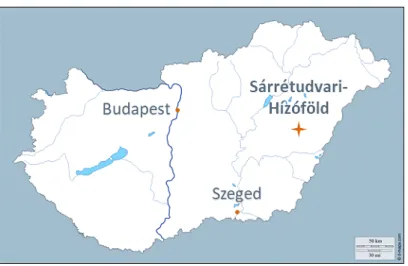
We selected a sample of 67 individuals from the 10th-century Hungarian cemetery of Sárrétudvari-Hízóföld, in order to analyze the individuals according to the presence or absence of riding deposit in their grave. A modern comparison group of 47 presumed non-rider individuals from the documented collection of Lisbon was also selected. Only adult males were included to limit the effect of sex and age on the changes. The main objectives were to identify skeletal changes reliably related to the practice of horse riding and to improve our understanding of the populations from the Hungarian Conquest period.
Various types of skeletal changes were analyzed, including some entheseal changes (at muscles attachment sites), joint changes, vertebral changes, morphological variants, and traumatic lesions. Measurements of the lower limb bones were also used to calculate indices of shape and robusticity.
Statistical analyses mostly revealed significant differences between the Hungarian groups with or without riding deposit and the comparison group from Lisbon. They concerned especially some entheseal changes at the coxal bone, femur, tibia, and calcaneus, a morphological adaptation on the femoral neck, intervertebral disc herniations at the thoracolumbar junction, or the ovalization of the acetabulum on the coxal bone. All these traits
7
individuals without deposit in their grave were likely riding horses as well.
Among the limitations calling for caution is the restricted size of our archaeological samples, which is one of the points that should be improved in the future. In addition, some skeletal changes, such as the entheseal changes, have a multifactorial etiology, which represents a limitation for their interpretation. In that regard, we performed the exploratory analysis of the microarchitecture of an enthesis, the radial tuberosity. Using micro-CT acquisitions and 3D reconstructions of the canals of the cortical bone, we observed that some microstructural variations could allow, with further research, distinguishing entheseal changes related to activity from those related to other factors, thus contributing to more reliable reconstructions of the activities in past populations.
In the end, we emphasize that the selection of a pertinent anthropological collection, with direct evidence of the practice of an activity, and the application of strict methodological criteria, are determinant factors for the reliable identification of activity-related skeletal changes.
8
Certaines modifications observées sur les os humains peuvent permettre de reconstituer les activités des populations anciennes. L'équitation représente notamment un intérêt particulier, ayant apporté des changements profonds et durables dans l'histoire de l'évolution culturelle humaine. Cependant, divers facteurs de biais et l'absence de données contextuelles claires liées aux restes osseux donnent souvent lieu à des interprétations limitées ou peu fiables des modifications osseuses en termes d’activités spécifiques.
Les sources archéologiques et historiques attestent que des tribus de populations semi-nomades ont conquis le bassin des Carpates à l’aide d’armées de cavaliers-archers au tournant des 9e et 10e siècles, conduisant ainsi à la fondation du Royaume de Hongrie en l'an 1000/1001. Les cimetières de cette période fournissent des cas de dépôts de matériel lié à l’archerie et à l’équitation ainsi que des ossements de chevaux associés aux individus dans les tombes. Ces populations sont ainsi parmi les plus pertinentes pour mener des études méthodologiques sur les modifications osseuses liées aux activités, et notamment à la pratique cavalière.
Nous avons sélectionné 67 individus issus du cimetière hongrois de Sárrétudvari-Hízóföld (10e siècle), pour les analyser selon la présence ou l'absence de dépôt lié au cheval dans leurs tombes. Un échantillon moderne de comparaison de 47 individus présumés non-cavaliers a également été sélectionné au sein de la collection documentée de Lisbonne. Seuls les sujets adultes masculins ont été inclus afin de limiter l'influence de variations en lien avec le sexe et l'âge. Les objectifs étaient d’identifier des modifications squelettiques liées à la pratique cavalière et d’améliorer nos connaissances sur les populations de la Conquête hongroise.
Nous avons analysé diverses modifications osseuses, au niveau des enthèses (points d'attache des muscles), articulations et vertèbres, ainsi que des variations morphologiques et lésions traumatiques. Des mesures des os des membres inférieurs ont aussi servi à calculer des indices de forme et de robustesse.
Les analyses statistiques ont principalement révélé des différences significatives entre les groupes hongrois avec ou sans mobilier et le groupe de comparaison. Celles-ci concernent notamment les modifications de certaines enthèses de l’os coxal, du fémur, du tibia et du calcanéus, une adaptation morphologique sur le col du fémur, les hernies discales à la jonction thoraco-lombaire, ou encore l'ovalisation de l’acétabulum de l'os coxal. Ces traits peuvent tous être liés à la posture du cavalier et semblent donc être des indicateurs prometteurs pour la
9
Parmi les limitations, appelant malgré tout à la prudence, figure la taille restreinte de nos échantillons archéologiques, qui est l’un des points qui devront être améliorés à l'avenir. En outre, certaines modifications osseuses, comme celles des enthèses, ont une étiologie multifactorielle, limitant ainsi leur interprétation. À cet égard, nous avons mené l’analyse exploratoire de la microarchitecture d'une enthèse, la tubérosité du radius. À l’aide d’acquisitions micro-CT et de reconstructions 3D des canaux de l'os cortical, nous avons observé que des variations microstructurales pourraient permettre, avec des recherches supplémentaires, de distinguer les modifications des enthèses liées aux activités de celles liées à d’autres facteurs, contribuant ainsi à de plus fiables reconstructions des activités des populations anciennes.
Au final, le choix d'une collection anthropologique pertinente, avec des preuves directes de la pratique d'une activité, ainsi que l'application de critères méthodologiques stricts, sont autant d’éléments déterminants pour l'identification fiable de modifications squelettiques liées aux activités.
10
TABLE OF CONTENTS
ACKNOWLEDGMENTS ... 2 ABSTRACT ... 6 RÉSUMÉ COURT ... 8 TABLE OF CONTENTS ... 10 LIST OF ABBREVIATIONS ... 15 CHAPTER 1. INTRODUCTION ... 16The reconstruction of activities in ancient populations ... 17
The research on activity-related skeletal changes ... 17
Brief biological background... 17
Main limitations in the research on activity-related skeletal changes ... 19
The identification of horse riding ... 23
Why is horse riding so interesting? ... 23
What do we know from sports medicine and modern riders? ... 25
Archaeozoology and equestrian activity ... 28
Bioanthropology and horse riding... 28
II.D.1. Previous research on skeletal changes related to horse riding ... 28
II.D.2. Main limitations for the identification of reliable horse riding-related skeletal changes...40
II.D.2.a. Limitations related to the link between horse riding and the materials 40 II.D.2.b. The lack of a comparison group ... 42
II.D.2.c. Other methodological limitations ... 44
II.D.2.d. Implications for the identification of horse riding-related skeletal changes...46
Research objectives ... 47
A bioanthropological contribution ... 47
11
The cemeteries and populations from the Conquest period: archaeological
context...51
CHAPTER 2. MATERIALS & METHODS ... 55
Sárrétudvari-Hízóföld: A unique cemetery from the Hungarian Conquest period .... 56
Presentation of the cemetery and the collection... 56
Selection of the samples from the Hungarian Conquest period ... 58
I.B.1. Methodological considerations ... 58
I.B.2. Reassessment of the sex of the individuals ... 59
I.B.3. Reassessment of the age-at-death of the individuals ... 60
I.B.4. Composition of the samples of early Hungarians based on the archaeological deposits... 61
Out-sample comparison: the documented collection of Lisbon ... 62
Why a comparative sample? ... 62
Selection of a collection ... 63
The documented collection of Lisbon ... 64
II.C.1. Presentation of the collection ... 64
II.C.2. Selection of the sample ... 64
Methods for the study of horse riding-related skeletal changes ... 67
Macromorphological analyses ... 67
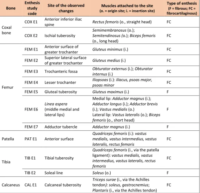
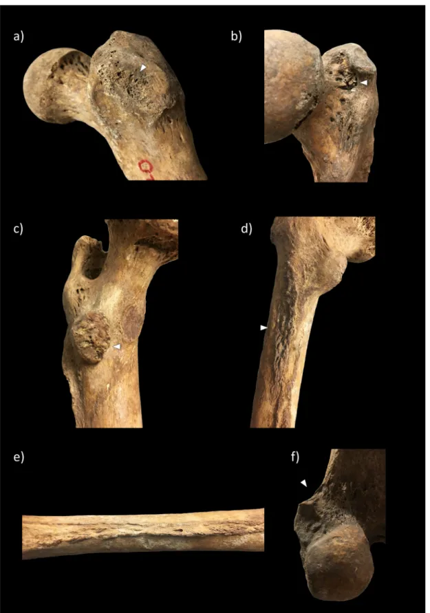
III.A.1. Entheseal changes ... 67
III.A.2. Joint changes ... 72
III.A.3. Morphological variants of the femur ... 76
III.A.4. Vertebral changes ... 80
III.A.4.a. Schmorl’s nodes ... 80
III.A.4.b. Spondylolysis ... 81
12
III.B.1. The index of ovalization of the acetabulum ... 85
III.B.2. Other indices of shape and robusticity ... 87
Limiting bias ... 89
III.C.1. The influence of pathological conditions ... 89
III.C.2. The influence of age ... 91
III.C.3. Other aspects ... 93
Statistical analyses ... 94
III.D.1. General considerations ... 94
III.D.2. Qualitative analyses ... 95
III.D.3. Quantitative analyses ... 95
CHAPTER 3. RESULTS ... 97
Macromorphoscopic analyses ... 98
Results of the analysis of entheseal changes ... 98
I.A.1. Intergroup comparisons ... 98
I.A.1.a. Young and mature adult individuals ... 98
I.A.1.b. Individuals in all adult age categories ... 101
I.A.2. Bilateral asymmetry ... 104
Results of the analysis of joint changes ... 106
I.B.1. Intergroup comparisons ... 106
I.B.1.a. Young and mature adult individuals ... 106
I.B.1.b. Individuals in all adult age categories ... 108
I.B.2. Bilateral asymmetry ... 110
Results of the analysis of morphological variants of the femur ... 112
I.C.1. Intergroup comparisons ... 112
I.C.2. Bilateral asymmetry ... 115
13
Results of the analysis of traumatic lesions ... 122
Results of the osteometric analyses ... 124
Index of ovalization of the acetabulum ... 124
II.A.1. Inter- and intraobserver agreement evaluation ... 124
II.A.2. Intergroup comparisons ... 125
II.A.3. Bilateral asymmetry ... 127
Other indices of shape and robusticity ... 129
II.B.1. Intergroup comparisons ... 129
II.B.2. Bilateral asymmetry ... 132
CHAPTER 4. DISCUSSION ... 135
Entheseal changes ... 136
Young and mature adult individuals ... 136
Individuals in all adult age categories ... 138
Further interpretations and considerations ... 139
Joint changes ... 141
Morphological variants of the femur ... 142
Vertebral changes ... 145
Schmorl’s nodes ... 145
Spondylolysis ... 148
Traumatic lesions ... 148
The ovalization of the acetabulum ... 151
The shape and robusticity of lower limb bones ... 153
The identification of reliable horse riding-related skeletal changes ... 157
The horse riders from the Hungarian Conquest period ... 160
14
investigation ... 166
The selection of the enthesis ... 167
Materials... 168
Methodology ... 170
Microstructural comparison of different types of entheseal changes ... 172
Could µ-CT and 3D reconstructions help to identify activity-related entheseal changes? ... 174 CONCLUSIONS ... 176 SUMMARY ... 178 RÉSUMÉ ... 181 ÖSSZEFOGLALÓ ... 185 REFERENCES ... 189 LIST OF FIGURES ... 217 LIST OF TABLES ... 222 APPENDICES ... 228
15
DISH diffuse idiopathic skeletal hyperostosis
EC entheseal changes
FPH foot phalanges
HPH hand phalanges
HRSC horse riding-related skeletal changes
IOA index of ovalization of the acetabulum
L1-5 first-fifth lumbar vertebra
LIS Lisbon (comparison group from the documented collection of Lisbon)
µ-CT micro-computed tomography (or also micro-CT)
NRD no riding deposit (group of individuals from the Hungarian Conquest period)
RD riding deposit (group of individuals from the Hungarian Conquest period)
S1 first sacral vertebra
SN Schmorl’s nodes
T1-12 first-twelfth thoracic vertebra
17
The reconstruction of activities in ancient populations
The research on activity-related skeletal changes
The study of the activities and behaviors in ancient populations represents a particularly active research area in bioarchaeology, even considered by some scholars as the “Bioarchaeology’s Holy Grail” (Jurmain et al., 2012). Bone changes of different types have been considered and interpreted for decades as activity, occupational or stress markers, with the aim of providing information about the activities of the individuals. Their analysis has involved many bioanthropologists and paleopathologists, with an important step forward accomplished from the 1980’s (e.g., Angel, 1982; Merbs, 1983; Ruff et al., 1984; Dutour, 1986; Kennedy, 1989; Hawkey & Merbs, 1995; Pálfi & Dutour, 1996; Robb, 1998; Jurmain, 1999; Al-Oumaoui et al., 2004; Mariotti et al., 2004; Rhodes & Knüsel, 2005; Molnar, 2006; Mariotti et al., 2007; Perréard Lopreno, 2007; Weiss, 2007; Villotte et al., 2010; Jurmain et al., 2012; Niinimäki, 2012b; Alves Cardoso & Henderson, 2013; Henderson & Alves Cardoso, 2013; Milella et al., 2015; Henderson et al., 2016).
In some cases, material archaeological remains such as artifacts or built structures are not indicative of the activities performed by individuals during their life. The analysis of the individuals’ skeletal remains represents the most direct or sometimes the only way to address the question, even if many limitations must be considered for the reconstruction of activities. The main questions addressed in this research area concern mostly locomotion, mobility, and asymmetry patterns as well as the sexual division of labor, the socio-economic organization, and, more generally, the reconstruction of the lifestyles and behaviors in past societies.
Brief biological background
Intense and regular physical activity can lead to some pathological or non-pathological bone changes of different types. The most promising research areas in the reconstruction of activities concern especially the musculoskeletal stress markers (MSMs), nowadays rather called entheseal changes (EC), as well as osteoarthritis and bone biomechanical properties, even though other types of changes are also used as activity-related markers (Kennedy, 1989; Larsen, 1997; Capasso et al., 1999; Jurmain et al., 2012).
The studies in the field of activity reconstruction are based on the bone’s reaction to loading and its ability to adapt its shape and structure in response to it (Niinimäki, 2012a). The performance of any physical activity involves the musculoskeletal system as the body is in motion. External loads and muscle loads are transferred to the tissues. Those mechanical
18
stimuli, or forces, generate signals to bone cells that are transduced into a biological response, including bone remodeling, the process that removes old and damaged bone and replaces it with new tissue. Bone resorption is initiated by osteoclasts, and bone formation is initiated then by osteoblasts. In normal bone, the balance between osteoblastic and osteoclastic activity is disturbed due to an increase in loading and new bone is deposited on periosteal and endosteal surfaces of cortical bone and on trabecular surfaces in cancellous bone (Mellon & Tanner, 2012). This results in bone mass and morphology changes that make the bone more suitable to resist to mechanical loading, according to Wolff’s law (1892). Bone cross-sectional geometry allows, for instance, to study the biomechanical properties that are adapted in response to physical activity, but it mostly provides information regarding general loading patterns (Niinimäki, 2012a; Tichnell, 2012).
Unlike cross-sectional geometry, the analysis of EC is more adapted if we are interested in the study of specific activities (Tichnell, 2012). Entheses are the insertion sites of tendons, ligaments, and joint capsules on the bone. A distinction can be made between two types of entheses. Fibrous entheses are mainly encountered at the metaphyseal or diaphyseal areas while fibrocartilaginous entheses include the insertions at the epiphyses and processes of long bones as well as the short bones of hands and feet and several ligaments in the spine (Benjamin & Ralphs, 1998; Benjamin & McGonagle, 2001). EC are pathological or non-pathological modifications at the insertion sites (La Cava, 1959; Niepel & Sit'aj, 1979; Lagier, 1991; Benjamin et al., 2002). These alterations are visually observable on dry bone and can take the form of new bone formation (raised margins, enthesophytes, irregular or rugose surfaces) or bone destruction (porosity, cavitations, cortical defects, erosive areas) (e.g., Hawkey & Merbs, 1995; Robb, 1998; Mariotti et al., 2004; Villotte, 2009; Henderson et al., 2016; Villotte et al., 2016). The precise etiology of these features remains however debated as many factors, age above all, need to be considered (Henderson et al., 2017a). Theoretically, the tissues forming the enthesis remodel in order to resist muscle tension (Tichnell, 2012) and osteophytic and osteolytic processes develop according to the type, level, and direction of loading applied and the deposition or resorption thresholds of the bones (Niinimäki, 2012a). Muscular activity is considered to stimulate bone remodeling by an increase in blood flow at the entheses resulting in hypertrophic muscle insertion sites, while osteophytes are thought to result from muscular tear (which leads to bone avulsion and ossification exostosis) (Hawkey & Merbs, 1995; Tichnell, 2012) and osteolytic processes might be due to continual microtrauma (Hawkey & Merbs, 1995). Regarding these, some scholars also suggest that they might be related to blood
19
vascularization in the entheses or that they could be due to the absence of a layer of cortical bone in some entheseal regions and represent remains from developmental phases during adolescence (Henderson et al., 2017a). In the end, although various determinant factors need to be considered, EC are more promising for studying the effect of specific activities as each activity involves specific muscles and continual stress of a muscle can presumably result in changes observed at the level of the involved entheses (Tichnell, 2012).
Osteoarthritis, or degenerative joint disease, represents the third main research area in the reconstruction of activities, partly because it has been used widely and relatively early as a marker of activity (Ortner, 1968; Jurmain, 1977; Angel, 1982; Merbs, 1983). Apart from dental lesions, osteoarthritis is also the most commonly observed condition in skeletal remains (Waldron, 2009). The bone changes observed at the synovial joints include marginal osteophytes and contour deformation, as well as porosity, bone formation, and eburnation on the joint surface (Buikstra & Ubelaker, 1994; Waldron, 2009; Jurmain et al., 2012). These alterations appear following the breakdown of the articular cartilage of the joint and the inflammation in the synovial membrane. Vascularization of the subchondral bone, in particular, is responsible for the formation of new bone (Waldron, 2009). The discussions on the link between osteoarthritis and activities are based on the fact that it has been considered as resulting from repetitive mechanical loading and that repetitive tasks could, therefore, lead to severe osteoarthritic features on specific joints (Weiss & Jurmain, 2007). Nowadays, it is acknowledged from clinical research that the etiology of osteoarthritis is multifactorial (Larsen, 1997; Resnick, 2002; Ortner, 2003; Weiss & Jurmain, 2007; Waldron, 2009; Jurmain et al., 2012).
Main limitations in the research on activity-related skeletal changes
Several authors reviewed the challenges related to the use of skeletal changes for the reconstruction of activities in past societies and suggest to use them with caution (Dutour, 1992, 2000; Weiss & Jurmain, 2007; Jurmain et al., 2012; Villotte & Knüsel, 2013). A major issue with activity-related skeletal changes (ARSC) is their lack of specificity, which is closely related to their multifactorial etiology: not only physical activity but also age, sex, body weight, body size, genetic factors, ancestry, diet or pathological conditions can indeed influence the expression of entheseal changes (EC), osteoarthritis, or bone geometry (e.g., Dutour, 1992; Rogers & Waldron, 1995; Wilczak, 1998; Knüsel, 2000b; Crubezy et al., 2002; Weiss & Jurmain, 2007; Niinimäki, 2011; Jurmain et al., 2012; Milella et al., 2012; Weiss et al., 2012; Niinimäki & Baiges Sotos, 2013; Ruff & Larsen, 2014; Schrader, 2019). These systemic factors
20
can expose an individual to risk for developing changes, while other factors can also have a local influence, such as musculoskeletal anomalies, joint alignment or injury, which can, for instance, expose directly a joint to risk for osteoarthritis (Dutour, 1992; Schrader, 2019) (Figure 1).
Figure 1. Summary of the potential risk factors for osteoarthritis (Johnson and Hunter, 2014)
Among all those factors, age is probably the most important one to be considered when dealing with ARSC. Older individuals are logically more likely to develop degenerative joint changes. This could be linked with “cumulative wear-and-tear (i.e., the repetitive use of a joint over one’s life course), sarcopenia, decreased bone turn over, as well as a reduced capacity for soft tissue repair” in the case of osteoarthritis (Schrader, 2019: 61). Regarding EC, while the high correlation with age is now widely recognized (e.g., Weiss, 2003, 2004; Alves Cardoso & Henderson, 2010; Milella et al., 2012), it is still unclear if it is the consequence of their accretion over life, an increased susceptibility to remodeling, a reduction in tendon vascularity, a decrease in cortical bone, or the loss of muscle mass (see Schrader, 2019). Sex must also be considered as females seem to be at higher risk of developing osteoarthritis. Possible reasons are a post-menopause reduction in estrogen levels, lessened bone mineral density, bone composition, ligament laxity, lower cartilage volume, pregnancy, and neuromuscular strength (see Schrader, 2019). Moreover, the differences in muscle mass between females and males could also affect entheses, with males being more susceptible to develop larger and severe EC (Weiss, 2003, 2004; Weiss et al., 2012; Nikita, 2017; Schrader, 2019). Besides sex and age, it has also been suggested that there is an inter-individual variation in bone production and bone resorption which may affect the expression of ARSC, with individuals who would be “bone-formers”, and
21
others who would rather be “bone-losers” (Rogers et al., 1997; Mays, 2016; Schrader, 2019). Furthermore, it is now widely acknowledged that metabolic and inflammatory disorders like diffuse idiopathic skeletal hyperostosis (DISH) or seronegative spondyloarthropathies can lead to the development of EC (e.g., Villotte & Knüsel, 2013). While there were early warnings of caution regarding this aspect (Dutour, 1986, 1992; Pálfi, 1992), it is only recently that studies relying on EC tend to exclude systematically the individuals affected by such diseases from samples.
A further limitation in the analysis of activity-related markers is the large variety of scoring methods, concerning osteoarthritis (Crubézy, 1988; Buikstra & Ubelaker, 1994; Rogers & Waldron, 1995) and entheseal changes (Crubézy, 1988; Hawkey & Merbs, 1995; Mariotti et al., 2004; Villotte, 2006; Mariotti et al., 2007; Villotte et al., 2010; Henderson et al., 2013), as well as the large scale of methods of statistical analysis used in this type of research (Nikita, 2017; Schrader, 2019). This makes very difficult the comparisons between studies. Regarding EC, in particular, their relation to activity has proven not to be straightforward and the scoring methods appear not to be entirely adequate, leading some to go further and investigate the microarchitecture of the entheses. The results of the study by Djukic and collaborators (2015), who scanned several entheses using microcomputed tomography, suggest that stages of scored macroscopic EC do not reflect the microscopic organization of entheses, except maybe in the most severe cases. Furthermore, Michopoulou and colleagues (2015; 2017) tested the correlation between several scoring methods and activity and observed that activity was rarely significant as a factor affecting EC expression (compared to age and body mass for instance), suggesting that the tested scoring methods do no effectively reflect activity. Regarding the analysis of bone geometrical features for research on activities, external macroscopic measurements are commonly used for quantifying robusticity but are limited compared to the potential of cross-sectional bone geometry, which is however characterized by technical, financial, and time challenges as it ideally requires the use of computed tomography, more accurate than plane radiography for example (Stock & Shaw, 2007; Jurmain et al., 2012; Ruff & Larsen, 2014). In general, the sample size has also often been recognized as problematic in the field of activity reconstruction (e.g., Henderson & Nikita, 2016) — being closely dependent on bone preservation — as well as, in particular, the lack of documented out-groups of sufficient size for comparison (Knüsel, 2000b).
Another issue that concerns EC, in particular, comes from the demonstration of the distinction between two types of entheses, fibrocartilaginous and fibrous ones, as mentioned
22
earlier (Benjamin & McGonagle, 2001; Benjamin et al., 2002). Several studies suggest that fibrocartilaginous entheses have a stronger correlation with activity than fibrous ones (Villotte et al., 2010; Havelková et al., 2011; Villotte & Knüsel, 2013; Weiss, 2015), which could be explained by differences in their structure and the way tendons attach to the bone (Benjamin et al., 2002). Fibrous entheses would also be less promising activity markers due to the difficulty to identify their limits and to describe their changes compared to their “normal” aspect, which is not clearly defined (Villotte & Knüsel, 2013; Villotte et al., 2016; Nikita, 2017).
Furthermore, besides the multifactorial etiology of ARSC, other general analytical pitfalls must be acknowledged and avoided in the attempt to reconstruct activities in past populations from the analysis of skeletal changes. They concern the problem of transposition between present and past activities (i.e., the manner in which they are and were performed), the lack of clinical evidence for validation of the link between a specific activity and a specific marker identified from archaeological materials, or the lack of analogy regarding activities that are no longer practiced today (Dutour, 1992, 2000). Besides the lack of specificity of the markers mentioned previously, which means that identical skeletal changes in different individuals can result from different activities or various other causes, the relative sensitivity of the markers should also be acknowledged. A specific activity may not systematically lead to the expression of similar skeletal changes in different individuals, or even to the expression of any skeletal changes at all (Dutour, 2000). In addition, a specific activity rarely results in a single type of change, therefore a single specific marker should not be enough to confirm an activity hypothesis; only the combination of several different markers, consistent with each other, may allow making a reliable association with a particular activity (Dutour, 1992, 2000). The existence of relevant archaeological data that can be linked whether in a conclusive or a compatible way to an activity is determinant for the selection of any study material in order to limit the influence of the methodological biases mentioned above (Dutour, 1992, 2000).
Finally, we should keep in mind that bone changes do not reflect the consequences of a single activity performed during life, but that “the skeleton registers a mosaic of activities over the course of each individual’s lifetime” (Kennedy, 1998: 308).
23
The identification of horse riding
Why is horse riding so interesting?
Among all activities, horse riding brought profound and lasting changes in the history of human cultural evolution concerning major aspects such as trade, settlement, warfare, subsistence, social organization and political ideology (Anthony & Brown, 1991; Anthony, 2007). The use of horses for transportation also considerably contributed to the circulation of languages, Indo-European ones in particular, as well as cultures and diseases, among other things (Anthony, 2007; Outram et al., 2009; Librado et al., 2016; de Barros Damgaard et al., 2018; Gaunitz et al., 2018).
Archaeological evidence suggests that horse domestication likely started in the Copper Age Botai hunter-herder culture, in the northern Kazakh steppe, around 3500-3000 years BCE (Outram et al., 2009; de Barros Damgaard et al., 2018; Gaunitz et al., 2018). There, not only horse burials were discovered but also remains of tools associated with the production of leather thong and traces of equine milk fats in ceramics. These elements, combined with bit-related pathologies on the teeth in horse remains tend to support the practice of milking and harnessing, and thus, of pastoral husbandry (Outram et al., 2009; Gaunitz et al., 2018).
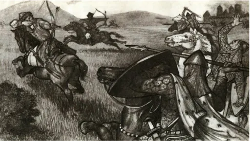
Horses can be involved as much for transportation than for agricultural and warfare activities. Their importance for humans can be detected in numerous artistic representations, in the material culture, in historical texts as well as in the funerary practices (Hyland, 1996; Garofalo, 2004; Outram et al., 2011; Baillif-Ducros, 2018). As an example of their social importance, we can mention the development of relay stations for horses in different societies since Antiquity. They allowed the fast transportation of people (for private or professional purposes) and materials through territories over long distances and thereby contributed to the shaping of road networks, such as in France between the 17th and the 19th centuries (Bretagnolle & Verdier, 2014). On another note, the Bayeux Tapestry, which depicts the events that lead to the Norman Conquest of England by William the Conqueror in 1066, offers a vivid illustration of the importance of horses in warfare. Through numerous animal representations, the tapestry reveals the significant place of horses in the Norman society and the fundamental role played by the cavalry (especially mounted lancers) during the Battle of Hastings, after the transportation of several thousands of horses across the Channel (Bouet, 2015) (Figure 2).
24
The practice of depositing complete horses in graves seems to go back to the late Roman Iron Age (1-375 CE) while separate horse burials can be found in central and northern Europe from the early and the late Iron Age (Jennbert, 2003). When an animal is deposited in a separate pit, it can be considered as an animal grave. In such cases, it is possible to state that the people responsible for this burial cared for the animal and had a purpose (Jennbert, 2003).
Replacing oxen, onagers, and donkeys as draught animals, horses were considered as a prestige animal (Garofalo, 2004). During different eras, like in the Middle Ages, riding a horse was often a sign of social distinction, as owning horses required specific infrastructures, equipment, and assigned workforce for care and maintenance (Hyland, 1999; Baillif-Ducros, 2018). Horse riding was regularly performed by particular subgroups in societies and was, therefore, an activity associated with the identities of those specific groups and was bearing symbolic meaning (Hyland, 1999; Tichnell, 2012). Furthermore, in many cultures (e.g., Scythians, Celts, Gauls, Avars, Magyars), the horse had strong spiritual importance and played a role during various funerary rituals and practices (Garofalo, 2004; Bede, 2015).
With this in mind, it is clear that a better understanding of who exactly were horse riders during their life can shed light on different aspects of past societies. For instance, for some ancient populations of nomadic pastoralists, the sparseness of archaeological remains and the lack of internal textual evidence, among other causes, may result in a partial or oversimplified understanding regarding different aspects of the societal organization (Tichnell, 2012). Considering the extreme importance of horse riding in their life, the analysis of this practice in those populations can bring elements of answers, and, while ethnographic studies can be very
Figure 2. Norman horsemen depicted on the Bayeux Tapestry, ca. 1080. Embroidery, Bayeux Tapestry Museum, Bayeux, France. From http://www.hs-augsburg.de
25
informative, the examination of skeletal remains for horse riding-related bone changes represents the most direct source of information (Tichnell, 2012).
What do we know from sports medicine and modern riders?
Although the techniques and style of riding, the equipment, and the morphology of the horses used nowadays by modern riders are not directly comparable with those used by past populations in different regions of the world, clinical sources can bring relevant complementary data (McGrath, 2015). For both modern and ancient riders, some features must, indeed, be common in relation to the posture on the horse and the muscles needed to maintain it, notably if stirrups were used.
In the position privileged by riders, the pelvis is in retroversion (tilted backward), the back is vertical and straight, and the lumbar lordosis is diminished, allowing better absorption of vertical stresses (Auvinet, 1999; Humbert, 2000). Sitting astride forces the femurs into abduction while pointing the toes ahead rotates them medially and gripping the horse engages the adductors. While it is recommended that the heel be aligned with the shoulder and the hip, leading to a flexed knee, variations in the riding style also see the knee being semi-extended, with the heel put forward (Garofalo, 2004).
The muscles that are engaged in horse riding cover a wide range of movements and include especially the adductors (hip adduction), the gluteals (hip extension, abduction, medial and lateral rotation), the quadriceps (knee extension, hip flexion), the hamstrings (knee flexion, hip extension), the hip rotators (hip lateral rotation) and the calf muscle (plantarflexion). Among them, the adductors are often presented as particularly important for riding as they keep the legs together on the horse, which is not a common action otherwise in daily life (Baillif-Ducros et al., 2012; Willson, 2013; Djukic et al., 2018). The abdominal muscles are also determinant for the balance and the control and stabilization of the posture. Finally, the shoulder and trunk stabilizers are important notably for accurate movements of the arms that control the reins (when used) and for the head posture (Willson, 2013). The regular and intensive use of those muscles for the highly demanding activity that is horse riding is likely to lead to muscular injuries. Furthermore, due to the speed, height, weight, and strength of the horse, riding represents – and has always represented – a potentially dangerous activity that puts the rider at risk for injuries of different types (Bixby-Hammett & Brooks, 1990; Garofalo, 2004; Ball et al., 2007). In terms of potential of injuries, horse riding is even more dangerous than activities such as automobile racing, motorcycle riding, skiing, football, and rugby, and it is also considered to be the sport with the highest mortality (Ball et al., 2007).
26
Sports medicine describes the injuries affecting modern riders, whether they perform horse riding in a professional or a leisure context. Based on numerous studies published between 1959 and 1987, concerning European and American individuals, the described injuries concern mostly the upper extremity (from 24 to 61 % of the reported cases depending on the sources) followed by the lower extremity (18–36 %), while about 20 % of cases concern the head and the face and from 4 to 27 % are spinal injuries (Bixby-Hammett & Brooks, 1990). The second most common type of injuries, after the soft tissue injuries, and before the concussions, are fractures. While the majority of the accidents occur when the horse is mounted, mostly being due to a fall, 1 to 27 % occur while handling or taking care of the horse on foot and are related to the animal itself (kicking, biting, or stepping on the rider). It was also noted that it is rather common (37 %) to suffer several times horse-related injuries during the course of a lifetime (Bixby-Hammett & Brooks, 1990).
In a different study that relied on a survey addressed to adult patients who had suffered major equestrian injuries in Alberta, Canada, between 1995 and 2005, that percentage (subjects who had already suffered previous equestrian injuries) reached 47 % (Ball et al., 2007). This study also reveals that, as for the cause of traumas, 60 % of patients were thrown off or fell off the horse while 16 % were crushed by a falling horse, 8 % were kicked, 4 % were stepped on, and the remaining 13 % cases were injured by other mechanisms. It can also be noted that 45 % of the patients required surgery, while 7 % died because of their equestrian injuries. Furthermore, in this sample, the injuries concerned the chest (54 %), head (48 %), abdomen (22 %) and extremities (17 %), with, in particular, skull (18 %), extremity (17 %), spinal (17 %) and pelvic fractures (15 %) (Ball et al., 2007).
Other studies focusing more precisely on traumas revealed that the extremities were the most commonly affected regions (46,1–54,4 %), followed by the trunk (28,9 %) and the head and neck (16,6–23,8 %) in samples of patients from Sweden and the United States analyzed in the early 2000s (Loder, 2008; Altgärde et al., 2014; Ki et al., 2018). Various additional sources put the frequency of the traumas in the following descending order: the upper limbs, skull, neck, lower limbs, vertebral column, thorax, and the pelvis (Baillif-Ducros, 2018). Traumas on the upper extremities, in particular, are essentially related to the attempt of self-protection that occurs during a fall. Moreover, pubic symphysis diastasis can also be observed, although rarely, on horse riders. A violent impact on the horse, with the pommel of the saddle, or from a fall, can, indeed, lead to a partial or complete rupture of the joint, without fracture (Garofalo, 2004; Baillif-Ducros, 2018).
27
Sports medicine also describes other types of injuries, including some being the result of overuse. In polo players from Argentina, for instance, despite fractures (39 % of injuries) were also recorded sprains and strains (19 %), head concussions (19 %), muscular injuries (13 %), such as hip adductor tendinitis, and also dislocations (5 %), post-traumatic back pain (5 %) and complex lesion of the eye (2 %), as well as facial lacerations (Costa-Paz et al., 1999; Garofalo, 2004).
Baillif-Ducros (2018) delivers a review of the bone lesions that can be observed on equestrian athletes. They include, on the vertebral column, lumbar hyperlordosis, spondylolysis (stress fracture on the vertebral arch) and spondylolisthesis (forward displacement of a vertebra compared to the inferior one), osteoarthritis, disc deteriorations and Schmorl’s nodes (or intraosseous disc herniations), and Scheuermann’s disease (kyphosis of the lower thoracic region, with wedging of the anterior portion of the vertebral bodies) (e.g., Auvinet, 1980b; Lang, 1995; Auvinet, 1999; Humbert, 2000; Pugh & Bolin, 2004).
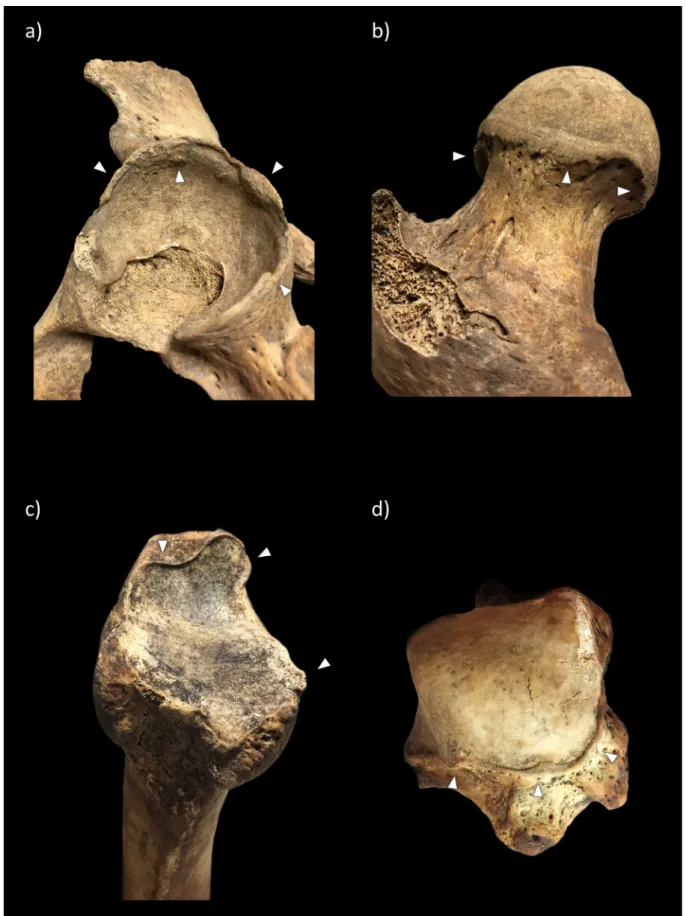
The hip, a determinant region for the posture and the communication between the rider and the horse, is also affected (Baillif-Ducros, 2018). Microtraumatic injuries, due to muscular overuse, are recorded: adductor strain, myositis ossificans in the adductors (or rider’s bone, although this lesion is not observed anymore in modern riders), and enthesopathies of the adductors (e.g., Auvinet, 1980a; Auvinet & Ginet, 1980). In the riding position, the retroversion of the pelvis can also be the cause of the femoroacetabular impingement (FAI) (Baillif-Ducros, 2018). In this position, the femoral head-neck junction is abnormally in contact with the acetabular rim. The repetition of this contact can lead to coxarthrosis and, presumably, to the presence of non-metric traits such as the Poirier’s facet (extension of the femoral head articular surface onto the neck) and the fossa of Allen (pitted area or cortical erosion with exposition of the trabecular bone) (Villotte & Knüsel, 2009; Radi et al., 2013). Furthermore, perifoveal osteophytes can also be observed, as well as pubic inguinal lesions (Baillif-Ducros, 2018).
The use of stirrups provides a point of stability for the rider. The subsequent loading for the knee joint can lead to gonarthrosis. The level of pressure and the possible lesions would notably depend on the length of the stirrups as it influences the degree of flexion of the knee (Baillif-Ducros, 2018).
However, some of those lesions have been observed only in low frequencies in riders and are not specific to horse riding. Scheuermann’s disease, the FAI, or the perifoveal osteophytes, for instance, are common in other high-level athletes (Auvinet, 1999; Baillif-Ducros, 2018). Despite problems of transposition between modern and ancient riders, clinical data from sports
28
medicine are a good indicator of what skeletal changes should be investigated in particular in archaeological populations.
Archaeozoology and equestrian activity
Various studies have attempted to infer activities from the paleopathological analysis of horse skeletal remains (e.g., Levine et al., 2000; Pluskowski et al., 2010; Bulatović et al., 2014; Marković et al., 2014; Taylor et al., 2014; Bindé et al., 2018). Among several promising markers that could be identified, the presence of bit wear is even often considered as a direct and unmistakable sign of riding (Brown & Anthony, 1998; Anthony, 2007; Bendrey, 2007; Taylor et al., 2015; Greenfield et al., 2018). While the analysis of entheseal changes for the reconstruction of activities in human past populations is largely spread, their study on animal skeletal remains was often unconsidered. With the aim of a better understanding of their status and the activities in which they were involved in past societies, scholars recently focused their interest on the entheseal changes developed by riding, draft and pack animals, including, especially, horses (Bendrey, 2008; Taylor et al., 2015; Bertin et al., 2016; Bindé et al., 2019) and reindeers (Niinimäki & Salmi, 2016; Salmi & Niinimäki, 2016). Other approaches focusing on the horses include the study of different vertebral paleopathological conditions, such as ankylosing spondylitis (or “bamboo spine”) or deforming spondyloarthrosis (Jeffcott, 1980; Lignereux et al., 1998; Levine et al., 2000; Bartosiewicz & Bartosiewicz, 2002; Janeczek et al., 2014; Lignereux & Bouet, 2015), as well as the analysis of morphological changes on the skulls, such as on the nasal bones and the premaxilla (Taylor et al., 2015; Taylor & Tuvshinjargal, 2018), even if such changes may depend on the equipment used and the riding style.
Although those bone changes are promising for the identification of riding from horse skeletal remains, the existence of a direct link between specific changes and the practice of horse riding has not yet been unarguably demonstrated with regard to human skeletal remains.
Bioanthropology and horse riding
II.D.1. Previous research on skeletal changes related to horse riding
Bioanthropological research on horse riding in ancient populations has developed in the past decades, with a growing interest from the beginning of the 1990s, especially. Among the early contributions on this topic, the work of Miller and collaborators on historic Native American Omaha and Ponca skeletal remains (Miller & Reinhard, 1991; Miller, 1992; Reinhard et al., 1994) and the paleopathological investigation performed by Pálfi and Dutour on a Hungarian cemetery of the 10th century CE (Pálfi, 1992; Pálfi & Dutour, 1996; Pálfi, 1997) particularly played an influential role.
29
A combination of skeletal changes, repeatedly observed and interpreted as signs of riding in past populations, is sometimes referred to as horse riding syndrome or horseback riding
syndrome in the literature since Pálfi (1992, “syndrome du cavalier”). It covers pathological
and nonpathological changes of various types, most of them having previously been reviewed (Garofalo, 2004; Baillif-Ducros et al., 2012; McGrath, 2015; Baillif-Ducros, 2018). To synthetize, those skeletal changes can be categorized in several groups: (a) spinal changes; (b) extraspinal joint changes; (c) entheseal changes; (d) morphological variants; and (e) extraspinal traumatic lesions. For clarity, we group them under the term horse riding-related skeletal
changes (abbreviated as HRSC), even though their association with the practice of horse riding
is, in the great majority of cases, simply assumed and not demonstrated.
We present here in distinct tables a summary of the main HRSC (Table 1, 2, 3, 4, and 5). Those are the skeletal changes previously observed and described by scholars on anthropological samples from archaeological context for which the practice of horse riding was presented as a possible or compatible cause. Our aim with this synthetic review is to be as exhaustive as possible. The signs that were only mentioned but not directly observed and described by the scholars are not referred to, along with the changes that were observed on the skeletons of presumed riders but not explicitly associated with the practice of horse riding. Some studies that were presented as conference communications were available to us only through their abstracts, which sometimes did not provide enough data or details to be included here (e.g., Jacquemard et al., 2011; Eng, 2013; Storch et al., 2013). The studies that mention HRSC but that are not specific enough regarding their nature or their localization are briefly discussed under the relevant tables when it is relevant. Furthermore, studies focusing on HRSC but that could not obtain conclusive results (i.e., a probable association between specific changes and horse riding) are not referred to (e.g., Eng, 2013; Sarry et al., 2016, on the shape of the acetabulum). Finally, when the authors describe the same horse riding-related changes on the same skeletal series in different publications, the earliest reference that was available to us is given.
30
Table 1. Summary of the spinal skeletal changes associated with horse riding in the anthropological literature
Spinal changes observed References
Intervertebral disk degeneration; Facet joint osteoarthritis (including at the cervical level)
Edynak, 1976 (lumbar); Bradtmiller, 1983; Angel et al., 1987; Miller & Reinhard, 1991; Pálfi, 1992; Sandness & Reinhard, 1992; Blondiaux, 1994; Willey, 1997; Courtaud & Rajev, 1998; Charlier, 2006; Langlois & Gallien, 2006; Fornaciari et al., 2007; Wentz & de Grummond, 2009; Bagagli et al., 2012; Baillif-Ducros et al., 2012; Anđelinović et al., 2015; Eng, 2016; Khudaverdyan et al., 2016; Sarry et al., 2016; Panzarino & Sublimi Saponetti, 2017 (apophyses, asymmetrical, post-traumatic after a fall); Karstens et al., 2018
Schmorl’s nodes/herniations Miller & Reinhard, 1991; Sandness & Reinhard, 1992; Willey, 1997; Reinhard & Wall, 2002; Langlois & Gallien, 2006; Wentz & de Grummond, 2009; Üstündağ & Deveci, 2011; Bagagli et al., 2012; Fornaciari et al., 2014; Anđelinović et al., 2015; Serna et al., 2015; Antikas & Wynn-Antikas, 2016; Sarry et al., 2016; Karstens et al., 2018
Accentuated thoracic kyphosis and lumbar lordosis/Scheuermann’s disease; Compression fractures
Blondiaux, 1994; Üstündağ & Deveci, 2011; Baillif-Ducros et al., 2012; Fornaciari et al., 2014; Sarry et al., 2016
Spondylolysis Miller & Reinhard, 1991; Pálfi, 1992; Dutour & Buzhilova, 2014; Fornaciari et al., 2014
Vertebral fusion Miller & Reinhard, 1991; Wentz & de Grummond, 2009 Kissing spine (or Baastrup’s disease) Miller & Reinhard, 1991
Sandness and Reinhard (1992) also describe a higher frequency of spondylolysis in historic Omaha and Ponca series compared to prehistoric ones. They associate this observation with the activities that predisposed the individuals “either to greater habitual stress, or incidences of trauma” (p. 306), without, however, explicitly mentioning horse riding, unlike they do as a possible explanation for the higher frequency of Schmorl’s nodes.
31
Table 2. Summary of the extraspinal joint changes associated with horse riding in the anthropological literature
Anatomical
region extraspinal joint changes Localization of observed References
Pelvis Sacroiliac joint Bradtmiller, 1983; Üstündağ & Deveci, 2011
Hip
Coxofemoral joint Bradtmiller, 1983; Miller & Reinhard, 1991; Pálfi, 1992; Courtaud & Rajev, 1998; Garofalo, 2004; Charlier, 2006; Fornaciari et al., 2007; Robin, 2011 (asymmetrical); Fornaciari et al., 2014; Sarry et al., 2016; Baillif-Ducros, 2018 Perifoveal osteophytes on the femoral head
(mentioned apart from other hip joint changes)
Blondiaux, 1994; Fornaciari et al., 2003; Garofalo, 2004; Fornaciari et al., 2007; Baillif-Ducros et al., 2012; Sarry et al., 2016; Panzarino & Sublimi Saponetti, 2017; Baillif-Ducros, 2018
Knee
Patellofemoral joint Robin, 2011 (asymmetrical); Baillif-Ducros & McGlynn, 2013; Baillif-Ducros, 2018
Tibiofemoral joint (left medial condyle) Molleson, 2007; Robin, 2011
Unspecified Bradtmiller, 1983; Miller & Reinhard, 1991; Eng, 2016
Ankle
Talocrural joint Panzarino & Sublimi Saponetti, 2017
Unspecified Bradtmiller, 1983
Foot Distal first metatarsal (“toe stirrup”) Miller & Reinhard, 1991 Shoulder
Glenohumeral joint Anđelinović et al., 2015; Karstens et al., 2018 Acromioclavicular joint Karstens et al., 2018
Elbow Unspecified Miller & Reinhard, 1991; Anđelinović et al., 2015; Eng, 2016 Thorax Sternal ends of ribs and clavicles Miller, 1992
It should be clarified that the authors who mention extraspinal joint changes of the upper part of the body explain it, in most cases, as being the consequence of rein or bridle tension (Eng, 2016; Karstens et al., 2018).
Considering the relatively early date of the following study, we should also mention that Edynak (1976), referred to by Larsen (1987) and Gibbon (1984), examined a series of skeletons from Yugoslavian mounds dated from Iron Age to the medieval period, and “found that articular joints of the males’ pelvic regions were more severely affected by osteoarthritis than any other joints” (Larsen, 1987: 391). The author associated this observation with the absorption of shock that occurs in horse riding, as the population included presumed riders. However, there is no precision regarding which joint, in particular, is concerned (i.e., sacroiliac or coxofemoral joint). According to Gibbon (1984), Edynak observed that “males exhibited a pattern of more
32
direct stress on all joints, and pelvic-lumbar degenerative arthritis related to the absorption of shock from below” (p. 206).
Finally, Panzarino and Sublimi Saponetti (2017), studying the remains of a Southern Italian (Apulia) individual from the 15th century, attribute the presence of osteochondritis dissecans at the acetabulum, femoral head, and talus to the stress related to galloping.
33
Table 3. Summary of the entheseal changes associated with horse riding in the anthropological literature (1/2)
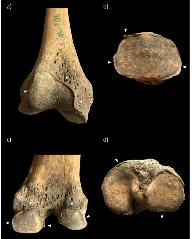
Bone Site of the observed entheseal changes (o. = origin site; i. = insertion site) Muscles attached to the site References
Coxal bone
Ilium posterior (superior and posterior
to the inferior gluteal line) Gluteus maximus (o.); Gluteus medius (o.); Gluteus minimus (o.) Miller, 1992; Pálfi, 1992; Anđelinović et al., 2015; Ciurletti, 2017
Pubic body Adductor brevis (o.) Miller, 1992
Ischial tuberosity Semimembranosus (o.); Semitendinosus (o.); Biceps femoris
(o., long head) Pálfi, 1992; Molleson & Hodgson, 1993; Ciurletti, 2017; Djukic et al., 2018 Inferior pubic ramus and ramus of
ischium (medial border) Adductor magnus (o.) Miller, 1992; Pálfi, 1992; Ciurletti, 2017
Femur
Greater trochanter Gluteus medius (i.); Gluteus minimus (i.) Miller & Reinhard, 1991; Pálfi, 1992; Molleson & Hodgson, 1993; Blondiaux, 1994; Molleson & Blondiaux, 1994; Knüsel, 2000a (medius); Fornaciari et al., 2003; Garofalo, 2004 (medius); Üstündağ & Deveci, 2011; Anđelinović et al., 2015; Antikas & Wynn-Antikas, 2016; Khudaverdyan et al., 2016; Ciurletti, 2017; Panzarino & Sublimi Saponetti, 2017
Trochanteric fossa Obturator externus (i.); Obturator internus (i.) Blondiaux, 1989; Molleson & Hodgson, 1993; Blondiaux, 1994; Molleson & Blondiaux, 1994; Belcastro et al., 2001; Fornaciari et al., 2007; Bartsiokas et al., 2015; Khudaverdyan et al., 2016; Ciurletti, 2017
Lesser trochanter Iliopsoas (i.): iliacus, psoas major, psoas minor Pálfi, 1992; Molleson & Hodgson, 1993; Belcastro et al., 2001; Charlier, 2006; Fornaciari et al., 2014; Panzarino & Sublimi Saponetti, 2017; Djukic et al., 2018
Gluteal tuberosity Gluteus maximus (i.) Pálfi, 1992; Blondiaux, 1994; Molleson & Blondiaux, 1994; Belcastro et al., 2001; Fornaciari et al., 2003; Charlier, 2006; Uerpmann et al., 2006; Tichnell, 2012; Pulcini, 2014; Bartsiokas et al., 2015; Ciurletti, 2017