HAL Id: tel-02890626
https://tel.archives-ouvertes.fr/tel-02890626
Submitted on 6 Jul 2020HAL is a multi-disciplinary open access archive for the deposit and dissemination of sci-entific research documents, whether they are pub-lished or not. The documents may come from teaching and research institutions in France or abroad, or from public or private research centers.
L’archive ouverte pluridisciplinaire HAL, est destinée au dépôt et à la diffusion de documents scientifiques de niveau recherche, publiés ou non, émanant des établissements d’enseignement et de recherche français ou étrangers, des laboratoires publics ou privés.
biotoxin immunosensing
Lu Zhang
To cite this version:
Lu Zhang. Design of plasmonic nanoparticles and their use for biotoxin immunosensing. Or-ganic chemistry. Sorbonne Université; Nanyang Technological University, 2018. English. �NNT : 2018SORUS439�. �tel-02890626�
Sorbonne Université
Nanyang Technological University
Physique et Chimie des Matériaux
School of Materials Science and Engineering
Design of plasmonic nanoparticles and their use for
biotoxin immunosensing
Submitted by Lu ZHANG
For the dual degree of Doctor of Philosophy (Ph.D.) of
Sorbonne Université and Nanyang Technological University
Defense scheduled for the 16 November 2018 In front of a jury composed of:
Mrs. Sabine SZUNERITS Professor Reviewer Mr. Borja SEPULVEDA Researcher Reviewer Mr. Shuzhou LI Associate Professor Examiner Mrs. Sierin LIM Associate Professor Examiner Mrs. Michèle SALMAIN Research Director Examiner Mr. Ali ABOU-HASSAN Associate Professor Examiner Mrs. Souhir BOUJDAY Professor Supervisor Mr. Bo LIEDBERG Professor Co-supervisor
Acknowledgments
The work presented in this thesis has been carried out from October 2015 to September 2018 enrolled under the dual degree PhD programme between Sorbonne Université in France and Nanyang Technological University in Singapore.
This work would not have been possible without numerous support. I thus acknowledge a great number of people.
Foremost, I would like to acknowledge and extend my heartfelt gratitude to my supervisors Pr. Souhir Boujday and Pr. Bo Liedberg who have accepted me as their student in this exciting and challenging project and kept me on track throughout the whole of this journey. Although they have given me the freedom to find my own way sometimes, they have always been around for guidance and for sharing their great knowledge and brilliant ideas. And my sincere thanks undoubtedly go to Dr. Michèle Salmain. I appreciate a lot that she is always there for discussions concerning scientific matters and everything else. Her encouragement for me along all these three years has lighted my journey of research and made me more confident achieving the goals.
I sincerely thank Pr. Sabine Szunerits and Dr. Borja Sepulveda for being reviewers of my thesis, as well as Assoc. Pr. Shuzhou Li, Assoc. Pr. Sierin Lim, and Assoc. Pr. Ali Abou-Hassan for agreeing to be members of the jury of my thesis.
I am very grateful to all the people that have come and gone throughout these three years in the three labs, Laboratoire de Réactisvité de Surface (LRS) and Institut Parisien de Chimie Moléculare (IPCM) in Sorbonne Université and Centre for Biomimetic Sensor Science (CBSS) in Nanyang Technological University. All of you have made my time inside as well as outside the labs very pleasurable and instructive.
I want to thank Pr. Hélène Pernot, director of LRS, that has received me in this lab. I would also thank Dr. Juliette Blanchard that recommended me to this project and also for her support through these years. Thanks also go to Dalil Brouri and Sandra Casale for helping me with the electron microscopy, Clément Guibert for sharing the knowledge of colloidal nanoparticles, Sonia M’Barek, Annie Mettendorff, Sabine Même and Isabelle Vuillaume for administrative support. I’m also very grateful to my colleagues Maroua Ben Haddada, David Hu, Alexis
Loiseau, Médéric Lequeux, Yacine Mazouzi, Vincent Pellas and Somia Tomane for their help, support, and sharing the pleasurable time in lab. Thanks also Elisa Silva-Gomes and Ricardo Garcia de Castro for their understanding and support especially during the period of redaction of my thesis.
I would also thank Anne Vessieres, Nathalie Duran, Yong Wang, Liang Chang from IPCM for sharing the experience of research and life.
I also acknowledge my colleagues from CBSS, Xueling Feng, Peng Chen, James Ho, Gaurav Sinsinbar, Garima Goyal, Gopal Ammanath, Nevena Klisara, and Amit Kumar. With their help and support, I could have had a good time in Singapore. Special thanks to Meixia Tay for her help with administrative activities.
Finally, I would like to thank my parents, my brother, my sister-in-law, my super lovely niece and nephew for supporting, encouraging and believing in me. And Christoph Steininger for all patience and love.
Thanks to all again. Without the invaluable help and support from all of you, none of this work would have been possible.
Paris, October 2018.
Contents
General Introduction ... 3 Paper I: Antibody-Gold Nanoparticle Bioconjugates - Applications in Optical Biosensing (Literature Review) ... 7 Paper II: Direct Quantification of Surface Coverage of Antibody in IgG-Gold Nanoparticles Conjugates ... 51 Paper III: Naked Eye Immunosensing of a Food Biotoxin using Gold Nanoparticles-Antibody Bioconjugate. ... 85 Paper IV: Spatially Controlled Reduction and Growth of Silver in Hollow Gold Nanoshell Particles ... 115 Paper V: Core-shell Gold Silver Nanoparticles for Plasmonic Biosensing of Toxins: Towards Naked-eye Detection ... 143 General Conclusions and Perspectives ... 171
General Introduction
Biotoxins are toxic substances produced by microorganisms (bacteria, fungi, microalgae etc.) for predation or defense of predation from other species. Food contaminated by these microorganisms and/or biotoxins at some stages of the food production chain and subsequently ingested by human is a threat to the human health. For instance, staphylococcal enterotoxins (SEs) produced by some Staphylococcus aureus strains are a major cause of food poising and especially represent the second cause of foodborne diseases in France.1 It is generally admitted that ingestion of as low as 100 ng is sufficient to cause intoxication symptoms in the form of severe gastroenteritis. Until now, at least 21 different serotypes have been identified with serotype A (SEA) being the most frequently encountered biotoxin in food poisoning outbreaks by S. aureus.2 SEA is a small monomeric protein (28 kDa) with high thermal and proteolytic stability3, so that even consumption of cooked food can be deleterious to health if contaminated. For example, in year 2000, SEA caused the poisoning of more than 14 000 Japanese who consumed contaminated milk from Japan’s biggest dairy factory.4 Thus, control of food safety all along the production chain and early detection of biotoxin are required to prevent food poisoning outbreaks.
Detection of SEA in food matrices is rather difficult since it is usually present at very low concentrations and the matrix (milk, cheese etc.) contains many other potentially interfering proteins that may lead to false results. Enzyme immunoassay techniques for SEA detection 5-6 provide good sensitivity. However, the multi-step procedure is generally time-consuming. Radioimmunoassay of SEA7 also provides good sensitivity, but the handling of the radioactive waste remains problematic. As an alternative, biosensors operating with optical8-10, acoustic 11-14 or electrical15 transduction modes for the detection of SEA were also developed with variable working range and sensitivity. These analytical methods provided results in a much shorter time than ELISA-type assays. However, these tests requiring expensive instruments and well-trained technicians are therefore needed to be carried out in dedicated laboratories.
Nanomaterials have drawn a lot of interest in the development of toxin biosensing.16 The high surface area-to-volume ratio of nanomaterials provides more active surface regions and thus potentially improves the biosensing performances. Among the nanomaterials, gold nanoparticles (AuNPs) are especially attractive17 because of their ease of synthesis and functionalization, biocompatibility, and inertness and, more importantly, their unique optical
properties owing to the Localized Surface Plasmon Resonance (LSPR) phenomenon18-19. The so-called LSPR phenomenon is generated by an incident light wave trapped within conductive nanoparticles smaller than the wavelength of incident light. The conduction electrons in nanoparticles oscillate collectively at a resonance frequency induced by the incident light. As a result, AuNPs absorb or scatter the light very intensely at a certain wavelength.
The extremely high extinction coefficient of AuNPs in the visible spectral range promoted the development of lateral flow immunoassays to visually detect SEA20. However, they do not usually provide quantitative information and display limited sensitivity. Another family of AuNP-based biosensing assays relies on the sensitivity of the LSPR band to local refractive index change on AuNP upon binding SEA.16 Although this biosensing assay was fast and provided high sensitivity, the detection required a spectrometer with high spectral resolution. Clearly, there is an urgent need of specific, sensitive, as well as rapid, easy-to-use and cost-effective analytical methods to detect and quantify SEA in food matrices.
My work, presented in this thesis, has been focused on the design of plasmonic nanoparticles and their applications for SEA immunosensing in milk, especially aiming at the development of biosensing devices from which SEA detection could be achieved by naked-eye readout. This thesis is comprised of the following chapters written as scientific publications.
I. Antibody-Gold Nanoparticle Bioconjugates - Applications in Optical Biosensing (Literature Review).
II. Direct Quantification of Surface Coverage of Antibody in IgG-Gold Nanoparticles Conjugates.
III. Naked-Eye Immunosensing of a Food Biotoxin using Gold Nanoparticles-Antibody Bioconjugate.
IV. Spatially Controlled Reduction and Growth of Silver in Hollow Gold Nanoshell Particles.
V. Core-Shell Gold Silver Nanoparticles for Plasmonic Biosensing of Toxins: Towards Naked-Eye Detection.
Globally, these papers are divided into two closely linked parts, where the first part is based on pure spherical gold nanoparticles (AuNPs) (papers I to III) and the second part is focused on gold-silver nanoparticles (papers IV-V). The work contained in each paper is briefly summarized before each paper.
REFERENCES
1. Kérouanton, A.; Hennekinne, J. A.; Letertre, C.; Petit, L.; Chesneau, O.; Brisabois, A.; De Buyser, M. L., Characterization of Staphylococcus aureus strains associated with food poisoning outbreaks in France. Int. J. Food Microbiol. 2007, 115 (3), 369-375.
2. Balaban, N.; Rasooly, A., Staphylococcal enterotoxins. Int. J. Food Microbiol. 2000, 61 (1), 1-10.
3. Schantz, E. J.; Roessler, W. G.; Woodburn, M. J.; Lynch, J. M.; Jacoby, H. M.; Silverman, S. J.; Gorman, J. C.; Spero, L., Purification and some chemical and physical properties of staphylococcal enterotoxin A. Biochemistry 1972, 11 (3), 360-366.
4. Asao, T.; Kumeda, Y.; Kawai, T.; Shibata, T.; Oda, H.; Haruki, K.; Nakazawa, H.; Kozaki, S., An extensive outbreak of staphylococcal food poisoning due to low-fat milk in Japan: estimation of enterotoxin A in the incriminated milk and powdered skim milk.
Epidemiol. Infect. 2003, 130 (1), 33-40.
5. Freed, R. C.; Evenson, M. L.; Reiser, R. F.; Bergdoll, M. S., Enzyme-linked immunosorbent assay for detection of staphylococcal enterotoxins in foods. Appl. Environ.
Microbiol 1982, 44 (6), 1349-1355.
6. Park, C. E.; Akhtar, M.; Rayman, M. K., Simple solutions to false-positive staphylococcal enterotoxin assays with seafood tested with an enzyme-linked immunosorbent assay kit (TECRA). Appl. Environ. Microbiol 1993, 59 (7), 2210-2213.
7. Miller, B. A.; Reiser, R. F.; Bergdoll, M. S., Detection of staphylococcal enterotoxins A, B, C, D, and E in foods by radioimnunoassay, using staphyloccal cells containing protein A as immunoadsorbent. Appl. Environ. Microbiol 1978, 36 (3), 421-426.
8. Medina, M. B., A biosensor method for detection of Staphylococcal enterotoxin A in raw whole egg. J. Rapid Methods Autom. Microbiol. 2006, 14 (2), 119-132.
9. Rasooly, L.; Rasooly, A., Real time biosensor analysis of staphylococcal enterotoxin A in food. Int. J. Food Microbiol. 1999, 49 (3), 119-127.
10. Tsai, W.-C.; Li, I.-C., SPR-based immunosensor for determining staphylococcal enterotoxin A. Sens. Actuators B 2009, 136 (1), 8-12.
11. Salmain, M.; Ghasemi, M.; Boujday, S.; Spadavecchia, J.; Técher, C.; Val, F.; Le Moigne, V.; Gautier, M.; Briandet, R.; Pradier, C.-M., Piezoelectric immunosensor for direct and rapid detection of staphylococcal enterotoxin A (SEA) at the ng level. Biosens. Bioelectron.
2011, 29 (1), 140-144.
12. Salmain, M.; Ghasemi, M.; Boujday, S.; Pradier, C.-M., Elaboration of a reusable immunosensor for the detection of staphylococcal enterotoxin A (SEA) in milk with a quartz crystal microbalance. Sens. Actuators B 2012, 173, 148-156.
13. Karaseva, N.; Ermolaeva, T., A regenerable piezoelectric immunosensor on the basis of electropolymerized polypyrrole for highly selective detection of Staphylococcal Enterotoxin A in foodstuffs. Microchim. Acta 2015, 182 (7-8), 1329-1335.
14. Ben Haddada, M.; Salmain, M.; Boujday, S., Gold colloid-nanostructured surfaces for enhanced piezoelectric immunosensing of staphylococcal enterotoxin A. Sens. Actuators B
2018, 255, 1604-1613.
15. Pimenta-Martins, M. G. R.; Furtado, R. F.; Heneine, L. G. D.; Dias, R. S.; de Fátima Borges, M.; Alves, C. R., Development of an amperometric immunosensor for detection of staphylococcal enterotoxin type A in cheese. J. Microbiol. Methods 2012, 91 (1), 138-143. 16. Ben Haddada, M.; Hu, D.; Salmain, M.; Zhang, L.; Peng, C.; Wang, Y.; Liedberg, B.; Boujday, S., Gold nanoparticle-based localized surface plasmon immunosensor for staphylococcal enterotoxin A (SEA) detection. Anal. Bioanal. Chem. 2017, 409 (26), 6227-6234.
17. Li, Y.; Schluesener, H. J.; Xu, S., Gold nanoparticle-based biosensors. Gold Bulletin
2010, 43 (1), 29-41.
18. Faraday, M., X. The Bakerian Lecture.—Experimental relations of gold (and other metals) to light. Philos. Trans. R. Soc. London 1857, 147 (159), 145-181.
19. Mie, G., Beiträge zur Optik trüber Medien, speziell kolloidaler Metallösungen. Ann.
Phys. 1908, 330 (3), 377-445.
20. Wang, W.; Liu, L.; Xu, L.; Kuang, H.; Zhu, J.; Xu, C., Gold-Nanoparticle-Based Multiplexed Immunochromatographic Strip for Simultaneous Detection of Staphylococcal Enterotoxin A, B, C, D, and E. Part. Part. Syst. Charact. 2016, 33 (7), 388-395.
Paper I: Antibody-Gold Nanoparticle Bioconjugates -
Applications in Optical Biosensing (Literature Review)
Paper I
Antibody-Gold Nanoparticle Bioconjugates - Applications in
Optical Biosensing (Literature Review)
Preface
Taking advantage of the striking optical properties of gold nanoparticles (AuNPs) owing to the Localized Surface Plasmon Resonance (LSPR) phenomenon and the incomparable affinity and specificity of antibody (Ab) for its antigen, we were interested in applying AuNPs as transducer and Ab as bioreceptor to design biosensing platforms. In the first stage, a survey of the recent literature was carried out which highlighted the different strategies employed for conjugation of the Ab on AuNPs. The conjugation approaches include physisorption of Ab on AuNPs which is straightforward and chemisorption of Ab on AuNPs where chemical modifications of Ab or AuNPs are required. The chemical modifications of functional groups or sites in Ab and the modifications of AuNP surface have been presented in detail. Besides, the quantification methods of Ab surface coverage in AuNP-Ab conjugates were also summarized, including both direct and indirect approaches. We noticed that although the quantification of Ab surface coverage is important to assess the bioconjugation, it still remains challenging. In addition, selected applications of AuNP-Ab bioconjugates in optical biosensing were presented, which covered the LSPR, surface enhanced Raman scattering (SERS), fluorescence-enhancement or quenching/dequenching(recovery) biosensing platforms.
Figure I: Literature review contents: bioconjugation strategies, quantification of antibody
Antibody-Gold Nanoparticle Bioconjugates -
Applications in Optical Biosensing (Literature Review)
Lu Zhang1,2,3,4, Michèle Salmain2, Bo Liedberg3, and Souhir Boujday1*
1 Sorbonne Université, CNRS, Laboratoire de Réactivité de Surface (LRS), 4 place Jussieu, F-75005 Paris, France.
2 Sorbonne Université, CNRS, Institut Parisien de Chimie Moléculaire (IPCM), 4 place Jussieu F-75005 Paris, France.
3 Centre for Biomimetic Sensor Science, School of Material Science and Engineering, Nanyang Technological University, 637553 Singapore.
4 Sorbonne University and Nanyang Technological University Dual Degree PhD Programme.
Laboratoire de Réactivité de Surface, UMR CNRS 7197, Sorbonne Université, case 178, 4 Place Jussieu, 75252 Paris cedex 05, France.
Contents
Introduction ... 13
Bioconjugation methods ... 13
Physisorption ... 14
Chemisorption ... 15
Chemical modification of antibody ... 15
Binding through amino groups ... 16
Binding via carboxylate groups ... 19
Binding via carbohydrate groups ... 19
Binding via sulfhydryl groups ... 20
Binding through nucleotide-binding site (NBS) ... 24
Modification of AuNP ... 25
Carboxyl or NHS ester groups ... 25
AuNP-Fc binding protein ... 26
Modification of AuNP and antibody ... 26
Reductive amination ... 27
Biotin-Streptavidin (STV) association ... 27
Hybridization ... 27
Click chemistry ... 28
Quantification of antibody in AuNP-Ab conjugates ... 29
Indirect quantification ... 29
Direct quantification ... 30
Applications of AuNP-antibody conjugates in optical biosensing ... 31
AuNP-antibody bioconjugates in LSPR sensors ... 32
AuNP-antibody conjugates for enhanced SPR biosensors ... 33
AuNP-based plasmon resonance scattering sensing ... 34
AuNP-based fluorescence sensing ... 35
AuNP-based SERS sensing ... 37
SERS nanoprobe-based optical labeling ... 38
Analyte-induced SERS nanoprobe aggregation/anti-aggregation ... 39
Conclusions and perspectives ... 40
INTRODUCTION
Gold nanoparticles (AuNPs) are widely used in the fields of biosensing, diagnostics, nanomedicine etc.1-5 Because of the incomparable affinity and specificity to target, antibody (Ab) as bioreceptor long has been used in conjunction with AuNPs. A good conjugation method should produce stable AuNP-Ab conjugates while maintaining the ability of the Ab to recognize and bind to its corresponding antigen. Thus, a robust conjugation method is of critical importance for improving the sensitivity of biosensors. To evaluate the coupling chemistry between Ab and AuNPs and quantify the Ab surface coverage on AuNPs, various quantification methods have been applied via direct or indirect approaches. In this review, we will focus on IgG-type antibodies conjugated to spherical AuNPs and present the strategies of conjugation, followed by quantification methods of Ab surface coverage, and finally selected applications in optical biosensing.
BIOCONJUGATION METHODS
Figure 1 : Conjugation of antibody on gold nanoparticles via physisorption and chemisorption
via (i). modification of antibody, (ii). modification of gold nanoparticle and (iii). modification of antibody and gold nanoparticle at same time.
physisorption
In this section, we will describe different strategies that have been applied to AuNP-Ab conjugation. These strategies are based on physisorption or chemisorption via modification of Ab and/or of AuNPs. (Figure 1)
Physisorption
Physisorption is generally established through electrostatic, hydrogen bonding, hydrophobic, and Van der Waals attractive forces between AuNPs and Ab. It is the simplest and most straightforward conjugation method. It requires minimal expertise in synthesis. Its easiness is based on the fact that it is not necessary to chemically modify the Ab nor the AuNPs.
During the preparation of AuNP-Ab conjugates by physisorption, several parameters should be considered: (a) the isoelectric point (pI) of the Ab, (b) the pH of adsorption solution, and (c) the added quantity of Ab.6 It is generally agreed that maximal adsorption of proteins occurs when the pH is close to or slightly above their pI.7-11 For many proteins, especially antiserum-derived immunoglobulins, the average pI spans over a broad range of pH values. The optimal coupling pH for a given Ab should be determined through measurement of the relative pI range of Ab. In practice, conjugation is tested at different pH to find the optimal value. However, many antibodies are best adsorbed at a pH 8-9. Next, the added quantity of Ab for adsorption is determined through the flocculation test. This test allows to determine the minimum amount of Ab to maintain AuNPs stability against salt-induced aggregation, e.g. 10% NaCl. Aggregation may be monitored by the color change of the colloidal solution or by measuring the absorbance of the solution at its λmax.12 In practice, the final added amount of Ab is the minimum amount plus 10% - 20%13-14 to produce the AuNP-Ab conjugates. Finally, many protocols include a subsequent step to stabilize the conjugate suspension by addition of polyethylene glycol (PEG, 1%)12-13, 15 or BSA (0.25%)14, 16. These blocking agents also mask any remaining free sites on AuNPs, thus preventing nonspecific binding during assaying.17 Most protocols of Ab physisorption to AuNPs were established as mentioned above.18-20
Tokuyasu reported that addition of large excess of protein to AuNPs yields conjugates with high specific activity.21 Nevertheless, there was evidence that overloading may cause progressive loss of loosely bound protein.22 Byzova et al. found that Ab concentration used for conjugation can be reduced below the stabilizing concentration without losing antigen-capturing activity of the conjugates.23 Hence, the optimal Ab concentration should be chosen
the factor of Ab concentration, Geng et al. showed that additional purification of synthesized AuNPs prior to conjugation to polyclonal Ab at acidic pH improved the reproducibility of conjugate preparation and this conjugate displayed excellent analytical performances.24
Physisorption of Ab to AuNPs displays two main drawbacks: (1) the Ab is randomly immobilized on AuNPs; (2) the attachment is weak. Under binding equilibrium, AuNP-bound and free Ab co-exist in the conjugate sample. And multilayer adsorption of polyclonal antibodies may occur. The loosely bound layers except first and second layers may desorb, thus making the conjugates less stable over time.
Chemisorption
Chemisorption of Ab is not as straightforward as physisorption, but the former provides higher stability and better reproducibility. It requires several steps, for instance, chemical modification of Ab or/and AuNPs prior to conjugation depending on the coupling chemistry selected.
Chemical modification of antibody
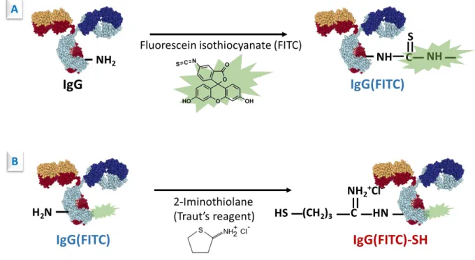
The sulfhydryl group is very attractive for conjugation of Ab to AuNPs since it forms a strong Au-S bond. However, there are no free sulfhydryl groups in antibodies. Thus, the sulfhydryl groups need to be created (by reaction of appropriate thiolation reagent systems) or generated (by reduction of endogenous disulfides). This section will discuss modification of the functional groups on antibodies used for covalent binding, including common groups such as amine, carboxylate, carbohydrate moieties, disulfides, and also special sites like the nucleotide binding site (NBS). (Figure 2)
Binding through amino groups
Amino groups are abundant in antibodies and are mostly located at the surface owing to their polarity and charge (positive at physiological pH). They are found on the side chain of lysines and at the N-terminal position of the light and heavy chains of IgG. What is more, amines are very reactive towards many reagents without any previous activation owing to their nucleophilicity.
For example, Traut’s reagent (2-iminothiolane, Figure 3) undergoes ring-opening reaction with a primary amine to generate a sulfhydryl group (Figure 4). This process is very efficient and proceeds rapidly at slightly basic pH.25-27 Another similar reagent, N-acetyl homocysteine thiolactone (also called citiolone or 2-acetamido-4-mercaptobutyric acid, Figure 3) can be used alternatively. The corresponding thiolation reaction is much like the reaction of Traut’s reagent. But Ab modification often results in much lower yields unless the reaction is done for extended period of time and at very basic pH (10-11).25
Figure 3 : Thiolation reagents: Traut’s reagent (2-iminothiolane) and N-acetyl homocysteine
thiolactone (citiolone or 2-acetamido-4-mercaptobutyric acid).
Figure 4 : Traut’s reagent reacts with amino group of antibody.
Since Bragg and Hou introduced N-hydroxysuccinimide (NHS) esters as activated carboxylic acids in 1980,28 they became the major amine-coupling reagents in bioconjugation chemistry
N-succinimidyl S-acetylthioacetate (SATA, Figure 5) and N-succinimidyl S-acetylthiopropionate (SATP, Figure 5) react with proteins amino groups forming stable amide linkages and protected sulfhydryl groups in the form of thioesters (Figure 6). This protecting group can be released with hydroxylamine to form free –SH just before conjugation to AuNPs.25
Figure 5 : Reagents: N-succinimidyl acetylthioacetate (SATA) and N-succinimidyl
S-acetylthiopropionate (SATP).
Figure 6 : SATA reacts with available amino groups of antibody.
In addition, disulfide bond-containing NHS esters are very attractive as the disulfide self-assembles on AuNPs by formation of Au-S bonds. For example, sulfoNHS-activated modifier 3,3'-dithiobis(sulfosuccinimidylpropionate) (DSPPT),29 N-succinimidyl 3-(2-pyridyldithio)propionate (SPDP)30 and N-succinimidyl 6-(3(2-pyridyldithio)propionamido)hexanoate) (LC-SPDP)31 (Figure 7) were used to modify amino groups of Ab prior to AuNPs chemisorption. Reagent including poly(ethylene glycol) (PEG) linker, like OPSS-PEG-NHS (Figure 7) was also applied.32 The length of the PEG linker could
be controlled, thus the Ab could be a bit far away from AuNP, having less risk of steric hindrance. At the same time, PEG also helps to prevent nonspecific protein binding on AuNPs.
Figure 7 : Reagents: disulfide bond containing NHS esters.
However, amine reactive coupling chemistry via NHS esters presents a major drawback owing to the rapid degradation of NHS esters by hydrolysis whose rate is comparable to the acylation rate.33 Thus, large excess of Ab is required to favor the acylation reaction. This strategy, binding via amino groups, results in randomly oriented Ab molecules on AuNPs. (Figure 8) This may cause partial loss of antigen binding capacity due to direct binding of the antigen binding site on AuNPs or steric hindrance with ‘head-on’ and ‘side-on’ spatial orientations.
Binding via carboxylate groups
Carboxylate groups (present on the side chain of glutamic acid and aspartic acid and at the C-terminal position of the light and heavy chains of IgG) are not as reactive as amino groups, so they need to be previously activated for example by NHS/EDC. However, after the carboxylate group is activated, it may react with the amino groups of another Ab molecule resulting in antibody crosslinking. Thus, it is not suitable for the following conjugation to AuNPs.
Binding via carbohydrate groups
The Fc region of polyclonal IgG produced by the major species (rabbit, goat and so on) contains carbohydrate chains. (Figure 2) Binding through these sugar moieties, the antibody will be immobilized on AuNPs in oriented fashion. This strategy requires a first step in which the carbohydrates are mildly oxidized by sodium periodate (NaIO4) to create reactive aldehydes. Then aldehyde-activated sugars can react with hydrazide, for example 2-acetamido-4-mercaptobutyric acid hydrazide (AMBH), forming hydrazone linkage (also called Schiff base) and leaving a free terminal sulfhydryl group. The Schiff base is further stabilized by reduction with sodium cyanoborohydride (Figure 9).25 Other heterobifunctional PEG linkers, like monothiol-PEG-hydrazide or dithiol-PEG- hydrazide (Figure 10) have been widely used by different research groups.34-38
A major limitation of this strategy is that Ab should be glycosylated. Polyclonal antibodies (from antisera) are usually glycosylated, but some other Ab preparations may not have carbohydrates such as recombinant antibodies fermented in bacteria, or some monoclonal antibodies that did not undergo post-translational glycosylation.
Figure 10 : Commercial thiol-PEG- hydrazide linkers.
Binding via sulfhydryl groups
UV-PIT. Ab can be directly immobilized on gold surfaces by using a UV light-induced
approach, known as photochemical immobilization technique (PIT).39 (Figure 11) Briefly, tryptophan residues absorb UV light and relax by transferring the absorbed energy to their neighbors. If a cystine (formed by 2 cysteines bound together by a disulfide bridge) is close enough to the tryptophan, the photon energy is virtually absorbed by cystine, leading to cleavage of disulfide bridge. The resulting free thiol groups can anchor efficiently to gold substrates. The native structural and functional properties of Ab are preserved. An increase of sensor sensitivity and of linear range has been shown using PIT method.40 This method was shown effective also for the conjugation of Ab to AuNPs.41
Figure 11 : Location of tryptophan residues on antibody and photochemical immobilization
technique (PIT). Picture is adapted from ref.39.
Fragmentation. In antibodies, the sulfhydryl groups borne by the cysteine residues are all
engaged in disulfide bridges, and they are important to the Ab function as they: i) contribute to the tertiary structure of each Ab subunit (intrachain S-S bridges), and covalently connect ii) the heavy to the light chains and iii) the two heavy chains at the hinge region (interchain S-S bridges). (Figure 12) As only free sulfhydryl groups of Ab can be conjugated directly to AuNPs, the native disulfide bonds must be selectively cleaved by reducing agents while preserving the antigen-binding capacity. The reduction of Ab is affected by several factors, for example, Ab host, pH, reductant and reductant concentration. By careful manipulation of these factors, high yield of half-Ab fragments can be obtained.42
Figure 12 : Reduction of antibody: (1) antibody fragments with functional paratope; (2)
For instance, at moderate concentration of dithiothreitol (DTT, Figure 13 and Figure 14-A) and absence of denaturant (such as urea, guanidine or SDS), limited cleavage of disulfide bridges of antibodies can result in reducing mainly the ones between the two heavy chains in the hinge region (rabbit and goat IgGs have two disulfide bridges in this region). This produces two half-antibodies, each containing one antigen binding site and free sulfhydryls at the hinge region (Figure 12-1).26, 43-44 Other reducing reagents, such as 2-mercaptoethanolamine (2-MEA, Figure 13 and Figure 14-B)45-47 and tris(2-carboxyethyl)phosphine (TCEP, Figure 13 and Figure 14-C)48-50, are widely used for Ab reduction. Both 2-MEA and DTT contain thiol groups that may compete with the reduced antibodies in the adsorption step to AuNPs. Therefore, the reducing agents 2-MEA and DTT need to be removed from the reduced Ab solution prior to conjugation to AuNPs. Such a separation adds one more step to the protocol that may lead to loss of the reduced Ab and trace of reducing agent may still remain. TCEP is a more stable and stronger reducing agent than 2-MEA and DTT. Since TCEP is sulphur-free reagent, it does not compete with the reduced Ab during adsorption to AuNPs, and therefore a separation step is not essential. This makes it a more convenient reducing reagent of Ab for chemisorption to AuNPs.
Figure 13 : Commonly used reductants for antibody fragmentation: dithiothreitol (DTT),
Figure 14 : Reduction of antibody to half-antibody fragments using dithiothreitol (DTT) (A),
2-mercaptoethanolamine (2-MEA) (B) and tris(2-carboxyethyl)phosphine (C).
Through this strategy, the antigen binding site is directed away from AuNPs, and because of the reduced size, a higher number of antigen binding sites per nm2 on the AuNP surface can be achieved, thus giving a better antigen binding capacity. Balevicius et al. demonstrated that Ab fragment modified surface interacted with antigens more efficiently in comparison with that modified by whole Ab.51 However recent studies showed that this strategy induced inactivation of the antigen binding capacity of the Ab fragments due to conformational and/or orientation change of fragments on the gold surface with time. The activity disappeared almost completely 60 min after immobilization. This inactivation can be slowed down if the Ab fragments are co-adsorbed with polyethylene glycol (PEG).52 (Figure 15)
Figure 15 : Illustration of time-dependent inactivation of antibody fragments. Picture is
adapted from ref.52.
Binding through nucleotide-binding site (NBS)
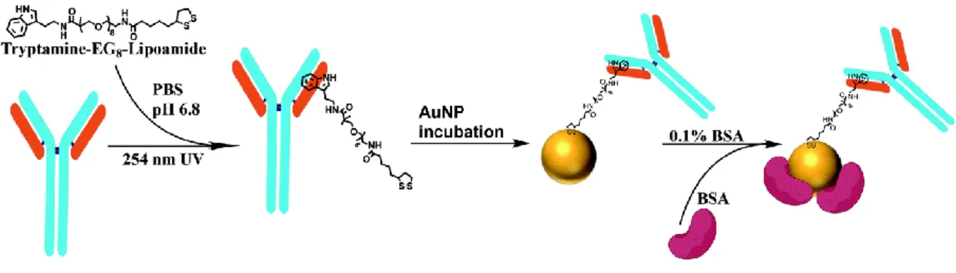
Even across different isotypes, antibodies contain a largely conserved sequence between the heavy and light chains of the Fab region known as nucleotide binding site (NBS, location shown in Figure 16). Targeting the NBS using a small indole molecule and UV irradiation, oriented immobilization of Ab on surface could be achieved. Alves et al. found that this method provided 8-fold better antigen sensitivity compared to physical adsorption.53 Mustafaoglu et al. applied this method to conjugation of Ab to AuNPs.49 (Figure 17) In practice, the indole-containing molecule, tryptamine-PEG8-lipoamide, was photo-cross-linked to the NBS of Ab upon exposure to UV light. Then functionalized antibodies were immobilized on AuNPs in an oriented manner via S–Au bonds. They also compared the antigen recognition sensitivity of this system to other conjugation methods, e.g. physisorption, EDC-NHS method, thiol reduction (Ab fragmentation), and concluded this UV-NBS method gave enhanced antigen detection capability and sensitivity with a high level of selectivity.
Figure 16 : Location of the nucleotide binding site (NBS) in antibody is shown in a cartoon
Figure 17 : Indole derivative used for UV-NBS method and conjugation scheme adapted from
ref.49.
Modification of AuNP
AuNPs are readily modified by low molecular weight thiols or disulfides. Various heterobifunctional ligands containing thiols or disulfides were used for stable anchoring onto AuNPs with another functional terminal group, e.g. carboxylate group, for binding antibodies. Carboxyl or NHS ester groups
AuNPs have been decorated with carboxyl functions by binding a COOH-terminated crosslinker though Au-S bond. In some crosslinkers, carboxyl groups are already activated in the form of NHS/sulfo-NHS esters, whereby direct chemisorption of Ab could be realized through formation of amide bonds; while some are native carboxylates, in this case, NHS (or sulfo-NHS)/EDC chemistry should be applied to activate the carboxylate group prior to Ab adsorption.
NHS (or sulfo-NHS) heterobifunctional crosslinker, such as 3,3’-dithiobis(sulfosuccinimidyl propionate) (DTSSP) (Figure 7) was chemisorbed on AuNPs followed by direct immobilization of Ab.29, 54 Special chemical groups could be implanted in the crosslinker for appropriate application. For example, Grubisha et al. synthesized 5,5’-dithiobis(succinimidyl-2-nitrobenzoate) (DSNB, Figure 18) containing NO2 group which gives a strong Raman signal.55 Meanwhile, heterobifunctional PEG linkers, HS-PEG-COOH, were also used, for instance, monothiol-PEG-COOH30, 37, 56-59 and dithiol-PEG-COOH linker57, 60. After self-assembling on AuNPs though Au-S bond, NHS (or sulfoNHS)/EDC chemistry was used to activate the carboxyl group to N-succinimidyl ester, which then reacted with amine group on the Ab forming stable amide bond between PEG and Ab.
Figure 18 : Reagent : 5,5’-dithiobis(succinimidyl-2-nitrobenzoate) (DSNB).
Coupling of Ab to the NHS-activated AuNPs proceeds in a random orientation via any free amino groups present on Ab. In this way an Ab can react with more than one AuNP and thus induce AuNPs cross-linking.
AuNP-Fc binding protein
Ab-binding proteins such as protein G/A specifically bind to the Fc region of Ab. Ab immobilized on Fc-binding protein-coated surfaces is therefore properly oriented for optimal antigen binding. What is more, protein G/A could bind various IgG subclasses like from rabbit, mouse, human and other mammalian species. As Ab modification is not required for immobilization, bound Ab fully retains its binding ability. In practice, protein A was first reacted with N-succinimidyl 3-(2-pyridyldithio)-propionate (SPDP).30, 61 Upon addition of the SPDP‐modified protein A to AuNPs, the thiolated ligand displaced the citrate layer on AuNPs. Ab was subsequently immobilized on protein A via Fc region in an oriented configuration on AuNPs. Alternatively, protein G was physically adsorbed on AuNPs prior to Ab immobilization.58 Another Fc specific polyclonal Ab, for instance, rabbit anti-mouse IgG (RAM) was widely used in SPR sensor for oriented immobilization of Ab.62
One of the major concerns associated with this method is the additional immobilization process of Fc-binding protein coating on AuNP surface prior to Ab immobilization. Protein G/A-Ab binding is also less stable than the other covalent immobilization methods.
Modification of AuNP and antibody
Reductive amination
The sugar moieties of Ab are oxidized with NaIO4 creating aldehyde groups. On the other hand, AuNPs were decorated with primary amino groups using for instance NH2-PEG-SH.The stable attachment of Ab on AuNPs was realized in presence of NaCNBH3 (reaction mechanism shown in Figure 9).30
Biotin-Streptavidin (STV) association
Biotinylated Ab is commercially available. It can interact with streptavidin that has been attached to AuNPs.63-64
Hybridization
Ab conjugation to AuNPs can be also achieved via DNA hybridization using specific Watson-Crick base pairing of two complementary nucleic acids. This strategy basically requires a two-step process: conjugation between Ab and DNA single strand sequence, and conjugation between AuNPs and complementary DNA sequence. Combined with biotin-streptavidin association (STV),65-66 directional attachment of Ab on AuNPs was achieved. (Figure 19-A) Experimentally, DNA-STV was prepared from thiolated oligonucleotides and recombinant streptavidin using heterobifunctional cross-linker sulfosuccinimidyl 4-(N-maleimidomethyl) cyclohexane-1-carboxylate (sulfoSMCC). Then DNA-STV was conjugated to biotinylated Ab through high affinity between biotin and STV. On the other hand, complementary capture oligonucleotides were chemisorbed to AuNPs through Au-S bond. In the end, the DNA-STV Ab bound to DNA-AuNPs via Watson-Crick base pairing. Similarly, instead of biotin-STV, protein G was also combined to DNA base pairing for oriented attachment of Ab.67 (Figure 19-B)
Figure 19 : DNA sequence combined with biotin-streptavidin (A)65 or protein G (B) 67. Click chemistry
Ab was reacted with azido-PEG8-NHS ester to introduce azido groups on the Ab in a random fashion. On the other hand, a synthetic copolymer coating was grown on AuNP surface, one of the monomers bearing an alkyne functionality. This thin polymer layer on the surface stabilized the colloidal suspension.68 Azido-containing Ab was then conjugated to alkyne-carrying AuNPs by copper-catalyzed azide-alkyne cycloaddition. (Figure 20)
Figure 20 : Schematic representation of surface modification of AuNPs with a synthetic
copolymer and subsequent conjugation with azido-modified antibody. Picture is adapted from ref.68.
Current methods concerning the conjugation of Ab to AuNPs, via strategies of physisorption and chemisorption, have been presented. The particular characteristics of antibodies, including their shape, function and chemical stability, make their attachment to AuNPs a challenging task.
The orientation of Ab molecules and how they are linked are important to keep the Ab activity intact for the desired application of the conjugates.
QUANTIFICATION OF ANTIBODY IN AUNP-AB CONJUGATES
The performance of AuNP-based biosensors is markedly affected by the applied conjugation strategy between Ab and AuNPs that in turn controls surface coverage and Ab orientation.38, 69-71 In the previous section, we have presented various conjugation strategies. To assess these strategies, information about orientation and surface coverage of Ab on AuNPs is highly desirable. Characterization techniques of Ab orientation on planar sensor surface have been reported in refs.71-72. In addition, several quantitative methods to measure surface coverage of Ab on AuNPs have been reported to date. We will present the different methods in this section.
Indirect quantification
Most quantification methods rely on the indirect analysis of Ab surface coverage via the quantitation of unbound Ab remaining in the supernatant after adsorption to AuNPs. In practice, excess unbound Ab in supernatant can be quantified with bicinchoninic acid (BCA)24 or Bradford total protein assays.34, 54, 70 Then the total number of Ab adsorbed on AuNPs is calculated as the difference between the initial amount of Ab added to the AuNP suspension and the amount of unbound Ab that remained in the supernatant. However, Ab conjugation to AuNPs is generally followed by a blocking step with another protein, typically BSA14, 73, to prevent aggregation of the nanoparticles in saline environment and increase the long-term stability of the colloidal solution. This additional step, leading to the presence of another protein, makes the previous classical protein assays inapplicable. In this case, Ab assay in the supernatant can be achieved by enzyme-linked immunosorbent assay (ELISA) using a microtiter plate coated with the corresponding antigen.27 Alternatively, prior to conjugation, Ab can be labeled with a fluorescent dye to achieve sufficient sensitivity of detection and non-bound Ab in supernatant is measured by spectrofluorimetry at the excitation/emission wavelengths of the fluorescent dye.35, 38 It should be noted that Ab quantification by these indirect methods implies no loss of Ab during the purification process, e.g. during transfer of the solution from one container to another, or Ab adsorption to the container walls. This is why indirect methods often lead to an overestimation of Ab surface coverage on AuNP.54
Direct quantification
Ab adsorption on AuNPs has been quantified from the LSPR peak shift due to the local refractive index (RI) change caused by Ab adsorption.41, 74 Although this approach provides a direct quantification of the adsorbed Ab, it requires accurate knowledge of RI of proteins at the nanoparticles surface. This is quite challenging because the RI depends on the surface coverage, protein orientation, and water content, none of which are constant.75-77 Other methods, like dynamic light scattering (DLS) and nanoparticle tracking analysis (NTA) measure the increase in hydrodynamic diameter after adsorption of protein on AuNPs to derive information on surface coverages.32, 77-78 The thickness of the bound protein layer does not directly provide accurate information on bound proteins per nanoparticles. Some models were derived allowing to correlate the average number of bound protein to the hydrodynamic diameter increment, for instance, Taylor dispersion analysis (TDA).79
BCA protein assay31 and ELISA26, 30, 32, 58, 70 were also used to directly quantify the adsorbed Ab. Even with some careful calibration treatment, i.e. removal of the nanoparticle contribution by subtracting a spectrum of pure nanoparticle solution, overestimation of the Ab on nanoparticle was obtained, demonstrating that nanoparticles interfere chemically and/or electromagnetically with the BCA assay.31 Similar problems may happen for fluorescence36 -based direct quantification Ab in conjugates.
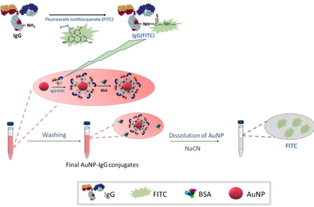
To avoid interference from the matrix, dissolution of nanoparticles or digestion of Ab strategy has been reported. The former method relies on the dissolution of the AuNPs in AuNP-Ab conjugates by KI/I2 mixture, followed by labeling of recovered Ab by the fluorescent dye NanoOrange and spectrofluorimetric measurement.54 Though this method gives more reliable results than the indirect protein assay methods, it is not applicable to BSA-blocked AuNP-Ab conjugates. The latter method is based on the complete digestion of the protein in AuNP-protein conjugates in 6 N HCl followed by fluorescent labeling of the generated aminoacids and assay of glycine derivative by HPLC-fluorescence detection.80 To note, this method is not applicable to BSA-blocked AuNP-Ab conjugates either.
On the other hand, radioactive labeling of Ab in quantification is a robust method52, however, the handling of the radioactive waste remains challenging.
By comparing different quantification methods, we see well that accurate quantification of surface coverage of Ab still remains challenging.
APPLICATIONS OF AUNP-ANTIBODY CONJUGATES IN OPTICAL
BIOSENSING
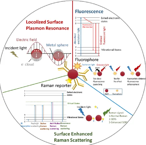
Figure 21 : Optical properties of AuNPs: Localized Surface Plasmon Resonance (LSPR),
Surface Enhanced Raman Scattering (SERS) and fluorescence enhancement and quenching. Historically, highly light absorbing and color dense Ab-conjugated AuNPs were shown to provide high capability for sensitive staining techniques. For instance, AuNP-Ab conjugates have long been used as indicators in lateral flow assays (LFA) using immunochromatographic test strips,81-82 that provide simple, rapid, low cost, and user-friendly detection of various analytes. In addition, these conjugates are widely used in immunohistochemical (IHC) staining combined with electron microscopy.12-14, 21, 83 Here we will present recent applications of AuNP-Ab conjugates based on the optical properties of AuNPs, i.e. Localized Surface Plasmon Resonance (LSPR), surface enhanced Raman scattering (SERS) and fluorescence enhancement
or quenching/dequenching. (Figure 21) According to the signal transduction type, the current well-developed immunoassays based on AuNP-Ab conjugates can be divided into four broad categories: LSPR, SPR, fluorescence, and SERS immunoassay. (Figure 22)
Figure 22 : AuNP-antibody bioconjugates in optical biosensing.
AuNP-antibody bioconjugates in LSPR sensors
The general principle behind LSPR-based sensors is the LSPR peak shift arising from local refractive index (RI) change caused by binding event between AuNP-Ab and antigen or from antigen-induced crosslinking aggregation. (Figure 23) LSPR-based assays have been conducted both in homogeneous solution and on planar surfaces coated with AuNP monolayer.
For example, when AuNP-Ab binds to the target with only one epitope (Figure 23A), the LSPR peak of AuNP red-shifts because of an increase in the RI close to AuNPs. What is more, the peak shift is proportional to the target concentration. Thus, this assay is useful for quantitative detection.84-85 In case there are multiple binding sites on the target (epitopes), the target acts as a linker and interparticle crosslinking-induced aggregation of AuNPs occurs (Figure 23B).27, 41, 86-87 Moreover, the aggregation induces interparticle surface plasmon coupling of AuNPs, leading to the change of the absorbance spectrum and even to a color change of the colloidal solution. The degree of AuNPs aggregation (or redispersion of aggregates) is proportional to the target concentration, thus allowing this assay to be useful in quantitative detection. Alternatively, a non-crosslinking aggregation of AuNP-Ab induced by an increasing salt concentration can be used as in ref.88.
Other sensors based on LSPR band shift have been fabricated by coating AuNPs onto substrate surface. (Figure 24) For example, AuNPs were attached to quartz,89 ITO-glass,90 polystyrene64,
etc. Ab was then immobilized on AuNPs, consequently, the sensing platform was employed to
quantitatively detect antigens based on the LSPR peak shift which resulted from local RI change of binding target. These fabricated LSPR immunosensors showed high sensitivity and selectivity. For instance, the limit of detection (LOD) of HIV-1 was estimated at 200 fg/mL,90 which is 70 fold better than that of previously reported detection method91. Inci et al. applied this technology for quantitative detection of intact virus in whole blood sample at clinically relevant concentrations.64
Figure 24 : LSPR sensor on planar surface: AuNPs are coated on substrates, followed by
antibody immobilization.
AuNP-antibody conjugates for enhanced SPR biosensors
AuNPs have been incorporated in SPR biosensors, providing an effective way to increase sensor response, which results from the electronic coupling interaction of the propagating surface plasmons with localized surface plasmons of AuNPs. There are two different types, (i) AuNPs
are immobilized on SPR sensor surface directly, and (ii) AuNPs are immobilized on top of the sensing layer. (Figure 25)
For example, a layer of AuNPs was first coated on SPR sensor surface, followed by coating another layer of AuNP-Ab. The two layers of AuNPs enhanced the sensitivity of the following detection of target.92 Alternatively, AuNP-Ab conjugates were bound at the last step forming a sandwich format. (Figure 25-B) Briefly, a layer of capture Ab was immobilized on SPR sensor surface. After sample injection, AuNP-Ab finally bound to sensor surface. Signal amplification and lower limit of detection were evidenced. This strategy was applied for detection of human IgG93, prostate-specific antigen94-96 and carcinoembryonic antigen (CEA).57
Figure 25 : AuNP-based enhanced SPR sensors: (A) AuNPs are immobilized on SPR sensor
surface directly; (B) AuNPs are immobilized on top of sensing layer.
AuNP-based plasmon resonance scattering sensing
The light-scattering cross-section of a AuNP with a diameter of 60 nm is 200-300 times stronger than that of a polystyrene bead of same size, and 4-5 orders of magnitude stronger than that of a fluorescent dye, e.g., fluorescein.97-98 Dynamic light scattering (DLS) is a technique routinely used to analyze the size and size distribution of nanoparticles. Combining the use of AuNPs as light scattering enhancer and DLS as readout provides a useful sensor design.
In practice, when AuNP-Ab is mixed with a sample containing the target, depending on its concentration, the target binding will cause AuNPs to form dimers, oligomers, or aggregates. The relative ratio of different forms can be measured quantitatively through DLS analysis. This ratio increases accordingly with increased amount of antigen in sample solution, and such a correlation forms the analytical basis of a homogeneous immunoassay. By exploiting this sensing platform, human IgG99, prostate-specific antigen49, 100 and virus17 have been detected.
The one-step assay does not involve any washing step and yields highly sensitive, specific and rapid results. Moreover, extremely small amounts of samples are needed for the assay.
AuNP-based fluorescence sensing
Figure 26 : Schematic illustration of the effect of distance between AuNP and fluorophore on
fluorescence emission.
AuNP can lead to both fluorophore quenching and enhancement which can be explained by a well-established Förster resonance energy transfer (FRET) mechanism.101 (Figure 26) When AuNP and fluorophore are close to each other, FRET occurs from the fluorophore to AuNP and thus quenching happens.102 In contrast, at an appropriate distance to AuNP, fluorescence enhancement can happen, which is typically referred to as plasmon-enhanced fluorescence (PEF).103 Especially, Anger et al.104 experimentally and theoretically investigated the relationship between the fluorescence rate and the distance between AuNP and fluorophore and demonstrated a continuous transition from fluorescence enhancement to fluorescence quenching of a single molecule on AuNP.
Generally, an AuNP-based fluorescence probe system consists of two functional components: the recognition element, here Ab, which provides specific binding of target, and the fluorescent dye, which is used for analyzing the binding event. The high loading efficiency of the fluorescent dye on AuNP surface with high area-to-volume ratio and high fluorescence enhancement or quenching/unquenching abilities of the fluorescent dye-AuNP pairs make this probe ultrasensitive. Assays are typically designed via fluorescence-nanoprobe labeling and analyte-induced fluorescence quenching/dequenching (recovery).
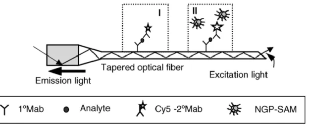
Alternative assay platforms, for instance, fiber-optic biosensors based on AuNP-fluorescent dye enhancement, have been applied by different research groups.91, 105-106 Briefly, a capture Ab was immobilized on the surface of an optical fiber. After sample injection, fluorescence probe consisting of fluorophore-labeled Ab-conjugated AuNPs was added. Subsequently, the fluorescence signal was then excited and measured by the photomultiplier tube. (Figure 27) In this assay, two factors were considered to enhance the assay sensitivity. First, electromagnetic field around the AuNP surface efficiently enhanced the fluorescence signal. Second, there were multiple fluorophores labeled on each AuNP allowing several fluorophores to be simultaneously excited during measurement. These two effects combine, producing a significant improvement in the sensitivity of the system compared to a conventional fluorescence probe or ELISA.
Figure 27 : Schematic diagram of fluorescence measurement in biosensor: (I) regular
immunosensing with fluorophore-labeled antibody; (II) immunosensing using AuNPs as fluorescence enhancer. Picture is adapted from ref.105.
Based on the latter format, the dequenching format, porcine reproductive and respiratory syndrome virus was detected.107 A fluorophore-labeled Ab was immobilized on protein A-AuNPs. In such case was the fluorescence quenched by A-AuNPs. Upon binding of antigen, conformational change within the biosensor complex increased the distance between the fluorescent dye and AuNP, leading to a dequenching (recovery) effect by the AuNP.
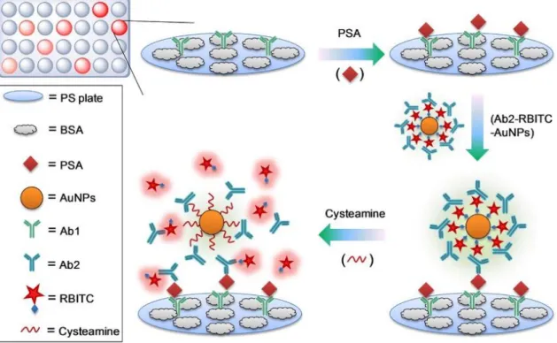
Liu et al. also explored an AuNP-based fluorescence-activatable immunoassay for detecting ultralow levels of cancer biomarker (PSA) in patient serum samples (Figure 28).108 Each AuNP was labeled with thousands of fluorescent dyes (rhodamine B isothiocyanate) and the dye layer on AuNPs was covered with Ab that was bound through electrostatic interactions.
the substrate and the fluorescence of dye on AuNP surface was highly quenched via the nanoparticle surface energy transfer effect. After addition of cysteamine into this detection system, the fluorescent dyes were competitively displaced from the AuNP surface, leading to significant restoration of dye fluorescence. Thus, the concentration of PSA in serum samples was quantified according to the intensity of the recovered fluorescence. The LOD of the developed probe for PSA was more than 2 orders of magnitude lower than that of the conventional fluorescence probe.
Figure 28 : Schematic illustration of dequenching (recovery) of fluorescent dyes. Picture is
adapted from ref.108.
AuNP-based SERS sensing
SERS is a powerful spectroscopic technique that provides in situ nondestructive fingerprint information. More importantly, its ultrahigh signal enhancement is up to 1014 that is capable for single-molecule detection. In the AuNP-based SERS sensing, upon exciting the LSPR of AuNPs, the large electromagnetic field that localized at AuNP surface is exploited to measure enhanced vibrational molecular signatures. SERS nanoprobes have been widely applied in immunoassays. As the probe, the essential design is the use of AuNPs with Ab and Raman reporter molecules. The total enhancement factor results from (i) LSPR-enhanced Raman scattering and (ii) chemical enhancement. The assays are typically designed as
SERS-nanoprobe based optical labeling and analyte-induced SERS nanoprobe aggregation/antiaggregation assays.
SERS nanoprobe-based optical labeling
The SERS substrate, here AuNPs offer a stronger Raman enhancement than individual nanostructures due to the formation of hotspots at AuNPs junctions. The SERS nanoprobe could be used in direct optical labeling.
For example, Grubisha et al. reported a sandwich format immunoassay using SERS nanoprobes as optical labels. As the probe, AuNPs were functionalized with a monolayer of intrinsically strong Raman reporter (DSNB, Figure 18) followed by coating Ab layer.55 In this case, the distance between the Raman reporter and the AuNP surface was minimal and the number of the Raman-reporters on each AuNP was maximized. These two factors led to an enhanced sensitivity of this assay.
Li et al. presented a SERS-based immunochromatographic assay (ICA) for ultrasensitive and quantitative detection of phenylethanolamine A (PA) (Figure 29).109 The principle of this new sandwich assay is similar to conventional ICA based on AuNPs but using a SERS probe which consisted of Au-Ag core-shell nanoparticles labeled with Ab and 4-mercaptobenzoic acid (MBA, Raman reporter) (AuMBA@Ag-Ab). After the ICA procedure, the specific Raman scattering intensity of MBA on the test line was measured for quantitative detection of PA. The LOD of this assay was improved by 1−3 orders of magnitude compared with other immunoassays.
Qian et al. successfully applied pegylated SERS nanoprobe for tumor targeting and detection.110 Large optical enhancement was achieved under in vivo conditions for tumor detection in live animal. Similarly, Cho et al. applied it in isolation and analysis of circulating cancer stem cells through Raman imaging based on SERS nanoprobe.111
Analyte-induced SERS nanoprobe aggregation/anti-aggregation
Another assay format is based on antigen-induced aggregation/anti-aggregation of SERS nanoprobes.
Wang et al. reported the application of SERS nanoprobes in target-induced aggregation assay.50 In the system, there were two SERS nanoprobes, spherical AuNPs and gold nanorod (AuNR). Both were decorated with Raman reporter and half-fragment Ab (as shown in Figure 30). In the presence of target, the SERS nanoprobes aggregated leading to strong SERS enhancement arising from plasmonic coupling, and SERS spectra were acquired directly on the sample solution. Instead, after the SERS nanoprobes reacted with target, the aggregated nanoparticles were captured and concentrated through a membrane filter. SERS analysis was acquired from membrane surface.29, 112
Figure 30 : Schematic illustration of single-step SERS immunoassay via target-controlled
assembly of SERS nanoprobes. Picture is adapted from ref.50.
Wu et al. presented a SERS immunoassay in anti-aggregation format for ultrasensitive detection of folic acid (FA) by controlling the assembly of AuNPs and silver nanoparticles (AgNPs) heterodimers through antigen-Ab binding (Figure 31).113 In practice, the AuNPs were coated with Ab (against FA) and Raman reporter (4-aminothiophenol (4-ATP)); the AgNPs were coated with antigen (FA-BSA conjugates) and Raman reporter (4-ATP). Without target in sample, the Ab/Raman reporter-labeled AuNPs and the antigen/Raman reporter-labeled AgNPs assembled completely, forming heterodimers and showing strong SERS intensity. However, in the presence of FA, free FA competed with the AgNP-labeled antigen for binding to
AuNP-labeled Ab, thus reducing the assembly of heterodimers. Consequently, the concentration of target FA was revealed by the significantly reduced Raman intensity of the Raman reporter.
Figure 31 : Schematic illustration of the SERS immunoassay for detection of folic acid (FA)
based on AuNP−AgNP heterodimers formation. Picture is adapted from ref.113.
Besides the applications of AuNP-Ab in optical biosensing mentioned above, AuNP-Ab conjugates have also been used in other transduction techniques, for example, piezoelectric transduction using quartz crystal microbalance (QCM). The sensing design is the same as for AuNP-enhanced SPR sensor and the incorporation of AuNPs into QCM-based sensing systems enhanced detection sensitivity by their high surface area47, 114 and by serving as a “mass enhancer.115-116
CONCLUSIONS AND PERSPECTIVES
Here, we have summarized and discussed various conjugation strategies, quantification methods of surface coverage and applications of AuNP-Ab conjugates that have been designed and successfully applied in optical biosensing. With the excellent properties of AuNPs, as well as various signal transduction approaches, nanomaterials have paved the way for the development of easy, rapid, low-cost immunoassays. The optimization of conjugation for the sensing systems is still required to meet the demands of clinical diagnostics in complex biological fluids such as urine, serum, and blood. For example, further miniaturization of Ab allows increment of binding sites on AuNP surface. Assays can be further integrated into mobile phones or computers for real-time monitoring. The fundamental advantages of AuNPs will continue to contribute to the development of biosensors in the following years.
REFERENCES
1. Saha, K.; Agasti, S. S.; Kim, C.; Li, X.; Rotello, V. M., Gold Nanoparticles in Chemical and Biological Sensing. Chem. Rev. 2012, 112 (5), 2739-2779.
2. Wilson, R., The use of gold nanoparticles in diagnostics and detection. Chem. Soc. Rev.
2008, 37 (9), 2028-2028.
3. Chen, P. C.; Mwakwari, S. C.; Oyelere, A., Gold nanoparticles: From nanomedicine to nanosensing. Nanotechnol. Sci. Appl. 2008, 1, 45-66.
4. Tang, L.; Li, J., Plasmon-Based Colorimetric Nanosensors for Ultrasensitive Molecular Diagnostics. ACS Sensors 2017, 2 (7), 857-875.
5. Sepulveda, B.; Angelome, P. C.; Lechuga, L. M.; Liz-Marzan, L. M., LSPR-based nanobiosensors. Nano Today 2009, 4 (3), 244-251.
6. Geoghegan, W. D., The effect of three variables on adsorption of rabbit IgG to colloidal gold. J. Histochem. Cytochem. 1988, 36 (4), 401-407.
7. Bagchi, P.; Birnbaum, S. M., Effect of pH on the adsorption of immunoglobulin G on anionic poly(vinyltoluene) model latex particles. J. Colloid Interface Sci. 1981, 83 (2), 460-478.
8. Demanèche, S.; Chapel, J.-P.; Monrozier, L. J.; Quiquampoix, H., Dissimilar pH-dependent adsorption features of bovine serum albumin and α-chymotrypsin on mica probed by AFM. Colloids Surf. B 2009, 70 (2), 226-231.
9. Geoghegan, W. D.; Ackerman, G. A., Adsorption of horseradish peroxidase, ovomucoid and anti-immunoglobulin to colloidal gold for the indirect detection of concanavalin A, wheat germ agglutinin and goat anti-human immunoglobulin G on cell surfaces at the electron microscopic level: a new me. J. Histochem. Cytochem. 1977, 25 (11), 1187-1200.
10. Hook, F.; Rodahl, M.; Kasemo, B.; Brzezinski, P., Structural changes in hemoglobin during adsorption to solid surfaces: Effects of pH, ionic strength, and ligand binding. Proc. Natl
Acad. Sci. 1998, 95 (21), 12271-12276.
11. Peng, Z. G.; Hidajat, K.; Uddin, M. S., Adsorption of bovine serum albumin on nanosized magnetic particles. J. Colloid Interface Sci. 2004, 271 (2), 277-283.
12. Horisberger, M.; Rosset, J.; Bauer, H., Colloidal gold granules as markers for cell surface receptors in the scanning electron microscope. Experientia 1975, 31 (10), 1147-1149. 13. Horisberger, M.; Rosset, J., Colloidal gold, a useful marker for transmission and scanning electron microscopy. J. Histochem. Cytochem. 1977, 25 (4), 295-305.
14. De Mey, J.; Moeremans, M.; Geuens, G.; Nuydens, R.; De Brabander, M., High resolution light and electron microscopic localization of tubulin with the IGS (immuno gold staining) method. Cell Biol. Int. Rep. 1981, 5 (9), 889-899.
15. El-Sayed, I. H.; Huang, X.; El-Sayed, M. A., Surface Plasmon Resonance Scattering and Absorption of anti-EGFR Antibody Conjugated Gold Nanoparticles in Cancer Diagnostics: Applications in Oral Cancer. Nano Lett. 2005, 5 (5), 829-834.
16. Hermanson, G. T., Preparation of Liposome Conjugates and Derivatives. In
Bioconjugate techniques, Elsevier: 2008; pp 858-899.
17. Lai, Y. H.; Koo, S.; Oh, S. H.; Driskell, E. A.; Driskell, J. D., Rapid screening of antibody–antigen binding using dynamic light scattering (DLS) and gold nanoparticles. Anal.
Methods. 2015, 7 (17), 7249-7255.
18. Rayavarapu, R. G.; Petersen, W.; Ungureanu, C.; Post, J. N.; van Leeuwen, T. G.; Manohar, S., Synthesis and Bioconjugation of Gold Nanoparticles as Potential Molecular Probes for Light-Based Imaging Techniques. Int. J. Biomed. Imaging 2007, 2007, 1-10. 19. Safenkova, I. V.; Zherdev, A. V.; Dzantiev, B. B., Correlation between the composition of multivalent antibody conjugates with colloidal gold nanoparticles and their affinity. J.
Immunol. Methods 2010, 357 (1-2), 17-25.
20. Lou, S.; Ye, J.-y.; Li, K.-q.; Wu, A., A gold nanoparticle-based immunochromatographic assay: The influence of nanoparticulate size. Analyst 2012, 137 (5), 1174-1181.
21. Tokuyasu, K. T., Present state of immunocryoultramicrotomy. J. Histochem. Cytochem.
1983, 31 (1A), 164-167.
22. Horisberger, M.; Clerc, M. F., Labelling of colloidal gold with protein A. Histochem.
1985, 82 (3), 219-223.
23. Byzova, N. A.; Safenkova, I. V.; Slutskaya, E. S.; Zherdev, A. V.; Dzantiev, B. B., Less is More: A Comparison of Antibody–Gold Nanoparticle Conjugates of Different Ratios.
Bioconjugate Chem. 2017, 28 (11), 2737-2746.
24. Geng, S. B.; Wu, J.; Alam, M. E.; Schultz, J. S.; Dickinson, C. D.; Seminer, C. R.; Tessier, P. M., Facile Preparation of Stable Antibody–Gold Conjugates and Application to Affinity-Capture Self-Interaction Nanoparticle Spectroscopy. Bioconjugate Chem. 2016, 27 (10), 2287-2300.
25. Hermanson, G. T., Fluorescent Probes. In Bioconjugate techniques, Elsevier: 2008; pp 396-497.