HAL Id: tel-01532886
https://tel.archives-ouvertes.fr/tel-01532886
Submitted on 4 Jun 2017HAL is a multi-disciplinary open access archive for the deposit and dissemination of sci-entific research documents, whether they are pub-lished or not. The documents may come from teaching and research institutions in France or abroad, or from public or private research centers.
L’archive ouverte pluridisciplinaire HAL, est destinée au dépôt et à la diffusion de documents scientifiques de niveau recherche, publiés ou non, émanant des établissements d’enseignement et de recherche français ou étrangers, des laboratoires publics ou privés.
Transcriptomic approach of the response to metals in
the hydrothermal mussel Bathymodiolus azoricus
Gonzalo Fuenzalida del Rio
To cite this version:
Gonzalo Fuenzalida del Rio. Transcriptomic approach of the response to metals in the hydrothermal mussel Bathymodiolus azoricus. Molecular Networks [qbio.MN]. Université Pierre et Marie Curie -Paris VI, 2016. English. �NNT : 2016PA066487�. �tel-01532886�
! !
Université Pierre et Marie Curie
Ecole doctorale Sciences de la Nature et de l’Homme, Ecologie et Evolution
Equipe Adaptation et Biologie des Invertébrés en Conditions Extrêmes (ABICE)Laboratoire Adaptation et Diversité en Milieu Marin UMR7144
Transcriptomic approach of the response to metals
in the hydrothermal mussel Bathymodiolus azoricus
Par FUENZALIDA Gonzalo
Thèse de doctorat de Biologie
Dirigée par Arnaud Tanguy et François Lallier
Présentée et soutenue publiquement le 2 décembre 2016
Devant un jury composé de :
Pr. DANGER Jean-Michel, Université du Havre, Rapporteur. Pr. SERGENT Odile, Université Rennes I, Rapporteur.
Dr. PAIN DEVIN Sandrine, Université de Lorraine, Examinateur. Dr. GAGNAIRE Beatrice, Centre de Cadarache, Examinateur. Pr. CORMIER Patrick, UPMC, Examinateur.
Para mi hija y esposa, por todo el tiempo invertido en esto y no al estar con ustedes.
Acknowledgement
First I want to thank Arnaud for the confidence that he put on me for the development of this work and for all the tips that he gave me in the laboratory which allowed for a better and fast laboratory experience which I appreciate, and also his help to solve my car problems… a very complete PhD supervisor. Also thanks to François for all the corrections made in posters, papers, presentations and of course in this manuscript and specially thanks for helping me in the last months with the administrative things with CNRS.
Second thanks to all the members of the ABICE team: Ann, Dominique, Anne Sophie, Camille, Kevin, Perrine, Bérénice, Stéphane, Didier, Jean, Jean-Yves and Alexis for the good reception and disposal to help me, making the lab a pleasant place to work.
Also I say thank you to Leyla Cardenas, who trained me as a marine biologist teaching me how to work as a scientist and also helped me to come to Roscoff to begin my PhD work.
Thanks also to all the french and not French people that I met in party’s or playing football Remy, Kevin, Marie, Pierre, Raphael and Vanessa, Angelique, Hugo, Jaromir, Zujaila, Adriana, Cristina, Andres, Ainoa, Pinocho, Javier, Fuli, Yuna, Wanek, Misharl and all the people that I met at the Station Biologique (technician and students) in different courses and formations with which I share more than one beer. Also thanks to the members of the AJC for the organization of formation in Roscoff required by the doctoral school.
To my parents, brother, cousins, nephews, uncles, aunts and Chilean friends who have always been supporting me in all my decisions (even when they have not been very good), I hope I can give back all that have helped me.
Thanks also to my Chilean friend Jano and Katy for all the good moments that we share and for joining us before and after the firsts moths of my daughter birth, I hope this friendship will continue over time.
Finally, thanks to my wife, which has been an important pillar in the last year and in my training as a father, a function which I will be able to devote more time.
1
Table of contents
CHAPTER I 3 INTRODUCTION 1. BIOMARKERS 5 2. ECOTOXICOLOGY IN AQUATIC ECOSYSTEMS 7 3. ECOTOXICOLOGY AND METALS IN AQUATIC SYSTEMS 10 4. RESPONSE TO HEAVY METALS IN MARINE INVERTEBRATES. 19 5. OMICS ERA IN POLLUTION RESPONSE 25 6. HYDROTHERMAL ECOSYSTEMS AS NATURAL LABORATORY FOR HEAVY METAL ADAPTATION 28 7. OBJECTIVES 37 CHAPTER II 39 METAL ACCUMULATION AND REGULATION OF METAL RELATED GENE EXPRESSION IN THE HYDROTHERMAL VENT MUSSEL BATHYMODIOLUS AZORICUS AS A SIGNATURE OF ENVIRONMENTAL CONTAMINATION CHAPTER III 93 DIFFERENTIAL EXPRESSION OF SUPEROXIDE DISMUTASE ISOFORMS AS INDICATOR OF OXIDATIVE STRESS IN RESPONSE TO METALS IN THE HYDROTHERMAL MUSSEL BATHYMODIOLUS AZORICUS: APPLICATION TO FIELD AND EXPERIMENTAL POPULATIONS. CHAPTER IV 141 IDENTIFICATION AND REGULATION OF FERRITINS IN THE HYDROTHERMAL VENT MUSSEL BATHYMODIOLUS AZORICUS IN NATURAL AND EXPERIMENTAL POPULATIONS CHAPTER V 171 LARGE SCALE TRANSCRIPTOMIC ANALYSIS OF BATHYMODIOLUS AZORICUS DISCUSSION, CONCLUSION, PERSPECTIVES 245 BIBLIOGRAPHY 251 ABSTRACT BACK COVER3
Chapter I
Introduction
In 1962, the marine biologist and conservationist Rachel Carson published her book "A Silent Spring" in which she warned harmful effects of pesticides (DDT) in the environment mainly in birds and humans, establishing a relationship between toxicology and environment. In 1969, René Truhaut introduced the term "ecotoxicology" as the combination of two very different subjects: ecology (defined as the scientific study of interactions that determine the distribution and abundance of organisms" ‘Krebs 1985) and toxicology (defined as the study of injurious effects of substances on living organisms). If the limit of the toxicological studies is the organisms, ecotoxicology aims to assess the impact of chemicals on a larger scale considering different levels of organization from cell to ecosystems (Truhaut, 1977). At the beginning, the study of environmental toxicology was limited to the detection of toxic residues in the environment or in individual organisms and the use of tests to estimate the toxic effects of chemicals on animals other than human. One main limitation of these approaches was the impossibility to extrapolate those experimental results obtained at an individual level to the understanding of a more complex, multivariate environment. At that point, ecotoxicology was developed in the aim to measure/quantify and predict the impact of pollutants on populations, communities and whole ecosystems rather than on individuals. The exact scope and definition of ecotoxicology still remains on debate. In 1994, Forbes and Forbes defined ecotoxicology as the field of study which integrates the ecological and toxicological effects of chemical pollutants on populations, communities and ecosystems with the fate (transport, transformation and breakdown) of such pollutants in the environment. They also identified three main objectives for ecotoxicology: 1) obtaining data for risk assessment and environmental management, 2) meeting the legal requirements for the development and release of new chemicals into the environment and 3) developing empirical or theoretical principles to improve knowledge of the behavior and effects of chemicals in living systems. To fill these objectives, different steps have to be considered like the identification of the entry, distribution and fate of pollutants within the environment, in living (biota) organisms within an ecosystem and the harmful effects of the chemical pollutants on the constituents (biotic & abiotic) of ecosystems. However, ecotoxicology is different from stress ecology which considers a broader range of natural stressors such as oxygen depletion or the effects of temperature, populations and communities that directly impact on toxicity.
4
There is further separation between environmental toxicology and ecotoxicology, with a tendency of ecotoxicology to focus on the level of communities and ecosystems and environmental toxicology being more focused on the level of individual organisms or cells. Ecotoxicology aims to understand the mechanisms/mode of action by which environmental contaminants modify normal biological performance in order to develop appropriate measures to prevent adverse outcomes. There are numerous contaminant effects that can compromise the ecological fitness of individuals or populations and the impact of a toxic contaminant or complex contaminant mixture depends on the relative sensitivity of a species, community or ecosystem, but also the intensity and timing of exposure. Ecotoxicology faces the challenge of predicting and assessing the effects of an increasing number of chemical stressors on aquatic species and ecosystems. With increasing ecological relevance the reproducibility, specificity and thus suitability for standardization of methods tends to diminish. A more simple definition has been proposed by Walker et al., (1996) defining ecotoxicology as "the study of the harmful effects of chemicals upon ecosystems". There are 6 main types of toxicants that are studied in a variety of species including carcinogens that cause cancer, mutagens that cause mutations in DNA, teratogens that cause birth defects, allergens that cause unnecessary immune response, neurotoxins that damage nervous system and endocrine
disruptors that interfere with hormones. More, toxicity is affected by many parameters such
as the sensitivity to toxicant that vary with sex, age and weight. Exposure to toxicant is defined either as acute (high exposure in short period of time) or chronic (low concentration of toxicants with a long period of time of exposure). In both cases, results of both type of exposure are used in some form of weight of evidence assessment, but without clear guidance as to how to interpret deferential responses and intensities of response.
One of the main objectives of ecotoxicology is the monitoring of environmental pollution, which includes the development of bioassays or single-species tests to obtain a fast and easy access to tools of evaluation of chemicals toxicity and/or effects in both terrestrial and marine ecosystems. However, study of ecotoxicological parameters using only single-species, single toxic and laboratory tests do not represent the complexity of ecosystems and the diversity of responses, so it remains fundamental to understand how populations respond to these environmental changes in their natural habitats. Field studies showed that the ecosystem health mainly depends on three factors: differences in the fate and transport of the toxic substance, the complexity of systems and individual responses of organisms (Burger, 1997).
5
Figure 1: Flow chart for effect of pollutants on ecosystems
1.
Biomarkers
During the Nato workshop in 1993, the term biomarker was defined as a biological response that can be related to an exposure to, or toxic effect of, an environmental chemical or chemicals. In the same time, Depledge (1994) defined the term biomarker as a biochemical, cellular, physiological or behavioural variation that can be measured in tissue or body fluid samples at the level of the whole organism (either individuals or populations) that provides evidence of exposure (exposure biomarkers) to and/or effects (health biomarkers) of one or more chemical pollutants. Depledge also gives some indications such as:
* an ideal health biomarker is sensitive to chemical stress and is irrefutably linked to the Darwinian fitness of the organism
* Darwinian fitness is the combined relative probability of survival and rate of reproduction of the individual
Pollutant Biochemical changes Physiological changes Whole organism responses Population changes Community composition Ecosystems Response time Importance Difficulty of linkage to specific chemicals
6
* an ideal exposure biomarker is both sensitive and specific to exposure by a single chemical or group of chemicals
* The ideal biomarker in ecotoxicology combines the properties of both types
Several definitions have been given to define what a “biomarker” is. Newman and McIntosh (1991) defines the term biomarkers as a cellular, tissue, body fluid, physiological or biochemical change in extant individuals that is used quantitatively during biomonitoring to either imply the presence of significant pollutant or as an early warming system of imminent effect. Walker et al (1996) defined biomarkers as a change in biological response, ranging from molecular through cellular and physiological responses to behavioral changes, which can be related to exposure to or toxic effects of environmental chemicals. The National Academy of Sciences defines a biomarker or biological marker as a xenobiotically induced alteration in cellular or biochemical components or processes, structures, or functions that is measurable in a biological system or sample (ENTOX/TIWET, 1996). Silbergeld et al (1994) defines biological markers as physiological signals that reflect exposure, early cellular response or inherent or acquired susceptibilities, which provide a new strategy for resolving some toxicological problems. Sensu stricto, we define a biomarker as a biological response to a chemical or a group of chemical agents (Walker et al., 1996) but not the presence of the agent or its metabolites within the body (internal dose). However, there is no doubt that the measurement of a xenobiotic in a biological system or sample is a bioindicator of exposure, and thus, it could be considered like a biomarker. The biomarkers have been classified into three main categories based on the effect in the organisms (Silins and Högberg, 2011).
Biomarkers of exposure: these biomarkers measure the internal dose of the toxic
compound or metabolite (or any product of the interaction between xenobiotic agent and a target molecule) in one, several body parts compartments of an organism or in the whole body. Bernard and Lauwerys (1986) classified the biomarkers of exposure into two main subgroups according to their selectivity test, which is based on the direct measurement of the unchanged chemicals or their metabolites in biological media. The non-selective tests are used as non-specific indicators of exposure to a group of chemicals. One example is the concentration of 1-hydroxypyrene in the urine of organisms exposed to polycyclic aromatic hydrocarbons (PAHs). PAH are a group of highly carcinogenic elements where the Benzo(a)pyrene is the most studied as a biomarker of pollution, however it is not the only one, has even been reported that other elements are much more carcinogenic, reason by why
7
many studies preferred report the excretion levels of 1-hydroxypyrene as a biomarker of PAH exposure (Silins and Högberg, 2011).
Biomarkers of susceptibility: they are indicators of a particular sensitivity of
individuals to the effect of a xenobiotic or to the effects of a group of such compounds. They correspond to indicator of inherited or acquired trait from an organism in response to exposure substance. Genetic markers are included in this group such as the polymorphisms of specific genes associated with the metabolism of toxic material in the body (Norppa, 2004), alteration in chromosomal structure.
Biomarkers of effect: these biomarkers assess the biochemical, physiological or
behavioral disturbance produced in an organism exposed to any toxic substance that can be associated with disease or death. Some examples are the biomarkers of cytotoxicity (Lieggi et al., 2010), levels of necrotic cells (Ward et al., 2008) or unbalance antibody levels (Whitehead and Crawford, 2006) well described in human diseases. The ideal biomarkers of effect should be early detected and be able to show adverse effects before they become irreversible. They represent the most studied biomarkers in the literature and they include different classes of parameters of blood composition, alterations of specific enzyme activities, DNA-adducts appearance, mRNA expression and protein concentrations variations. One of the most emblematic example is the formation of DNA adduct as a consequence of exposure to xenobiotics.
2.
Ecotoxicology in aquatic ecosystems
The number of ecotoxicological studies conducted in aquatic ecosystems showed a important growth during the last twenty years principally due to the increasing pollution as a result of an indiscriminate dumping of industrial, urban and agricultural sources into natural aquatic ecosystems. The main contaminants that can be detected in aquatic ecosystems include poly aromatic hydrocarbons (HAPs), pharmaceutical substances, radionuclides and toxic metals. Once contaminants are incorporated into the ecosystem, they can be stored in the sediment, increasing their persistence, be directly incorporated into the organisms and immediately exert their toxic effect like disease, suppression of immune systems, oxidative stress, mutation on DNA or can be bio-accumulated in different tissues and consequently in the food chain.
8
2.1 Microorganisms
The ability of microorganisms to adapt quickly to environmental changes is one of the main characteristics which has allowed them to colonize different types of ecosystems around the world. Bacteria especially have been associated with a wide variety of functions like antibiotic production, anti-biofilm activity, biodegradation of organic matter and are an important component in the biogeochemical cycle of different elements like carbon and nitrogen. Another characteristic is that they developed a metabolic potential to transform or mineralize organic contaminant into less harmful substance cleaning the contaminated environment, which has allowed the development of bioremediation technology (Dash and Mangwani, 2013).
Petroleum and plastic are one of the major pollutants of anthropogenic origin which are found in large amounts in aquatic ecosystems and some bacteria are able to degrade these pollutants. They can be degraded by oil-eating microbes from different genera that use petroleum as a carbon and energy source (Sakalle and Rajkumar, 2010). Polyethylene, polystyrene and polyvinyl chloride are the main plastics reported in aquatic environments in the last decades and some studies of exposure to plastic in laboratory condition demonstrated the ability of specific bacteria isolated in deep sea ecosystems to degrade those plastics in non toxic forms (Sekiguchi et al, 2010). The biodegradation of Polyaromatic hydrocarbons (PAHs) is also carry out by aerobic and anaerobic bacteria in which the metabolic intermediates in respiratory process like NADPH+ or oxidoreductases enzyme activity play a important role (Cerniglia, 1992). High levels of metal bioaccumulation in bacteria has also been reported, like in the bioluminescent bacteria Vibrio harveyi that shows a high concentration of cadmium with measured values near to 23.3 mg/g dry cell (Abd-elnaby et al., 2011). A similar tolerance has been reported in other bacteria (Enterobacter cloaceae) associated with chelation capacity in different metal as cadmium and copper, removing them from the contaminated environment by the secretion of exo-polysacacharides (Iyer et al., 2005).
The molecular microbiology and genetics have been developed in the last years, which has led to the development of genetically modified microorganisms for the biodegradation of xenobiotic substances, mainly based on the introduction of vectors, which contain specific gene sequences associated to toxic tolerance that are then transformed in the marine bacteria (Dash and Mangwani, 2013) but its applicability has not been tested in field studies.
9
2.2 Fishes
Fish also have been described as a good model for study in ecotoxicology mainly because they are primary predators in the food chain in aquatic ecosystems and also because many species are commercially exploited for human consumption. Some of the principal commonly species used are zebrafish (Danio rerio), rainbow trout (Oncorhynchus mykiss), tilapia (Oreochromis sp.), three-spined stickleback (Gasterosteus aculeatus), guppy (Poecilia
reticulata), killfish (Fundulus heteroclitus). The available information on these species
concern different aspects of their biology including behavior and evolutionary history, geographic distribution, genomic, transcriptomic and proteomic data, cell lines and mutants and cultivation.
Dispersal is one of its main features of fishes as a response to pollution. However when there are barriers to dispersion, fishes have developed a phenotypic plasticity or evolutionary adaptations to respond to any stressor agent. Numerous studies have been conducted to better understand the molecular mechanisms involved in the response of fishes to different environmental conditions like salinity, hypoxia and temperature. Specific adaptations has been evidenced like the activation of Na+, K+ ATPase, shift toward anaerobic metabolism via glycolysis during O2 tension which has is characterized by elevated concentrations of lactate in blood, differential levels of transcription in lactate dehydrogenase (LDH) related to differences in the sequence of promoter region of this gene in populations exposed to different temperature gradient (Cochran and Burnett, 1996; Evans, 2008; Schulte et al., 2000). Fishes can also tolerate high levels of toxic xenobiotic, metals and PAHs with an accumulation principally reported in gill, liver, muscle, kidney, blood. Among the mechanisms of response associated to the exposure of those compounds, case of the activation of the cytochrome P4501A1 (CYP1A1), glutathione S-transferases (GST), superoxide dismutase (SOD), metallothionein (MT), transcription factors (Nrf2) which is involved in the control of a variety of antioxidant defenses, have been reported (Giulio et al., 2015). Pollution in Antarctic area was also examined recently in notothenioids fishes which are well distributed genera in high latitudes and important species in trophic chain. The results obtained suggest different metabolic response between two species (Notothenia rossii and
Notothenia coriiceps) exposed to the same toxic, principally evidenced by variations in
glucose-6-10
phosphatase (G6Pase) enzymes and activities of the enzymatic antioxidant defense (Rodrigues et al., 2015).
2.3 Invertebrates
The invertebrates represent a great majority of the macrospecies living from estuaries to deep sea. In the intertidal environmental, it is possible to find a large diversity of invertebrates from different taxa (crustaceans, mollusks, porifera, annelidas) which are exposed to large changes in environmental parameters that also vary in predictability and intensity, given valuable models system to understand the diversity of adaptive mechanisms developed by these organisms to deal with these fluctuating environments. Bivalves are among the best studied models because they present several characteristic allowing their use in ecotoxicological studies: a diversity of species (mussels, oysters, clams, …), a worldwide distribution, a commercial importance, an easy use in laboratory experiment, and a presence in many contrasted environments.
3.
Ecotoxicology and metals in aquatic systems
Ecotoxicology of trace metal in aquatic systems correspond to raise the question « how does toxic and non-toxic metal vary over space and time in and between aquatic habitats and show how they affect living organisms? The term ‘trace metal’ is used to characterize metals that are present at low (trace) concentrations (sometimes defined as 0.01% dry weight) in the environment, in both physical and biotic components. However, some of those metals are detected at high concentration in organisms. Heavy metals term is mainly used for metals that are above a threshold atomic weight, typically incorporating all transition metals of the periodic table. Other chemical characteristics are also considered such as the similar chemical characteristics that make them biologically relevant. These metals become toxic to biota when present in high bioavailability but many are essential to the metabolism of life, consistently across the eukaryotes with sometimes an excellent but not perfect agreement between eukaryotes and prokaryotes. Numerous biochemical pathways underlying life processes are conserved in all organisms and require the same elements to function. Based on another criteria, the term ‘trace metal’ is restricted to essential metals with essential metabolic function then excluding the non-essential metals that have no metabolic function.
11
For the chemists, a strict definition has been proposed to characterize the term “trace metal”. Nieboer and Richardson (1980) proposed a chemical classification system based on the Lewis acid properties of metal ions. Metals are separated into Class A, Class B or Borderline according to their degree of ‘hardness’ or ‘softness’ as acids and bases. Class A metal ions are Lewis hard acids, readily form cations, and have a ligand affinity order O > N > S. Class B metal ions are Lewis soft acids, ‘more covalent’ and have an affinity order S > N > O. Borderline metal ions present intermediate properties. Metals with Class B or Borderline ions also fit into the category of trace metals. For the non-chemists, the affinity of trace metals for sulphur and nitrogen promotes their binding to molecules in cells especially proteins, and makes some of them essential and all of them toxic link to their ability to bind in the wrong place at the wrong time when available in excess.
There are three major categories of trace metal concentration data that are currently measured in order to compare differences in trace metal pollution in aquatic habitats over space and time: 1) trace metal concentrations in water, 2) trace metal concentrations in sediment and 3) trace metal concentrations in resident biota. Evaluation of dissolved trace metal concentration in water is very important to determine if these concentrations are close from those shown to present a toxic effect on organisms using toxicity tests. Dissolved concentrations usually vary over time, particularly for example in estuaries with differential inputs of river and sea water at different states of the tide, and differential river flow according to recent rainfall in the catchment often varying seasonally. Each measurement represents a single time point that may be very different from the dissolved concentration present at that exact location the day before or the day after
3.1 Biological significance of metals
Living organisms store and transport various transition metals to provide appropriate concentrations for basic metabolism (mainly enzyme cofactors). The normal concentration range varies according to metals and is difficult to determine in biological systems. More, metal deficiencies and excesses both contribute to generate pathological changes. The complexity of cell types in multicellular organisms also participates to the dynamics of metals distribution; the storage and the transport of transition metals being not carried by all cells but specialized ones. The form of the metals remains always ionic but the oxidation state may vary based on cell biological needs. The main heavy metals in environmental monitoring studies are mercury, cadmium, lead and copper.
12
Mercury (Hg) is a heavy metal present under three chemical forms: elemental (Hg
without any additional atoms attached to it), organic, and inorganic that are interconvertible, and can all produce systemic toxicity (Graeme and Pollack, 1998). Origin of Hg in the environment are both natural and from anthropogenic sources like mining, fossil fuels combustion, incineration, emission from smelters, fungicides and catalyst activities. Although mainly present in the atmosphere, a large part of Hg returns into the coastal sea as precipitates. Hg is also present at high concentrations in sediments of aquatic environment since both inorganic and organic Hg are linked to particles, colloids and high molecular weight organic matter (Schiff, 2000). Inorganic Hg can be converted by specific bacteria into methylmercury which represent the most toxic chemical species able to provoke deleterious effects to the central nervous system, deficiencies in the immune system and development [Harada et al., 1998). Dissolved methylmercury is easily bioavailable and can bioaccumulated and biomagnified into the marine food chains to reach very high concentrations in upper levels of the chain. Toxicity of Hg has been studied in several marine invertebrates species such as the clam Ruditapes philippinarum, (Liu et al., 2011), the crustacean Ligia italic, in which ultrastructural alterations in the hepatopancreas epithelium have been observed (Longo et al., 2013) or Scylla serrata in which modifications of several immune related parameters (total haemocyte count, lysosomal membrane stability, phenoloxidase, superoxide generation and phagocytosis) have been reported (Singaran et al., 2013). In embryos of the sea urchin
Strongylocentrotus purpuratus exposed to mercury, the inhibition of specific molecular
transporters increases intracellular accumulation of inorganic Hg but had no effect on accumulation of organic Hg. These results illustrated the existence of a specific elimination of inorganic Hg and a differential accumulation and potency of the two major forms of Hg found in marine environments (Bosnjak et al., 2009). Accumulation of Hg in the soft tissues of the oysters Saccostrea cucullata (Shimeshan et al., 2012) and Crassostrea angulata illustrated the interest of such organism for monitoring of Hg mercury in the aquatic system. Proteomic analysis conducted on gonads of oysters following food-chain contamination with HgCl evidenced different proteins such as 14 - 3 - 3 protein, GTP binding protein, arginine kinase and 71 kDa heat shock cognate protein) as good candidate biomarkers for environmental Hg contamination (Zhang et al., 2013).
Cadmium (Cd), is a heavy metal released both from natural sources and
anthropogenic activities resulting from its large utilization in some industrial and agricultural activities (e.g. pigments, nickel-cadmium batteries, smelting and refining of metals and many
13
other sources). Cd is a highly toxic environmental pollutant and potent cell poison which induce different types of damage including cell death. Cd toxicity is amplified in organisms as a consequence of the metal’s long biological half-life which range from 15 to 30 years according to species and tissues. Cd easily penetrates the cells, via transport mechanisms normally used for other purposes, but is eliminated very slowly (Jarup et al., 1998). Since Cd is a non-essential metal which present no physiological function that is irreversibly accumulated into cells and strongly interact with various cellular components and molecular targets. Cd may enter cells via divalent ion transporters, such as zinc transporters (Kingsley and Frazier, 1979), can cross the plasma membrane as divalent ions, exerting an agonistic role against calcium ionic channels (Foulkes, 2000). Toxicity of Cd is also modulated by various abiotic factors (De Lisle and Roberts, 1988). Among Cd effects, cases of teratogenesis and carcinogenesis, due to cytotoxic concentrations of the ion, have been reported both in invertebrates and in higher organisms. Accumulated evidence has also shown that Cd increased not only cellular ROS levels, but also lipid peroxidation and alteration in glutathione (GSH) levels in various cell types, suggesting that Cd-induced apoptosis may be connected with oxidative stress (Rana, 2008). In invertebrates it up-regulates the expression of antioxidant enzymes, metallothioneins and heat shock proteins (HSPs) and down-regulates the expression of digestive enzymes, esterases and phospholipase A2. Cd also interferes with tissue organization, immune responses and cell cycles by inducing apoptosis (Sokolova et al., 2004). Due to its high level of resilience, Cd is a contaminant which accumulate in the food-chain involving that for many aquatic predators, Cd comes largely from food and the ease with which Cd penetrate tissues mainly depends on the form in which this metal is bound in prey cells (Cd present in the cytosol being more available than Cd associated with insoluble prey components (Dubois and Hare, 2009). High concentrations of Cd has been reported in aqueous organisms including invertebrates, such as sponges, mollusks, crustaceans, echinoderms with very often the existence of significant differences in Cd concentration in tissues in differently contaminated sites. Various effects of Cd have been reported in different species. For example, modifications in cell morphology and cell aggregation in Scopalina
lophyropoda, by enhancing pseudopodia/filopodia formation which promotes cell movement
have been described (Cebrian and Uriz, 2007). High concentrations of Cd are detected in the gills and digestive gland of the mussel Mytilus galloprovincialis associated with alteration in the physiology of respiration and feeding processes (Viarengo et al., 1994), in the digestive gland and kidney of mussel Crenomytilus grayanus (Podgurskaya and Kavun, 2006), in the renal tissue of Antarctic bivalve Laternula elliptica (Rodrigues et al., 2009) or in the
14
hepatopancreas of Mytilus edulis and body wall of echinoderms such as Asterias rubens. . Similar observations have been made in few species of Antarctic molluscs highlighting the importance of food in the primary pathway for Cd bio-accumulation (Nigro et al., 1997). Data on the effect of Cd on cellular and molecular defense strategies such as apoptosis, autophagy, metal detoxication and stress proteins have been obtained in Paracentrotus lividus embryos (Rochherri and Matranga, 2010).
Lead (Pb) is a heavy metal naturally present in the environment that becomes highly
toxic when ingested and cause severe damages to the nervous system and causing different disorders. Pb compounds exist in two main oxidation states, +2 and +4 (Nava-Ruiz et al., 2012).
Pb is a bio-persistent pollutant mainly originating from human activities that accumulates at the top of the food chain. In marine invertebrates, the Pb toxicity varies according to species but also to their life stage in a dose and time dependent manner. A characterization of the cytosolic distribution of Pb was carried out in the digestive gland of Mytilus galloprovincialis, Pb is present in molecule with high molecular weight but when Pb concentrations are elevated, Pb is also present in low molecular weight biomolecules, illustrating suitability of the distribution of selected metals among different cytosolic ligands as potential indicator for metal exposure (Strizak et al., 2014). Sea urchin embryos of the sea urchin Paracentrotus
lividus exposed to Pb exhibit alterations of morphology at gastrula and pluteus stages (Geraci
et al., 2004) and a reduction of calcium accumulation is observed in embryos of
Strongylocentrotus purpuratus (Tellis et al., 2014). In the mussel Perna viridis exposed to
environmental Pb concentrations, several enzymes classically used in toxicology (catalase, reduced glutathione, glutathione S-transferase, and lipid peroxides) showed differences in activities (Hariharan et al., 2014).
Copper (Cu) is a metal present in all environments and is present in aqueous solution
as copper(II) under the form [Cu(H2O)6]2+. Copper occurs naturally at low levels in air, soil
and water but activities associated to mining and smelting of copper, industrial emissions, municipal wastes, fertilizers, and pesticides have increased copper levels in our biosphere (Eisler 2000). Atmospheric Cu originates primarily from human activities (73%) Precipitation of atmospheric copper is a significant source of Cu to the aquatic environment in mining and industrial areas and deposition patterns vary relative to prevailing winds and intensity of
15
industrial activity (Nriagu, 1979). Due to its biostatic properties, copper has been used as antifouling to protect against barnacles and mussels in marine paints but also in the aquaculture industry because of its antimicrobial properties. Many proteins need copper as a cofactor. Copper proteins play various roles in biological electron transport and oxygen transportation (that use the interconversion of Cu(I) and Cu(II) (Vest et al., 2013). Copper is also essential in the aerobic respiration of all eukaryotes and is present in mitochondria where the cytochrome c oxidase, (the last protein in oxidative phosphorylation) binds the O2
between a copper and an iron. Copper is also found in many superoxide dismutases, proteins that catalyze the decomposition of superoxides by converting it to oxygen and hydrogen peroxide. Copper is also present in the major protein involved in the oxygen carrier, hemocyanins, in most mollusks and some arthropods such as the horseshoe crab (Limulus
polyphemus). Copper enzymes are involved in vital processes such as the control of cellular
energetics for synthetic activities, for muscular activity and heat production, for the structural organisation of basement membranes and connective tissues essential for the integrity of both skeletal and soft tissues.
3.2 Metal uptake
The gills of bivalves constitute a key interface for the uptake of dissolved metal ions from water and in a less extent the mantel. To enter the cell metals first must cross the cellular membrane, this process is given by three ways: transport membrane protein of relative wide specificity, transport by intrinsic proteins which metal ions pass selectively or for endocytosis, figure 2 (Khan et al., 2015; Marigómez et al., 2002; Wang and Fisher, 1999).
Transmembrane pumps then play an pivotal role, because they regulate the ionic homeostasis in the cell. Ca2+ pump and Na+/H+ antiporter exchange are the most studied mechanisms of ion homeostasis, the first regulating the calcium levels which is important in shell formation and is taken actively from the environment principally for transmembrane Ca2+ pump, and the second is responsible of intracellular pH and cellular volume. However the presence of toxic metals can disturb the uptake by direct competition with this metal, as the reported for zinc and cadmium that have one of the higher level of assimilation between different invertebrates species and a very slowly removal rate (Ahearn et al., 2001; Machado and Lopes-Lima, 2011; Wang and Fisher, 1999). For example, cadmium may enter the gills
16 Figure 2. Schematic diagram describing potential uptake pathways during waterborne exposure of Peringia ulvae to Ag NPs. Ag NP dilution in the external media (17 salinity estuarine water) would lead to the presence of aqueous Ag which may enter the cell via ion transport channels, such as the proton-coupled Na+ channels. Intact Ag NPs could be potentially internalised via endocytotic pathways; clathrin-mediated endocytosis, caveolae
mediated endocytosis or macropinocytosis. Adapted from Khan et al 2014.
by passive diffusion or across Ca2+-channels probably through a rapid binding to intracellular ligands that ensures removal of cytosolic Cd2+, this uptake process depending on the availability of ATP (Figure 3). Other metal such as iron, when present as insoluble particulate compounds, may be absorbed by pynocytosis. Case of endocytic vesicles in epithelial cells containing lead has been reported in the gills. Similarly, in several freshwater mussels Cd is localized in endocytic vesicles of gill epithelial cells. However metals enter the cytosol either across membrane channels or by endocytosis and can be incorporated into lysosomes or transported in vesicles across the epithelial cells to be later exocytosed basally into the blood and incorporated into circulating hemolymph/blood cells
17 Figure 3. Representation of the ways used by Cd ions to cross the gill cells in bivalves (modified from Carpené and George, 1981).
Some differences exist between filter and grazer species. For example, in grazers, iron is not be endocytosed by gill epithelial cells and the uptake of Zn appears to be directly from water without the use of ion pumps and could be transported across the gill epithelia into the blood. More, mucus layers adhering to the gill may promote epithelial uptake by trapping and concentrating metals and may, thereby, establish a diffusion gradient to favor uptake of the metal. Mucous may also play a role in the fixation of metals ions participating to the decrease of their bio-disponibility. The mantle of molluscs is involved in the metabolism of divalent cations mainly by the active incorporation of Ca2+ and some other elements from the extracellular pallial fluid into the shell. The mantle epithelial cells of bivalves can accumulate Cd, Cu, Fe, Hg and Zn in lysosomes. The shell also serves as a storage matrix for toxic metals even concentrations remain much lower than in soft tissues. Metal ions are replacing calcium in the carbonate complex or are bind by adsorption to the organic component of the shell. Byssus also participate to metal fixation mainly iron since 30% of the Fe from ferric hydroxide can be removed by deposition in the byssal threads of mussels through the fixation of metal on diphenols or semiquinones that are able to form stable ligands with various metals.
2"2$=(+,-;)(5$)9$.15+8-$&,$68))>$;8+-.+$$
In molluscs, haemocytes (blood cells) participate to the metals transportation either associated with cytosolic proteins or within lysosomes and blood plasma proteins may bind metals in a non-specific way. In marine mussels and clams, Zn and Cd are transported primarily in blood plasma, associated with high molecular weight proteins (HMWP) and in
18
the flat oyster Ostrea edulis, Cu and Zn are mainly transported within haemocytes. Metals are accumulated in haemocyte lysosomes as non-digested products. Under exposure to Cd, Cu or Pb, a migration of haemocytes can be observed towards gills or digestive tract and other detoxification organs suggesting that blood cells may constitute the most relevant system for metal transport between tissues in molluscs. The presence of lysosomal vesicles of brown cells (cell having an oval or round shape, a presence of membrane fenestrations and yellowish-brown cytoplasmic granules) that can accumulate various metals has been reported in mussels, clams and oysters.
3.4 Metal accumulation
The digestive gland of molluscs is the main center for metabolic regulation but is also involved in immune defense and homeostatic regulation of the internal medium (calcium, haemolymphatic pH, cell volume...), as well as in the mechanisms of xenobiotics detoxification and elimination. When bound to particulate material, metals go through the digestive gland. For example, iron enters the digestive cells via microvilli plasma and pynosomes, and then transferred to lysosomes and further to residual bodies. According to metals, different parts of the digestive gland are involved in ion transport. In the freshwater gastropod, Lymnaea truncatula, epithelial cells present in the peritentacular area and the alimentary canal participate to the binding of iron, and copper only enters through cells of the peritentacular area. Cd is heterogeneously distributed along the different regions of the digestive tract in freshwater mussels. Accumulation of metals in lysosomes of the digestive cells occurs by two different pathways: with food or from haemocytes by phagocytosis and directly incorporated in lysosomes or dissolved metals may also be incorporated from either the lumen in the bottom of the cell after having previously immobilized by cytosolic metallothioneins. Endocytic metal-protein complexes can also fuse with primary lysosomes to give heterophagolysosomes in which the biological material is hydrolyzed. At that point, ions are partially available for the cell and partially bound to the undigested material that remains in the endo-lysosomal system. Metal ion can then be later eliminated from the cell via exocytosis of residual bodies.
3.5 Excretion of metals
Elimination of metals may occur by different complementary ways according to metals. Cu can be extruded with mucous secretion or released by exocytosis to the external
19
medium from frontal cells and Zn may be released by secretory cells in the postlateral zone or by basal exocytosis of lysosomes in the abfrontal epithelial cells. Haemocytes also participate to metal elimination, releasing ions by diapedesis across the epithelia of the mantle and digestive tract. In the freshwater bivalves, the excretory system include the pericardial gland that is involved in ultrafiltration, and under Cd exposure, Cd can be detected in the pericardial gland, bound to small plasma protein-complexes that pass throughout the ultrafiltration barrier to the primary urine. Metal detoxification from digestive gland cells may occur via feces or via basal lamina release towards haemocytes dispersed by the connective tissue of the visceral mass.
4.
Response to heavy metals in marine invertebrates.
Exposure to different heavy metals and bioaccumulation in natural population has been reported in many bivalves (Marigómez et al., 2002), showing the high tolerance of these species and given great potential for assess the status of chemical pollutants in aquatic ecosystems (biomarkers of toxicity). Heavy metals have been described as one of the main selective pressure acting in marine invertebrates, affecting embryonic development, stress protein induction, immune system, DNA, RNA and lipid damages with subsequent cellular apoptosis (Chiarelli and Roccheri, 2014). The degree of sensitivity of species depends on the metal type and its concentration exposure, which strongly vary between species. For example, exposure to copper in limpet (Patella vulgata), crabs (Carcinus maenas) and mussels (Mytilus
edulis) reveals that at high concentration, metals interfere with different physiological
parameters such as survival ability, heart rate, proteins levels in hemolymph, lysosomal stability, neurotoxic effect (acetylcholinesterase activity) and specific biomarkers of metal exposure (metallothionein) and a gradient in sensitivity is observed where limpet is more sensitive than crab and mussel (Brown et al., 2004).
Bivalves species have been chosen as model organisms especially in marine environments to understand the mechanisms involved in the response of metal exposure, because they can accumulate different types of essential and non essential metals in high levels and the major models used are oysters, scallops, clams and mussels (Chandurvelan et al., 2015; Lavradas et al., 2016; Paul-Pont et al., 2012; Sakellari et al., 2013; Varotto et al., 2013; Wang et al., 2009). In bivalves used as metal bioindicators of aquatic contamination different methodologies have been developed to localize and quantify metal concentrations, from histochemistry, spectrophotometric techniques and electronic microscopy, allowing
20 understanding the uptake pathways from the ambient water (Sakellari et al 2013; Chandurvelan et al, 2015).
Once the metal is acumulated in the organism have the potential to be incorporated in different physiological pathways (normally for the essencial metals, Cu, Zn) but when his concentration exeed the levels of tolerance or are toxic (non essencial metals, Cd, Pb, As, Hg) metals can be stored in compartimentalizated vacuoles or can be begin detoxification mechanism of manner temporaly or permanently, however both process require specific metal binding enzymes or proteins that permit the transport internally to a particular organ for be excreted from the organism (Rainbow, 1997).
At molecular level different specific and non specific biomarkers of metal pollution has benn described, showing a conserved mechanis across different taxas but that vary in the level of efficiency and network complexity.
?"#$C1+'5&41$)BD*1,$-;1'&1-$ECFGH$
Metals increases the production of reactive oxygen species (ROS) including superoxide anion (O2-), hydrogen peroxide (H2O2) and the highly reactive hydroxyl radical
(.OH), which may interfere in many metal-related processes and generate modification of lipids, proteins and nucleic acids (Lushchak, 2011). The ROS formation occurs through the following reactions: Metalox + O 2- Metalred + O2 Metalred+ H 2O2 OH + OH-+ Metalox O2-+ H2O2 OH + OH-+ O2 and
with the net result
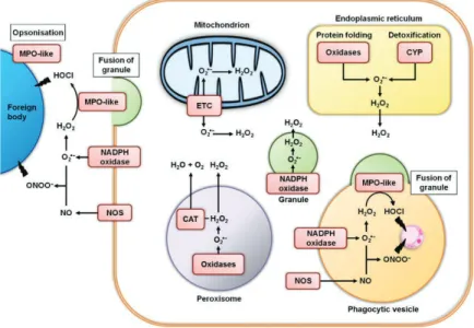
In response to ROS, an antioxidant machinery as evolved in many species from bacteria to humans, including activation of different low molecular weight scavengers like superoxide dismutase (SOD), catalase (CAT) and glutathione (GSH), ascorbic acid (vitamin C) that interact in a complex network to participate for example to the reduction of the membrane lipid peroxidation (Regoli and Giuliani, 2014; Canesi 2015). ROS are also naturally produced by different cellular compartments including the mitochondria (Figure 4)
21 as a product of the oxygen metabolism, the endoplasmic reticulum, peroxisomes and phagocytic vesicles that generate oxidative stress as the result of protein folding which requires oxygen as the final oxidant, H2O2 production and degradation, transferring electrons
across biological membranes through NADPH-oxidases activity (Donaghy et al, 2015).
Figure 4. Major sites of reactive oxygen and nitrogen species generation in generalized haemocytes in marine bivalves. Abbreviations: CAT, catalase; CYP, cytochrome P-450; ETC, electron transfer chain; MPO, myeloperoxidase; NOS, nitric oxide synthase. Adapted form Donaghy et al 2015.
?"!$G:;1()B&>1$>&-.:5+-1$EGFIH$
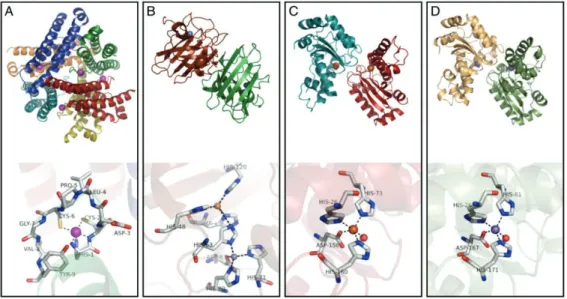
The superoxide dismutase is a dimeric enzyme present in almost all the aerobic
organism which principal function is the catalytic conversion of the O2- to oxygen (O2) and
hydrogen peroxide (H2O2) in presence of H+ (Fridovich, 1995). SODs are classified in two
categories according to their cellular localization, cytosolic and mitochondrial but both types require the presence of specific metals as cofactor for his activation, the specificity been indicated on the active site (figure 5). Manganese SOD (MnSOD) are found in the mitochondria, copper/zinc SODs (Cu/Zn SOD) which is the most abundant form can be found in the cytoplasm such as the iron SOD (FeSOD), usually found in prokaryotes, and the recently described nickel SOD (NiSOD) (Abreu and Cabelli, 2010). The mechanism by SOD activity involves the sequential reduction and oxidation of the metal center, with the
22 subsequent oxidation and reduction of superoxide radicals. Ecotoxicology studies in marine species such as Crassostrea gigas, Mytilus edulis, Mytilus galloprovincialis highlight the activation of SOD as a biomarker of oxidative stress in experimental exposure to xenobiotic agent and both activity (determined at protein level and activity) and gene expression level have been explored (Kim et al., 2015; Boutet et al., 2004; Lewis et al., 2016; Lushchak, 2011). The role of SOD in natural populations has been also studied in a context of naturally anthropogenic contaminated areas to validate the use of this specific biomarker in field context (Orbea et al., 2002; Rodriguez-Ariza et al., 1993).
Figure 5. A comparison of the enzyme structures and active sites for the four SODs, (A)
Streptomyces coelicolor NiSOD, (B) human CuZnSOD, (C) E. coli FeSOD and (D) MnSOD
Adopted from Abreu and Cabelli, 2010.
?"2$%15+88)50&),1&,-$E%=-H$
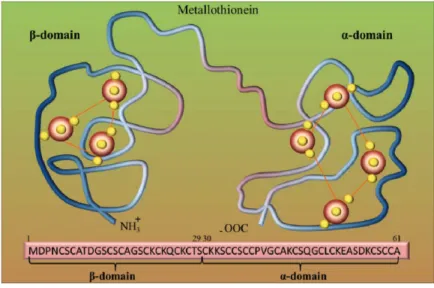
The metallothioneins (MTs) are one of the best studied stress response associated to metal exposure in invertebrates. Metallothioneins (MTs) are low molecular weight (6 to 7 kDa) cytoplasmic metal-binding proteins with high cysteine content (close to 30% of their amino acid content) that give the ability of the thiol group to capture free metal ions reducing cellular damage ) are that bind essential (copper, zinc, iron, manganese) and non essential (cadmium, mercury, lead, silver) metal ions that are sequestered into a metal/sulphur cluster (Amiard et al., 2006; Isani and Carpenè, 2014;Cherian and Goyer, 1978, Kojima and Kägi 1978). Although amino acid composition is variable, the cysteine content is more conserved with characteristic motif like Cys-Cys, Cys-X-Cys and Cys-X-Y-Cys, where X and Y are
23 amino acids other than cysteine (Paul-Pont et al., 2012). , however not all the function of the Mts as been describes, also free radical scavenger activity, Normally the cysteines are arranged in two metal thiolate cluster (figure 6) which are able to specifically bind metals that later can be exported out of the cellule. Important biological functions of MTs are 1) trace metal homeostasis, 2) protective role against excess reactive heavy metal ions, 3) free radical scavengers, 4) reservoir of essential metals that can be donated to other metalloproteins and 5) cell protection against intracellular oxidative damage (Karin, 1985; Hamer, 1986) and they have been linked to modulations of immune response, genotoxicity and carcinogenicity (Asselman et al., 2012; Nordberg et al., 2009).
Several studies have shown that MTs display a specific tissue expression pattern that is correlated with development and differentiation (Banerjee et al., 1982; Chatterjee and Maiti, 1990; Nemer et al., 1991). A large number of MT isoforms have been isolated from vertebrates and invertebrates, apparently originating from events of gene duplication that may have occurred many times independently (Griffith et al. 1983, Hunziker et al. 1995). Nine MT isoforms have been characterized in the blue mussel Mytilus edulis (Barsyte et al. 1999), seven in the sea urchin Strongylocentrotus purpuratus (Nemer et al. 1984, Wilkinson & Nemer, 1987), one each in the tropical green mussel Perna viridis (Khoo & Patel 1999) and the Eastern oyster Crassostrea virginica (Unger et al. 1991), and four in the Pacific oyster
Crassostrea gigas (Tanguy et al. 2001, Tanguy and Moraga 2001, David et al., 2012) and two
in the flat oyster(Boutet et al., 2002).
Figure 6. Metallothionein (MT) structure. Model of two binding sites of metallothionein. Red big beads are metal atoms (e.g., Zn), and small yellow beads are sulfur
24 atoms. Adopted from Ruttkay-Nedecky et al., 2013.
MT transcription is also regulated by a set of binding sites for transcription factors localized in the promoter region as metal responsive elements (MRE), metal transcription factor (MTF) and zinc finger protein (Janssens et al., 2009).
?"?$/1((&5&,$E/1(H$
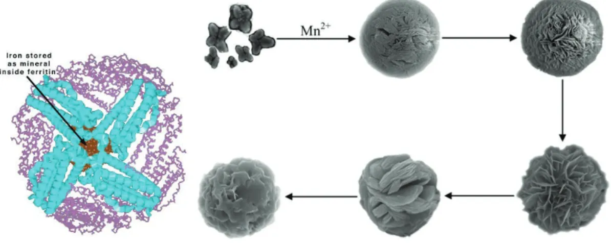
Ferritin which is the principal protein involved in iron storage, is a structurally
complex protein formed by 24 subunits of two types of polypeptide chains giving a globular shape that can accommodate large quantity of iron in his interior (Figure 7), and is usually present in the cytosol (Lobreaux et al., 1992). The process by which iron is stored begin in the H (heavy) chain which oxidizes Fe2+ in a specific ferroxidase site, and the L (Light) chain that performs the entry of the metal into the protein, while more metals enter the more Fe3+ cluster are formed in the cavity of the protein (Strange et al., 1993).
Figure 7. Tridimentional structure of ferritin (left) and manganese ferritin aggregate formation after exposure to manganese (right). Adopded from Chet et al 2015
The principal function of ferritin consists in the storage of free iron when his concentration exceeds the normal levels and its release when needed. In marine invertebrates the mechanism of activation of these proteins is less documented compared to other detoxification proteins but recent works showed that the transcriptionally and translational expression of this protein is positively correlated with the exposure of iron in several bivalves (Zhang et al., 2013; Zhou et al., 2014), but it has also been reported that this protein could store other types of heavy metals (Chen et al., 2015; Zapata et al., 2009).
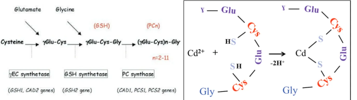
25 ?"@$J0D5)'018+5&,$EJK-H$
The phytochelatin (PC) is another type of metal binding peptides which is produced from glutathione by the enzyme phytochelatin synthase (PCS) (Figure 8). It has been described primarily in plants and terrestrial invertebrates to present an equivalent function of the MTs in metal detoxification because they also are characterized by the presence of cysteine motif (Cobbett, 2000; Inouhe, 2005; Vatamaniuk et al., 2005). The general structure of PCs consists only in three amino acids Glutamic acid (Glu), Cysteine (Cys) and Glycine (Gly) where the first two are linked through a !-carboxylamide bond, forming the structure (!-Glu-Cys)n-Gly where n can vary from 2 to 5 repetitions. They are normally found in the
cytosol but when they have interacted with a toxic metal, the complex PC-metal are sequestered to the vacuole (Cobbett, 2000; Rodrigo et al., 2013).
Figure 8. Phytochelatin syntthesis (left) and structural conformation when interact with metals (right). Adopded from Inohue, 2005 and Merlos et al 2013.
In marine invertebrates only a few sequences are reported for the phytochelatin synthase (PCS), particularly in the genome of the pacific oyster Crassostrea gigas and recently in the transcriptome of the hydrothermal mussel Bathymodiolus platifrons (Wong et al., 2015). The role of PCs could be a potential efficient mechanism in heavy metal sequestration in marine invertebrates which it has so far been less explored.
@"
A&$/+,*(',$5,<%2270$%5,(*+<%5+*,
The Omics term refers to the field of biology that includes genomic, transcriptomic and metabolomics which has considerably increased in the last years due the development of high throughput sequencing techniques as 454, Illumina, Solid and Heliscope which strongly increased the amount of DNA and RNA information, with a low error rate and cost. This opportunity to get a large amount of genomic data represented one of the most important
26
evolution for the study of non-model species (Pavlopoulos et al., 2013). This amount of genetic information has revolutionized areas of ecology and evolution, greatly expanded the scientific possibilities to assess the mechanisms that govern ecological interactions (Straalen and Feder, 2012). The field of genomics (the study of how an individual’s entire genetic make-up, the genome, translates into biological functions) should strongly contribute to provide tools that may assist our understanding of how chemicals can impact on organisms and ecosystem health. Genomic tools can facilitate ecology and evolutionary biology studies, providing useful data to identify ecological performance-regulated gene loci; to perform functional analysis of ecological performance-related traits; evaluate individual, population, community, and ecosystem responses to the environment and examine the degree and significance of genetic variation among ecological performance-related traits (Snape et al., 2004).
The term ‘ecotoxicogenomics’ has been proposed to describe the integration of genomics (transcriptomics, proteomics and metabolomics) into ecotoxicology and has been defined as the study of gene and protein expression in non-target organisms that is important in responses to environmental toxicant exposures (Figure 9).
Figure 9: Conceptual framework for ecotoxicogenomics
The way populations can adapt to environmental toxicants also has been explored using omics techniques, where traditionally the response to any toxic agent was studied by a limited number of genes but at present, many genes can be analyzed simultaneously in term of transcript regulation and interaction, protein concentration and activities and how specific mutation can change the protein function or activity. One application of transcriptomics and
27
proteomics in ecotoxicology that show an increasing interest is the characterization of the mode of action (MOA) profiling of environmental toxicants, in order to identify particularly sensitive subsets of ecological populations, and discover new molecular biomarkers of interest. The use of gene (or protein) expression profiling-based MOA will allow the identification of new toxic substances based on their expression profiling comparison to already known specific toxicants MOAs. The development of this approach is one of the priority of the European program REACH (European Union regulation on Registration Evaluation and Authorization of Chemicals,) that has been established to both optimize resources and to limit the use of animals for testing purposes. The new molecular biomarkers (single gene or complex gene expression pattern) should contribute to provide an early detection of environmental stress, to infer mechanism of action and allow a more relatively efficient monitoring of the environment. But, all this biomarkers have be integrated with phenotypic end points (activities, metabolites concentration, chemical profiles) to establish robust correlations between molecular biomarkers and phenotypic end points which is one of the major criticisms on the application of transcriptomics and proteomics to ecotoxicology.
Three main methods dominate transcriptomic analyses: the single gene expression study using quantitative PCR, the microarray technique that has existed since the mid-1990s and that allow the analysis of the expression of hundreds genes (Schena et al., 1995) and the RNA-seq that utilizes the power of high-throughput sequencing (HTS) for transcriptomics (Wang et al., 2009; Schirmer et al., 2010). Both methods initially use RNA and both result in a data matrix indicating relative expression levels of each transcript in each individual analyzed but both techniques require biological replications to allow robust statistical analysis (Lee et al., 2005). The microarrays use short gene sequences representing part of the DNA that are fixed to a surface and the levels of expression of the genes are estimated by hybridization with complementary DNA (cDNA) transcribed from mRNA transcripts previously labelled with fluorescent dye. After the hybridization, the array is scanned with lasers where the level of fluorescence detected is proportional to the number of cDNA transcript that are hybridizing at particular probe. The RNA-seq use high-throughput sequencing technique to generate hundreds of thousands of fragments of a cDNA library, the length, sequencing depth and quality depending on the sequencing platform used. The main differences with microarrays is that RNA-seq can detect all expressed genes (even new genes) when microarray only provide information on the genes present on the array and that RNAseq
28
requires a bioinformatics language for assemble and align the huge amount of transcripts generated (Alvarez et al., 2015).
Bivalve genomic resources remain not well annotated or well described, with the exception of some species classically used in biomonitoring studies such as Pacific oyster, C.
gigas, whose genome has been recently published (Zhang et al., 2012) or the pearl oyster, Pinctada fucata, in which genome annotation is still at the draft level (Takeshi et al., 2016).
Other bivalve transcriptomes are publicly available for M. galloprovincialis (Rosani et al., 2011; Gerdol et al., 2014), Patinopecten yessoensis (Hou et al., 2011), Ruditapes
philippinarum (Milan et al., 2011; Moreira et al., 2012) and Corbicula fluminea (Chen et al.,
2013). Several reviews papers have been published to provide some examples on how -omic tools have been used in the study of immunity and host–pathogen interactions in mollusc bivalves (Cancela et al., 2010; Gestal et al., 2008; Guo et al., 2008; Romero et al., 2012; Wang et al., 2013; Yue, 2014, Gomez-Chiarri et al., 2015). The use of large scale transcriptomics approach (ie microarray or RNAseq) to monitor pollutant effects is growing and has been mainly developed in the clam R. philippinarum in a population context (Milan et al., 2016) or in M. edulis and M. galloprovincialis in which correlation between metals concentration in tissues and gene expression profile has been reported (Venier et al., 2006; Dondero et al., 2010, 2011; Poynton et al., 2014, Varotto et al., 2013). These studies reported evidence serves to validate the use of this molecular tools in ecotoxicological studies but the major challenge still remains to get a better understand of how this molecular changes caused by toxic exposure in the organism modulate biological changes in the ecosystem (Schirmer et al., 2010).
6.
Hydrothermal ecosystems as natural laboratory for heavy
metal adaptation
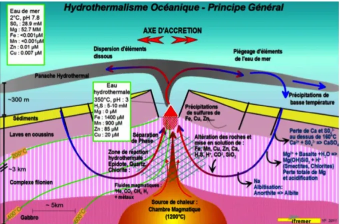
Deep-sea hydrothermal vents ecosystems are distributed among all the oceans, principally along mid-ocean ridges where new oceanic crust is generated. Vent fluid is formed by infiltration of deep-sea water into fractures of the oceanic plate and chemical transformation at high temperature deep in the crust, under the influence of the underlying magmatic chamber. The vent fluid returns to the ocean through diffuse or focused venting, with typically high temperature (up to 400°C depending on sites), loaded with heavy metals and reduced gases such as carbon dioxide (CO2), sulfide (H2S), hydrogen (H2) and methane
29
high temperature is reduced, the pH becomes more basic, some metals precipitate forming the classical chimney structures and some remain in the water column. However these characteristics are very variable for each vent site, providing a biotope with different environmental conditions (Demina and Galkin, 2016; Von Damm, 1995).
Figure 10. Geological formation of an hydrothermal vent. Adapted from Fouquet and Lacroix, 2012.
The initial discovery of luxuriant vent ecosystems in 1977 around the Galapagos (Corliss et al, 1979) revolutionized the understanding of life in the oceans because until that date the paradigm was that life in the deep ocean was scarce due to the absence of light, and consequently photosynthetic primary production, limiting food availability to what came from the surface layers. In the last forty years the development of new exploratory technology, principally manned submersibles and remotely operated vehicles (ROV), has enabled the discovery of many of these ecosystems across the oceans (Figure 11), revealing the occurrence of local, chemosynthetic primary production supporting very abundant communities of endemic species (Desbruyères et al. 2006), comprising arthropods, molluscs and annelids which have adapted to these ecosystems, and challenging major evolutionary, ecological, physiological hypotheses and paradigms (Danovaro et al., 2014).
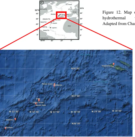
30 /&*:(1$##"$I&-5(&6:5&),$)9$0D>()501(.+8$41,5$-&51-$+'()--$501$)'1+,-"$ L>);51>$9().$3,51(C&>*1M$41(-&),$!"#$
31 $$N"#$OD>()501(.+8$41,5-$&,$%&>PL58+,5&'$C&>*1$E%LCH$
Ten years after the discovery of Corliss and colleague’s, exploration in the MAR were performed (Rona et al., 1986), evidencing that active hydrothermal vents also exist on slow spreading ridge axes. Until now, many scientific expeditions have been devoted to the MAR where several vent fields have been described between 10º N and 40º N (Figure 12), and some more recently in the south Atlantic. The most studied are the three vent fields located near the Azores Triple Junction (ATJ), characterized by contrasted physicochemical characteristics associated with their depth and the composition of oceanic crust (Charlou et al., 2000; 2002): Menez Gwen (MG, -850m), Lucky Strike (LS; -1700m), and Rainbow (RB; -2300m). An indication of the characteristics of the vent fluid (end-member) is given in Table 1, but one must keep in mind that due to the chaotic nature of hydrothermal venting, large excursions around these values can be measured at a given date.
!
Figure 12. Map of north MAR hydrothermal vent fields. Adapted from Charlou et al 2002. !