HAL Id: tel-01754646
https://hal.univ-lorraine.fr/tel-01754646
Submitted on 30 Mar 2018
HAL is a multi-disciplinary open access
archive for the deposit and dissemination of sci-entific research documents, whether they are pub-lished or not. The documents may come from teaching and research institutions in France or abroad, or from public or private research centers.
L’archive ouverte pluridisciplinaire HAL, est destinée au dépôt et à la diffusion de documents scientifiques de niveau recherche, publiés ou non, émanant des établissements d’enseignement et de recherche français ou étrangers, des laboratoires publics ou privés.
Comparative genomic analysis of Carnobacterium
maltaromaticum: Study of diversity and adaptation to
different environments
Christelle Iskandar
To cite this version:
Christelle Iskandar. Comparative genomic analysis of Carnobacterium maltaromaticum: Study of diversity and adaptation to different environments. Food and Nutrition. Université de Lorraine, 2015. English. �NNT : 2015LORR0245�. �tel-01754646�
AVERTISSEMENT
Ce document est le fruit d'un long travail approuvé par le jury de
soutenance et mis à disposition de l'ensemble de la
communauté universitaire élargie.
Il est soumis à la propriété intellectuelle de l'auteur. Ceci
implique une obligation de citation et de référencement lors de
l’utilisation de ce document.
D'autre part, toute contrefaçon, plagiat, reproduction illicite
encourt une poursuite pénale.
Contact : [email protected]
LIENS
Code de la Propriété Intellectuelle. articles L 122. 4
Code de la Propriété Intellectuelle. articles L 335.2- L 335.10
http://www.cfcopies.com/V2/leg/leg_droi.php
Université de Lorraine
École Nationale Supérieure d’Agronomie et des Industries Alimentaires
Ecole Doctorale Sciences et Ingénierie des Ressources, Procédés, Produits, Environnement (RP2E)
Laboratoire d’Ingénierie des Biomolécules (LIBio)
THESE
présentée devant L’Université de Lorraine
pour obtenir le grade de Docteur de l’Université de Lorraine
Spécialité : Procédés Biotechnologiques et Alimentaires
Par
Christelle ISKANDAR
Analyse par génomique comparée de Carnobacterium maltaromaticum : Etude de la diversité et de l’adaptation à différents environnements Comparative genomic analysis of Carnobacterium maltaromaticum:
Study of diversity and adaptation to different environments
Prof. Xavier DOUSSET UMR INRA-SECALIM - ONIRIS Nantes Rapporteur
Dr. Marie-Christine
CHAMPOMIER-VERGES
UMR 1319 MICALIS – INRA Jouy en Josas Rapporteur
Dr. Veronique DELCENSERIE
FARAH - Fundamental and Applied Research for Animals & Health - University of Liege
Examinateur
Dr. Fréderic BORGES LIBIO Laboratoire d'Ingénierie des
Biomolécules ENSAIA - Université de Lorraine
Examinateur
Prof. Anne-Marie REVOL-JUNELLES
LIBIO Laboratoire d'Ingénierie des
Biomolécules ENSAIA - Université de Lorraine
Directrice de thèse
Dr. Catherine CAILLIEZ-GRIMAL
LIBIO Laboratoire d'Ingénierie des
Biomolécules ENSAIA - Université de Lorraine
II
Dédicace
Je dédie ce travail
A mes parents,
qui ont tout sacrifié pour me voir réussir.
Vous êtes la source de mes efforts et la flamme de mon
cœur.
A mes frères
lumière de mes jours, mon soutien moral, source de joie et
de bonheur.
Vous m’avez toujours aidée et encouragée durant tout le
chemin.
III
Remerciements
Ce travail de thèse a été réalisé au sein du Laboratoire d’Ingénierie des Biomolécules LIBio. La réalisation de ce mémoire a été possible grâce au concours de plusieurs personnes à qui je voudrais témoigner toute ma reconnaissance.
Je remercie tout d’abord les membres du jury, Monsieur Xavier DOUSSET Professeur à ONIRIS
Nantes et Madame Marie-Christine CHAMPOMIER-VERGES, Directrice de recherche à l’INRA,
rapporteurs de thèse et Madame Veronique DELCENSERIE, assistant- professeur à l’université de Liège,
examinatrice des travaux de thèse, pour avoir accepté de juger ce travail..
J’exprime ensuite toute ma reconnaissance à mes encadrants, Madame Anne-Marie REVOL-JUNELLES, directrice de thèse, Madame Catherine CAILLIEZ-GRIMAL co-directrice et Monsieur Fréderic BORGES pour avoir accepté de diriger ce travail. Merci pour votre aide, votre soutien et spécialement pour votre confiance en moi.
Je tiens à remercier aussi tout le personnel du LIBio, Monsieur Michel LINDER, directeur du
LIBio, enseignant-chercheurs, doctorants, techniciens et stagiaires. C’était un grand plaisir de vous
connaitre. Un grand merci à tous les membres de l’équipe de microbiologie, Myriam, Sylvie et Arnaud,
pour leur soutien et leur bonne humeur. Je suis tout aussi redevable à Monsieur Emmanuel RONDAGS, Monsieur Fabrice BLANCHARD et Monsieur Cédric PARIS pour leur disponibilité et leurs judicieux conseils, qui ont contribué à alimenter ma réflexion.
Je voudrais adresser un merci à ERASMUS MUNDUS qui m’a donné la chance de réaliser ce parcours en me procurant le financement nécessaire durant mes deux ans en France. Un grand merci à Madame Delphine LAURANT pour sa patience.
Je voudrais exprimer ma reconnaissance envers les amis, spécialement Georges et Elie, qui m’ont
apporté leur support moral et intellectuel malgré la distance. Un très grand merci à Christelle SALAMEH
pour sa confiance et son support inestimable. Tu es plus qu’une amie, tu es une sœur, un compagnon de
route dans les moments agréables, et un soutien dans les moments pénibles. Une pensée particulière à mon cher ami Ibrahim. Ta présence m’a donnée la force et le courage pour pouvoir toujours avancer.
Enfin, un vif remerciement s’adresse à mes parents, mes frères et ma tante Roula, pour leur encouragement, leur confiance et l’entraide dans les moments difficiles.
IV
Résumé
Carnobacterium est un genre bactérien ubiquiste appartenant au groupe des bactéries lactiques (LAB). Les
souches de Carnobacterium maltaromaticum sont présentes dans une grande diversité d’environnements et de produits alimentaires. Elles présentent de nombreuses propriétés, dont un effet positif en fabrication fromagère et des potentialités probiotiques qui en font un microorganisme à forte potentialité industrielle. L’objectif de ce travail de thèse est d’approfondir les connaissances sur C. maltaromaticum par une étude génomique. Neuf génomes de bactéries du genre Carnobacterium, dont 5 de C. maltaromaticum, disponibles sur la plateforme MicroScope ont été analysés et comparés. Carnobacterium maltaromaticum DSM20342 MX5 possède un génome de 3,85 Mbp, le plus grand génome parmi les LAB connus, de 1 Mbp de plus que celui des autres Carnobacterium n’appartenant pas à l’espèce
C. maltaromaticum. L’analyse génomique détaillée indique la présence de quelques voies métaboliques spécifiques
et suggère que les souches de C. maltaromaticum et C. divergens présenteraient une surface cellulaire caractérisée par une grande diversité moléculaire.Les trois autres souches de Carnobacterium présenteraient une surface fortement dépourvue en molécules de surfaces dont les protéines. Les propriétés de surface qui en découlent seraient à relier à la capacité de ces deux espèces à coloniser différents habitats. Par ailleurs, les souches de C. maltaromaticum isolées de différents produits laitiers se caractérisent par une grande diversité génomique. Afin de caractériser le niveau de diversité, les gènes impliqués dans les voies métaboliques d’utilisation du lactose (voie du Tagatose-6Phosphate, gènes
lac) et du galactose (voie de Leloir, gènes gal) ont été caractérisés par analyse génomique au sein du genre Carnobacterium. De plus, ces gènes ont été recherchés par PCR chez 42 souches de C. maltaromaticum. Les deux
voies sont présentes et organisées différemment chez les différentes souches de C. maltaromaticum d’origine laitière et non laitière L’analyse de ces gènes au niveau de la population révèle une dissémination des deux voies au sein des 4 lignées de cette population. La voie de Leloir est présente dans les lignées I, II et III et la voie du Tagatose-6Phosphate dans les lignées I, II et IV. Ces résultats suggèrent que les gènes lac et gal ont évolué selon un schéma complexe, qui est le reflet du haut niveau de diversité génétique de la population. La recherche de ces gènes au sein des 237 génomes de LAB disponibles sur la plateforme MicroScope Mage indique que les gènes lac et gal présentent un niveau de diversité élevé parmi les différents genres bactériens constituant le groupe des LAB, avec une dominance de la présence de la voie de Leloir. Ce niveau de diversité génétique n’est retrouvé qu’à l’échelle du genre chez les LAB et non de l’espèce, comme c’est le cas de C. maltaromaticum.
Mots-clés : Carnobacterium maltaromaticum, diversité, adaptation, comparaison génomique, lactose.
Summary
Carnobacterium bacteria belong to the lactic acid bacteria (LAB) and are ubiquitous. Carnobacterium maltaromaticum strains are present in a wide variety of environments and foods. They have many positive properties
in cheese manufacturing that make them microorganisms of industrial interest. The objective of this thesis is to deepen the knowledge on C. maltaromaticum by a comparative genomic approach. Nine Carnobacterium genomes, including 5 C. maltaromaticum, available on the platform MicroScope were analyzed and compared. Carnobacterium
maltaromaticum DSM20342 MX5 is the largest genome among LAB, 1Mbp more than other Carnobacterium species.
Detailed analysis indicates the presence of some specific metabolic pathways, and the presence of a high molecular diversity of cell surface proteins in C. maltaromaticum and C. divergens, while the three other Carnobacterium species present only few proteins on their surfaces. This can be related to the ability of these two species to colonize different habitats. Furthermore, C. maltaromaticum strains isolated from various dairy products are characterized by a large genomic diversity. In order to characterize this diversity, genes involved in lactose (Tagatose-6Phosphate pathway,
lac genes) and galactose (Leloir pathway, gal genes) metabolic pathways were identified by genomic analysis within Carnobacterium genus and detected by PCR in 42 C. maltaromaticum strains. Both pathways are present and
organized differently in the C. maltaromaticum strains. The analysis of these genes at the population level shows a spread of the two pathways within the 4 lineages of this population. The Leloir pathway is present in the lineages I, II and III and the Tagatose-6Phosphate in the lineages I, II and IV. These results suggest that the lac and gal genes have evolved in a complex pattern, which reflects the high level of genetic diversity of the population. These genes were characterized in 237 LAB genome and exhibit a high degree of diversity among the different bacterial genera of the LAB group, with a dominance of the presence of the Leloir pathway. This level of genetic diversity is found at the genus level in LAB and not at the species level, as for C. maltaromaticum.
V
Table of content
Dédicace ... II Remerciements ... III Résumé ... IV Summary ... IV Table of content ... V List of Tables ... VII List of Figures ... VIII List of abbreviations ... XList of Annexes ... 1
INTRODUCTION ... 2
CHAPTER I: BIBLIOGRAPHY ... 6
1. Lactic Acid Bacteria and Carnobacterium 7 1.1. Taxonomy and genomics of Lactic Acid Bacteria ... 7
1.2. The genus Carnobacterium ... 11
2. Bacterial Cell Wall Proteins 24 2.1. Proteins anchorage to the Cell Surface ... 25
2.2. The function of Cell Wall Proteins ... 29
3. Bacterial adaptation to dairy products: lactose and galactose metabolism 36 3.1. Introduction ... 36
3.2. Sugar uptake systems ... 38
3.3. Lactose molecule cleavage: beta-galactosidase ... 39
3.4. Lactose/ galactose metabolism pathways ... 39
VI
3.6. Genes mutation effects ... 41
3.7. Specificity and genes organization in LAB strains ... 42
4. Objectives 49 CHAPTER II: RESULTS AND DISCUSSION ... 51
1. Comparative genomic analysis reveals contrasting differences between Carnobacterium species 52 1.1. Introduction ... 52
1.2. Material and Methods ... 54
1.3. Results and discussion ... 55
1.4. Conclusion ... 72
2. Adaptation to dairy environment: lactose metabolism pathways in LAB and Carnobacterium 73 2.1. Genes associated to lactose metabolism illustrate the high diversity of Carnobacterium maltaromaticum ... 73
2.2. Diversity of lactose/galactose metabolic pathways within Lactic Acid Bacteria 87 CHAPTER III: CONCLUSIONS AND PERSPECTIVES ... 102
REFERENCES ... 107
ANNEXES ... 137
Résumé ... 156
VII
List of Tables
Table 1: Ecology of eight Carnobacterium sp. ... 13
Table 2: Ecology of Carnobacterium divergens and Carnobacterium maltaromaticum. ... 14
Table 3: Genes implicated in lactose and galactose transportation systems, Tagatose-6P and Leloir pathways. ... 37
Table 4: lac/gal genes organization in some LAB strains. ... 43
Table 5: Origins of Carnobacterium sp. ... 54
Table 6 : General features of Carnobacterium genomes. ... 57
Table 7: Comparative analysis of the Pan/Core genome (50-80) between A: all the Carnobacterium genomes, B: Carnobacterium sp. and C. inhibens subsp. gilichisnkyi WN1359 genomes and C: Carnobacterium divergens and C. maltaromaticum genomes. ... 60
Table 8: Primer sequences used in this study ... 77
Table 9: Lactose/galactose metabolism putative genes in Carnobacterium sp. ... 79
Table 10: Presence of lac and gal genes within a collection of 42 Carnobacterium maltaromaticum strains. ... 83
VIII
List of Figures
Figure 1: Schematic phylogenetic tree of some lactic acid bacteria, including some Gram-positive bacteria of the low GC subdivision, Bacillus, Listeria and Staphylococcus. ... 8 Figure 2: Genome size of some LAB genera. ... 9 Figure 3: eBURST analysis of 47 C. maltaromaticum strains. ... 19 Figure 4 : Cell envelope of lactobacilli with a schematic representation of cell wall and membrane-associated proteins. ... 25
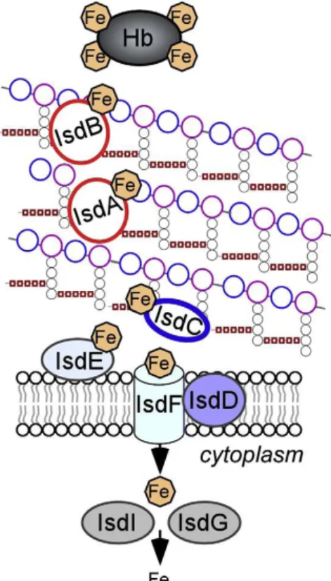
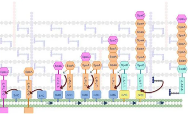
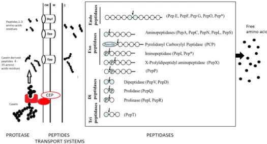
Figure 5 : Isd-mediated heme-iron uptake across the cell wall of Staphylococcus aureus. ... 30 Figure 6 : Model of pilus biogenesis. ... 32 Figure 7 : Proteolysis pathway by LAB, with schematic representation of the action of peptidases found in lactic acid bacteria. ... 34
Figure 8: Schematic representation of the lactose and galactose catabolic pathways. ... 37 Figure 9: Relationship between Carnobacterium genus. A- phylogenetic tree of the sequenced genomes of Carnobacterium based on the conserved nucleic sequence of 10 essential genes (dnaK gyrA polA lepA dnaB gyrB secA ftsZ recG ileS). B- eBURST analysis of 49 C.
maltaromaticum strains. ... 56
Figure 10: COG distribution in the different classes for 5 strains of Carnobacterium. C.
maltaromaticum LMA28, C. divergens V41, Carnobacterium sp. 17.4, C. inhibens subsp. gilichinskyi WN1359. ... 59
Figure 11: Pan/Core genome analysis of Carnobacterium maltaromaticum strains with a cut-off of 70% amino-acid identity on 80% coverage. ... 61
Figure 12: Comparison of the distribution of the Core genome and the intraspecific Core genome in the different COG classes... 62
Figure 13: Secretome size in the different Carnobacterium genomes, LAB genomes and other Gram-positive bacteria... 64
IX Figure 15: Gene cluster organization and synteny between C. maltaromaticum LMA28,
L. rhamnosus GG and E. faecalis V583. ... 72
Figure 16: Organization and synteny between lac genes among Carnobacterium
maltaromaticum LMA28, DSM20342-MX5, ATCC35586, Streptococcus mutans, and Staphylococcus aureus. ... 78
Figure 17: Organization and synteny between gal genes in Carnobacterium
maltaromaticum 3.18, C. inhibens subsp. gilichinskyi WN1359, Carnobacterium sp. 17.4 and Carnobacterium sp. AT7. ... 81
Figure 18: Diversity of the lactose and galactose metabolic pathways within A. genus and B. species of the different genus analyzed. ... 98
X
List of abbreviations
°C degree celcius µl micro litre µM micro Mole 16S rDNA 16S ribosomal Desoxyribo Nucleic Acid 16S rRNA 16S ribosomal RiboNucleic Acidaa amino acid
ADN Acide
DesoxyriboNucléique AOP Appélation d’Origine Protégée
BBH Bidirectionnal Best Hit
CBL Club des Bactéries Lactiques
CC Clonal Complex CDS Coding DNA Sequence
CEP Cell Envelope Protease
CFU.g-1 Colony Forming Unit per Gramme
CM Cell Membrane
COG Cluster of Orthologuous Genes CRISPES-cas Cluster Regularly Interspaced Short Palindromic Repeats – associated genes
CWP Cell Wall Proteins CWSS Cell Wall Signal Sequence
EC Enzyme Commission EPS Exopolysaccharids FBP Fructose Bi Phosphate G6P Glucose 6 Phosphate GC Guanine and Cytocine GRAS Generally Recognized As Safe
Hb Hemoglobin
HGT Horizontal Gene Transfer
Isd Iron Surface Determinant
KEGG Kyoto
Encyclopedia of Genes and Genomes
LAB Lactic Acid Bacteria
LTA LipoTeichoic Acid LysM Lysine Motif
MAP Modified
Atmosphere Packaging mbar milli bar
Mbp Mega base pair MLST Multi Locus Sequence Typing NEAT NEAr-Transporter ng nanogramme P phosphate PCR Polymerase Chain Reaction PEP PhosphoEnolPyruvate PG PeptidoGlycan PRD PTS Regulatory Domains PTS PhosphoTransferase System SDP Sortase Dependant Protein sec second
Sec Secretion system SP Signal Peptide
List of Annexes
Annexe 1: CDS distribution of the intraspecific core genome in the different COG ... 138 Annexe 2: Summary of the analysis of the cell wall proteins ... 142 Annexe 3: Genes encoding surface proteins of the 9 Carnobacterium strains, and the genes involved in the proteolysis. ... 143
Annexe 4: Percentage of identity between lac and gal genes within LAB. ... 148 Annexe 5: List of C. maltaromaticum LMA28 megaplasmid genes grouped in the different COG ... 149
2
INTRODUCTION
3 Les travaux de recherche effectués au sein du LIBio sont centrés sur la valorisation d’agro-ressources à des fins alimentaires et non alimentaires, en alliant des compétences en biochimie, physico-chimie et microbiologie. L’objectif de la recherche développée au laboratoire est de comprendre et de maitriser la structuration et la fonctionnalisation de la matière molle, les mécanismes de transferts et de relargage dans des systèmes complexes, ainsi que les interactions au sein des différents systèmes étudiés. L’ensemble des travaux prend en compte l’impact des paramètres biotique et abiotique afin de stabiliser les systèmes et/ou de concevoir des vecteurs et des matrices à fonctionnalité ciblée. Les composés actifs peuvent être des biomolécules - lipides polaires, acides gras polyinsaturés, antioxydants, composés antibactériens - ou des bactéries.
Des systèmes glucidiques et protéiques sont conçus pour la stabilisation et/ou la vectorisation des composés actifs particuliers que sont les bactéries. Le choix du système matrice/composé actif intègre les interactions avec la matrice ainsi que l’impact que pourrait avoir la bactérie incorporée sur la structuration des communautés microbiennes de l’écosystème considéré.
Dans ce contexte, la matrice alimentaire et la bactérie lactique Carnobacterium
maltaromaticum servent de modèle dans le cadre de cette étude.
Carnobacterium appartient au groupe des bactéries lactiques (LAB). Sur les onze espèces
que contient le genre Carnobacterium, les plus fréquemment isolées des aliments sont C.
mataromaticum et C. divergens (Leisner et al., 2007). Carnobacterium maltaromaticum a été
isolée d’une grande variété d’aliments dont des produits laitiers (Afzal et al., 2010). En effet, les travaux réalisés au LIBio ont démontré que C. maltaromaticum fait partie de la microflore complexe des fromages (Cailliez-Grimal et al., 2007, 2005; Millière et al., 1994; Milliere and Lefebvre, 1994; Rahman et al., 2014a). La présence de cette bactérie n'est pas négligeable car elle peut atteindre des concentrations élevées en fin d’affinage dans des fromages au lait de chèvre ou de vache (jusqu'à 109 UFC.g-1) (Cailliez-Grimal et al., 2007) où elle jouerait un rôle positif (Afzal
et al., 2010). C’est une bactérie lactique psychrotrophe, capable de se développer à des valeurs de pH alcalin et produisant un arome malté (Afzal et al., 2013b, 2012). De nombreuses souches de cette espèce sont connues pour la production d’une large gamme de bactériocines (Afzal et al., 2010). Les derniers travaux réalisés au laboratoire ont mis en évidence des potentialités probiotiques de C. maltaromaticum LMA28 (Rahman et al., 2014b) ainsi qu’une grande diversité
4 génétique au sein d’une collection de 42 souches de C. maltaromaticum d’origines diverses (Rahman et al., 2014a).
Le séquençage des génomes bactériens est de plus en plus rapide et accessible. Ceci ouvre la voie à de nouvelles possibilités d’analyse des propriétés des bactéries. Récemment, le génome de C. maltaromaticum LMA28 a été séquencé au laboratoire (Cailliez-Grimal et al., 2013). Les génomes C. maltaromaticum 3.18 et ML.1.97 ainsi que celui de C. divergens ont été mis à notre disposition grâce à des collaborations respectivement avec J. Leisner (University of Copenhagen, Denmark) et H. Prévost (ONIRIS, Nantes). Ceux de deux autres souches de Carnobacterium sp. AT7 et 17.4, de C. maltaromaticum DSM20342 MX5 et ATCC35586, ainsi que celui de
C. inhibens subsp. gilichinsky WN1359 sont en accès libre.
C’est dans ce contexte que s’est déroulé ce travail de thèse, dont l’objectif est d’approfondir les connaissances sur C. maltaromaticum par une étude génomique.
Cette étude devrait permettre de répondre à différentes questions :
- Carnobacterium maltaromaticum est isolée d’une grande diversité d’environnement et la population présente une grande diversité. Une analyse a montré que les génomes des souches de C. maltaromaticum sont de plus grande taille que ceux des autres bactéries du même genre. Que renferme cet ADN ? Quelle est la fonction des gènes spécifiquement présents chez
C. maltaromaticum ?
- Carnobacterium maltaromaticumest très représentée dans l’environnement laitier, avec
une grande diversité de souches. Cette diversité peut-elle se retrouver au niveau des propriétés physiologiques, et notamment au niveau des voies métaboliques d’utilisation du lactose, composé clé en technologie fromagère ?
Afin de répondre à ces interrogations, une revue bibliographique, rédigée en anglais, a été réalisée sur la génomique des bactéries lactiques, l’écologie des bactéries du genre
Carnobacterium et les voies de métabolisation du lactose et du galactose. Les analyses de
génomique comparée ont montré la particularité des bactéries du genre Carnobacterium vis-à-vis de leur surface cellulaire. Une partie de la bibliographie est relative à la structure des surfaces bactériennes des bactéries à Gram-positive.
5 Les résultats sont rédigés en anglais et présentés en deux chapitres :
Le premier chapitre est dédié à l’analyse génomique comparée des différentes bactéries du genre Carnobacterium.
Le deuxième chapitre est dédié à la caractérisation des gènes impliqués dans les voies de métabolisation du lactose et du galactose chez les bactéries du genre Carnobacterium, chez
C. maltaromaticum plus spécifiquement et chez l’ensemble des LAB.
Les conclusions générales et les perspectives que laissent entrevoir ce travail sont rédigées en français et présentées dans le dernier chapitre de cette thèse.
Ce travail a fait l’objet :
- d’une communication affichée au Club des Bactéries Lactiques (CBL) Lille, 17-19 juin 2015, France
Christelle F. ISKANDAR, Frédéric BORGES, Abdur RAHMAN, Emmanuel RONDAGS, Fabrice BLANCHARD, Benoît REMENANT, Monique ZAGOREC, Jorgen J. LEISNER, Catherine CAILLIEZ-GRIMAL, Anne-Marie REVOL-JUNELLES. Analyse génomique et diversité de Carnobacterium maltaromaticum : un exemple, le métabolisme du lactose
- d’une publication soumise à Food Microbiology
Christelle F. ISKANDAR, Catherine CAILLIEZ-GRIMAL, Abdur RAHMAN, Emmanuel RONDAGS, Benoît REMENANT, Monique ZAGOREC, Jorgen J. LEISNER, Frédéric BORGESand Anne-Marie REVOL-JUNELLES Genes associated to lactose metabolism illustrate the high diversity of Carnobacterium maltaromaticum
7
1.
Lactic Acid Bacteria and Carnobacterium
1.1.Taxonomy and genomics of Lactic Acid Bacteria
The interest for lactic acid bacteria (LAB) and foods begins with Pasteur’s work on lactic acid fermentation in 1857 and the first isolation of a pure culture, Bacterium lactis (now called Lactococcus lactis subsp. lactis), by Leister in 1873. According to Orla-Jensen in 1919, the “true lactic acid bacteria” form a natural group of Gram-positive, non sporulating rods or cocci that ferment carbohydrates and higher alcohols to form lactic acid (Axelsson, 2004; Stackebrandt and Teuber, 1988; Stiles and Holzapfel, 1997).
The definition and classification of LAB change with the introduction of molecular biology and was the focus of intense taxonomic study with approaches involving both phenotypic and phylogenetic characterization of bacteria (Axelsson, 2004; Pot, 2008).
Actually, this term LAB relates to the metabolic capabilities of microorganisms to ferment various nutrients predominantly into lactic acid (Klaenhammer et al., 2005; Liu, 2003). The general characteristics of LAB are that they are Gram-positive, anaerobic, acid-tolerant and non-sporulating (Klaenhammer et al., 2005), catalase negative, although some LAB strains have a catalase activity mediated by a non-heme “pseudocatalase”(Engesser and Hammes, 1994), microaerophilic, rods and cocci (Axelsson, 2004).
LAB form a heterogeneous group of microorganisms, and the different genera included in the term LAB have been subject to several controversies. Historically, the four genera Lactobacillus, Leuconostoc, Pediococcus and Streptococcus form the core of the group, corresponding to the current genera Enterococcus, Carnobacterium, Lactobacillus,
Lactococcus, Leuconostoc, Oenococcus, Pediococcus, Streptococcus, Vagococcus and Weissella (Axelsson, 2004; Pot, 2008; Stackebrandt and Teuber, 1988; Stiles and Holzapfel,
1997).
Several taxonomic revisions of these genera and the description of new genera lead to several change. Among domesticated bacteria studied and exploited, LAB are found in two distinct phyla, namely Firmicutes and Actinobacteria. LAB in Actinobacterium phylum include only Atopobium and Bifidobacterium genera (which produce lactic acid but always in combination with acetic acid), with a Guanine-Cytosine (GC) content of 36-46% and 58-61%, respectively (de Vos, 2011; Horvath et al., 2009; Makarova et al., 2006). Within the Firmicutes phylum, LAB belong to order Lactobacillales and include the genera Aerococcus, Alliococcus,
8
Carnobacterium, Enterococcus, Lactobacillus, Lactococcus, Leuconostoc, Oenococcus, Pediococcus, Streptococcus, Tetragenococcus, Vagococcus and Weissella which are all low
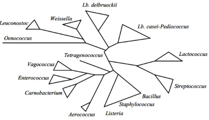
GC content organisms (31-49%) (de Vos, 2011; Pot, 2008). In a phylogenetic tree of LAB published in 2004, Carnobacterium genus appeared to be more related to Vagococcus and
Enterococcus than to any other LAB (Figure 1) (Axelsson, 2004). However, phylogenetic
relationships among LAB are still the subject of many discussions (Zhang et al., 2011).
Figure 1: Schematic phylogenetic tree of some lactic acid bacteria, including some Gram-positive bacteria of the low GC subdivision, Bacillus, Listeria and Staphylococcus (Axelsson, 2004).
The first LAB that was completely sequenced, L. lactis subsp. lactis IL1403 came out in 2001 (Bolotin et al., 2001). Then in 2003, Kleerebezem et al. sequenced the first and complete
Lactobacillus strain genome, L. plantarum WCFS1 (Kleerebezem et al., 2003). In 2014,
approximately 6,800 bacterial genome sequences are available of which 182 Lactobacillus genomes, 14 Lactococcus genomes, 113 Enterococcus genomes, and much more Streptococcus genomes (Wassenaar and Lukjancenko, 2014).
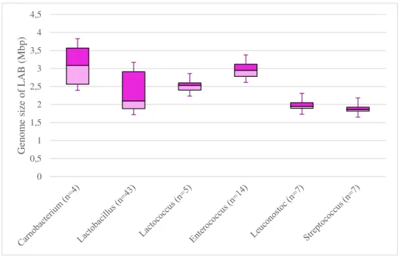
Genome sequencing is a relatively new discipline allowing a rapid bacterial exploration. It offers a wealth of information to well-understand LAB when investigating their gene content, their properties and their role in human health and food fermentation (Johnson and Klaenhammer, 2014). The study of LAB sequenced bacteria showed that Lactobacillales present a diversity in genome size ranging from 1.8 Mbp for Streptococcus to 3.7 Mbp for
Carnobacterium (Figure 2). It was hypothesized that Lactobacillales diverged from their
common ancestor with Bacilli causing the loss of 600-1200 genes, some of them encoding biosynthesis enzymes (Makarova and Koonin, 2007). Genome size within a genus is homogenous in almost all LAB except in Lactobacillus (Wassenaar and Lukjancenko, 2014)
9 and Carnobacterium genera. Genome size of Lactobacillus range from 1.3 Mbp for L. iners AB-1 (Macklaim et al., 2011) to 3.3 Mbp for L. plantarum WCFS1 (Kleerebezem et al., 2003). The genome size of Carnobacterium species is estimated ranging from 1.9 (for C.
alterfunditum) (Pikuta et al., 2005) to 3.7 Mbp (for C. maltaromaticum) (Cailliez-Grimal et al.,
2013). Carnobacterium maltaromaticum strains possess bigger genomes compared to other LAB (Hols et al., 2005; Kelly et al., 2010; van de Guchte et al., 2006). It has been suggested that the large size of Carnobacterium genome may be the reason of its well adaptation to environmental challenges (Leisner et al., 2007). Indeed, in order to survive and grow in a variety of environments, a bacterium will need a large number of encoded genes, and thus a larger genome. Therefore, the number of predicted protein genes strongly correlates with genome size; that means that a larger genome holds a larger number of CDS (Coding DNA Sequence) (Wassenaar and Lukjancenko, 2014).
Figure 2: Genome size of some LAB genera.
according to (Cailliez-Grimal et al., 2013; Douillard and de Vos, 2014; Leisner et al., 2012; Leonard et al., 2013; Voget et al., 2011; Wassenaar and Lukjancenko, 2014). n = number of strains
LAB are found naturally in a variety of environmental habitats, including plant (fruits, vegeTable, cereal), meat and milk environment, and are involved in a large number of industrial and spontaneous food fermentations, notably those based on raw materials derived from these natural habitats. The traditional roles for many LAB have been as starter cultures (Klaenhammer et al., 2005), which lead to their widespread human consumption. Indeed, the LAB used in food are considered non-pathogenic and are awarded the qualification of
Anglo-0 0,5 1 1,5 2 2,5 3 3,5 4 4,5 Gen om e size of L A B ( Mb p)
10 Saxon agencies Generally Regarded As Safe (GRAS) (Aguirre and Collins, 1993). Their primary contribution is in rapid acid production and acidification of foods, but metabolic processes accompanying the growth of LAB impact also flavor, nutrition, and texture quality of a variety of fermented foods (Axelsson, 2004; Klaenhammer et al., 2005; Kleerebezem and Hugenholtz, 2003; Pot, 2008). The sequenced and annotated bacterial genomes permit the prediction of certain important characteristics researched by industrials. Thanks to comparative genomics, it became easy to search for metabolic pathways (Cogan et al., 2007). Indeed,
Lactobacillus helveticus CNRZ32 had been discovered to possess homologs of genes involved
in the proteolysis of casein found in milk (Broadbent et al., 2013). The research project consists in predicting the formation of metabolic compounds via the exploration of the genomic content of CDS in comparison to previously annotated genomes. This is the case also of the identification of flavor molecules (G. Smit et al., 2005), decarboxylation of branched-chain α-Keto acids in L. lactis (B. A. Smit et al., 2005). In addition, LAB have the ability to avoid the food product deterioration by inhibition of bacterial growth by the production of lactic acid and growth-inhibiting compounds such as acids, H2O2, CO2 and bacteriocins (Cotter et al.
2005).
Some LAB are also closely associated with the mucosal surfaces of animals and human environment, including the gastrointestinal tract, the oral, the respiratory and the vaginal cavities. Moreover, many species of LAB are considered to be important components of the normal intestinal microbiota, which contribute to a variety of functions including intestinal integrity, immunomodulation, and pathogen resistance. Selected groups of LAB are used as probiotics for human consumption (Gilliland, 1989; Klaenhammer et al., 2005; Pot, 2008; Stiles and Holzapfel, 1997).
LAB colonize a great diversity of habitats. Adaptation to a particular niche is allowed by genomic remodeling. Comparative genomics of LAB revealed that loss and gain of genes occurred during the evolution of these bacteria, and it is accepted that these events allowed to improve their ecological performance (Makarova et al., 2006).
The more striking examples of evolution by gene loss are Streptococcus thermophilus (Bolotin et al., 2004), Lactobacillus bulgaricus (Kafsi et al., 2014; van de Guchte et al., 2006),
L. helveticus (Callanan et al., 2008) and L. lactis subsp. cremoris UC509.9 (Ainsworth et al.,
2013). The genome of these bacteria present many pseudogenes linked to amino acids biosynthesis and those of the fermentation of plant derived sugars (Cavanagh et al., 2015). It
11 was hypothesized that these lineages experienced a massive gene loss resulting in genome decay.
At another extent, the gene gain has been detected in several cases. For organisms mainly present in gastrointestinal tracts, such as L. acidophilus (Altermann et al., 2005), L.
plantarum (Kleerebezem et al., 2003) and L. gasseri (Azcarate-Peril et al., 2008), many genes
were suspected to be gained by Horizontal Gene Transfer (HGT) and contributed to the interaction of the bacterium with intestinal mucosa and its survival in gastric conditions. In this connection, many mucin-binding proteins and gene clusters for transport of a diverse group of carbohydrates are found in those genomes.
Gain of genes has been also well documented with the acquisition of plasmids holding metabolic pathways. This is the case of Lactococcus garvieae (Collins et al., 1983; Vendrell et al., 2006; Wyder et al., 2011), L. lactis (Kelly et al., 2010) and Streptococcus macedonicus (Malkin et al., 2008) isolated from different environments. They are known to possess plasmids containing genes responsible of the lactose and galactose utilization via the Tagatose-6Phosphate pathway (Cavanagh et al., 2015; Flórez and Mayo, 2015; Papadimitriou et al., 2015; Passerini et al., 2010).
1.2. The genus Carnobacterium 1.2.1. Taxonomy
The genus Carnobacterium was first proposed to clarify the taxonomic position of atypical heterofermentative Lactobacillus species (groupe III) isolated from chicken meat. They suggested classifying Lactobacillus divergens and Lactobacillus piscicola in a new genus,
Carnobacterium, as Carnobacterium divergens and Carnobacteriun piscicola. Two new
species, Carnobacterium gallinarum and Carnobacterium mobile were also added in this new genus (Collins et al., 1987).
The first Carnobacterium strain was isolated in 1974 and designated as Lactobacillus
maltaromicus by Miller (Miller et al., 1974). Based on phenotypic and genotypic comparisons,
this strain and C. piscicola described in 1987 were reclassified in 2003 as C. maltaromaticum (Mora et al., 2003).
12 The Carnobacterium genus forms a phylogenetically coherent group belonging to the LAB, which is quite distinct from other LAB, as shown by 16S rDNA sequencing (Wallbanks et al., 1990). It belongs to the phylum Firmicutes, Class Bacilli, order, Lactobacillales, family
Carnobacteriaceae. This new family was created in 2009 (Ludwig, et al., 2009) with Carnobacterium as genus type and was formed of two paraphyletic groups: the first one
consisted of eleven genera, Alkalibacterium, Allofustis, Alloiococcus, Atopococcus,
Atopostipes, Carnobacterium, Desemzia, Dolosigranulum, Isobaculum, Marinilactibacillus
and Trichococcus, and the second one included Atopobacter and Granulicatella. The phylogenetic position of this second group is still under discussion (Pikuta, 2014).
Strains of Carnobacterium are Gram-positive, non-spore-forming rods or coccobacilli, motile or not. These bacteria are catalase negative, but some species exhibit catalase activity in the presence of heme. Initially described as heterofermentative, carnobacteria could be considered to be homofermentative organisms that produce lactic acid from glucose (except for the species C. pleistocenium) or facultative heterofermentative. Some species could metabolize hexoses and pentoses to L(+)-lactic acid and, depending on the access of oxygen, may produce acetic acid, ethanol, CO2 and formic acid in varying amounts. Respiration might occur in the
presence of hematin. They do not reduce nitrate to nitrite. Production of NH4+ from arginine is
a result of its catabolism catalyzed by the arginine deaminase pathway. Some species possess the ability of converse tyrosine to tyramine. They are mesophilic and some species may be psychrotolerant and grow at 0 °C. NaCl is not required for growth. Some strains are halotolerant until 8% NaCl and alkaliphilic with growth until pH 9.5 (Cailliez-Grimal et al., 2014; Pikuta, 2014; Pikuta and Hoover, 2014).
The genus Carnobacterium included 11 species: Carnobacterium alterfunditum, C.
divergens, C. funditum, C. gallinarum, C. iners, C. inhibens, C. jeotgali, C. maltaromaticum, C. mobile, C. pleistocenium, and C. viridans. Recently, C. gilichinskyi was reclassified as C. inhibens subsp. gilichinskyi, and the subspecies C. inhibens subsp. inhibens was created
automatically for C. inhibens (Nicholson et al., 2015).
1.2.2. Ecology
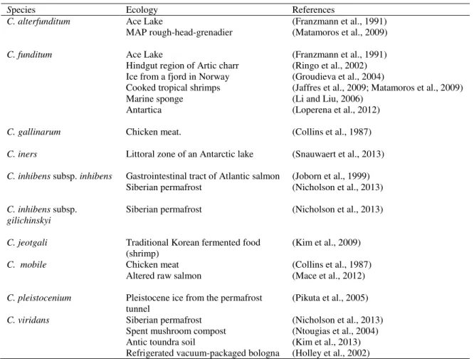
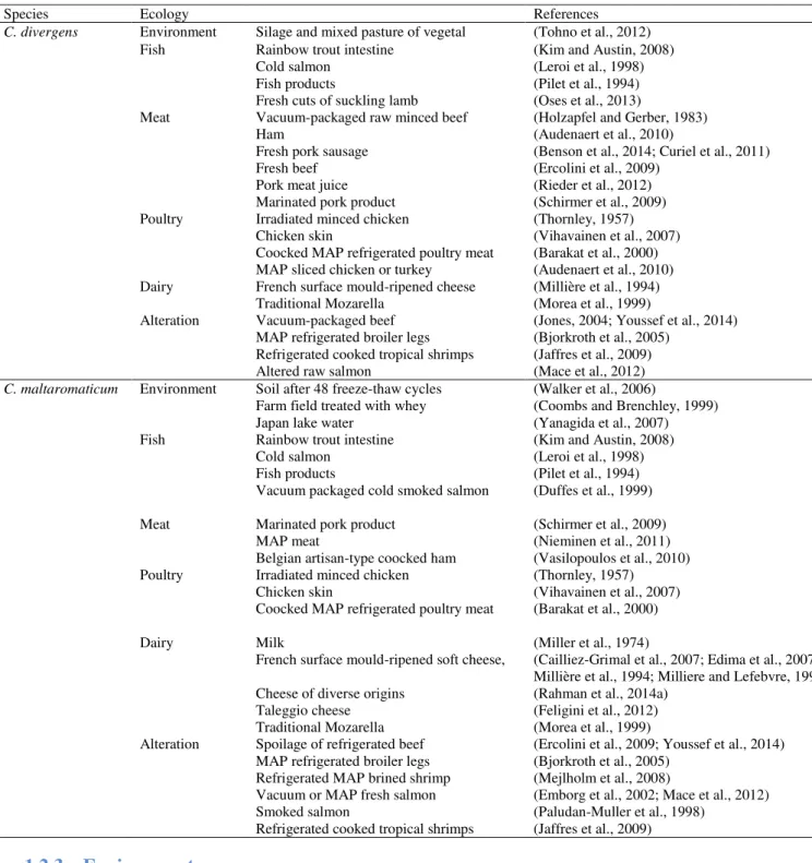
Carnobacteria are ubiquitous LAB which have been frequently isolated from cold and temperate environment and from animals and foods of animal origin such as seafood, meat and dairy products. Table 1 lists the carnobacteria species and Table 2 focuses on ecology of the two species C. divergens and C. maltaromaticum.
13 The wide distribution in nature is due to their particular physiology. Moreover, these bacteria produce several bacteriocins, which allow them to compete with other bacteria for colonization of the environment.
A potential pathogenicity is associated with some Carnobacterium species.
Table 1: Ecology of eight Carnobacterium sp.
Species Ecology References
C. alterfunditum Ace Lake (Franzmann et al., 1991)
MAP rough-head-grenadier (Matamoros et al., 2009)
C. funditum Ace Lake (Franzmann et al., 1991)
Hindgut region of Artic charr (Ringo et al., 2002) Ice from a fjord in Norway (Groudieva et al., 2004)
Cooked tropical shrimps (Jaffres et al., 2009; Matamoros et al., 2009) Marine sponge (Li and Liu, 2006)
Antartica (Loperena et al., 2012)
C. gallinarum Chicken meat. (Collins et al., 1987)
C. iners Littoral zone of an Antarctic lake (Snauwaert et al., 2013)
C. inhibens subsp. inhibens Gastrointestinal tract of Atlantic salmon (Joborn et al., 1999) Siberian permafrost (Nicholson et al., 2013)
C. inhibens subsp. gilichinskyi
Siberian permafrost (Nicholson et al., 2013)
C. jeotgali Traditional Korean fermented food
(shrimp) (Kim et al., 2009)
C. mobile Chicken meat (Collins et al., 1987)
Altered raw salmon (Mace et al., 2012)
C. pleistocenium Pleistocene ice from the permafrost
tunnel
(Pikuta et al., 2005)
C. viridans Siberian permafrost (Nicholson et al., 2013) Spent mushroom compost (Ntougias et al., 2004) Antic toundra soil (Kim et al., 2013) Refrigerated vacuum-packaged bologna (Holley et al., 2002)
14
Table 2: Ecology of Carnobacterium divergens and Carnobacterium maltaromaticum.
Species Ecology References
C. divergens Environment Silage and mixed pasture of vegetal (Tohno et al., 2012)
Fish Rainbow trout intestine (Kim and Austin, 2008) Cold salmon (Leroi et al., 1998) Fish products (Pilet et al., 1994) Fresh cuts of suckling lamb (Oses et al., 2013)
Meat Vacuum-packaged raw minced beef (Holzapfel and Gerber, 1983) Ham (Audenaert et al., 2010)
Fresh pork sausage (Benson et al., 2014; Curiel et al., 2011) Fresh beef (Ercolini et al., 2009)
Pork meat juice (Rieder et al., 2012) Marinated pork product (Schirmer et al., 2009) Poultry Irradiated minced chicken (Thornley, 1957)
Chicken skin (Vihavainen et al., 2007) Coocked MAP refrigerated poultry meat (Barakat et al., 2000) MAP sliced chicken or turkey (Audenaert et al., 2010) Dairy French surface mould-ripened cheese (Millière et al., 1994)
Traditional Mozarella (Morea et al., 1999)
Alteration Vacuum-packaged beef (Jones, 2004; Youssef et al., 2014) MAP refrigerated broiler legs (Bjorkroth et al., 2005)
Refrigerated cooked tropical shrimps (Jaffres et al., 2009) Altered raw salmon (Mace et al., 2012)
C. maltaromaticum Environment Soil after 48 freeze-thaw cycles (Walker et al., 2006)
Farm field treated with whey (Coombs and Brenchley, 1999) Japan lake water (Yanagida et al., 2007) Fish Rainbow trout intestine (Kim and Austin, 2008)
Cold salmon (Leroi et al., 1998) Fish products (Pilet et al., 1994) Vacuum packaged cold smoked salmon (Duffes et al., 1999) Meat Marinated pork product (Schirmer et al., 2009)
MAP meat (Nieminen et al., 2011) Belgian artisan-type coocked ham (Vasilopoulos et al., 2010) Poultry Irradiated minced chicken (Thornley, 1957)
Chicken skin (Vihavainen et al., 2007) Coocked MAP refrigerated poultry meat (Barakat et al., 2000) Dairy Milk (Miller et al., 1974)
French surface mould-ripened soft cheese, (Cailliez-Grimal et al., 2007; Edima et al., 2007; Millière et al., 1994; Milliere and Lefebvre, 1994) Cheese of diverse origins (Rahman et al., 2014a)
Taleggio cheese (Feligini et al., 2012) Traditional Mozarella (Morea et al., 1999)
Alteration Spoilage of refrigerated beef (Ercolini et al., 2009; Youssef et al., 2014) MAP refrigerated broiler legs (Bjorkroth et al., 2005)
Refrigerated MAP brined shrimp (Mejlholm et al., 2008)
Vacuum or MAP fresh salmon (Emborg et al., 2002; Mace et al., 2012) Smoked salmon (Paludan-Muller et al., 1998)
Refrigerated cooked tropical shrimps (Jaffres et al., 2009)
1.2.3. Environment
Cold environment
Carnobacteria are mesophilic psychotolerant species able to grow at 0-28°C and this psychrotolerance could explain their distribution in cold natural environments. Some
Carnobacterium sp. are remarkably resistant to freezing. In fact, a Carnobacterium sp., closed
15 Some strains are able to grow under low temperature (0°C), low pressure (7 mbar) and anoxic CO2-dominated atmosphere (Nicholson et al., 2015).
Three species were initially isolated from cold environment with low nutrients contents in Alaska. Carnobacterium funditum and C. alterfunditum were isolated from the water of Ace Lake (Franzmann et al., 1991) and C. pleistocenium from Pleistocene ice from the permafrost tunnel (Pikuta et al., 2005). Carnobacterium funditum was also isolated from ice taken from a fjord in Norway (Groudieva et al., 2004), in Antartica (Loperena et al., 2012), and six Carnobacterium strains were isolated from the Siberian permafrost. Five of these strains were closely related to C. inhibens and one isolate to C. viridans (Nicholson et al., 2013).
Carnobacterium iners was isolated from a microbial mat actively growing in the
littoral zone of an Antarctic lake (Forlidas Pond) in the Pensacola mountain (Snauwaert et al., 2013). Carnobacterium sp. are present in Lake Vanda, a permanently ice-covered Antartic lake (Bratina et al., 1998), in deep surface sediments (Newberry et al., 2004). Several new strains have been identified in cold-preserved tissue sample from a Siberian baby mammoth (Pikuta et al., 2011). However, the culture origin is not clear and their presence could be due to contamination during transport (Pikuta and Hoover, 2014).
Terrestrial environment
Some Carnobacterium strains closed to C. maltaromaticum are present in terrestrial environment such as a farm field treated with whey (Coombs and Brenchley, 1999).
Carnobacterium strains closed to C. viridans are part of the Anctic toundra soil (Kim et al.,
2013) and of Spent mushroom compost, composed of thermally treated cereal straw and animal manure mixture colonized by fungi (Ntougias et al., 2004). Carnobacterium sp. are one of the dominant strains found in biogas slurry compost and cow manure compost (Hong-yan et al., 2013). Carnobacterium is also present in freshwater habitats such as lake water in Japan (Yanagida et al., 2007).
16 Vegetal
Carnobacterium divergens is part of LAB strains isolated from mixed pasture of
timothy (Phleum pratense) and orchadgrass (Dactylis glomerata) and its badly preserved silages (Table 2) (Tohno et al., 2012).
Carnobacterium funditum is present in the bacterial community of marine sponge, Craniella australiensis (Li and Liu, 2006) and a Carnobacterium sp. was isolated from a
Sphagnum pond (Leisner et al., 2007).
Recently, a rhizobacterial isolate, Carnobacterium sp. SJ-5 (Jain and Choudhary, 2014) was characterized and it was demonstrated that it protected soybean against infection by
Fusarium oxysporum.
Animals and products of animal origin
For years, the presence of Carnobacterium in foods was underreported due to the inability of these bacteria to grow in acetate containing media, such as Rogosa or MRS agar, usually used for LAB numeration and screening. Some modifications and adaptation of conventional microbial techniques (Edima et al., 2007; Holzapfel, 1992; Millière et al., 1994; Wasney et al., 2001) and development of molecular biology techniques adapted to detection of this genus (Brooks et al., 1992; Cailliez-Grimal et al., 2005; Connil et al., 1998; Nissen et al., 1994; Rachman et al., 2004), led to the detection and isolation of these bacteria in a great variety of products.
Due to their ability to grow at refrigerated temperatures and survive at elevated levels of CO2 in the modified atmospheres, they can proliferate in multiple refrigerated food
ecosystems and they frequently dominate in modified atmosphere-packed (MAP) products. Their presence and roles have been extensively studied these last years.
Fish and seafood
Carnobacterium are present in sea and fresh water and are usually found in the normal
intestinal microbiota of fish (Pilet and Leroi, 2011). Carnobacterium inhibens subsp. inhibens was first isolated from the gastrointestinal tract of Atlantic salmon (Joborn et al., 1999).
17
Salvelinus alpinus L. (Ringo et al., 2002) and C. maltaromaticum and C. divergens from the
rainbow trout intestine (Kim and Austin, 2008).
Carnobacterium funditum is present in the microbial ecosystem of cooked tropical
shrimps (Jaffres et al., 2009; Matamoros et al., 2009) and one C. alterfunditum strain was isolated in MAP rough-head-grenadier (Matamoros et al., 2009).
Carnobacterium maltaromaticum and C. divergens are the predominant Carnobacterium species isolated from seafoods and eventually dominate the LAB in cold
salmon (Leroi et al., 1998). Carnobacterium maltaromaticum was frequently isolated from different seafoods including fish products (Pilet et al., 1994), vacuum packaged cold smoked salmon (Duffes et al., 1999), and C. divergens was isolated from fish products (Pilet et al., 1994) and fresh cuts of suckling lamb (Oses et al., 2013).
Carnobacterium maltaromaticum is the only Carnobacterium species found in altered
refrigerated MAP brined shrimp (Mejlholm et al., 2008), vacuum or MAP fresh (Emborg et al., 2002) or smoked salmon (Paludan-Muller et al., 1998), and is present with C. divergens in altered refrigerated products such as cooked tropical shrimps (Jaffres et al., 2009) or altered raw salmon (Mace et al., 2012).
Carnobacterium jeotgali was reported to be present in a traditional Korean fermented
food, jeotgal, made with freshwater shrimp and salt (Kim et al., 2009).
Meat and meat products
No scientific report indicates the presence of Carnobacterium species in the gastro-intestinal or in the skin of animals. Leisner et al., (Leisner et al., 2007) suggested that the source of carnobacteria in meat products is most probably the processing plant. However, DNA from
C. maltaromaticum was found in the faeces of polar bears, cheetahs and humans and DNA from
other Carnobacterium species, including C. divergens, was found in human infant faeces and in cow rumen (Rahman, 2013).
Meats, meat products and poultry are substrates rich in proteins with water activity and neutral pH favorable for the growth of carnobacteria, reaching high levels (106-108 cfu.g1). They
are found in vacuum packaged meat and related products stored at low temperatures.
Carnobacterium viridans could be responsible of discoloration of refrigerated
18
Carnobacterium divergens was initially isolated from vacuum-packaged raw minced
beef (Holzapfel and Gerber, 1983) and was further detected in other meats such as ham (Audenaert et al., 2010), fresh pork sausage (Benson et al., 2014; Curiel et al., 2011), beef (Ercolini et al., 2011, 2009), pork meat juice (Rieder et al., 2012), marinated pork product (Schirmer et al., 2009) and has been associated with alteration of vacuum-packaged beef (Jones, 2004; Youssef et al., 2014). Carnobacterium maltaromaticum is also associated with marinated pork product (Schirmer et al., 2009), MAP meat (Nieminen et al., 2011), spoilage microbiota of Belgian artisan-type cooked ham refrigerated at 7°C for four weeks (Vasilopoulos et al., 2010) or refrigerated beef where it can contribute to meat spoilage (Ercolini et al., 2009; Youssef et al., 2014). These two strains dominate among the spoilage LAB of MAP refrigerated broiler legs (Bjorkroth et al., 2005).
Poultry
Thornley in 1957 has isolated atypical Lactobacillus strains from irradiated minced chicken. These strains were identified as C. maltaromaticum and C. divergens in 1987 (Collins et al., 1987).
Carnobacterium maltaromaticum and C. divergens are the two most abundant Carnobacterium species identified in poultry and in poultry foods. These two species are
present in the chicken skin in three manufacturers (Vihavainen et al., 2007) or in coocked MAP refrigerated poultry meat (Barakat et al., 2000). Carnobacterium maltaromaticum is also detected in the plant environment. This suggests that it was the origin of the chicken contamination. Carnobacterium divergens is detected in MAP sliced chicken or turkey (Audenaert et al., 2010).
19 Dairy product
Association of Carnobacterium with milk is very old. In 1974, the presence of C.
maltaromaticum in milk is associated with a distinct malty or chocolate like flavor and aroma
which correspond to the presence of aldehydes (Miller et al., 1974).
The presence of C. maltaromaticum in Brie cheeses, a variety of AOP (protected designation of origin) French surface mould-ripened soft cheese, was reported for the first time in 1994 (Millière et al., 1994; Milliere and Lefebvre, 1994). Out of 30 French soft-ripened or red-smear cheeses made from cow’s, ewe’s or goat’s milk (raw or pasteurized), 10 contains this species (Edima et al., 2007). This species was also isolated from cheese of diverse origins (Rahman et al., 2014a) and associated with Taleggio cheese (Feligini et al., 2012). Two species,
C. divergens and C. maltaromaticum is present in the dominant bacterial community of
traditional Mozarella (Morea et al., 1999). Carnobacterium divergens is also isolated in French surface mould-ripened cheese (Millière et al., 1994).
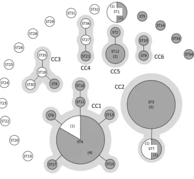
Figure 3: eBURST analysis of 47 C. maltaromaticum strains (Rahman et al., 2014).
Each sequence type ST is represented by a circle, the size of the circle is proportional to the number of strains belonging to a given ST. The number of strains is also indicated in brackets when the ST is represented by more than one strain. Dairy and non-dairy strains are represented by dark grey and white circles, respectively. Solid lines connect SLVs (Single Locus Variants) and broken lines connect DLVs (Double Locus Variants). Clonal Complexes (CC) are indicated in light grey.
20 Although Carnobacterium sp. can be isolated from diverse environments, little is known about their population structure. An MLST scheme based on the analysis of fragments of the genes dapE, ddlA, glpQ, ilvE, pyc, pyrE, and leuS was applied to a collection of 47 C.
maltaromaticum strains of diverse origins (Figure 3) (Rahman et al., 2014a). The scheme
allowed detecting 36 sequence types (STs) in the collection with eighty percent of the STs represented by a unique strain. Among the remaining STs, several are represented by strains from very diverse biotopes. Besides, eBURST analysis revealed 15 singletons and six clonal complexes. The two major clonal complexes CC1 and CC2 contained 11 and 7 strains, and connected 7 and 2 STs, respectively. The whole population is clustered in four deeply branched lineages, in which the dairy strains were spread.
Pathogenicity
There are some reports in the literature that consider Carnobacterium species as a fish pathogen, and C. maltaromaticum is usually associated with fish disease. In 1984, Hiu et al., (Hiu et al., 1984) proposed the name Lactobacillus piscicola for 17 strains isolated from diseased rainbow trout (Salmo gairdneri) and diseased cutthroat trout (Salmon clarki) (Michel et al., 1986). Some C. maltaromaticum strains were associated with mortalities of cultured striped bass and channel catfish (Baya et al., 1991) and caused important mortalities in market-size rainbow trout (Toranzo et al., 1993). More recently, Carnobacterium sp. was isolated from Chinook salmon from Michigan Lake affected with bacterial infections (Loch et al., 2012) and
C. maltaromaticum seemed to be a significant cause of morbidity and mortality in juvenile
salmon shark due to brain infection (Schaffer et al., 2013). The virulence often appears to be low and stressed fish more susceptible (Leisner et al., 2012). Carnobacterium maltaromaticum seems to be a common opportunistic pathogen (Michel et al., 1986).
Carnobacterium maltaromaticum was isolated from a case of multi-bacterial
synergistic gangrene (Xu et al., 1997) and from the pus from traumatic amputation of the right hand (Chmelar et al., 2002). Carnobacterium sp. was also isolated from human blood culture (Hoenigl et al., 2010). However, the causal association with the clinical symptoms remains uncertain. The patient might have become infected through a micro-lesion in his hand while consuming raw or cooked fish (Hoenigl et al., 2010).
21 No Carnobacterium species were direct human pathogen. Their optimal growth temperature below 36.6 °C and growth maxima within 28-40 °C suggest that these bacteria are not pathogenic for warm-blood animals and humans (Pikuta and Hoover, 2014).
The presence of virulence factors in carnobacteria is not well documented.
Carnobacterium viridans showed -haemolytic activity on sheep blood agar (Holley et al.,
2002). The presence of putative virulence genes encoding products involved in adhesion, capsule synthesis, haemolysis invasion and resistance to toxic compounds were identified in the genome of C. maltaromaticum ATCC 35586, isolated from a disease salmon (Leisner et al., 2012).
1.2.4. Carnobacterium : positive or negative flora ?
The presence of Carnobacterium in foods resulted in an increased number of scientific investigations to study their role and effect on these products. Among the 11 species of
Carnobacterium genus, only C. maltaromaticum and C. divergens are frequently isolated from
foods. They can be present as natural flora and/or as spoilers under specific storage conditions. The question was asked by Laursen et al., (Laursen et al., 2005), C. divergens and C.
maltaromaticum as spoilers or protective cultures in meat and seafood? Leisner et al., in 2007
(Leisner et al., 2007) noted that Carnobacterium can have positive or negative effects in the environment and in foods. However, other authors underlined its positive technological role in cheese making (Afzal et al., 2010).
Spoilers in meat and seafood
Carnobacteria are frequently associated with food spoilage, but spoilage activity shows intraspecies and interspecies variation (Leisner et al., 2007), and depends on the product.
Carnobacterium divergens and C. maltaromaticum are involved in the spoilage of
meat and seafoods products (Table 1 and 2). They can dominate the spoilage microbiota, particularly in refrigerated food packaged under modified or vacuum atmosphere (Remenant et al., 2015). These bacteria cause sensory spoilage due to the production of volatile compounds (Laursen et al., 2006; Leisner et al., 2007). In refrigerated meat, the alteration caused by C.
maltaromaticum is due to production of volatile organic compounds, aldehydes, lactones and
22 produce 2,3-butanedione and 2,3-pentanedione, substances which give off a strong butter odor and are not unpleasant (Joffraud et al., 2001).
Some reports noticed the production of biogenic amine by Carnobacterium, but the level of tyramine produced varies depending on the food, the environmental conditions and the strains (Leisner et al., 2007; Masson et al., 1996). Carnobacterium divergens strains are able to produce tyramine in meat (Curiel et al., 2011; Masson et al., 1999) or seafood product (Laursen et al., 2006). Carnobacterium maltaromaticum produces tyramine, detected at the end of the shelf life, in fresh and MAP salmon (Emborg et al., 2002). However, histamine or tyramine are not produced by C. alterfunditum in seafood products (Matamoros et al., 2009) or in cheese artificially contaminated with C. maltaromaticum LMA28 (Edima et al., 2007).
In conclusion, the presence of Carnobacterium strains can be sometimes associated with spoilage of seafood products, but in many cases they are not directly responsible for off-flavors (Pilet and Leroi, 2011).
Positive role in dairy products
Carnobacterium maltaromaticum is the predominant Carnobacterium species isolated
in dairy products, and has a positive role (Afzal et al., 2010). This strain did not cause off-flavours and could possibly even play a beneficial role in texture and taste during cheese ripening (Afzal et al., 2010; Millière et al., 1994; Milliere and Lefebvre, 1994). In cheeses tested, C. maltaromaticum constituted the main psychrotrophic LAB flora. Its growth was able to continue even at an alkaline pH value, reaching high levels (108–109 cfu.g−1) at the end of a
further cold (4 °C) storage period (Cailliez-Grimal et al., 2007). This species constitutes 70% of the curd of Mozzarella cheese and is a citrate-fermenting member of the microflora involved in fermentation (Morea et al., 1999). Carnobacterium maltaromaticum LMA28 is a slow acidifying species compared to commercial starter LAB, such as L. lactis or S. thermophilus (Edima et al., 2008) but can sustain low pH values in co-culture with these starters (Edima et al., 2008). Because of their slow acidifying activity, Carnobacterium strains cannot be used as a starter and can therefore be considered as NS (Non Starter) LAB. Carnobacterium
maltaromaticum produces α-ketoisocaproic acid, 3-methylbutanal and 3-methylbutanol from
leucine, 3-methylbutanoic acid and methylbutanal from isoleucine, and methylbutanol, 2-methylpropanal and 2-methylpropanol from valine (Afzal et al., 2013b, 2012). It has been
23 suggested that these aldehydes convey the malty aroma that is the characteristic of
C. maltaromaticum in milk.
Protectrice culture
The genus Carnobacterium is well known for its ability to produces bacteriocins. Several
Carnobacterium strains are known to produce anti-Listeria bacteriocins (Drider et al., 2006).
Six bacteriocins belonging to class IIa, IIc and one cyclic, have been described for different
C. maltaromaticum strains (Afzal et al., 2010).
Carnobacterium strains have been extensively studied for their role as protective flora
against Listeria monocytogenes in dairy, meat or fish foods (reviewed by (Leisner et al., 2007)) and cold smoked salmon (review by (Pilet and Leroi, 2011)).
Potential probiotic bacteria?
Carnobacterium maltaromaticum and C. divergens are well known as a normal flora of fish
intestine and induce the expression of genes encoding IL-1b and TNF- of head kidney leucocytes of rainbow trout suggesting that these bacteria can stimulate the innate immunity of this fish species (Kim and Austin, 2008). In addition, feeding rainbow trout with these bacteria enhances the cellular and humoral immune response in rainbow trout (Kim and Austin, 2008).
Carnobacterium maltaromaticum LMA28 was able to survive during the transit through the
digestive tract in a murin model and to interact with the host in a cell line model (Rahman et al., 2014b) suggesting that the immunomodulatory properties of Carnobacterium are not restricted to interaction with fish and could be extended to mammals.
24 Carnobacteria, and notably C. maltaromaticum, show environmental diversity. Cell Wall Proteins (CWP) are well-known to be responsible of the bacterial interaction with its environment. They are involved in various important processes (Bierne and Cossart, 2007; Burgain et al., 2014a, 2014b). Their characterization would permit to understand why
Carnobacterium are able to colonize such different environments.
2.
Bacterial Cell Wall Proteins
Gram-positive bacteria possess a cell wall composed of several components, i.e. peptidoglycan polymer, teichoic acids, lipoteichoic acids and proteins.
Proteins are synthetized in the cytoplasm and possess a signal peptide (SP) on their N-terminal in order to be addressed to the membrane (Blobel, 1980). The secretion through the cytoplasmic membrane is possible via several pathways. They are conserved within bacteria: secretion system (Sec), twin-arginine translocation (Tat), flagella export apparatus (FEA), frimbrilin-protein exporter (FPE), pore forming (holin), peptide-efflux ABC and WXG100 secretion system (Wss) (for reviews Desvaux et al., 2009; Driessen and Nouwen, 2008; Lee et al., 2006; van Wely et al., 2001).
The Sec system assures i) the secretion of unfolded proteins across the membrane and ii) the insertion of proteins into the cytoplasmic membrane (Luirink et al., 2005). Proteins are synthetized as precursors containing the maturation protein with an N-terminal SP essential for the signature of protein secretion. The SP has a tripartite structure including a positively charged N-terminus, a hydrophobic core and a neutral or negatively charged C-terminus containing SP cleavage site (von Heijne, 2001).
What differ the secreted proteins and the anchored proteins is the presence of a conserved domain on the C-terminal of the protein. This binding is possible through either the covalent or the non-covalent interactions with the peptidoglycan or secondary wall polymers such as teichoic acids. CWP display a variety of functions. Some are involved in structural support and movement, others in enzymatic activity, and others in interaction with the environment.
25
2.1. Proteins anchorage to the Cell Surface
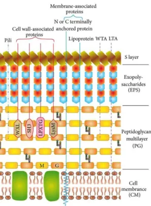
Several types of anchorage systems are used by Gram-positive bacteria for protein attachment to the cell surface. Each type is characterized by the presence of a conserved sequence. Some proteins are non-covalently associated with the cell wall containing GW modules, WxL or LysM domains. Others are designated to be covalently attached to the peptidoglycan layer by sortases via a sortase dependent motif, usually called the LPxTG motif (Desvaux et al., 2006; Scott and Barnett, 2006). Several proteins are anchored to the membrane by a hydrophobic tail (Sutcliffe and Harrington, 2002) and some are attached through unknown domains and believed to have cytoplasmic functions (Doyle, 2007; Schaumburg et al., 2004; Trost et al., 2005) (Figure 4).
Figure 4 : Cell envelope of lactobacilli with a schematic representation of cell wall and membrane-associated proteins. The Figure was adapted from (Kleerebezem et al., 2010; Sengupta et al., 2013).
The cell membrane (CM) with embedded proteins is covered by a multilayered peptidoglycan (PG) decorated with lipoteichoic acids (LTA), wall teichoic acids (WTA), pili, proteins, and lipoproteins. Exopolysaccharides (EPS) form a thick covering closely associated with PG and are surrounded by an outer envelope of S-layer proteins. The proteins are attached to the cell wall either covalently (LPxTG proteins) or noncovalently (exhibiting LysM, SH3, or WXL domains), lipid are anchored to the CM (lipoproteins) or attached to the CM via N- or C-terminal transmembrane helix. M: N-acetyl-muramic acid; G: N-acetyl-glucosamine.