via the Xps Type II Secretion System and Outer Membrane Vesicles
Magali Solé,* Felix Scheibner, Anne-Katrin Hoffmeister, Nadine Hartmann,* Gerd Hause,* Annekatrin Rother,* Michael Jordan, Martine Lautier,* Matthieu Arlat,* Daniela Büttner
Institute of Biology, Department of Genetics, Martin Luther University Halle-Wittenberg, Halle (Saale), Germany
ABSTRACT
Many plant-pathogenic bacteria utilize type II secretion (T2S) systems to secrete degradative enzymes into the extracellular milieu. T2S
sub-strates presumably mediate the degradation of plant cell wall components during the host-pathogen interaction and thus promote bacterial
virulence. Previously, the Xps-T2S system from Xanthomonas campestris pv. vesicatoria was shown to contribute to extracellular
protease activity and the secretion of a virulence-associated xylanase. The identities and functions of additional T2S substrates
from X. campestris pv. vesicatoria, however, are still unknown. In the present study, the analysis of 25 candidate proteins from
X. campestris pv. vesicatoria led to the identification of two type II secreted predicted xylanases, a putative protease and a lipase
which was previously identified as a virulence factor of X. campestris pv. vesicatoria. Studies with mutant strains revealed that
the identified xylanases and the protease contribute to virulence and in planta growth of X. campestris pv. vesicatoria. When
analyzed in the related pathogen X. campestris pv. campestris, several T2S substrates from X. campestris pv. vesicatoria were
secreted independently of the T2S systems, presumably because of differences in the T2S substrate specificities of the two
patho-gens. Furthermore, in X. campestris pv. vesicatoria T2S mutants, secretion of T2S substrates was not completely absent,
suggest-ing the contribution of additional transport systems to protein secretion. In line with this hypothesis, T2S substrates were
de-tected in outer membrane vesicles, which were frequently observed for X. campestris pv. vesicatoria. We, therefore, propose that
extracellular virulence-associated enzymes from X. campestris pv. vesicatoria are targeted to the Xps-T2S system and to outer
membrane vesicles.
IMPORTANCE
The virulence of plant-pathogenic bacteria often depends on TS2 systems, which secrete degradative enzymes into the extracellular
milieu. T2S substrates are being studied in several plant-pathogenic bacteria, including Xanthomonas campestris pv. vesicatoria,
which causes bacterial spot disease in tomato and pepper. Here, we show that the T2S system from X. campestris pv. vesicatoria
se-cretes virulence-associated xylanases, a predicted protease, and a lipase. Secretion assays with the related pathogen X. campestris pv.
campestris revealed important differences in the T2S substrate specificities of the two pathogens. Furthermore, electron microscopy
showed that T2S substrates from X. campestris pv. vesicatoria are targeted to outer membrane vesicles (OMVs). Our results, therefore,
suggest that OMVs provide an alternative transport route for type II secreted extracellular enzymes.
M
any Gram-negative plant-pathogenic bacteria utilize
spe-cialized protein secretion systems to deliver virulence
fac-tors, including DNA or bacterial effector proteins, into plant cells
(
1
). The efficient trans-kingdom transport of bacterial effector
proteins is often hindered by the rigid plant cell wall, which mainly
consists of cellulose, hemicellulose, and pectin. Bacteria,
there-fore, often secrete cell wall-degrading enzymes, which contribute
to bacterial virulence and include, e.g., cellulases, xylanases,
po-lygalacturonases, and amylases (
2–6
). It is assumed that the
deg-radation of plant cell wall components by bacterial enzymes
facil-itates the acquisition of nutrients as well as the translocation of
virulence factors into the host cell (
7–14
).
Bacterial extracellular enzymes are often secreted by a type II
secretion (T2S) system, which is a major virulence factor of many
Gram-negative plant-pathogenic bacteria, including Ralstonia
so-lanacearum and species of Erwinia and Xanthomonas (
8
,
15–18
).
T2S is a two-step process that often requires the Sec system for
protein transport across the inner membrane (IM) (
18
,
19
).
Sec-dependent transport of proteins depends on an N-terminal signal
peptide with an average length of 20 amino acids that is usually
cleaved by a peptidase during or after Sec-dependent transport
(
20
,
21
). The subsequent delivery of T2S substrates across the
outer membrane (OM) is mediated by the T2S apparatus, which
consists of 12 to 15 proteins that are located in the IM, the
Received 28 April 2015 Accepted 19 June 2015 Accepted manuscript posted online 29 June 2015
Citation Solé M, Scheibner F, Hoffmeister A-K, Hartmann N, Hause G, Rother A, Jordan M, Lautier M, Arlat M, Büttner D. 2015. Xanthomonas campestris pv. vesicatoria secretes proteases and xylanases via the Xps type II secretion system and outer membrane vesicles. J Bacteriol 197:2879 –2893.doi:10.1128/JB.00322-15. Editor: P. J. Christie
Address correspondence to Daniela Büttner, daniela.buettner@genetik.uni-halle.de. M.S. and F.S. contributed equally to the work.
* Present address: Magali Solé, Federal Environment Agency Department IV 1.3, Pesticides Ecotoxicology and Environmental Risk Assessment, Dessau-Rosslau, Germany; Nadine Hartmann, La Jolla Institute for Allergy and Immunology, La Jolla, California, USA; Gerd Hause and Annekatrin Rother, Biocenter of the Martin Luther University Halle-Wittenberg, Halle (Saale), Germany; Martine Lautier and Matthieu Arlat, INRA, Laboratoire des Interactions Plantes-Microorganismes, UMR441, Castanet-Tolosan, France, and Université de Toulouse, Université Paul Sabatier, Toulouse, France.
Supplemental material for this article may be found athttp://dx.doi.org/10.1128 /JB.00322-15.
Copyright © 2015, American Society for Microbiology. All Rights Reserved.
doi:10.1128/JB.00322-15
on January 18, 2016 by INRA - France
http://jb.asm.org/
periplasm, and the OM (
18
,
19
,
22
). T2S substrates are recognized
in the periplasm by a yet-unknown signal and are presumably
pushed through the OM secretin channel by the continuous
as-sembly and disasas-sembly of a periplasmic pseudopilus (
18
,
19
).
T2S systems and their cognate substrates have been intensively
studied in several bacterial model organisms, including bacteria of
the genus Xanthomonas, which belong to the gamma subdivision
of proteobacteria and comprise economically important plant
pathogens (
23
,
24
). Many Xanthomonas spp. contain two T2S
sys-tems that are encoded by homologous xps and xcs gene clusters
(
25
). While Xcs-T2S systems appear to be dispensable for
viru-lence, a virulence function has been reported for Xps-T2S systems
(
7–9
,
11–14
,
26–29
). In agreement with their contribution to
vir-ulence, xps-T2S genes from Xanthomonas spp. are often activated
in planta and coregulated with genes encoding components of the
type III secretion (T3S) system (
8
,
10
,
13
,
14
,
30
,
31
). T3S systems
are essential pathogenicity factors of many Gram-negative
plant-and animal-pathogenic bacteria plant-and serve as delivery systems for
bacterial effector proteins into eukaryotic host cells (
32
). Given
the coregulation of T2S and T3S genes, it has been postulated that
the local degradation of the plant cell wall by T2S substrates
facil-itates the formation of the extracellular T3S pilus, which serves as
a transport channel for effector proteins to the host plasma
mem-brane (
8
,
33
). A contribution of the T2S system to T3S-mediated
effector protein delivery was previously shown for Xanthomonas
campestris pv. vesicatoria, which is the causal agent of bacterial
spot disease in pepper and tomato. The only known T2S substrate
from X. campestris pv. vesicatoria to date is the xylanase XCV0965,
which contributes to virulence and is required for the extracellular
xylanase activity of X. campestris pv. vesicatoria (
8
). In contrast,
additional tested candidate proteins with homology to type II
se-creted extracellular enzymes from other pathogens are sese-creted
T2S independently in X. campestris pv. vesicatoria (
8
). This
find-ing contradicts the anticipated conservation of T2S substrate
rep-ertoires and suggests pathogen-specific differences in T2S
sub-strate specificities, which might reflect bacterial adaptations to
certain environments or host plants.
The aim of the present study was the identification of type II
secreted virulence factors from X. campestris pv. vesicatoria and
the analysis of their potential contribution to the host-pathogen
interaction. Secretion assays with candidate T2S substrates led to
the identification of two type II secreted xylanases (XCV4358 and
XCV4360) and a putative protease (XCV3671) that contribute to
the virulence of X. campestris pv. vesicatoria. Furthermore, we
identified a previously described virulence-associated lipase
(XCV0536) as a substrate of the Xps-T2S system. Secretion of
several T2S substrates from X. campestris pv. vesicatoria was T2S
independent in the related pathogen Xanthomonas campestris pv.
campestris, suggesting differences in the T2S substrate specificities
of the two pathovars. Interestingly, immunoelectron microscopy
showed that type II secreted proteins from X. campestris pv.
vesi-catoria are also present in outer membrane vesicles (OMVs),
which might provide an alternative transport route for secreted
virulence factors across the OM.
MATERIALS AND METHODS
Bacterial strains and growth conditions. Bacterial strains and plasmids
used in this study are listed inTable 1. Escherichia coli cells were grown at 37°C in lysogeny broth (LB) medium. X. campestris pv. vesicatoria and X.
campestris pv. campestris strains were cultivated at 30°C in nutrient-yeast
extract-glycerol (NYG) medium (34). Plasmids were introduced into E.
coli by electroporation and into X. campestris pv. vesicatoria and X. camp-estris pv. campcamp-estris by electroporation or conjugation, using pRK2013 as
the helper plasmid in triparental matings (35). Antibiotics were added to the media at the following final concentrations: ampicillin, 100g ml⫺1; gentamicin, 15g ml⫺1; kanamycin, 25g ml⫺1; rifampin, 100g ml⫺1; spectinomycin, 100g ml⫺1.
Infection studies. For infection studies, X. campestris pv. vesicatoria
strains were inoculated with a needleless syringe into the intercellular spaces of leaves of the near-isogenic pepper lines Early Cal Wonder (ECW) and ECW-10R (36) at concentrations of 1⫻ 108CFU/ml in 1 mM
MgCl2if not stated otherwise. Disease symptoms were scored over a
pe-riod of 10 days postinoculation (dpi). For in planta growth curves, bacte-ria were inoculated at a density of 1⫻ 104CFU/ml into leaves of ECW
pepper plants. Bacterial growth was determined as described previously (37). Experiments were repeated at least twice.
Protease and xylanase activity assays. For the analysis of extracellular
protease and xylanase activities, bacteria were grown overnight in liquid NYG medium, adjusted to an optical density at 600 nm (OD600) of 1.0,
and incubated at 30°C for 1 to 2 days on NYG plates with 1% agar and containing 1% skimmed milk (AppliChem; for analysis of protease activ-ity) or 0.1% remazol brilliant blue (RBB) xylan (Sigma-Aldrich; for anal-ysis of xylanase activity) (38). Bacteria were inoculated into holes in the agar and removed before the documentation of the halos. Experiments were repeated at least twice.
Generation of expression constructs. Gene fragments were amplified
by PCR from X. campestris pv. vesicatoria strain 85-10 or X. campestris pv. campestris strain LMG 568 and cloned into plasmid pBRM in a one-step restriction/ligation reaction using the type IIS restriction enzyme BsaI and ligase (39). For fragments with an internal BsaI site, the reaction mixture was incubated with ligase for an additional 20 min at 37°C after heat inactivation (39). Alternatively, internal BsaI sites were removed by PCR using primers that introduced silent mutations. For the generation of a pBRM-P0536 construct, XCV0536 including a 530-bp upstream region was amplified by PCR and cloned into plasmid pBRM-P using BsaI and ligase. pBRM-P is a derivative of pBRM which lacks the lac promoter (Table 1). Similarly, XCV3671, XCV4358, and XCV4360 were amplified by PCR and cloned individually together with a 494-bp fragment span-ning the predicted promoter of XCV4361 into pBRM-P in a single restric-tion/ligation reaction. Alternatively, XCV3671, XCV4358, and XCV4360 were cloned into the Golden Gate-compatible suicide vector pLAND-P downstream of the predicted promoter of XCV4361 by using BsaI and ligase. To obtain an expression construct encoding XCV0536⌬2–29– c-Myc, the 530-bp upstream regions of XCV0536 and XCV0536 lacking codons 2 to 29 were amplified by PCR and cloned into pBRM-P in a single restriction/ligation reaction. All primer sequences are listed in Table S1 in the supplemental material.
Generation of deletion constructs. To delete XCV4358 and XCV4360
from the genome of strain 85-10, 700- to 800-bp flanking regions of both genes were amplified by PCR using genomic DNA from X. campestris pv. vesicatoria strain 85-10 as the template. The amplicons were introduced into the Golden Gate-compatible suicide vector pOGG2 in a single restric-tion/ligation reaction using BsaI and ligase. For the deletion of XCV3671, the first 265 and the last 268 codons of XCV3671 were amplified by PCR and cloned into the suicide vector pOGG2. The resulting construct con-tained a deletion of codons 266 to 360 of XCV3671 and an additional frameshift mutation after codon 266. Deletion constructs were intro-duced into the genome of X. campestris pv. vesicatoria strains by homol-ogous recombination as described previously (40). Double-crossover events resulted in the deletion mutant strains that are described inTable 1.
Secretion experiments and protein analysis. For in vitro T2S assays,
bacteria were grown overnight in NYG medium, resuspended at a cell density of 1.5⫻ 108CFU/ml, and incubated on a rotary shaker at 30°C for
1 h (41). Culture supernatants were separated from bacterial cells by fil-tration, and secreted proteins were precipitated by trichloroacetic acid as
on January 18, 2016 by INRA - France
http://jb.asm.org/
TABLE 1 Bacterial strains and plasmids used in this study
Strain or plasmid Relevant characteristic(s)a Reference or source Xanthomonas strains
85-10 X. campestris pv. vesicatoria wild-type strain; pepper race 2; Rifr 63
85* 85-10 derivative containing hrpG* 49
85-10⌬xpsE xpsE deletion mutant of strain 85-10 8
85-10⌬xpsD xpsD deletion mutant of strain 85-10 8
85-10⌬xcsD xcsD deletion mutant of strain 85-10 8
85-10⌬xpsD⌬xcsD xpsD xcsD double deletion mutant of strain 85-10 8
85-10⌬0965 Derivative of strain 85-10 with XCV0965 deleted 8
85-10⌬EEE Triple deletion mutant of strain 85-10 lacking xpsE, xcsE, and XCV4312 8
85-10⌬4358 Derivative of strain 85-10 with XCV4358 deleted This study 85-10⌬4360 Derivative of strain 85-10 with XCV4360 deleted This study 85-10⌬4358⌬4360 XCV4358 XCV4360 double deletion mutant of strain 85-10 This study 85-10⌬3671 Derivative of strain 85-10 with codons 266 to 360 of XCV3671 deleted and containing a
frameshift mutation after codon 265
This study
85-10⌬eps Derivative of strain 85-10 deficient in EPS production Bonas et al., unpublished LMG 568 X. campestris pv. campestris wild-type strain
LMG 568⌬xpsD Derivative of strain LMG 568 with xpsD deleted M. Lautier, unpublished LMG 568⌬xcsD Derivative of strain LMG 568 with xcsD deleted M. Lautier, unpublished LMG 568⌬xpsD⌬xcsD Derivative of strain LMG 568 with xpsD and xcsD deleted M. Lautier, unpublished
E. coli strains
DH5␣ F⫺recA hsdR17(rk⫺mk⫹)80dlacZ⌬M15 Bethesda Research Laboratories
DH5␣ pir F⫺recA hsdR17(rk⫺mk⫹)80dlacZ⌬M15 [pir] 64
Plasmids
pBluescript(II) KS Phagemid, pUC derivative; Apr Stratagene
pBRM Golden Gate-compatible derivative of pBBR1MCS-5, lacP, 3⫻ c-Myc epitope-encoding sequence; Gmr
8
pBRM-P Derivative of pBRM lacking lac promoter 8
pBRM0007 pBRM derivative encoding XCV0007–c-Myc This study pBRM0024 pBRM derivative encoding XCV0024–c-Myc This study pBRM0027 pBRM derivative encoding XCV0027–c-Myc This study pBRM0536 pBRM derivative encoding XCV0536–c-Myc This study pBRM-P0536 pBRM-P derivative encoding XCV0536–c-Myc under control of native promoter This study pBRM-P0536⌬2–29 pBRM-P derivative encoding XCV0536⌬2–29–c-Myc under control of native promoter This study pBRM0583 pBRM derivative encoding XCV0583–c-Myc This study pBRM0670 pBRM derivative encoding XCV0670–c-Myc This study pBRM0673 pBRM derivative encoding XCV0673–c-Myc This study pBRM0730 pBRM derivative encoding XCV0730–c-Myc This study pBRM0805 pBRM derivative encoding XCV0805–c-Myc This study pBRM0845 pBRM derivative encoding XCV0845–c-Myc This study pBRM0889 pBRM derivative encoding XCV0889–c-Myc This study pBRM0961 pBRM derivative encoding XCV0961–c-Myc This study pBRM0965⌬2–24 pBRM derivative encoding XCV0965⌬2–24–c-Myc This study pBRM1033 pBRM derivative encoding XCV1033–c-Myc This study pBRM1064 pBRM derivative encoding XCV1064–c-Myc This study pBRM1075 pBRM derivative encoding XCV1075–c-Myc This study pBRM1202 pBRM derivative encoding XCV1202–c-Myc This study pBRM1311 pBRM derivative encoding XCV1311–c-Myc This study pBRM1335 pBRM derivative encoding XCV1335–c-Myc This study pBRM1372 pBRM derivative encoding XCV1372–c-Myc This study pBRM2747 pBRM derivative encoding XCV2747–c-Myc This study pBRM2993 pBRM derivative encoding XCV2993–c-Myc This study pBRM3283 pBRM derivative encoding XCV3283–c-Myc This study pBRM3425 pBRM derivative encoding XCV3425–c-Myc This study pBRM3639 pBRM derivative encoding XCV3639–c-Myc This study pBRM3671 pBRM derivative encoding XCV3671–c-Myc This study pBRM-P3671 pBRM-P derivative encoding XCV3671–c-Myc under control of XCV4361 promoter This study pBRM4096 pBRM derivative encoding XCV4096–c-Myc This study pBRM4355 pBRM derivative encoding XCV4355–c-Myc This study pBRM4358 pBRM derivative encoding XCV4358–c-Myc This study pBRM4358⌬2–22 pBRM derivative encoding XCV4358⌬2–22-c-Myc This study
(Continued on following page)
on January 18, 2016 by INRA - France
http://jb.asm.org/
described previously (41). Equal protein amounts of bacterial cell extracts and culture supernatants (adjusted according to cell density) were ana-lyzed by SDS-PAGE and immunoblotting using an antibody specific for the c-Myc epitope (Roche) or a polyclonal XopA-specific antibody (42). Secondary antibodies were horseradish peroxidase-labeled anti-mouse or anti-rabbit antibodies (Amersham Pharmacia Biotech). Antibody reac-tions were visualized by enhanced chemiluminescence. Experiments were repeated at least twice.
For experiments with proteinase K, bacteria were resuspended at a cell density of 1.5⫻ 108CFU/ml in NYG medium and incubated on a rotary
shaker at 30°C for 1 h or overnight. Two-milliliter aliquots of bacterial culture supernatants were incubated with 200g proteinase K (Thermo Scientific) for 2 h at 37°C. As a control, supernatants were incubated without proteinase K for 2 h on ice or at 37°C. To degrade cellular pro-teins, bacteria were grown overnight in 200 ml NYG medium, and the cell pellet after centrifugation was dissolved in 3 ml 1⫻ phosphate-buffered saline (PBS) and lysed with a French press. Ten microliters of a 1:10 dilution of the cell lysate was incubated with 1g proteinase K for 2 h at 37°C. Proteins were analyzed by SDS-PAGE and immunoblotting using c-Myc epitope-specific antibodies. Experiments were repeated at least twice.
RNA analysis. For transcript analyses via quantitative reverse
tran-scription-PCR (qRT-PCR), bacteria were grown overnight in NYG me-dium, adjusted to an OD600of 0.15, and incubated at 30°C until the
culture reached an OD600of 0.6 to 0.8. RNA was extracted using a
TRIzol-based protocol, and cDNA was synthesized using 2g RNA in a RevertAid H Minus first-strand cDNA synthesis kit (Fermentas). XCV0536, XCV4358, XCV4360, XCV3671, and 16S rRNA were amplified with a CFX Connect real-time PCR detection system (Bio-Rad) using Absolute Blue SYBR green fluorescein mix (Thermo Scientific) and a 1:100 dilution of cDNA solution. The efficiency of each PCR was determined by standard curves based on dilution series of template and melting curve analysis. Mean transcript levels were determined based on values obtained from technical triplicates and the levels of constitutively expressed 16S rRNA transcripts as described elsewhere (ABI user bulletin 2; Applied Biosys-tems). Individual experiments were performed with three different RNA preparations for each strain and were repeated twice with similar results.
Immunoelectron microscopy. For immunoelectron microscopy,
bacteria were cultivated overnight at 30°C in NYG medium and rapidly frozen with a high-pressure freeze fixation apparatus (HPM 010; BAL-TEC, Liechtenstein). The material was cryosubstituted with 0.25% glutar-aldehyde (Sigma) and 0.1% uranyl acetate (Chemapol) in acetone for 2 days at⫺80°C using cryosubstitution equipment (FSU; BAL-TEC) and embedded in HM20 (Polysciences Europe) at⫺20°C. For immunolabel-ing, ultrathin sections (80 nm) were treated with a monoclonal c-Myc epitope-specific antibody (generous gift of U. Conrad) and a secondary anti-mouse antibody conjugated with 10-nm gold particles (Sigma-Al-drich). The sections were poststained with uranyl acetate and lead citrate in an EM-Stain apparatus (Leica) and subsequently observed with a Zeiss Libra 120 tranmission electron microscope (TEM) operating at 120 kV (Carl Zeiss Microscopy). Images were obtained as described above.
For negative staining of isolated OMVs, 4-l aliquots of isolated OMVs were placed on copper grids covered with a Formvar film for 1 min. Excess liquid was removed with a filter paper, and the grids were air dried for 10 min, washed three times with H2O, and stained with 2% aqueous
uranyl acetate solution. The samples were inspected with an EM 900 TEM (Zeiss SMT). Micrographs were taken with an slow-scan charge-coupled-device SM-1k-120 camera (TRS). For the analysis of OMV formation in
planta, infected plant tissue was analyzed as described previously (43).
Isolation of OMVs. For the isolation of OMVs, bacteria were
incu-bated overnight in 10 ml NYG medium at 30°C. At an OD600of 0.8 to 1.0,
cells were pelleted by centrifugation (10,000⫻ g for 10 min at 4°C), and culture supernatants were passed through a filter with a pore size of 0.45 m and subsequently through a filter with a pore size of 0.20 m to remove residual cells. Filtrates were centrifuged at 38,000⫻ g for 3 h at 4°C, supernatants were removed, and the samples were centrifuged again at 41,000⫻ g for 1 h at 4°C. The pellet was resuspended in PBS and centrifuged at 100,000⫻ g for 6 h at 4°C. After removal of the supernatant, the pellet was resuspended in 60l PBS and analyzed by immunoblotting and electron microscopy.
RESULTS
Secretion assays with T2S candidate substrates from X.
campes-tris pv. vesicatoria strain 85-10. To identify type II secreted
viru-TABLE 1 (Continued)Strain or plasmid Relevant characteristic(s)a Reference or source
pBRM-P4358 pBRM-P derivative encoding XCV4358–c-Myc under control of XCV4361 promoter This study pBRM4360 pBRM derivative encoding XCV4360–c-Myc This study pBRM-P4360 pBRM-P derivative encoding XCV4360–c-Myc under control of XCV4361 promoter This study pBRM4437 pBRM derivative encoding XCV4437–c-Myc This study pBRMhrpB1 pBRM derivative encoding HrpB1–c-Myc 45
pBRMhrpB1Stop pBRM derivative encoding HrpB1 45
pBRMXCC0857 pBRM derivative encoding XCC0857–c-Myc 47
pBRMXCC4115 pBRM derivative encoding XCC4115–c-Myc 47
pBRMXCC4118 pBRM derivative encoding XCC4118–c-Myc 47
pBRMXc0705 pBRM derivative encoding Xc0705–c-Myc (PghAxc-c-Myc) 8
pBRMXc1849 pBRM derivative encoding Xc1849–c-Myc (PghBxc-c-Myc) 8
pBRMxopJ pBRM derivative encoding XopJ–c-Myc D. Büttner, unpublished pDGW0965 pDGW4 M derivative encoding XCV0965–c-Myc 8
pDSK602 Broad-host-range vector; contains triple lacUV5 promoter; Spr 65
pDSK604 Derivative of pDSK602 with modified polylinker 66
pDxpsE pDSK604 derivative carrying xpsE This study pOK1 Suicide vector; sacB sacQ mobRK2 oriR6K; Spr 40
pOGG2 Golden Gate-compatible derivative of pOK1 52
pOGG4358 Derivative of pOGG2 containing flanking regions of XCV4358 This study pOGG4360 Derivative of pOGG2 containing flanking regions of XCV4360 This study pOGG3671 Derivative of pOGG2 containing first 265 and last 268 codons of XCV3671 This study pRK2013 ColE1 replicon; TraRK⫹Mob⫹Kmr 35
pUC119 ColE1 replicon; Apr 67
aAp, ampicillin; Gm, gentamicin; Km, kanamycin; Rif, rifampin; Sp, spectinomycin; r, resistant.
on January 18, 2016 by INRA - France
http://jb.asm.org/
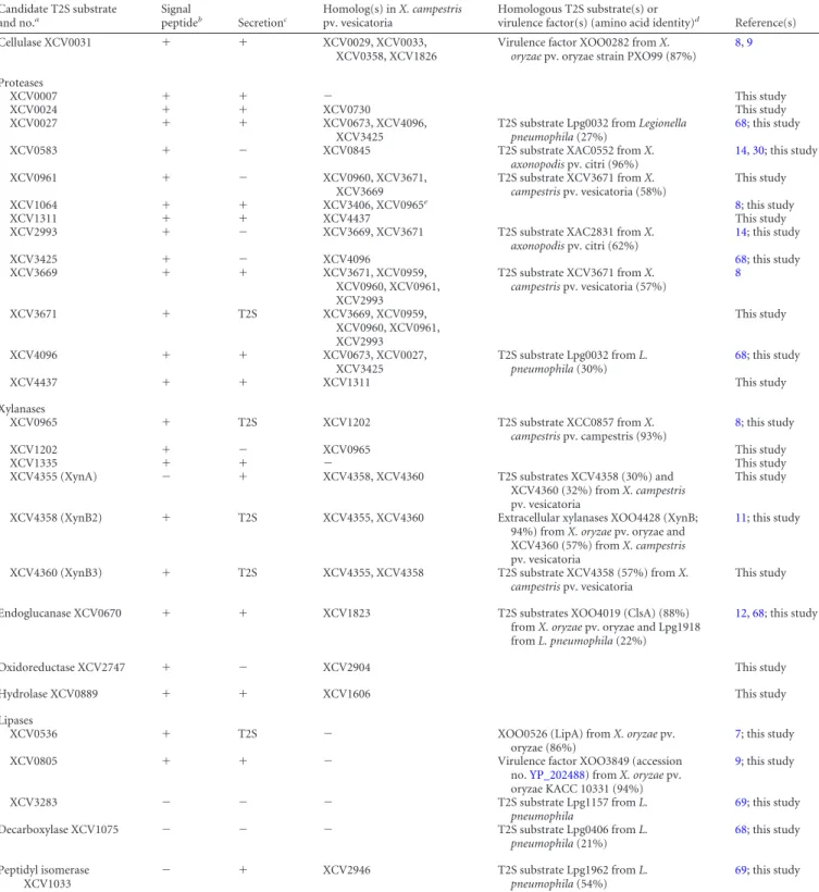
lence factors from X. campestris pv. vesicatoria strain 85-10, we
analyzed the secretion of 25 individual candidate substrates that
were mainly selected based on homologies to known T2S
sub-strates and/or virulence factors from Xanthomonas spp. (
Table 2
).
The corresponding genes were expressed in fusions with a
C-ter-minal triple c-Myc epitope-encoding sequence in X. campestris pv.
vesicatoria strain 85-10 and the T2S mutant 85-10⌬EEE, from
which the putative ATPase-encoding genes of the xps- and xcs-T2S
clusters and the homologous XCV4312 gene had been deleted (
8
).
The analysis of bacterial culture supernatants by immunoblotting
revealed that eight of the C-terminally c-Myc epitope-tagged
pro-teins were not, or were not efficiently, secreted by the wild-type
and the T2S mutant strain when the bacteria were incubated in
complex NYG medium (
Table 2
; see also Fig. S1 in the
supplemen-tal material). However, a negative influence of the C-terminal
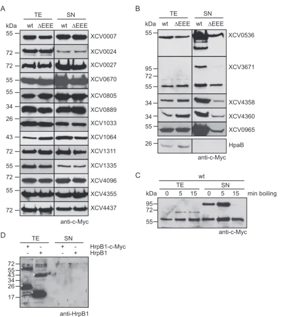
c-Myc epitope on secretion cannot be excluded. Seventeen proteins
were secreted, including putative proteases, xylanases, and the
lipase XCV0536 (also referred to as LipA), which was previously
identified as a virulence factor of X. campestris pv. vesicatoria (
44
)
(
Fig. 1A
and
B
). Reduced amounts of XCV0536 – c-Myc, the
pu-tative protease XCV3671– c-Myc, and the predicted xylanases
XCV4358 – c-Myc and XCV4360 – c-Myc were present in the
cul-ture supernatant of the T2S mutant strain 85-10⌬EEE compared
with strain 85-10, suggesting that the efficient secretion of these
proteins depends on the T2S systems (
Fig. 1B
). A similar finding
was observed for the previously identified T2S substrate XCV0965
when the blot was overexposed (
Fig. 1B
). We observed a size shift
for XCV3671– c-Myc, which has a molecular mass of
approxi-mately 70 kDa. The predominant signal detected by the c-Myc
epitope-specific antibody corresponded to a protein of
approxi-mately 55 kDa and thus presumably to a degradation product of
XCV3671– c-Myc. However, an additional signal at
approxi-mately 100 kDa was detected in the culture supernatants (
Fig. 1B
).
When the samples were boiled for 15 min instead of 5 min, the
intensity of this signal decreased (
Fig. 1C
). Notably, we previously
observed a similar size shift for the secreted predicted proteases
XCV0959 and XCV3669, which share sequence similarities with
XCV3671 (
Table 2
) (
8
).
The detection of proteins in the culture supernatants was
pre-sumably not caused by cell lysis or a general release of periplasmic
proteins into the extracellular medium, because the cytoplasmic
T3S chaperone HpaB– c-Myc and the periplasmic HrpB1 protein,
which were analyzed as a C-terminally c-Myc epitope-tagged
pro-tein and an untagged propro-tein, respectively, were detected in the
cell extracts but not in the culture supernatants (
Fig. 1B
and
D
)
(
45
,
46
). Furthermore, the detection of proteins in the culture
supernatants did not result from unspecific binding of the c-Myc
epitope-specific antibody to bacterial proteins (see Fig. S1 in the
supplemental material). As additional controls, we analyzed the
secretion of XCV0536 – c-Myc and XCV4358 – c-Myc in X.
camp-estris pv. vesicatoria strain 85-10
⌬eps, which is deficient in the
production of extracellular polysaccharides (EPS). Comparable
amounts of XCV0536 – c-Myc and XCV4358 – c-Myc were
de-tected in the culture supernatants of X. campestris pv. vesicatoria
strains 85-10 and 85-10⌬eps (see Fig. S1 in the supplemental
ma-terial), suggesting that the secretion of both proteins was not
af-fected by a possible attachment of the proteins to EPS. Taken
together, we conclude from these data that the predicted protease
XCV3671, the lipase XCV0536, and the putative xylanases
XCV4358 and XCV4360 are at least partially secreted by one or
both T2S systems.
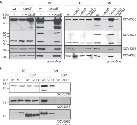
Efficient secretion of XCV0536, XCV3671, XCV4358, and
XCV4360 depends on a functional Xps-T2S system and the
pre-dicted Sec signals. To investigate whether secretion of XCV0536,
XCV3671, XCV4358, and XCV4360 is mediated by the Xps-T2S
system, we performed secretion assays with X. campestris pv.
vesi-catoria strain 85-10⌬xpsE, which lacks the putative
ATPase-en-coding gene of the Xps-T2S system. Reduced amounts of
XCV0536 – c-Myc, XCV3671– c-Myc, XCV4358 – c-Myc, and
XCV4360 – c-Myc were detected in the culture supernatants of
strain 85-10⌬xpsE compared with those of strain 85-10 (
Fig. 2A
).
Wild-type levels of secretion in strain 85-10
⌬xpsE were restored
upon ectopic expression of xpsE, suggesting that the observed
se-cretion deficiency was specifically caused by the lack of xpsE
(
Fig. 2A
). Similarly to strain 85-10⌬xpsE, reduced secretion of
XCV0536 – c-Myc, XCV3671– c-Myc, XCV4358 – c-Myc, and
XCV4360 – c-Myc was observed in strains 10⌬xpsD and
85-10
⌬xpsD⌬xcsD, from which the secretin-encoding gene of the xps
T2S gene cluster had been deleted. In contrast, wild-type secretion
levels were detected in strain 85-10
⌬xcsD (
Fig. 2A
).
Sequence analysis revealed that most tested candidate T2S
sub-strates contained an N-terminal predicted Sec signal (
Table 2
) that
presumably targets the proteins for Sec-dependent transport
across the IM. To analyze the contribution of the predicted
N-ter-minal Sec signals to the secretion of selected T2S substrates, we
generated expression constructs encoding N-terminal deletion
derivatives of the type II secreted predicted xylanases XCV0965
and XCV4358 and the lipase XCV0536. When XCV0965
⌬2–24–
c-Myc, XCV4358
⌬2–22– c-Myc, and XCV0536
⌬2–29– c-Myc were
an-alyzed for secretion in strains 85-10 and 85-10
⌬EEE, they were not
detectable in the culture supernatants (
Fig. 2B
), suggesting that
the predicted N-terminal Sec signals are required for the efficient
secretion of XCV0965, XCV4358, and XCV0536 into the
extracel-lular milieu.
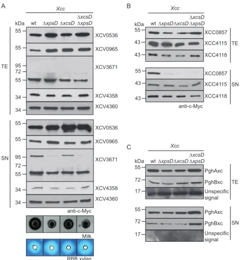
Secretion of XCV0965, XCV4358, and XCV4360 is type II
in-dependent in X. campestris pv. campestris. We previously
re-ported that the secretion of the T2S substrates PghBxc (Xc1849)
and PghAxc (Xc0705) from X. campestris pv. campestris is type II
independent in X. campestris pv. vesicatoria (
8
). To further
inves-tigate possible differences in T2S substrate specificities, we
ana-lyzed the secretion of T2S substrates from X. campestris pv.
vesi-catoria in X. campestris pv. campestris strain LMG 568 and T2S
mutant derivatives thereof with deletions of xpsD and/or xcsD.
When X. campestris pv. campestris strains were incubated in
com-plex NYG medium, similar amounts of XCV0965– c-Myc,
XCV0536 – c-Myc, XCV4358 – c-Myc, and XCV4360 – c-Myc
were detected in the culture supernatants of wild-type and T2S
mutant strains, suggesting that they were secreted independently
of the T2S systems in X. campestris pv. campestris (
Fig. 3A
). In
contrast, secretion of XCV3671– c-Myc was reduced in X.
camp-estris pv. campcamp-estris xpsD deletion mutants (
Fig. 3A
). As was
ob-served for X. campestris pv. vesicatoria, we detected an
XCV3671-specific signal at approximately 100 kDa in the culture
supernatant (see above) (
Fig. 3A
). We conclude from these
find-ings that the recognition of T2S substrates can vary in different
pathovars of X. campestris. This hypothesis is supported by the
finding that the T2S systems from X. campestris pv. campestris are
not essential for the extracellular protease and xylanase activities,
as was observed for X. campestris pv. vesicatoria (
Fig. 3A
) (
8
).
on January 18, 2016 by INRA - France
http://jb.asm.org/
TABLE 2 Candidate T2S substrates from X. campestris pv. vesicatoria strain 85-10 Candidate T2S substrate and no.a Signal peptideb Secretionc Homolog(s) in X. campestris pv. vesicatoria Homologous T2S substrate(s) or
virulence factor(s) (amino acid identity)d Reference(s)
Cellulase XCV0031 ⫹ ⫹ XCV0029, XCV0033,
XCV0358, XCV1826
Virulence factor XOO0282 from X.
oryzae pv. oryzae strain PXO99 (87%)
8,9 Proteases XCV0007 ⫹ ⫹ ⫺ This study XCV0024 ⫹ ⫹ XCV0730 This study XCV0027 ⫹ ⫹ XCV0673, XCV4096, XCV3425
T2S substrate Lpg0032 from Legionella
pneumophila (27%)
68; this study
XCV0583 ⫹ ⫺ XCV0845 T2S substrate XAC0552 from X.
axonopodis pv. citri (96%) 14,30; this study XCV0961 ⫹ ⫺ XCV0960, XCV3671, XCV3669 T2S substrate XCV3671 from X. campestris pv. vesicatoria (58%) This study XCV1064 ⫹ ⫹ XCV3406, XCV0965e 8; this study XCV1311 ⫹ ⫹ XCV4437 This study
XCV2993 ⫹ ⫺ XCV3669, XCV3671 T2S substrate XAC2831 from X.
axonopodis pv. citri (62%) 14; this study XCV3425 ⫹ ⫺ XCV4096 68; this study XCV3669 ⫹ ⫹ XCV3671, XCV0959, XCV0960, XCV0961, XCV2993 T2S substrate XCV3671 from X. campestris pv. vesicatoria (57%) 8 XCV3671 ⫹ T2S XCV3669, XCV0959, XCV0960, XCV0961, XCV2993 This study XCV4096 ⫹ ⫹ XCV0673, XCV0027, XCV3425 T2S substrate Lpg0032 from L. pneumophila (30%) 68; this study XCV4437 ⫹ ⫹ XCV1311 This study Xylanases XCV0965 ⫹ T2S XCV1202 T2S substrate XCC0857 from X. campestris pv. campestris (93%) 8; this study XCV1202 ⫹ ⫺ XCV0965 This study XCV1335 ⫹ ⫹ ⫺ This study
XCV4355 (XynA) ⫺ ⫹ XCV4358, XCV4360 T2S substrates XCV4358 (30%) and XCV4360 (32%) from X. campestris pv. vesicatoria
This study
XCV4358 (XynB2) ⫹ T2S XCV4355, XCV4360 Extracellular xylanases XOO4428 (XynB; 94%) from X. oryzae pv. oryzae and XCV4360 (57%) from X. campestris pv. vesicatoria
11; this study
XCV4360 (XynB3) ⫹ T2S XCV4355, XCV4358 T2S substrate XCV4358 (57%) from X.
campestris pv. vesicatoria
This study
Endoglucanase XCV0670 ⫹ ⫹ XCV1823 T2S substrates XOO4019 (ClsA) (88%)
from X. oryzae pv. oryzae and Lpg1918 from L. pneumophila (22%)
12,68; this study
Oxidoreductase XCV2747 ⫹ ⫺ XCV2904 This study
Hydrolase XCV0889 ⫹ ⫹ XCV1606 This study
Lipases
XCV0536 ⫹ T2S ⫺ XOO0526 (LipA) from X. oryzae pv.
oryzae (86%)
7; this study
XCV0805 ⫹ ⫹ ⫺ Virulence factor XOO3849 (accession
no.YP_202488) from X. oryzae pv. oryzae KACC 10331 (94%)
9; this study
XCV3283 ⫺ ⫺ ⫺ T2S substrate Lpg1157 from L.
pneumophila
69; this study
Decarboxylase XCV1075 ⫺ ⫺ ⫺ T2S substrate Lpg0406 from L.
pneumophila (21%) 68; this study Peptidyl isomerase XCV1033 ⫺ ⫹ XCV2946 T2S substrate Lpg1962 from L. pneumophila (54%) 69; this study a
All candidate T2S substrates are conserved in Xanthomonas spp.
b⫹, there is a predicted Sec signal; ⫺, no predicted Sec signal. The presence of a putative Sec signal was analyzed using the SignalP program (www.cbs.dtu.dk/services/SignalP/). c⫹, secreted in wild-type and T2S mutant strains; ⫺, not detectable in culture supernatants; T2S, reduced secretion in strains with mutations in T2S genes.
dThe percent amino acid identity with the homologous candidate T2S substrate from X. campestris pv. vesicatoria listed in the first column. e
The homology is restricted to the N-terminal region of XCV1064.
on January 18, 2016 by INRA - France
http://jb.asm.org/
Protease activity of X. campestris pv. campestris, however, was
reduced in xpsD mutant strains compared with the wild-type
strain (
Fig. 3A
).
The Xps-T2S system from X. campestris pv. campestris
se-cretes the predicted xylanase XCC0857 but is dispensable for the
secretion of the putative xylanases XCC4115 and XCC4118.
Next, we analyzed the secretion of putative xylanases from X.
campestris pv. campestris that share homology with predicted
xy-lanases from X. campestris pv. vesicatoria. The analyzed proteins
include XCC0857 (93% amino acid sequence identity with
XCV0965), XCC4118 (62% and 83% amino acid sequence
iden-tities with XCV4358 and XCV4360, respectively), and XCC4115
(84% amino acid sequence identity with XCV4355) (
Table 2
).
XCC0857, XCC4115, and XCC4118 were previously shown to be
secreted by X. campestris pv. campestris (
47
). Secretion assays with
X. campestris pv. campestris wild-type and T2S mutant strains
revealed that secretion of XCC0857– c-Myc was reduced in
⌬xpsD
and
⌬xcsD deletion mutants (
Fig. 3B
). In contrast, similar
wt ΔEEE wt TE SN XCV0007 XCV0027 XCV0024 XCV1311 XCV3671 XCV4096 XCV4437 kDa 55 72 72 72 55 72 95 72 72 anti-c-MycA
D
wt wt TE SN kDa XCV0889 34 XCV4358 XCV4360 34 34 XCV0536 55 anti-c-MycΔEEE ΔEEE ΔEEE
HpaB TE SN wt 26 0 5 15 0 5 15 min boiling 55 72 95 kDa anti-c-Myc 55 XCV0670 XCV1064 43 XCV1033 26 XCV0805
B
XCV4355 55 XCV1335 55 55 anti-HrpB1 72 55 43 34 26 17 TE SN HrpB1-c-Myc HrpB1 + - +- +- +-C
XCV0965 55 * * * *FIG 1 The T2S systems from X. campestris pv. vesicatoria strain 85-10 contribute to the secretion of the lipase XCV0536, the predicted protease XCV3671, and
the putative xylanases XCV4358 and XCV4360. (A) In vitro secretion assays with individual candidate T2S substrates. Strains 85-10 (wt) and 85-10⌬EEE (⌬EEE) carrying expression constructs encoding C-terminally c-Myc epitope-tagged candidate T2S substrates as indicated were incubated in NYG medium. Total cell extracts (TE) and culture supernatants (SN) were analyzed by immunoblotting using a c-Myc epitope-specific antibody. The c-Myc epitope-specific antibody did not detect unspecific proteins in the cell extracts or culture supernatants, as shown in Fig. S1 in the supplemental material. (B) Secretion levels of the lipase XCV0536, the predicted protease XCV3671, and the putative xylanases XCV4358, XCV4360, and XCV0965 were reduced in strain 85-10⌬EEE. X. campestris pv. vesicatoria strains 85-10 and 85-10⌬EEE containing expression constructs encoding C-terminally c-Myc epitope-tagged derivatives of XCV0536, XCV3671, XCV4358, XCV4360, and XCV0965, respectively, as indicated were incubated in NYG medium. TE and SN were analyzed as described for panel A. For the detection of XCV0965– c-Myc in the SN, the blot was overexposed. (C) Secreted XCV3671– c-Myc was detected as part of a protein complex that dissolved upon prolonged boiling of protein extracts. Strain 85-10 containing XCV3671– c-Myc was incubated in NYG medium, and TE and SN were analyzed by immuno-blotting. Samples were boiled for 0, 5, or 15 min as indicated, and the blot was probed with a c-Myc epitope-specific antibody. (D) The periplasmic HrpB1 protein was not detected in culture supernatants. Strain 85-10 containing HrpB1– c-Myc and HrpB1, as indicated, was incubated in NYG medium. TE and SN were analyzed by immunoblotting using an HrpB1-specific antibody. HrpB1 and HrpB1– c-Myc have the expected molecular masses of approximately 16 and 21 kDa, respectively, and formed oligomeric complexes as described previously (45). HrpB1-specific signals are indicated by white asterisks.
on January 18, 2016 by INRA - France
http://jb.asm.org/
amounts of XCC4115– c-Myc and XCC4118 – c-Myc were
de-tected in the culture supernatants, suggesting that they were
se-creted independently of the T2S systems from X. campestris pv.
campestris (
Fig. 3B
). XCC4118 was previously shown to be
re-quired for the extracellular xylanase activity of X. campestris pv.
campestris (
47
). The T2S-independent secretion of XCC4118 is,
therefore, in agreement with the finding that the extracellular
xy-lanase activity of X. campestris pv. campestris is unaffected in T2S
mutant strains (see above). When analyzed in X. campestris pv.
vesicatoria, secretion of XCC0857 was reduced in
⌬xpsD and
al-most not detectable in
⌬xcsD ⌬xpsD deletion mutant strains,
whereas XCC4115 and XCC4118 were secreted independently of
the T2S systems (see Fig. S2 in the supplemental material).
We also performed secretion assays with X. campestris pv.
campestris strain LMG 568 and corresponding T2S mutant strains
encoding C-terminally c-Myc epitope-tagged derivatives of
PghAxc and PghBxc, which were previously identified as T2S
sub-strates of X. campestris pv. campestris strain 8004 (
10
).
Interest-ingly, we did not observe T2S-dependent secretion of either
pro-tein in X. campestris pv. campestris strain LMG 568 (
Fig. 3C
),
suggesting differences in the T2S substrate specificities of X.
camp-estris pv. campcamp-estris strains 8004 and LMG 568.
Secreted proteins from X. campestris pv. vesicatoria are
de-tectable in OMVs. Given the finding that T2S substrates from X.
campestris pv. vesicatoria were still partially secreted by T2S
mu-tant strains, we investigated whether extracellular proteins from
X. campestris pv. vesicatoria can be secreted by OMVs, which often
contribute to the delivery of bacterial virulence factors across the
OM (
48
). Electron microscopy analysis revealed that X. campestris
pv. vesicatoria strain 85-10 frequently formed OMVs, which were
isolated from bacterial culture supernatants (
Fig. 4A
; see also Fig.
S3 in the supplemental material). OMVs were also observed in
infected plant tissue (
Fig. 4B
) and after in vitro cultivation of the
bacteria (
Fig. 4C
; see also Fig. S4 in the supplemental material). To
investigate the presence of secreted proteins in OMVs, we
per-formed immunoelectron microscopy with strain 85-10, which
contained the putative protease XCV0007– c-Myc, the predicted
xylanase XCV4355– Myc, and the T2S substrates XCV0536 –
Myc, XCV3671– Myc, XCV4358 – Myc, and XCV4360 –
c-Myc. Binding of c-Myc epitope-specific antibodies was detected
by secondary antibodies that were coupled to gold particles.
Inter-estingly, all analyzed proteins were detected at the bacterial
mem-branes and inside vesicle-like structures (
Fig. 4C
). In contrast,
almost no gold particles were present in the surrounding milieu or
inside the bacterial cells, suggesting that the analyzed proteins
were preferentially targeted to the bacterial membranes and to
vesicles (
Fig. 4C
). As gold particles were present in the lumens of
the OMVs but not inside bacterial cells, we assume that these
A B wt ΔxpsE -wt ΔxpsE -kDa 34 34 XCV0536 anti-c-Myc 55 43 34 55 72 95 TE SN wt ΔEEE TE SN FL ΔSP
wt ΔEEE wt ΔEEE wt ΔEEE
XCV4358 anti-c-MycXCV0965 FL ΔSP XCV0536 wt ΔxpsDΔxcsDΔxpsD ΔxcsDwt ΔxpsDΔxcsDΔxpsD ΔxcsD TE SN XCV4358 XCV4360 XCV3671 anti-c-Myc + XpsE + XpsE kDa 43 55 34 43
FIG 2 Efficient secretion of XCV0536, XCV3671, XCV4358, and XCV4360 depends on the Xps-T2S system. (A) Efficient secretion of XCV0536, XCV3671,
XCV4358, and XCV4360 depends on xpsE and xpsD. Strains 85-10 (wt), 85-10⌬xpsE (⌬xpsE), 10⌬xpsD (⌬xpsD), 10⌬xcsD (⌬xcsD), and 85-10⌬xpsD⌬xcsD (⌬xpsD⌬xcsD) without expression constructs (⫺) or carrying XpsE and/or individual T2S substrates as indicated were incubated in NYG medium. Total cell extracts (TE) and culture supernatants (SN) were analyzed by immunoblotting using a c-Myc epitope-specific antibody. (B) Secretion of the T2S substrates XCV0536, XCV4358, and XCV0965 depends on the N-terminal regions, which contain predicted Sec signal peptides (SP). Strains 85-10 (wt) and 85-10⌬EEE (⌬EEE) carrying full-length XCV0536–c-Myc (XCV0536; FL), XCV0536⌬2–29– c-Myc (XCV0536;⌬SP), XCV4358–c-Myc (XCV4358; FL), XCV4358⌬2–22– c-Myc (XCV4358;⌬SP), XCV0965–c-Myc (XCV0965; FL), or XCV0965⌬2–24– c-Myc (XCV0965;⌬SP) as indicated were incubated in NYG medium. TE and SN were analyzed as described for panel A.
on January 18, 2016 by INRA - France
http://jb.asm.org/
structures were vesicles and not sections of bacteria (
Fig. 4C
). For
steric reasons, sections through the tips of bacteria, which were
located in a perpendicular orientation, would not be visible in
close proximity to another bacterial cell. The labeling was c-Myc
epitope specific because only a very few gold particles were
de-tected outside the vesicle structures in samples of strain 85-10
containing plasmid pBRM (see Fig. S4 in the supplemental
mate-rial). The analyzed T2S substrates were also detected in OMVs of
strain 85-10
⌬EEE (see Fig. S4). This is in agreement with the
ob-servation that XCV0536 – c-Myc, XCV3671– c-Myc, XCV4358 –
c-Myc, and XCV4360 – c-Myc were still detectable in the culture
supernatant of the T2S-deficient strain 85-10⌬EEE, albeit in
re-duced amounts (see above).
To further test the hypothesis that the analyzed proteins were
present in OMVs, we added proteinase K to the supernatants of
bacterial cultures that were incubated overnight in complex NYG
medium. Immunoblot analysis showed that the amounts of the
T2S substrates XCV0536 – c-Myc, XCV3671– c-Myc, XCV4358 –
c-Myc, and XCV4360 – c-Myc were only slightly affected after
in-cubation with proteinase K (
Fig. 4D
). As all proteins were
de-graded by proteinase K when analyzed in bacterial cell lysates (
Fig.
4D
), we assume that they were protected from degradation in the
culture supernatant. Notably, however, when bacteria were
incu-bated for 1 h in NYG medium, significantly reduced amounts of
XCV0536 – c-Myc, XCV3671– c-Myc, XCV4358 – c-Myc, and
XCV4360 – c-Myc were detected in the presence of proteinase K
(see Fig. S5 in the supplemental material). This was not the case
for XCV0007– c-Myc or XCV4355– c-Myc, which were protected
against degradation by proteinase K (see Fig. S5). We assume
that XCV0536 – c-Myc, XCV3671– c-Myc, XCV4358 – c-Myc, and
XCV4360 – c-Myc were not efficiently targeted to OMVs during the
1-h incubation time and were therefore degraded by proteinase K.
wtΔxpsDΔxcsDΔxcsDΔxpsD TE XCC4118 XCC4115 XCC0857 Xcc A C 43 43 55 kDa SN TE anti-c-Myc anti-c-Myc B Milk RBB xylan 34 kDa wt ΔxpsDΔxcsDΔxcsDΔxpsD 34 55 55 72 95 34 34 55 55 72 95 XCV0965 XCV4358 XCV4360 XCV3671 XCV0536 XCV0965 XCV4358 XCV4360 XCV3671 XCV0536 Xcc XCC4118 XCC4115 XCC0857 43 43 55 SN 55 55 55 PghAxc 72 PghBxc Unspecific signal 17 Unspecific signal PghAxc PghBxc TE SN wtΔxpsDΔxcsDΔxcsDΔxpsD Xcc kDa 55 72 17
FIG 3 Secretion assays with X. campestris pv. vesicatoria and X. campestris pv. campestris strains revealed differences in T2S substrate specificities. (A) Secretion
of XCV0536, XCV0965, XCV4358, and XCV4360 from X. campestris pv. vesicatoria was type II independent in X. campestris pv. campestris. X. campestris pv. campestris strains LMG 568 (wt), LMG 568⌬xpsD (⌬xpsD), LMG 568⌬xcsD (⌬xcsD), and LMG 568⌬xpsD⌬xcsD (⌬xpsD⌬xcsD) carrying XCV0536–c-Myc, XCV0965– c-Myc, XCV3671– c-Myc, XCV4358 – c-Myc, or XCV4360 – c-Myc as indicated were incubated in NYG medium. Total cell extracts (TE) and culture supernatants (SN) were analyzed by immunoblotting using a c-Myc epitope-specific antibody. For the analysis of extracellular protease and xylanase activities, bacteria were incubated on NYG agar plates containing milk proteins or RBB xylan. Halo formation was documented 2 dpi. (B) Efficient secretion of the predicted xylanase XCC0857 in X. campestris pv. campestris depends on xpsD and xcsD. X. campestris pv. campestris strain LMG 568 and deletion derivatives thereof lacking xpsD (⌬xpsD), xcsD (⌬xcsD), or both xpsD and xcsD (⌬xpsD⌬xcsD) and carrying XCC0857–c-Myc, XCC4115–c-Myc, or XCC4118–c-Myc as indicated were incubated in NYG medium. TE and SN were analyzed as described for panel A. (C) PghAxc and PghBxc were secreted independently of the T2S systems of X. campestris pv. campestris strain LMG 568. X. campestris pv. campestris strain LMG 568 and T2S mutants thereof carrying PghAxc– c-Myc and PghBxc– c-Myc as indicated were incubated in NYG medium. TE and SN were analyzed as described for panel A. The blot was reprobed with an antibody directed against the putative translocon protein XopA, which unspecifically detects intracellular proteins in X. campestris pv. campestris. One representative signal is shown.
on January 18, 2016 by INRA - France
http://jb.asm.org/
Ectopic expression of XCV4358 or XCV4360 restores
extra-cellular xylanase activity in strain 85-10⌬0965. Next, we
ana-lyzed a possible contribution of the identified T2S substrates to
extracellular enzyme activities. For this, XCV3671, XCV4358, and
XCV4360 were individually deleted from the genome of X.
camp-estris pv. vesicatoria strain 85-10. For the analysis of extracellular
protease and xylanase activities, wild-type and T2S substrate
mu-tant strains were cultivated on NYG agar plates containing milk
proteins or RBB xylan. On these media, the extracellular protease
or xylanase activity of X. campestris pv. vesicatoria results in a
cleared halo around the bacterial inoculation zone. Halo
forma-tion on milk and RBB xylan plates was severely reduced in strains
85-10
⌬EEE and 85-10⌬0965, respectively, as was observed
previ-ously (
8
) (
Fig. 5A
). In contrast, a wild-type halo on milk plates was
detected for strain 85-10
⌬3671, suggesting that XCV3671 does
not significantly contribute to the degradation of milk proteins
(
Fig. 5A
). Similarly, the extracellular xylanase activities of strains
85-10⌬4358 and 85-10⌬4360 were unaltered compared with
those of the wild-type strain 85-10 (
Fig. 5A
). Wild-type xylanase
activity was also observed for a double deletion mutant strain
XCV0007 XCV4355 XCV0536 XCV4358 XCV4360 XCV3671 A B CW CP CW CW CP CP C XCV0536 XCV3671 XCV4358 XCV4360 wt ΔEEE 4°C 37°C 37°C 4°C 37°C 37°C + + TE SN wt ΔEEE + protease - - -- - -Lysates 37°C 37°C -wt kDa 43 55 34 34 D
FIG 4 Extracellular proteins from X. campestris pv. vesicatoria detected in OMVs. (A) Electron microscopy analysis of isolated OMVs from X. campestris pv.
vesicatoria strain 85-10 cultivated in NYG medium. Bar, 100 nm. (B) OMV formation by X. campestris pv. vesicatoria in infected pepper leaves. X. campestris pv. vesicatoria strain 85-10⌬eps was inoculated at a bacterial density of 3.6 ⫻ 108into leaves of susceptible ECW plants, and samples were taken 6 h postinfiltration
for electron microscopy analysis. Bar, 500 nm; arrow heads, OMVs. CW, plant cell wall; CP, plant cell cytoplasm. (C) Predicted extracellular enzymes were detectable in OMVs. Strain 85-10 containing XCV0007– Myc, XCV4355– Myc, XCV0536 – Myc, XCV3671– Myc, XCV4358 – Myc, or XCV4360 – c-Myc as indicated was analyzed by immunoelectron microscopy using a c-c-Myc epitope-specific antibody and a secondary antibody coupled to gold particles. Bar, 250 nm. (D) Secreted proteins were protected from degradation by proteinase K. Strains 85-10 (wt) and 85-10⌬EEE (⌬EEE) containing expression constructs for XCV0536 – c-Myc, XCV3671– c-Myc, XCV4358 – c-Myc, or XCV4360 – c-Myc as indicated were incubated overnight in NYG medium. Culture supernatants (SN) and bacterial lysates were incubated on ice or at 37°C in the presence (⫹) or absence (⫺) of proteinase K as indicated. Total cell extracts (TE), SN, and lysates were analyzed by immunoblotting, using c-Myc epitope-specific antibodies. For the detection of proteins in the culture supernatants, the blots were overexposed.
on January 18, 2016 by INRA - France
http://jb.asm.org/
lacking both XCV4358 and XCV4360 (
Fig. 5A
). As halo formation
on RBB xylan plates depends on the type II secreted xylanase
XCV0965 (
8
) (
Fig. 5A
), we assumed that XCV4358 and XCV4360
did not significantly contribute to the extracellular xylanase
activ-ity. However, XCV4358 and XCV4360 restored halo formation
when ectopically expressed under the control of the lac promoter
in strain 85-10⌬0965 (
Fig. 5B
), suggesting that they encode active
xylanases. The expression levels of the native XCV4358 and
XCV4360 genes might therefore not be sufficient to confer
detect-able xylanase activity in strain 85-10.
HrpG induces the expression of XCV0536, XCV3671, and
XCV4358 but not of XCV4360. To analyze the expression levels of
T2S substrate genes in X. campestris pv. vesicatoria, we performed
transcript studies with strains 85-10 and 85-10hrpG* (85*), which
contains HrpG*, a constitutively active version of HrpG (
49
).
HrpG is the key regulator of the T3S genes and also activates the
expression of T2S genes (
8
,
50–52
). Yet, the expression of the T2S
substrate gene XCV0965 is suppressed by HrpG, as was shown in
our previous study (
8
). When bacteria were grown in NYG
me-dium, transcript amounts of XCV0536 were significantly
in-creased in strain 85* compared with strain 85-10 (
Fig. 5C
). This is
in agreement with the previous finding that expression of
XCV0536 is induced by the transcriptional activator HrpX, which
is required for the expression of many HrpG-regulated genes and
is itself activated by HrpG (
53
,
54
). Slight increases of XCV3671
and XCV4358 transcript accumulation were observed in strain
0 10 20 30 40 50 85-10 85*
Relative transcript levels
60 70 XCV0536 XCV4358 XCV4360 XCV3671 D -wt Δ3671 E - pnat3671in cis -wt Δ4358 - pnat4358in cis -wt Δ4360 - pnat4360in cis Δ0965 wt Δ4358 Δ4360 Δ4358Δ4360 ΔEEE wt Δ3671 A C wt Δ0965 - - + 4358 + 4360 milk xylan B xylan 0 1 2 3 4 5 6 7 8 9 0 2 4 6 8 10 12 14 Log ( C F U /c m ²)
Days post inoculation * * * * * * wt Δ4358 Δ4360 Δ3671
FIG 5 XCV4358, XCV4360, and XCV3671 contribute to virulence. (A) The predicted protease XCV3671 and the predicted xylanases XCV4358 and XCV4360
do not significantly contribute to the extracellular protease and xylanase activities, respectively, of X. campestris pv. vesicatoria. For the analysis of extracellular protease activity, strains 85-10 (wt), 85-10⌬EEE (⌬EEE), and 85-10⌬3671 (⌬3671) were incubated on NYG agar plates containing milk proteins. To study xylanase activity, X. campestris pv. vesicatoria strains 85-10 (wt), 85-10⌬0965 (⌬0965), 85-10⌬4358 (⌬4358), 85-10⌬4360 (⌬4360), and 85-10⌬4358⌬4360 (⌬4358⌬4360) were grown on NYG agar plates containing RBB xylan. Halo formation was documented 2 dpi. (B) Ectopic expression of XCV4358 or XCV4360 in strain 85-10⌬0965 restores extracellular xylanase activity. Strains 85-10 and 85-10⌬0965 without an expression construct (⫺) or carrying an expression construct encoding XCV4358 – c-Myc or XCV4360 – c-Myc as indicated were grown on NYG agar plates containing RBB xylan. Halo formation was documented 2 dpi. (C) T2S substrate genes from X. campestris pv. vesicatoria were induced by HrpG. XCV0536, XCV3671, XCV4358, and XCV4360 transcripts were amplified from cDNA derived from X. campestris pv. vesicatoria strains 85-10 and 85*. Transcript amounts were adjusted based on the transcript levels of the 16S rRNA. Data points refer to mean values of relative expression levels, and error bars show standard deviations derived from technical triplicates. (D) In planta growth of T2S substrate mutants. X. campestris pv. vesicatoria strains 85-10, 85-10⌬4358, 85-10⌬4360, and 85-10⌬3671 were inoculated into leaves of susceptible ECW pepper plants, and bacterial growth was analyzed over a period of 14 days. Values are the mean bacterial colony counts from three samples taken from three different plants. Error bars represent standard deviations. The asterisks indicate significant differences between the population sizes of the wild-type and the individual mutant strains (P⬍ 0.05) based on the results of an unpaired Student t test. The experiment was repeated twice with similar results. (E) The predicted protease XCV3671 and the putative xylanases XCV4358 and XCV4360 contribute to bacterial virulence. Strains 85-10, 85-10⌬3671, 10⌬4358, and 85-10⌬4360 without an expression construct (⫺) or encoding XCV3671–c-Myc, XCV4358–c-Myc, or XCV4360–c-Myc under the control of the native XCV4361 promoter (pnat) in the genome as indicated were inoculated into leaves of susceptible ECW pepper plants. Disease symptoms were photographed at 6 to 8 dpi.
Dashed lines indicate the inoculated areas.
on January 18, 2016 by INRA - France
http://jb.asm.org/
85* compared with strain 85-10. In contrast, comparable basal
transcript levels of XCV4360 were detected in both strains (
Fig.
5C
). We, therefore, assumed that transcription of the T2S
sub-strate genes XCV0536, XCV3671, and XCV4358 but not of
XCV4360 is induced by HrpG. The observed HrpG-dependent
expression is in agreement with the presence of a plant-inducible
promoter (PIP) motif (consensus sequence TTCG-N
16-TTCG) in
the promoter region of XCV0536 and a PIP box-like motif
(TTCG-N
9-TTCG) upstream of XCV3671. The PIP box motif is
present in the promoter regions of many HrpX-regulated genes
and presumably serves as a binding site for HrpX (
53
,
54
).
XCV3671, XCV4358, and XCV4360 contribute to bacterial
virulence. The HrpG-dependent expression of T2S substrate
genes suggests that the corresponding gene products contribute
to bacterial virulence. To investigate a virulence function of
XCV3671, XCV4358, and XCV4360, the corresponding X.
camp-estris pv. vesicatoria mutant strains were inoculated into leaves of
susceptible ECW pepper plants for the analysis of in planta
bacte-rial growth. Strain 85-10 grew to approximately 10
8CFU ml
⫺1at
14 dpi, whereas growth of XCV4358 and XCV4360 mutant strains
was decreased approximately 8- to 10-fold (
Fig. 5D
). An even
more pronounced reduction in bacterial multiplication was
ob-served for strain 85-10
⌬3671 (
Fig. 5D
). We also analyzed the in
planta phenotypes of wild-type and T2S substrate mutant strains.
For this, bacteria were inoculated at a density of 1
⫻ 10
8CFU/ml
into leaves of susceptible pepper plants. As expected, the wild-type
strain 85-10 induced disease symptoms in the form of
water-soaked lesions that became necrotic 4 to 5 dpi (
Fig. 5E
). Compared
with the wild-type strain, strains 85-10⌬3671, 85-10⌬4358, and
85-10⌬4360 induced reduced disease symptoms in susceptible
pepper plants, which was visible as a reduction in the area of
in-fected necrotic plant tissue (
Fig. 5E
). The reduced virulence of T2S
substrate mutants was in agreement with the observed reduction
in their in planta growth (
Fig. 5D
).
For complementation assays, we introduced expression
con-structs encoding XCV3671– c-Myc, XCV4358 – c-Myc, and
XCV4360 – c-Myc under the control of the lac promoter into
strains 85-10, 85-10⌬3671, 85-10⌬4358, and 85-10⌬4360,
respec-tively. Infection experiments revealed that the ectopic expression
of T2S substrate genes in both wild-type and deletion mutant
strains led to reduced water-soaked lesions in susceptible plants
(see Fig. S6 in the supplemental material), suggesting that elevated
levels of the analyzed T2S substrates interfere with bacterial
viru-lence. Similar results were obtained when the genes were
ex-pressed without a C-terminal c-Myc epitope tag-encoding
se-quence (data not shown) or under the control of the native
promoter of XCV4361, which presumably drives the expression of
XCV4358 and/or XCV4360 (see Fig. S6 in the supplemental
ma-terial). Ectopic expression of T2S substrate genes might have an
effect on the transport efficiency of the T2S system and thus
neg-atively affect bacterial virulence. To avoid dominant-negative
ef-fects, we inserted the genes under the control of the XCV4361
promoter into the genome of the corresponding deletion mutants
by using the suicide plasmid pLAND-P, which allows the
integra-tion of genes into the hpaFG region next to the T3S gene cluster
(
55
). As the in cis expression of XCV3671– Myc, XCV4358 –
c-Myc, and XCV4360 – c-Myc restored wild-type plant reactions in
the corresponding deletion mutant strains (
Fig. 5E
), we assume
that the observed phenotypes were specifically caused by the
dele-tion of the T2S substrate genes. We, therefore, conclude that
XCV3671, XCV4358, and XCV4360 contribute to virulence and
bacterial multiplication in planta.
DISCUSSION