Dietary and Metabolic Control of Stem
Cell Function in Physiology and Cancer
The MIT Faculty has made this article openly available.
Please share
how this access benefits you. Your story matters.
Citation
Mihaylova, Maria M., David M. Sabatini, and Ömer H. Yilmaz.
“Dietary and Metabolic Control of Stem Cell Function in Physiology
and Cancer.” Cell Stem Cell 14, no. 3 (March 2014): 292–305.
As Published
http://dx.doi.org/10.1016/j.stem.2014.02.008
Publisher
Elsevier
Version
Author's final manuscript
Citable link
http://hdl.handle.net/1721.1/103962
Terms of Use
Creative Commons Attribution-NonCommercial-NoDerivs License
Dietary and Metabolic Control of Stem Cell Function in
Physiology and Cancer
Maria M. Mihaylova1,2, David M. Sabatini1,2, and Ömer H. Yilmaz2,3
1Whitehead Institute for Biomedical Research, Cambridge, MA 02142 USA; Howard Hughes Medical Institute, MIT, Cambridge, MA 02139; Broad Institute of Harvard and MIT, Seven Cambridge Center, Cambridge, MA 02142
2The David H. Koch Institute for Integrative Cancer Research at MIT, Cambridge, MA 02139; Department of Biology, MIT, Cambridge, MA 02139
3Department of Pathology, Massachusetts General Hospital and Harvard Medical School, Boston, MA 02114
Abstract
Organismal diet has a profound impact on tissue homeostasis and health in mammals. Adult stem cells are a keystone of tissue homeostasis that alter tissue composition by balancing self-renewal and differentiation divisions. Because somatic stem cells may respond to shifts in organismal physiology to orchestrate tissue remodeling and some cancers are understood to arise from transformed stem cells, there is a likely possibility that organismal diet, stem cell function, and cancer initiation are interconnected. Here we will explore the emerging effects of diet on nutrient-sensing pathways active in mammalian tissue stem cells and their relevance to normal and cancerous growth.
Introduction
Organismal diet is one of several lifestyle factors that influences health and disease, such as cancer incidence and progression. Food consumption can directly affect the availability of nutrients and indirectly modulate growth factor and hormone levels, which in turn can regulate tissue homeostasis and tumor initiation (Rafalski et al., 2012). Historically, the effects of diet-induced physiology in rodents has been audited by using two extremes of nutrient availability: calorie restriction and dietary or genetic models of obesity. Calorie restriction (also referred to as dietary restriction) in many animal models is defined as a reduction in nutrient intake on the order of 20 to 40% of total caloric intake, without causing malnutrition (Johnson et al., 2013). Interestingly, calorie restriction is associated with improved health (i.e. delayed onset of age-related pathologies), enhanced lifespan, or both in
© 2014 ll Press. All rights reserved.
Corresponding authors: Sabatini, D.M. ([email protected]); Yilmaz, Ö.H. ([email protected]).
Publisher's Disclaimer: This is a PDF file of an unedited manuscript that has been accepted for publication. As a service to our
customers we are providing this early version of the manuscript. The manuscript will undergo copyediting, typesetting, and review of
NIH Public Access
Author Manuscript
Cell Stem Cell. Author manuscript; available in PMC 2015 March 06.
Published in final edited form as:
Cell Stem Cell. 2014 March 6; 14(3): 292–305. doi:10.1016/j.stem.2014.02.008.
NIH-PA Author Manuscript
NIH-PA Author Manuscript
multiple species ranging from worms to mammals (Colman et al., 2009; Kenyon, 2010; Signer and Morrison, 2013). In contrast, obesity is associated with multiple comorbidities that include heart disease, metabolic syndrome, and cancer incidence. Because calorie restriction and obesity are on the opposite ends of the spectrum in regards to calorie intake, the physiologic state induced by each intervention will likely have different or opposing effects on pathways that are engaged in adult mammalian stem cells.
In this review, we explore the influence of diet and metabolic pathway regulation on adult stem cell biology in mammals and the subsequent implications of these factors in cancer initiation. From epidemiologic data, it has been long observed that diet influences cancer initiation in multiple species including humans; yet, little is known about how diet at the cellular level regulates somatic stem cell function and how this influences the predisposition of these cells to undergo transformation. One can speculate that dietary interventions that increase the size of somatic stem cell pools may over the long-term augment the probability of developing a cancer whereas dietary interventions that reduce the size of the stem cell pool may be associated with a reduction in cancer incidence. From a preventative or regenerative medicine perspective, a goal will be to develop nutritional interventions that mimic the metabolic effects of diets that enhance tissue regeneration without increasing the risk of developing cancer.
Adult stem cells: units of tissue adaptation and early transformation
Many mammalian tissues are maintained by stem cells that possess two defining
characteristics: 1) the capacity to self-renew and generate more stem cells that persist for the life of an organism and 2) the ability to differentiate into downstream progenitor cells that engender the cellular diversity inherent to tissues. At the base of the intestinal crypt are rare Lgr5+ cells that mark the majority of intestinal stem cells (ISCs), which drive the three to five day turnover of the intestine (Barker et al., 2007; Barker et al., 2012). Hematopoietic stem cells (HSCs) reside in the bone marrow and give rise to all of the mature lymphoid, myeloid, and erythroid blood cell types. Muscle regeneration is mediated by satellite cells, which reside juxtaposed to mature myofibrils (Sherwood et al., 2004). The satellite cell pool contains self-renewing muscle stem cells that can regenerate muscle tissue in response to damage (Brack and Rando, 2012). Neural stem cells reside in the central (CNS) and peripheral nervous systems (PNS) and generate diverse neural subtypes important for memory and gastrointestinal motility during fetal development or in adult mammals (Kruger et al., 2002; Zhang et al., 2008). In addition, stem cells exist in many other tissues including the epidermal skin, sweat glands, liver, and stomach (Barker et al., 2010; Huch et al., 2013; Lu et al., 2012; Snippert et al., 2010). The balance between stem cell self-renewal and differentiation is a key determinant of tissue homeostasis and allows stem cells to dynamically remodel tissues in response to turnover, damage, and disease.
Often the initial steps leading to transformation occur in somatic stem cells (Magee et al., 2012b; Miyamoto et al., 2000a). Over time these premalignant stem cells, or restricted progenitors derived from them, accumulate additional genetic or epigenetic changes that lead to frank cancers. This process is best illustrated in the mammalian intestine where activation of β-catenin (an early step in colon carcinogenesis) in Lgr5+ ISCs, but not in
non-NIH-PA Author Manuscript
NIH-PA Author Manuscript
Lgr5+ cells, is sufficient to induce the precursor adenomatous lesions that evolve into intestinal carcinomas (Barker et al., 2009; Zhu et al., 2009). A similar paradigm exists in the hematopoietic system (Chao et al., 2008). For instance, in chronic myelogenous leukemia (CML), CML HSCs posses the BCR-ABL translocation and only these CML HSCs (and not other CML progenitor populations) can transfer disease in transplantation assays (Jamieson et al., 2004b; Passegue et al., 2004). Ultimately, transformation to CML-blast crisis or frank cancer is believed to occur in downstream progenitors of the CML-HSC (Jamieson et al., 2004a). Another example is the expression of the leukemia AML1/ETO translocation in non-leukemic HSCs (Miyamoto et al., 2000b). The presence of a leukemic translocation in non-leukemic HSCs bolsters the notion that initial oncogenic events occur in stem cells and that leukemia arises when the progeny of premalignant HSCs (i.e. stem cells or restricted progenitors) accumulate additional oncogenic insults. Collectively, these findings suggest that tissue stem cells may not only be key units of regeneration, but also substrates that undergo events leading to transformation.
Intrinsic and extrinsic physiology-sensing in stem cells
Diet-induced physiologic cues are likely to influence stem cell biology and these can be partitioned into short-range signals (local signals) and long-range signals (circulating growth factors) as reviewed in Nakada et al. (2011). Stem cells often reside in unique cellular neighborhoods or niches in diverse tissues, allowing for their precise regulation by local cues. Lgr5+ ISCs, for instance, are nestled between Paneth cells at the bottom of intestinal crypts. These Paneth cells, by elaborating key short-range growth factors like Wnt3, EGF, and Notch ligands, are necessary for the in vivo maintenance of Lgr5+ ISCs such that depletion of Paneth cells in genetic mouse models results in the coordinated loss of Lgr5+ stem cells (Geiser et al., 2012; Sato et al., 2011). Endothelial and perivascular cells near the endosteum provide necessary short-range growth factors to foster HSC maintenance where genetic deletion of Stem Cell Factor in these niche cells leads to the loss of stem cells (Ding et al., 2012; Kiel et al., 2005). Similarly, hair follicle stem cells require activating short-range signals from the dermal papilla to initiate growth and regeneration of the hair follicle (Folgueras et al., 2013; Greco et al., 2009). Thus, local cues emanating from the stem cell niche are key governors of diverse stem cell pools.
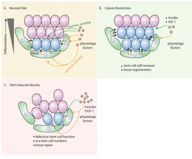
Although little is known about how mammalian stem cells integrate long-range systemic factors, stem cells may have intrinsic and extrinsic (via their niches) mechanisms for sensing diet-induced organismal physiology to alter tissue composition and growth (Figure 1). Neural stem cells possess intrinsic machinery for sensing IGF2 in the cerebral spinal fluid to fuel their proliferation (Lehtinen et al., 2011; Nakada et al., 2011). In other cases, the niche in response to long-range systemic factors extrinsically regulates stem cell function. For example, thyroid hormone promotes neurogenesis by modulating the adult neural stem cell niche (Lopez-Juarez et al., 2012). Another example is the intestine where Paneth cells control stem cell function in response to organismal feeding (discussed later) (Yilmaz et al., 2012). Also recent work demonstrates that sex influences how adult stem cells sense organismal cues. HSCs from males and females, for instance, differentially respond to circulating estrogen, likely allowing for enhanced hematopoiesis in females during
NIH-PA Author Manuscript
NIH-PA Author Manuscript
pregnancy, (Nakada et al., 2014). Thus, diverse mechanisms regulate stem cell fate in response to local and organismal demands.
Diet and stem cell homeostasis
Calorie Restriction
Given that calorie restriction delays age-related pathologies and increases lifespan in diverse species, it has been proposed that it does so in part by preserving stem cell numbers, function, or both in multiple tissues. Calorie restriction boosts the regenerative capacity of stem cells in multiple rodent tissues. In the central nervous system, for example, calorie restriction promotes the generation of new neurons in the dentate gyrus. In the blood system, calorie restriction prevents the decline of HSCs with age in certain genetic backgrounds (Chen et al., 2003; Ertl et al., 2008). From these studies, however, it is unclear whether calorie restriction mediates its effects intrinsically or extrinsically via short and/or long-ranged signals on stem cells.
Recent work by Wagers and colleagues has raised the possibility that both stem cell intrinsic and extrinsic regulatory mechanisms are at play in boosting rodent skeletal muscle stem cell activity in short-term calorie restriction (Figure 1B) (Cerletti et al., 2012). Short-term calorie restriction enhances skeletal muscle stem cell frequency in young and aged mice, improving muscle regeneration after injury. In functional assays, calorie restriction-derived stem cells are more potent inducers of muscle regeneration when transplanted into the injured muscle of control recipients, indicating that calorie restriction exerts at least some of its effects directly on stem cells. Muscle stem cells from calorie restricted mice not only had more mitochondria than their control counterparts, but these mitochondria were more adept at consuming oxygen for energy (ATP) production. These data suggest that metabolic plasticity allows stem cells to adapt to low calorie states by skewing their metabolism towards oxidative phosphorylation from glycolysis. This shift towards augmented oxidative phosphorylation promotes stem cell function in the muscle.
Interestingly, however, the physiological environment induced by calorie restriction may also play an integral role in mediating these effects in stem cells as isolated muscle stem cells from ad libitum fed mice manifested better engraftment and regeneration when transplanted into the injured muscle of calorie restricted hosts. Although it is still unclear mechanistically how the altered stem cell microenvironment in calorie restriction modulates muscle stem cell function, the authors speculate that a reduction in inflammation, a known effect of calorie restriction, boosts muscle repair after injury and the capacity of muscle stem cells to engraft in transplanted muscle.
Obesity
The obesity epidemic has increased significantly over the last several decades and underlies a multitude of “metabolic syndrome” associated disorders such as increased carbohydrate intolerance leading to insulin resistance and type II diabetes, elevated cholesterol and heart disease, hepatic steatosis, systemic inflammation, and increased incidence of certain types of cancers (Coughlin et al., 2004; Strickler et al., 2001; Vigneri et al., 2006). With high rates of adult and now childhood obesity on the rise, the metabolic syndrome epidemic has become
NIH-PA Author Manuscript
NIH-PA Author Manuscript
one of the leading causes of preventable death and continues to be an increasing burden on healthcare costs internationally.
Obesity, a chronic pathologic condition, is most frequently associated with surplus of caloric intake compared to caloric expenditure. One of the many consequences of the obese state is that individuals have an imbalance of endocrine hormonal signaling, such as increased insulin and leptin and decreased adiponectin, which collectively are associated with insulin resistance. Obesity is coupled with hormone and cytokine signaling deregulation that impede tissue repair after injury. For example, diabetic patients as well as diabetic and high fat diet (HFD)-induced obese mice exhibit delayed wound closure, explained partially by defects in myofibroblast differentiation (Seitz et al., 2010). Adipose-derived mesenchymal stem cells from obese patients showed skewed differentiation potential in ex vivo functional assays (Onate et al., 2012). In general, chronic obesity is associated with changes that decrease wound healing.
Recently, studies have implicated obesity and its associated disorders on the regulation of stem cell function in vivo. Both in rodent models of type 1 and 2 diabetes and in diabetic patients, HSCs were less effective at mobilizing out of their bone marrow environment into the systemic circulation (Ferraro et al., 2011; Rafalski et al., 2012). Further, when HSCs from ad libitum fed mice were transplanted into diabetic hosts, the HSCs displayed early proliferation, highlighting the extrinsic influence of the diabetic environment on HSC proliferation (Figure 1C). In addition, there is evidence suggesting that lipoproteins differentially control HSC proliferation, where low density lipids (LDLs) promote and high density lipids (HDLs) inhibit HSC numbers (Feng et al., 2012; Murphy et al., 2011). Also, hormones elevated in obesity, such as leptin, may enhance HSC numbers. Exogenous leptin augments phenotypic HSC numbers and function in ex vivo culture, but it is unclear whether leptin does so in transplantation assays or in obesity (Dias et al., 2013; Mukouyama et al., 1998). Interestingly, a component of the stromal HSC niche expresses the leptin receptor, raising the question of whether obesity-related changes in HSC function are mediated by the response of their niche to elevated leptin (Ding et al., 2012).
Another population of somatic stem cells that may be affected by HFD is hypothalamic neural stem cells. Two studies in 2012 demonstrated that there are Nestin and Sox2 positive neurogenic stem cells residing in the hypothalamus that are sensitive to the effects of high fat diet (Lee et al., 2012; Li et al., 2012). Li and colleagues demonstrated that HFD led to a reduction of hypothalamic stem cell numbers, as well as neurons produced from these cells. Interestingly prolonged HFD for 8 months led to a 10% decrease of major types of
hypothalamic neurons, which also include POMC neurons that regulate feeding behavior. Conversely, Lee and colleagues reported an increase of neuronal output and neurogenesis following a short-term HFD regimen. These studies indicate that the response to acute and chronic obesity may differentially regulate neural progenitor biology.
Altered organ function in obesity may correlate with increased intra-organ fat. Obese individuals are predisposed to hepatic steatosis or Non-Alchoholic Fatty Liver Disease (NAFLD). More advanced disease leads to liver cirrhosis, an irreversible state in which the liver can no longer regenerate and adequately repair itself. Repair to injury in the liver is
NIH-PA Author Manuscript
NIH-PA Author Manuscript
driven by bi- potent oval cells that generate new hepatocytes and bile duct epithelium (Yanger and Stanger, 2011). Oval cells accumulate in mouse models of fatty liver disease and other chronic liver diseases (Lowes et al., 1999; Roskams et al., 2003). A recent study also showed that injury in the liver leads to induction of Lgr5+ oval cells that were able to generate liver organoids in vitro and can differentiate into mature hepatocytes in
transplantation assays (Huch et al., 2013). Although the oval cell pool increases in response to obesity-induced liver damage, it is unclear whether this contributes to hepatocellular carcinoma.
HFD-induced obesity also hampers muscle repair after injury, resulting in smaller muscle fibers (Hu et al., 2010). These effects were noted to occur in the absence of impaired satellite cell proliferation, as assessed by Brdu incorporation, consistent with the notion that high fat diet may have extrinsic effects on muscle differentiation and maturation. One possibility is that HFD enhances inflammatory signals, which then delay proper muscle regeneration (Figure 1C). This model fits with the findings that calorie restriction may improve muscle regeneration upon injury through decreased inflammation.
Pathways regulated by organismal diet in stem cells
Because substrates such as glucose, amino acids and fatty acids will vary based on the composition of distinct dietary interventions, an important question is how are changes in nutrition translated to altered stem cell function. Recently, regulation of the
phosphatidylinositol 3-kinase/AKT (PI3K/AKT) growth factor pathway by insulin, the mechanistic target of rapamycin (mTOR) pathway by nutrients, the AMPK/LKB pathway by energy, and the Sirtuins through NAD+ levels, have been implicated in mediating many
of the beneficial and deleterious effects of calorie restriction and obesity on stem cell function and cancer initiation. These pathways have also been shown to play a critical role in the regulation of metabolic processes such as glycolysis and oxidative phosphorylation that are reviewed in Ochocki and Simon (2013), Shyh-Chang et al. (2013), Rafalski et al. (2012), and Folmes et al. (2012).
mTOR in HSCs and leukemia initiation
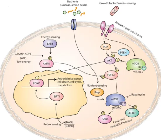
Many of the organismal benefits of calorie restriction on tissue function have been linked to the regulation of mTOR (Johnson et al., 2013). mTOR is the target of an FDA-approved compound named rapamycin and interacts with associated proteins to form two distinct complexes: mTOR complex 1 (mTORC1) and complex 2 (mTORC2) that have overlapping and distinct functions as well as different sensitivities to rapamycin inhibition (Laplante and Sabatini, 2012). mTOR has distinct functions depending on the protein complex that it nucleates. mTORC1 is a master integrator that couples cellular and organismal growth by responding to diverse upstream signals that includes oxygen, amino acids, and energy levels. Growth factors, such as insulin, activate the phosphatidylinositol 3-kinase (PI3K)/AKT pathway, which in turn leads to the stimulation of mTORC1 by inhibiting the tuberous sclerosis complex 1/2 (Inoki et al., 2002). The mTORC1 pathway promotes growth by stimulating protein and lipid synthesis. The mTORC2 pathway regulates survival and metabolism by phosphorylating AKT, which bridges signaling between the two distinct mTOR complexes (Figure 2).
NIH-PA Author Manuscript
NIH-PA Author Manuscript
Insights into the role of mTOR in stem cell fate regulation came from studies in which PTEN (phosphatase and tensin homologue), a phosphatase that negatively regulates
signaling through the PI3K/AKT, was deleted in HSCs (Yilmaz and Morrison, 2008; Yilmaz et al., 2006; Zhang et al., 2006). Deletion of PTEN or enforced genetic activation of AKT stimulated HSC proliferation and depletion, and led to the development of acute leukemia (Kharas et al., 2010; Yilmaz et al., 2006). Treatment with rapamycin reversed the decline of PTEN deficient HSCs and prevented the formation of acute leukemia, implicating mTORC1 as a key intrinsic regulator of not only HSC identity, but also of leukemia-initiating cells (cells capable of transferring leukemia upon transplantation). In subsequent studies, deletion of TSC1 (a negative regulator of mTORC1 but not of PI3K) in HSCs led to the loss of HSCs within weeks (Chen et al., 2008; Gan et al., 2008) by increasing mitochondrial biogenesis and reactive oxygen species (ROS). TSC1 deleted HSCs could be rescued by treatment with rapamycin or partially with antioxidant n-acetyl cysteine (NAC). In contrast to PTEN deficiency, TSC1 loss in HSCs did not lead to the generation of acute leukemia, indicating that pathways independent of mTORC1 are required for leukemogenesis.
An important question is how does chronic mTORC1 stimulation lead to stem cell depletion. Recent work by Morrison and colleagues demonstrated that Pten deletion activates a tumor suppressive response in HSCs (Lee et al., 2010). PTEN deletion induces expression of p16INK4a and p53 in HSCs during the depletion phase of stem cells, and this increase in tumor suppressor gene expression is ameliorated by treatment with rapamycin, placing this response downstream of mTOR. The activation of a senescence response is likely to protect the stem cell pool from acquiring secondary mutations that lead to the formation of
leukemia. Indeed, loss of tumor suppressors p16INK4a, p53, or both rescues the depletion of stem cells, but with the deleterious side effect of accelerating the generation of
transplantable leukemia-initiating cells (Figure 3).
Many of the effects of PTEN loss in HSCs can also be restored by targeting mTORC2, which acts upstream of mTORC1. mTORC2 phosphorylates AKT at Ser473, which in turn leads to the activation of mTORC1. Interestingly, deletion of Rictor, an essential mTORC2 component, has no effects on normal hematopoiesis, but prevents HSC loss and
leukemogenesis in PTEN-deficient HSCs. These findings indicate that although mTORC2 is dispensable for normal stem cell function, it is critical for leukemia-initiating cells in the setting of enforced PI3K/AKT signaling. Similar to HSCs, many tissues have stem cells that tolerate complete loss of mTORC2 signaling, such as the hematopoietic, intestine, muscle, and prostate, raising the question of whether mTORC2 inhibitors can target neoplastic growth with minimal toxicity to normal stem cells (Bentzinger et al., 2008; Guertin et al., 2009; Magee et al., 2012a; Yilmaz et al., 2012).
In contrast to mTORC2, inhibition of mTORC1 requires precise control in order to preserve stem cell function. Complete genetic loss of mTORC1 is detrimental for HSC maintenance: deletion of Raptor, a necessary component of the mTORC1 complex, causes progressive hematopoietic failure and is required for HSC regeneration (Hoshii et al., 2012; Kalaitzidis et al., 2012). However, partial inhibition of mTORC1 with rapamycin generally enhances stem cell self-renewal. For instance, rapamycin augments stem cell numbers in the intestine and blood and boosts the repair capacity of mucosal stem cells in radiation-induced
NIH-PA Author Manuscript
NIH-PA Author Manuscript
mucositis (Huang et al., 2012; Iglesias-Bartolome et al., 2012; Yilmaz et al., 2012). Further studies will be needed to elucidate the relationship of rapamycin-sensitive and insensitive mTORC1 substrates to stem cell maintenance.
Also, there is some evidence suggesting that sustained mTOR signaling accounts for some of the decline in stem cell function with age. HSCs isolated from one genetic strain of old mice showed evidence for increased mTOR signaling, although the mechanism of how mTOR signaling increases with age in HSCs is unknown (Chen et al., 2008). Interestingly, the age-related decline in the reconstituting capacity of HSCs, as well as defects in differentiation, were attenuated by rapamycin. However, the extent to which rapamycin actually rescues the aging phenotype is unclear as rapamycin also boosts the absolute numbers of HSCs in young mice (Huang et al., 2012; Nakada et al., 2010; Signer and Morrison, 2013). These beneficial effects of rapamycin on hematopoiesis may be secondary to its effect on increasing stem cell numbers rather than in rejuvenating aged HSC function; more studies will be needed to distinguish between these possibilities.
mTOR in other somatic stem cells
Sustained mTOR activity, outside of the hematopoietic system, also drives stem cell exhaustion. For example, persistent growth signaling by Wnt1 in the mouse epidermis caused hair follicle hypertrophy followed by loss of hair follicles and stem cells (Castilho et al., 2009). Although Wnt1 activated both β-catenin and mTORC1, treatment with rapamycin restored normal hair follicle growth and prevented stem cell depletion, highlighting the prevalent role of mTORC1 in this process. mTORC1 signaling also plays a dominant role in spermatogonial stem cell maintenance (Hobbs et al., 2010). The promyelocytic leukemia zinc finger (PLZF) transcription factor tunes stem cell mTORC1 activity via transcriptional control of Redd1, an mTORC1 inhibitor protein, in response to niche derived signals. Genetic deletion of Plzf results in spermatogonial stem cell depletion in an mTORC1-dependent manner that can be prevented with rapamycin. Critical control of mTOR signaling is essential for stem cell maintenance in diverse tissues.
The mTOR pathway is also important for the extrinsic regulation of stem cell fate. The mammalian small intestine, for example, undergoes structural changes (shorter villi with less numbers of differentiated enterocytes) in response to calorie restriction, indicating that diet-induced physiology alters tissue composition. At the level of the stem cell, calorie restriction in the mouse intestine dampens mTORC1 signaling in Paneth cells (a component of the stem cell niche), but not in stem cells themselves, resulting in an increase in both stem cell and Paneth cell numbers (Yilmaz et al., 2012). Mechanistically, reduced niche-dervied mTORC1 activity induces the expression of bone stromal antigen 1, an ectoenzyme that produces the paracrine factor cyclic ADP ribose (cADPR), in Paneth cells to bolster the proliferation of ISCs. Interestingly, instead of directly targeting mTORC1 signaling in ISCs, calorie restriction and rapamycin treatment create a more favorable stem cell niche. Preserving or increasing the intestinal stem cell pool in low calorie states may keep intestinal stem cells in a poised state, allowing them to rapidly regenerate the depleted differentiated cell types once nutrients become available. This poised state may be one mechanism that enables
mammalian tissues to adapt to intermittent famine.
NIH-PA Author Manuscript
NIH-PA Author Manuscript
LKB1/AMPK
Energy-sensing is differentially regulated between stem cells and their progeny and may also influence mammalian lifespan. The AMP-activated kinase (AMPK) is a master sensor of changes in the ratio of AMP to ATP (Hardie et al., 2012). During periods of low energy stress, such as food deprivation, AMPK becomes activated as the concentrations of AMP and ADP increase. Activated AMPK subdues anabolic cellular processes, such as mTOR-mediated growth and lipogenesis, and activates catabolic processes, such as autophagy, to preserve energy. Intriguingly, recent data indicate that metformin, a commonly prescribed drug for type 2 diabetes that activates AMPK, may phenocopy some of the health and lifespan benefits of calorie restriction in male rodents (Martin-Montalvo et al., 2013). Although the mechanism of how metformin activates AMPK is unclear and whether reduced mTOR signaling accounts for these beneficial effects, metformin treated rodents manifest calorie restriction-like gene expression changes and metabolic responses.
In addition to being a direct sensor of energy, AMPK is also under the control LKB1 kinase (also called STK11). LKB1 is a master regulator kinase that functions as a tumor suppressor and activates AMPK in response to low intracellular energy levels (Figure 2) (Shackelford and Shaw, 2009). In addition to phosphorylating AMPK, LKB1 also phosphorylates and regulates several other families of kinases related to AMPK, although phosphorylation of AMPKα1 and α2 are the only ones whose phosphorylation is regulated through changes of AMP/ADP to ATP levels (Hardie and Alessi, 2013; Mihaylova and Shaw, 2011). LKB1 has an essential role in HSC maintenance that is independent of the AMPK-mTOR pathway. In three separate studies, LKB1 deletion led to the transient expansion, but eventual depletion of HSCs, leading to pancytopenia (or loss of all blood cell types) (Gan et al., 2010; Gurumurthy et al., 2010; Nakada et al., 2010). LKB1 was intrinsically necessary for HSC function as LKB1-deleted HSCs or whole bone marrow cells were incapable of regenerating the hematopoietic system of control mice. LKB1 deficiency did not induce mTORC1 activity in HSCs and their function was not restored by rapamycin. Further, administration of small molecule AMPK activators did not prevent the decline of LKB1-deleted HSCs. Nakada et al. also tested the necessity of AMPK by deleting its two catalytic subunits in HSCs. Although AMPK deletion in HSCs reduces their numbers, AMPK was dispensable for HSC function in transplantation assays. Loss of either LKB1 or AMPK reduced mitochondrial membrane potential and function, but LKB1 played an AMPK-independent role in maintaining the genomic integrity of stem cells. It is important to note, however, that the effects of LKB1 on stem cell maintenance may be specific to certain types of tissues, as deletion of LKB1 in the intestine alters differentiation of secretory cells with no appreciable effect on ISC maintenance (Shorning et al., 2009). Future studies will need to address why some stem cell pools tolerate LKB1 loss while others do not.
Insulin signaling and FOXO
Insulin signaling regulates lifespan in diverse species, ranging from worms to mammals (Kenyon, 2010). Mutations in rodents that reduce insulin signaling extend lifespan, and reduced levels of circulating insulin and IGF-1 are hallmarks of calorie restriction (Kenyon, 2010). Insulin signaling via PI3K/AKT activates mTOR and non-mTOR dependent effectors. One set of substrates inhibited by AKT and not regulated by mTOR includes the
NIH-PA Author Manuscript
NIH-PA Author Manuscript
DNA-binding forkhead box O (FOXO) transcription factors. Upon phosphorylation by AKT, the FOXO transcription factors are excluded from the nucleus through 14-3-3 binding. FOXO transcription factors control critical physiological processes that include cell
proliferation, survival, and induce the expression of detoxifying enzymes, such as catalase and manganese superoxide dismutase (Figure 2) (Greer and Brunet, 2005). Sustained AKT activation, therefore, increases intracellular ROS levels, which leads to ROS-induced damage and apoptosis. Interventions that lead to reduced insulin signaling, such as calorie restriction, keep intact the FOXO transcription program that allow for oxidative stress response.
FOXO family members play an important role in stem cell maintenance. In the hematopoietic system, conditional ablation of FOXO1, FOXO3a, and FOXO4 or just FOXO3a led to the loss of HSCs through the increase of ROS production, but not to the generation of acute leukemia (Miyamoto et al., 2007; Tothova et al., 2007). Similar to PTEN- and TSC-deleted stem cells, FOXO-deleted HSCs were unable to regenerate the hematopoietic system of recipient mice. Many of the effects of FOXO deficiency in HSCs could be restored by treatment with the antioxidant NAC. FOXO3a suppression of ROS in HSCs required the tumor suppressor protein ataxia telangiectasia mutated (ATM), and induction of ROS in FOXO3-deficient HSCs led to the induction of the tumor suppressor p53 (Ochocki and Simon, 2013; Yalcin et al., 2008). While rapamycin treatment restores HSC function upon PTEN or TSC deficiency, rapamycin does not rescue the effects of FOXO deficiency in HSCs, indicating that the mechanism of HSC depletion with mTOR activation and FOXO deficiency are distinct.
FOXO transcription factors also play an important role in neural stem cell homeostasis in part through redox homeostasis (Paik et al., 2009; Renault et al., 2009; Webb et al., 2013; Yeo et al., 2013). For example, loss of FOXOs in neural stem and progenitor cells enhanced brain size and neural progenitor proliferation, which eventually caused the depletion of neural stem cells and reduced neurogenesis (Paik et al., 2009; Renault et al., 2009). These studies indicate that FOXOs constrain stem cell proliferation and maintain the self-renewal program in diverse adult stem cell populations.
Recent evidence also implicates FOXO3a in HSCs as an important mediator of a pro-autophagic response to acute food deprivation (Warr et al., 2013). HSCs, but not their downstream progeny, are exquisitely capable of activating autophagy, permitting them to withstand periods of nutrient and growth factor fluctuations. Although FOX3A is essential for poising stem cells for autophagy, there is considerable interaction between the LKB1/ AMPK pathway, insulin signaling and mTOR, which is also a potent inhibitor of autophagy. In addition to activation of FOXO3a with food deprivation, a potential mechanism may also involve the parallel attenuation of mTOR signaling by either reduced insulin signaling or activation of AMPK to initiate the autophagic response to allow stem cells to endure.
Sirtuins
There are seven members that comprise the mammalian Class III deacetylase family of Sirtuins. The Sirtuins, which have NAD dependent deacetylase activity to both histone and on histone targets, have been linked to regulating lifespan and healthspan in a number of
NIH-PA Author Manuscript
NIH-PA Author Manuscript
model organisms (Bordone et al., 2007; Fang and Nicholl, 2011; Herranz et al., 2010; Yu and Auwerx, 2009; Zhang et al., 2011). Recently, several studies have suggested that changes in NAD+ levels occur with aging and alterations of NAD+ production or
consumption can modulate healthspan and lifespan in a sirtuin-dependent manner (Canto et al., 2012; Mouchiroud et al., 2013; Yoshino et al., 2011). SIRT1 bears the greatest sequence homology to the yeast Sir2, which as been shown to increase yeast lifespan (Kaeberlein et al., 1999), and is the best-studied mammalian member in the context of stem cell biology and aging (reviewed in Rafalski et al. (2012)).
The role of Sirt1 in mammalian adult stem cell biology is perhaps best studied in the hematopoietic system. Multiple studies have examined the role of Sirt1 in HSCs. Sirt1-loss has little effect HSCs and downstream progenitor numbers and function (Leko et al., 2012). This perhaps could be explained by an up-regulation or compensation by another sirtuin family member (Narala et al., 2008). In contrast, Sirt1 has been implicated in the
homeostasis of neural stem cells. Deletion of Sirt1 was shown to lead to impaired neuronal differentiation and conversely improved differentiation upon overexpression (Hisahara et al., 2008). Further, Sirt1 deficient mice have been reported to exhibit neuronal defects in certain mouse backgrounds (Cheng et al., 2003; McBurney et al., 2003). Sirt1 has also been reported to limit the size of the oligodendrocyte progenitor pool (Rafalski et al., 2013). Recently it has been suggested that the mitochondrial sirtuin Sirt3 may regulate aspects of HSC aging. Sirt3 is dispensable for HSC maintenance in young mice but its expression declines with age. Interestingly, when re-expressed in aged cells it restores HSC function (Brown et al., 2013). Regulation of stem cells by the Sirtuins may contribute to aspects of tissue regeneration and aging.
Diet, stem cells, and cancer incidence
Calorie Restriction
Calorie restriction is a dietary intervention that not only promotes health and lifespan in diverse species, but also has potential anti-cancer effects. Moreschi first observed in 1909 that calorie restriction negatively influenced tumor progression, and since then the effects of calorie restriction on cancer incidence and progression have been extensively studied in numerous genetic mouse models and in xenograft experiments (Kritchevsky, 1999). It is generally observed that the calorie restriction state retards tumor growth, but its effects on cancer initiation are less certain (Longo and Fontana, 2010). There are models in which calorie restriction has been reported to reduce tumor initiation and others in which it has no effect (Longo and Fontana, 2010).
The mechanisms for the anti-cancer growth effects of calorie restriction are likely multifactorial. A dominant effect of calorie restriction involves a reduction in circulating systemic factors (e.g. hormones and growth factors), which may protect against cancer. Long-term calorie restriction, for instance, attenuates serum IGF-1 levels by 30–40%, and the anti-tumor initiation and progression effects of calorie restriction can be rescued by infusions of exogenous IGF-1 (Dunn et al., 1997; Longo and Fontana, 2010; Tomas et al., 1994). Consistent with the notion that reduced growth factor signaling curtails tumor
NIH-PA Author Manuscript
NIH-PA Author Manuscript
growth, tumors that harbored activating mutations in the insulin signaling pathway are resistant to calorie restriction (Kalaany and Sabatini, 2009). In addition, mouse FOXO1 was shown to be necessary for some of the anti-tumorigenic effects of calorie restriction, but whether FOXO is needed for increasing longevity in mammals in response to dietary restriction remains to be seen (Yamaza et al., 2010). Although sensitivity to insulin signaling is a key factor that predicts anti-growth responsiveness to calorie restriction, adaptations involving diverse hormones such as corticosteroids and sex hormones, DNA repair mechanism, and anti-oxidant proteins as reviewed in Longo and Fontana (2010) may also contribute.
Given that adult stems reside at the crossroads of responding to the physiologic consequences of diet and often accumulate mutations that lead to transformation, a prediction would be that dietary interventions in which there is a durable increase in stem cell numbers may over the long-term boost the number of target cells that can undergo mutagenesis (Figure 4B). A larger stem cell pool provides more potential substrates that are capable of acquiring mutations. As described earlier, paradoxically, calorie restriction bolsters the numbers, proliferation, or both of stem cells in young and old mice in multiple tissues, yet there are conflicting reports as to the effects of calorie restriction and tumor initiation in these same tissues. In the intestine, for example, calorie restriction enhances the numbers of Lgr5+ ISCs, which are also the cell-of-origin for intestinal precancerous adenomas. Studies assessing tumor initiation in the calorie restricted intestine in the same strain of mice and using the same or different (Tsao et al., 2002) genetic cancer models come to divergent conclusions. In general, all of these studies report a reduction in the overall size of adenomas consistent with the anti-growth effects of calorie restriction, but in terms of incidence some report either a neutral or negative effect (Kakuni et al., 2002; Mai et al., 2003). In another study by Weindruch and colleagues, calorie restriction, when started in middle aged mice, actually increased the incidence of spontaneous cancers (Pugh et al., 1999). Many factors such as composition and type of diet, the extent of calorie restriction, genetic model and background, and tissue type examined may differentially affect how calorie restriction influences tumor initiation.
In some cases it may be difficult to segregate the anti-growth and anti-initiation effects of calorie restriction, particularly with respect to how increased numbers of stem cells influence tumor initiation. Perhaps, the anti-growth effects of calorie restriction cloak some of its effects on tumor initiation. It is possible, for example, that in some instances calorie restriction increases tumor incidence, but that these lesions are below the threshold of detection because they are small in size. Another possibility is that stem cells under calorie restriction, although more numerous, have adapted autonomous strategies to enhance genomic stability, DNA repair, and reduce oxidative damage. Such adaptations may neutralize the effects of a larger, more robust stem cell pool. It is also possible that non-autonomous changes induced by calorie restriction, like improved immunosurveilance, compensate for increased tumor initiation by causing regression of early cancers (Figure 4B). More studies will be required to distill the mechanism of how the interplay between calorie restriction and a more active stem cell reservoir influence cancer incidence.
NIH-PA Author Manuscript
NIH-PA Author Manuscript
Ketogenic diets
Another type of diet that has been suggested to have an impact on cancer formation and progression is the ketogenic diet. Ketogenic diet (KD) is defined as a diet rich in fat, sufficient in protein and low in carbohydrates. The reduction of carbohydrates leads to a subsequent decrease of glucose utilization and an increase in the use of ketone bodies as an alternative energy source. One of the tissues impacted the most from a reduction of glucose utilization is the brain. In fact ketogenic diets have been used as an alternative therapy to reduce the incidence of seizures in children with epilepsy (Hartman and Vining, 2007). Some of the proposed mechanisms for the success of ketogenic diets is the inhibition of glycolysis, changes in activation of ATP-sensitive potassium channels and altered
transmission of glutamatergic synapses (Freeman et al., 2007; Lefevre and Aronson, 2000). Some studies utilizing a ketogenic diet have also supported its beneficial effects in vitro and in mouse models of cancer, although not always with positive effects.
Recently, ketogenic diets have been proposed as a therapeutic additive in the treatment of glioblastoma multiforme and several clinical trials have included KD in a combined therapy (Seyfried et al., 2012). The calorie restricted KD diet relies on shifting the energy source of glioblastoma cells to ketone bodies from glucose in conjuction to inducing pathways mimicking calorie restriction, which may be beneficial for survival of normal neurons but induce cancer cell death (Zuccoli et al., 2010). Several small studies have suggested that a calorie restricted ketogenic diet reduces tumor progression (Nebeling et al., 1995; Schmidt et al., 2011; Zuccoli et al., 2010). One obvious benefit of the combined ketogenic calorie restricted diet includes a reduction in insulin signaling. However, the differential effects of low calorie intake versus the composition of the ketogenic diet on stem cells and cancer needs more examination.
Obesity
In addition to predisposing to diabetes and cardiovascular disease, a number of
epidemiological studies over the past several decades have demonstrated that overweight and obese individuals have increased incidence of multiple different types of cancer (Finucane et al., 2011). Obesity and elevated visceral fat increase the incidence of gastro-esophageal reflux disease (GERD) and Barrett’s esophagus, which are risk factors for esophageal cancer (Lofdahl et al., 2011). The incidence of colonic adenomas and carcinomas correlate with increased body mass index (Calle and Kaaks, 2004; Ma et al., 2013; Okabayashi et al., 2012). Supporting these findings are studies in rodents that are subjected to high fat diet with or without genetic predispositions towards adenoma formation. Rodents fed a western or high fat diet have increased incidence in adenoma numbers compared to mice fed normal chow (Baltgalvis et al., 2009). Adenomas arising in mice fed a high fat diet are larger than those in calorie restriction, highlighting the opposing effects of calorie restriction and high fat diet on tumor growth (Mai et al., 2003). In addition, several other cancers have been associated with obesity such as postmenopausal breast and endometrial cancer (Brown and Simpson, 2010).
Unabated insulin signaling may account for some of the untoward effects of obesity on stem cells and cancer. Insulin is a mitogen that promotes the proliferation and growth of cancer
NIH-PA Author Manuscript
NIH-PA Author Manuscript
cells but also regulates tissue homeostasis (Figure 2) (Pollak, 2012). One possibility is that changes in growth factor and insulin signaling may increase the rate of senescence through increased division of somatic stem cells as observed in PTEN-deficient HSCs (Figure 3) (Lee et al., 2010). Chronic obesity may reduce stem cell function and once these stem cells acquire secondary mutations high levels of insulin and other systemic factors may drive their proliferation. Another possibility is that a non-stem cell population might compensate for reduced stem cell function in chronic obesity to maintain tissue homeostasis and serves as the cell-of-origin for some types of cancers, thus boosting the pool of cells capable of becoming transformed.
In addition to systemic factors, numerous studies have implicated the tumor
microenvironment as also contributing to the progression of cancer. Multiple studies have shown that adipocytes can secrete pro-growth factors to accelerate tumor growth (Dirat et al., 2011; Tokuda et al., 2003). Supporting this notion, it was recently reported that omental fat might attract ovarian cancer cells through the releases of cytokine factors, where inhibition of adipocyte-derived factors decreased tumor cell migration ex vivo (Nieman et al., 2011). The ovarian cancer cells reprogrammed their metabolism to feed off the lipid rich microenvironment.
Obesity-associated inflammation may also promote cancer initiation and progression. Chemical irritants, pathogens, chronic alcohol exposure, as well as dietary insults have all been shown to lead to chronic inflammation and in some cases, predispose to certain types of cancer (Grivennikov and Karin, 2010; Hotamisligil and Erbay, 2008). Inflammatory bowel disease, for example, enhances the risk for developing colorectal cancer (von Roon et al., 2007). It is interesting to note that not all persistent inflammatory conditions augment tumorigenesis: rheumatoid arthritis and psoriasis, for example, do not increase cancer risk. In fact, it has been suggested that low-grade inflammation in psoriasis may have inhibitory effects on cancer (Grivennikov and Karin, 2010; Nickoloff et al., 2005). At the molecular level, multiple different pathways feed into inflammation. One of the possible molecular connections between diet-dependent inflammation and tumor progression is the activation of nuclear factor- κB (NF-κB). NF-κB is activated in response to a number of signaling pathways including inflammatory cytokines, tumor necrosis factor α (TNF α), and activation of toll- like receptors. ROS-mediated activation of NF-kB in the intestine connects ISC proliferation to cancer initiation (Myant et al., 2013). Furthermore, activation of NF-kB in non-ISCs endowed these cells with the capacity to initiate intestinal cancers (Schwitalla et al., 2013) indicating that in inflammatory states cells capable of undergoing transformation are more diverse. One explanation for the increase in cancer with obesity may include having a larger pool of stem and non-stem cells that is susceptible to transformation (Figure 4C).
Conclusion
It has long been observed that diet has a profound influence on tissue regeneration and cancer incidence. A better appreciation for diet-sensing pathways and their effects on tissue homeostasis and cancer initiation may lead to safer anti- cancer and anti-aging therapies. Dietary interventions that sustainably drive regeneration by boosting stem cell numbers or
NIH-PA Author Manuscript
NIH-PA Author Manuscript
function may potentially run the risk of increasing cancer incidence. In order to minimize this risk, future work will need to carefully address not only how diet regulates stem cell fate, but also how these changes alter susceptibility to transformation in stem cell and non-stem cell populations. The goal will be to identify regulatory networks induced by dietary interventions that safely enhance tissue repair and regeneration, while minimizing cancer incidence. It will be also be interesting to see how pharmacologic compounds that mimic aspects of calorie restriction such as mTOR inhibitors or AMPK activators perform as agents for enhancing stem cell function and tissue repair in the elderly and obese. Lastly, given that some tumors harbor populations with heterogeneous tumorigenic potential of which some have cancer stem cell-like qualities, it will be interesting to elucidate how these dietary interventions or mimetics impact tumor heterogeneity and cancer stem cell biology.
Acknowledgments
We apologize for being unable to discuss multiple primary studies due to references and space limitations. M.M. is a Robert Black Fellow of the Damon Runyon Cancer Research Foundation, DRG-2146-13. D.M.S is an
investigator of the Howard Hughes Medical Institute and is supported by awards from the NIH (CA129105, CA103866, and AI047389), Koch Institute Frontier Research Program and the Ellison Medical Foundation. O.H.Y is supported by a K99/R00 Pathway to Independence Award from the NIH/NIA AG045144 and a CSIBD grant from the NIDDK/NIH (DK043351).
References
Baltgalvis KA, Berger FG, Pena MM, Davis JM, Carson JA. The interaction of a high-fat diet and regular moderate intensity exercise on intestinal polyp development in Apc Min/+ mice. Cancer prevention research. 2009; 2:641–649. [PubMed: 19549797]
Barker N, Huch M, Kujala P, van de Wetering M, Snippert HJ, van Es JH, Sato T, Stange DE, Begthel H, van den Born M, et al. Lgr5(+ve) stem cells drive self-renewal in the stomach and build long-lived gastric units in vitro. Cell stem cell. 2010; 6:25–36. [PubMed: 20085740]
Barker N, Ridgway RA, van Es JH, van de Wetering M, Begthel H, van den Born M, Danenberg E, Clarke AR, Sansom OJ, Clevers H. Crypt stem cells as the cells-of-origin of intestinal cancer. Nature. 2009; 457:608–611. [PubMed: 19092804]
Barker N, van Es JH, Kuipers J, Kujala P, van den Born M, Cozijnsen M, Haegebarth A, Korving J, Begthel H, Peters PJ, et al. Identification of stem cells in small intestine and colon by marker gene Lgr5. Nature. 2007; 449:1003–1007. [PubMed: 17934449]
Barker N, van Oudenaarden A, Clevers H. Identifying the stem cell of the intestinal crypt: strategies and pitfalls. Cell stem cell. 2012; 11:452–460. [PubMed: 23040474]
Bentzinger CF, Romanino K, Cloetta D, Lin S, Mascarenhas JB, Oliveri F, Xia J, Casanova E, Costa CF, Brink M, et al. Skeletal muscle-specific ablation of raptor, but not of rictor, causes metabolic changes and results in muscle dystrophy. Cell metabolism. 2008; 8:411–424. [PubMed: 19046572] Bordone L, Cohen D, Robinson A, Motta MC, van Veen E, Czopik A, Steele AD, Crowe H, Marmor
S, Luo J, et al. SIRT1 transgenic mice show phenotypes resembling calorie restriction. Aging cell. 2007; 6:759–767. [PubMed: 17877786]
Brack AS, Rando TA. Tissue-specific stem cells: lessons from the skeletal muscle satellite cell. Cell stem cell. 2012; 10:504–514. [PubMed: 22560074]
Brown K, Xie S, Qiu X, Mohrin M, Shin J, Liu Y, Zhang D, Scadden DT, Chen D. SIRT3 reverses aging-associated degeneration. Cell reports. 2013; 3:319–327. [PubMed: 23375372]
Brown KA, Simpson ER. Obesity and breast cancer: progress to understanding the relationship. Cancer research. 2010; 70:4–7. [PubMed: 20028864]
Calle EE, Kaaks R. Overweight, obesity and cancer: epidemiological evidence and proposed mechanisms. Nature reviews Cancer. 2004; 4:579–591.
NIH-PA Author Manuscript
NIH-PA Author Manuscript
Canto C, Houtkooper RH, Pirinen E, Youn DY, Oosterveer MH, Cen Y, Fernandez-Marcos PJ, Yamamoto H, Andreux PA, Cettour-Rose P, et al. The NAD(+) precursor nicotinamide riboside enhances oxidative metabolism and protects against high-fat diet-induced obesity. Cell metabolism. 2012; 15:838–847. [PubMed: 22682224]
Castilho RM, Squarize CH, Chodosh LA, Williams BO, Gutkind JS. mTOR mediates Wnt-induced epidermal stem cell exhaustion and aging. Cell stem cell. 2009; 5:279–289. [PubMed: 19733540] Cerletti M, Jang YC, Finley LW, Haigis MC, Wagers AJ. Short-term calorie restriction enhances
skeletal muscle stem cell function. Cell stem cell. 2012; 10:515–519. [PubMed: 22560075] Chao, MP.; Seita, J.; Weissman, IL. Cold Spring Harbor symposia on quantitative biology. 2008.
Establishment of a Normal Hematopoietic and Leukemia Stem Cell Hierarchy.
Chen C, Liu Y, Liu R, Ikenoue T, Guan KL, Zheng P. TSC-mTOR maintains quiescence and function of hematopoietic stem cells by repressing mitochondrial biogenesis and reactive oxygen species. The Journal of experimental medicine. 2008; 205:2397–2408. [PubMed: 18809716]
Chen J, Astle CM, Harrison DE. Hematopoietic senescence is postponed and hematopoietic stem cell function is enhanced by dietary restriction. Experimental hematology. 2003; 31:1097–1103. [PubMed: 14585375]
Cheng HL, Mostoslavsky R, Saito S, Manis JP, Gu Y, Patel P, Bronson R, Appella E, Alt FW, Chua KF. Developmental defects and p53 hyperacetylation in Sir2 homolog (SIRT1)-deficient mice. Proceedings of the National Academy of Sciences of the United States of America. 2003; 100:10794–10799. [PubMed: 12960381]
Colman RJ, Anderson RM, Johnson SC, Kastman EK, Kosmatka KJ, Beasley TM, Allison DB, Cruzen C, Simmons HA, Kemnitz JW, et al. Caloric restriction delays disease onset and mortality in rhesus monkeys. Science. 2009; 325:201–204. [PubMed: 19590001]
Coughlin SS, Calle EE, Teras LR, Petrelli J, Thun MJ. Diabetes mellitus as a predictor of cancer mortality in a large cohort of US adults. American journal of epidemiology. 2004; 159:1160–1167. [PubMed: 15191933]
Dias CC, Nogueira-Pedro A, Barbosa CM, Ribeiro-Filho AC, Wasinski F, Araujo RC, de Oliveira VX Jr, Miranda A, Paredes-Gamero EJ. Hematopoietic stem cell expansion caused by a synthetic fragment of leptin. Peptides. 2013; 50:24–27. [PubMed: 24090593]
Ding L, Saunders TL, Enikolopov G, Morrison SJ. Endothelial and perivascular cells maintain haematopoietic stem cells. Nature. 2012; 481:457–462. [PubMed: 22281595]
Dirat B, Bochet L, Dabek M, Daviaud D, Dauvillier S, Majed B, Wang YY, Meulle A, Salles B, Le Gonidec S, et al. Cancer-associated adipocytes exhibit an activated phenotype and contribute to breast cancer invasion. Cancer research. 2011; 71:2455–2465. [PubMed: 21459803]
Dunn SE, Kari FW, French J, Leininger JR, Travlos G, Wilson R, Barrett JC. Dietary restriction reduces insulin-like growth factor I levels, which modulates apoptosis, cell proliferation, and tumor progression in p53-deficient mice. Cancer research. 1997; 57:4667–4672. [PubMed: 9354418]
Ertl RP, Chen J, Astle CM, Duffy TM, Harrison DE. Effects of dietary restriction on hematopoietic stem-cell aging are genetically regulated. Blood. 2008; 111:1709–1716. [PubMed: 17947508] Fang Y, Nicholl MB. Sirtuin 1 in malignant transformation: friend or foe? Cancer letters. 2011;
306:10–14. [PubMed: 21414717]
Feng Y, Schouteden S, Geenens R, Van Duppen V, Herijgers P, Holvoet P, Van Veldhoven PP, Verfaillie CM. Hematopoietic stem/progenitor cell proliferation and differentiation is differentially regulated by high-density and low-density lipoproteins in mice. PloS one. 2012; 7:e47286. [PubMed: 23144813]
Ferraro F, Lymperi S, Mendez-Ferrer S, Saez B, Spencer JA, Yeap BY, Masselli E, Graiani G, Prezioso L, Rizzini EL, et al. Diabetes impairs hematopoietic stem cell mobilization by altering niche function. Science translational medicine. 2011; 3:104ra101.
Finucane MM, Stevens GA, Cowan MJ, Danaei G, Lin JK, Paciorek CJ, Singh GM, Gutierrez HR, Lu Y, Bahalim AN, et al. National, regional, and global trends in body-mass index since 1980: systematic analysis of health examination surveys and epidemiological studies with 960 country-years and 9.1 million participants. Lancet. 2011; 377:557–567. [PubMed: 21295846]
NIH-PA Author Manuscript
NIH-PA Author Manuscript
Folgueras AR, Guo X, Pasolli HA, Stokes N, Polak L, Zheng D, Fuchs E. Architectural niche organization by LHX2 is linked to hair follicle stem cell function. Cell stem cell. 2013; 13:314– 327. [PubMed: 24012369]
Folmes CD, Dzeja PP, Nelson TJ, Terzic A. Metabolic plasticity in stem cell homeostasis and differentiation. Cell stem cell. 2012; 11:596–606. [PubMed: 23122287]
Freeman JM, Kossoff EH, Hartman AL. The ketogenic diet: one decade later. Pediatrics. 2007; 119:535–543. [PubMed: 17332207]
Gan B, Hu J, Jiang S, Liu Y, Sahin E, Zhuang L, Fletcher-Sananikone E, Colla S, Wang YA, Chin L, et al. Lkb1 regulates quiescence and metabolic homeostasis of haematopoietic stem cells. Nature. 2010; 468:701–704. [PubMed: 21124456]
Gan B, Sahin E, Jiang S, Sanchez-Aguilera A, Scott KL, Chin L, Williams DA, Kwiatkowski DJ, DePinho RA. mTORC1-dependent and -independent regulation of stem cell renewal,
differentiation, and mobilization. Proceedings of the National Academy of Sciences of the United States of America. 2008; 105:19384–19389. [PubMed: 19052232]
Geiser J, Venken KJ, De Lisle RC, Andrews GK. A mouse model of acrodermatitis enteropathica: loss of intestine zinc transporter ZIP4 (Slc39a4) disrupts the stem cell niche and intestine integrity. PLoS genetics. 2012; 8:e1002766. [PubMed: 22737083]
Greco V, Chen T, Rendl M, Schober M, Pasolli HA, Stokes N, Dela Cruz-Racelis J, Fuchs E. A two-step mechanism for stem cell activation during hair regeneration. Cell stem cell. 2009; 4:155–169. [PubMed: 19200804]
Greer EL, Brunet A. FOXO transcription factors at the interface between longevity and tumor suppression. Oncogene. 2005; 24:7410–7425. [PubMed: 16288288]
Grivennikov SI, Karin M. Inflammation and oncogenesis: a vicious connection. Current opinion in genetics & development. 2010; 20:65–71. [PubMed: 20036794]
Guertin DA, Stevens DM, Saitoh M, Kinkel S, Crosby K, Sheen JH, Mullholland DJ, Magnuson MA, Wu H, Sabatini DM. mTOR complex 2 is required for the development of prostate cancer induced by Pten loss in mice. Cancer cell. 2009; 15:148–159. [PubMed: 19185849]
Gurumurthy S, Xie SZ, Alagesan B, Kim J, Yusuf RZ, Saez B, Tzatsos A, Ozsolak F, Milos P, Ferrari F, et al. The Lkb1 metabolic sensor maintains haematopoietic stem cell survival. Nature. 2010; 468:659–663. [PubMed: 21124451]
Hardie DG, Alessi DR. LKB1 and AMPK and the cancer-metabolism link - ten years after. BMC biology. 2013; 11:36. [PubMed: 23587167]
Hardie DG, Ross FA, Hawley SA. AMPK: a nutrient and energy sensor that maintains energy homeostasis. Nature reviews Molecular cell biology. 2012; 13:251–262.
Hartman AL, Vining EP. Clinical aspects of the ketogenic diet. Epilepsia. 2007; 48:31–42. [PubMed: 17241206]
Herranz D, Munoz-Martin M, Canamero M, Mulero F, Martinez-Pastor B, Fernandez-Capetillo O, Serrano M. Sirt1 improves healthy ageing and protects from metabolic syndrome-associated cancer. Nature communications. 2010; 1:3.
Hisahara S, Chiba S, Matsumoto H, Tanno M, Yagi H, Shimohama S, Sato M, Horio Y. Histone deacetylase SIRT1 modulates neuronal differentiation by its nuclear translocation. Proceedings of the National Academy of Sciences of the United States of America. 2008; 105:15599–15604. [PubMed: 18829436]
Hobbs RM, Seandel M, Falciatori I, Rafii S, Pandolfi PP. Plzf regulates germline progenitor self-renewal by opposing mTORC1. Cell. 2010; 142:468–479. [PubMed: 20691905]
Hoshii T, Tadokoro Y, Naka K, Ooshio T, Muraguchi T, Sugiyama N, Soga T, Araki K, Yamamura K, Hirao A. mTORC1 is essential for leukemia propagation but not stem cell self-renewal. The Journal of clinical investigation. 2012; 122:2114–2129. [PubMed: 22622041]
Hotamisligil GS, Erbay E. Nutrient sensing and inflammation in metabolic diseases. Nature reviews Immunology. 2008; 8:923–934.
Hu Z, Wang H, Lee IH, Modi S, Wang X, Du J, Mitch WE. PTEN inhibition improves muscle regeneration in mice fed a high-fat diet. Diabetes. 2010; 59:1312–1320. [PubMed: 20200318]
NIH-PA Author Manuscript
NIH-PA Author Manuscript
Huang J, Nguyen-McCarty M, Hexner EO, Danet-Desnoyers G, Klein PS. Maintenance of
hematopoietic stem cells through regulation of Wnt and mTOR pathways. Nature medicine. 2012; 18:1778–1785.
Huch M, Dorrell C, Boj SF, van Es JH, Li VS, van de Wetering M, Sato T, Hamer K, Sasaki N, Finegold MJ, et al. In vitro expansion of single Lgr5+ liver stem cells induced by Wnt-driven regeneration. Nature. 2013; 494:247–250. [PubMed: 23354049]
Iglesias-Bartolome R, Patel V, Cotrim A, Leelahavanichkul K, Molinolo AA, Mitchell JB, Gutkind JS. mTOR inhibition prevents epithelial stem cell senescence and protects from radiation-induced mucositis. Cell stem cell. 2012; 11:401–414. [PubMed: 22958932]
Inoki K, Li Y, Zhu T, Wu J, Guan KL. TSC2 is phosphorylated and inhibited by Akt and suppresses mTOR signalling. Nature cell biology. 2002; 4:648–657.
Jamieson CH, Ailles LE, Dylla SJ, Muijtjens M, Jones C, Zehnder JL, Gotlib J, Li K, Manz MG, Keating A, et al. Granulocyte-macrophage progenitors as candidate leukemic stem cells in blast-crisis CML. N Engl J Med. 2004a; 351:657–667. [PubMed: 15306667]
Jamieson CH, Weissman IL, Passegue E. Chronic versus acute myelogenous leukemia: a question of self-renewal. Cancer cell. 2004b; 6:531–533. [PubMed: 15607956]
Johnson SC, Rabinovitch PS, Kaeberlein M. mTOR is a key modulator of ageing and age-related disease. Nature. 2013; 493:338–345. [PubMed: 23325216]
Kaeberlein M, McVey M, Guarente L. The SIR2/3/4 complex and SIR2 alone promote longevity in Saccharomyces cerevisiae by two different mechanisms. Genes & development. 1999; 13:2570– 2580. [PubMed: 10521401]
Kakuni M, Morimura K, Wanibuchi H, Ogawa M, Min W, Hayashi S, Fukushima S. Food restriction inhibits the growth of intestinal polyps in multiple intestinal neoplasia mouse. Japanese journal of cancer research: Gann. 2002; 93:236–241. [PubMed: 11927003]
Kalaany NY, Sabatini DM. Tumours with PI3K activation are resistant to dietary restriction. Nature. 2009; 458:725–731. [PubMed: 19279572]
Kalaitzidis D, Sykes SM, Wang Z, Punt N, Tang Y, Ragu C, Sinha AU, Lane SW, Souza AL, Clish CB, et al. mTOR complex 1 plays critical roles in hematopoiesis and Pten-loss-evoked leukemogenesis. Cell stem cell. 2012; 11:429–439. [PubMed: 22958934]
Kenyon CJ. The genetics of ageing. Nature. 2010; 464:504–512. [PubMed: 20336132] Kharas MG, Okabe R, Ganis JJ, Gozo M, Khandan T, Paktinat M, Gilliland DG, Gritsman K.
Constitutively active AKT depletes hematopoietic stem cells and induces leukemia in mice. Blood. 2010; 115:1406–1415. [PubMed: 20008787]
Kiel MJ, Yilmaz OH, Iwashita T, Terhorst C, Morrison SJ. SLAM Family Receptors Distinguish Hematopoietic Stem and Progenitor Cells and Reveal Endothelial Niches for Stem Cells. Cell. 2005; 121:1109–1121. [PubMed: 15989959]
Kritchevsky D. Caloric restriction and experimental carcinogenesis. Toxicological sciences: an official journal of the Society of Toxicology. 1999; 52:13–16. [PubMed: 10630585]
Kruger GM, Mosher JT, Bixby S, Joseph N, Iwashita T, Morrison SJ. Neural crest stem cells persist in the adult gut but undergo changes in self-renewal, neuronal subtype potential, and factor
responsiveness. Neuron. 2002; 35:657–669. [PubMed: 12194866]
Laplante M, Sabatini DM. mTOR signaling in growth control and disease. Cell. 2012; 149:274–293. [PubMed: 22500797]
Lee DA, Bedont JL, Pak T, Wang H, Song J, Miranda-Angulo A, Takiar V, Charubhumi V, Balordi F, Takebayashi H, et al. Tanycytes of the hypothalamic median eminence form a diet-responsive neurogenic niche. Nature neuroscience. 2012; 15:700–702.
Lee JY, Nakada D, Yilmaz OH, Tothova Z, Joseph NM, Lim MS, Gilliland DG, Morrison SJ. mTOR activation induces tumor suppressors that inhibit leukemogenesis and deplete hematopoietic stem cells after Pten deletion. Cell stem cell. 2010; 7:593–605. [PubMed: 21040901]
Lefevre F, Aronson N. Ketogenic diet for the treatment of refractory epilepsy in children: A systematic review of efficacy. Pediatrics. 2000; 105:E46. [PubMed: 10742367]
Lehtinen MK, Zappaterra MW, Chen X, Yang YJ, Hill AD, Lun M, Maynard T, Gonzalez D, Kim S, Ye P, et al. The cerebrospinal fluid provides a proliferative niche for neural progenitor cells. Neuron. 2011; 69:893–905. [PubMed: 21382550]
NIH-PA Author Manuscript
NIH-PA Author Manuscript
Leko V, Varnum-Finney B, Li H, Gu Y, Flowers D, Nourigat C, Bernstein ID, Bedalov A. SIRT1 is dispensable for function of hematopoietic stem cells in adult mice. Blood. 2012; 119:1856–1860. [PubMed: 22219225]
Li J, Tang Y, Cai D. IKKbeta/NF-kappaB disrupts adult hypothalamic neural stem cells to mediate a neurodegenerative mechanism of dietary obesity and pre-diabetes. Nature cell biology. 2012; 14:999–1012.
Lofdahl HE, Lane A, Lu Y, Lagergren P, Harvey RF, Blazeby JM, Lagergren J. Increased population prevalence of reflux and obesity in the United Kingdom compared with Sweden: a potential explanation for the difference in incidence of esophageal adenocarcinoma. European journal of gastroenterology & hepatology. 2011; 23:128–132. [PubMed: 21178778]
Longo VD, Fontana L. Calorie restriction and cancer prevention: metabolic and molecular mechanisms. Trends in pharmacological sciences. 2010; 31:89–98. [PubMed: 20097433] Lopez-Juarez A, Remaud S, Hassani Z, Jolivet P, Pierre Simons J, Sontag T, Yoshikawa K, Price J,
Morvan-Dubois G, Demeneix BA. Thyroid hormone signaling acts as a neurogenic switch by repressing Sox2 in the adult neural stem cell niche. Cell stem cell. 2012; 10:531–543. [PubMed: 22560077]
Lowes KN, Brennan BA, Yeoh GC, Olynyk JK. Oval cell numbers in human chronic liver diseases are directly related to disease severity. The American journal of pathology. 1999; 154:537–541. [PubMed: 10027411]
Lu CP, Polak L, Rocha AS, Pasolli HA, Chen SC, Sharma N, Blanpain C, Fuchs E. Identification of stem cell populations in sweat glands and ducts reveals roles in homeostasis and wound repair. Cell. 2012; 150:136–150. [PubMed: 22770217]
Ma Y, Yang Y, Wang F, Zhang P, Shi C, Zou Y, Qin H. Obesity and risk of colorectal cancer: a systematic review of prospective studies. PloS one. 2013; 8:e53916. [PubMed: 23349764] Magee JA, Ikenoue T, Nakada D, Lee JY, Guan KL, Morrison SJ. Temporal changes in PTEN and
mTORC2 regulation of hematopoietic stem cell self-renewal and leukemia suppression. Cell stem cell. 2012a; 11:415–428. [PubMed: 22958933]
Magee JA, Piskounova E, Morrison SJ. Cancer stem cells: impact, heterogeneity, and uncertainty. Cancer cell. 2012b; 21:283–296. [PubMed: 22439924]
Mai V, Colbert LH, Berrigan D, Perkins SN, Pfeiffer R, Lavigne JA, Lanza E, Haines DC, Schatzkin A, Hursting SD. Calorie restriction and diet composition modulate spontaneous intestinal tumorigenesis in Apc(Min) mice through different mechanisms. Cancer research. 2003; 63:1752– 1755. [PubMed: 12702556]
Martin-Montalvo A, Mercken EM, Mitchell SJ, Palacios HH, Mote PL, Scheibye-Knudsen M, Gomes AP, Ward TM, Minor RK, Blouin MJ, et al. Metformin improves healthspan and lifespan in mice. Nature communications. 2013; 4:2192.
McBurney MW, Yang X, Jardine K, Hixon M, Boekelheide K, Webb JR, Lansdorp PM, Lemieux M. The mammalian SIR2alpha protein has a role in embryogenesis and gametogenesis. Molecular and cellular biology. 2003; 23:38–54. [PubMed: 12482959]
Mihaylova MM, Shaw RJ. The AMPK signalling pathway coordinates cell growth, autophagy and metabolism. Nature cell biology. 2011; 13:1016–1023.
Miyamoto K, Araki KY, Naka K, Arai F, Takubo K, Yamazaki S, Matsuoka S, Miyamoto T, Ito K, Ohmura M, et al. Foxo3a is Essential for Maintenance of the Hematopoietic Stem Cell Pool. Cell stem cell. 2007; 1:101–112. [PubMed: 18371339]
Miyamoto T, Weissman IL, Akashi K. AML1/ETO-expressing nonleukemic stem cells in acute myelogenous leukemia with 8;21 chromosomal translocation. Proceedings of the National Academy of Sciences of the United States of America. 2000a; 97:7521–7526. [PubMed: 10861016]
Miyamoto T, Weissman IL, Akashi K. AML1/ETO-expressing nonleukemic stem cells in acute myelogenous leukemia with 8;21 chromosomal translocation. Proc Natl Acad Sci USA. 2000b; 97:7521–7526. [PubMed: 10861016]
Mouchiroud L, Houtkooper RH, Moullan N, Katsyuba E, Ryu D, Canto C, Mottis A, Jo YS,
Viswanathan M, Schoonjans K, et al. The NAD(+)/Sirtuin Pathway Modulates Longevity through