An Ancient Mechanism Controls
the Development of Cells with a
Rooting Function in Land Plants
Benoît Menand,1Keke Yi,1,2Stefan Jouannic,1* Laurent Hoffmann,1† Eoin Ryan,1 Paul Linstead,1Didier G. Schaefer,3‡ Liam Dolan1§
Root hairs and rhizoids are cells with rooting functions in land plants. We describe two basic helix-loop-helix transcription factors that control root hair development in the sporophyte (2n) of the angiosperm Arabidopsis thaliana and rhizoid development in the gametophytes (n) of the bryophyte Physcomitrella patens. The phylogeny of land plants supports the hypothesis that early land plants were bryophyte-like and possessed a dominant gametophyte and later the sporophyte rose to dominance. If this hypothesis is correct, our data suggest that the increase in morphological complexity of the sporophyte body in the Paleozoic resulted at least in part from the recruitment of regulatory genes from gametophyte to sporophyte.
T
he invasion of land by plants in thePaleozoic was accompanied by marked changes in plant structure and life cycle and resulted in diversification of terrestrial ecosystems and pronounced climate change (1–3). One of the most important transforma-tions that occurred during the first 100 million
years after plants colonized the land was the rise to dominance of the diploid phase (sporo-phyte) of the life cycle (the land-plant life cycle comprises independent haploid and diploid organisms). The phylogenetic relationship among green algae and land plants suggests that the haploid phase (gametophyte) was morpho-logically more complex than the smaller dip-loid phase (sporophyte) in the earliest land plants (4). This changed over a period of ~100 million years to a situation in which the dip-loid phase became larger and more morpho-logically complex (4). This rise to dominance of the diploid phase of the life cycle was accom-panied by an enormous increase in morpho-logical diversity evident in Devonian floras and has persisted to the present day, when the land floras are largely dominated by diploid plants (3). To date, we have little understand-ing of the genetic basis of such a metamor-phosis of the land plant body. The characterization of the function of regulatory genes such as LEAFY (LFY) in both bryophytes and angio-sperms suggests that the increase in sporophyte diversity was brought about through the
modification of the activities of sporophyte-specific genes with sporophyte-sporophyte-specific func-tions (5). Here we show that genes that specifically promote the development of root hairs in diploid sporophytes of angiosperms also control the development of cells with similar functions in the haploid gametophytes of mosses. This suggests that genes with gametophyte functions in ancestral land plants were recruited to function in the sporophyte during the metamorphosis of the land plant body.
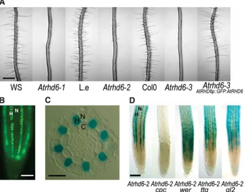
Root hairs are highly polarized cells that increase the surface area of the plant that is in contact with the growth substrate. They play important roles in nutrient acquisition and anchorage in those land plants that have roots (6, 7). The Arabidopsis thaliana root epidermis is organized in alternate rows of hair-forming cells (H cells) that produce a tip-growing pro-tuberance (root hairs) and rows of non–hair cells (N cells) that remain hairless. AtRHD6 (ROOT HAIR DEFECTIVE 6) positively regu-lates the development of H cells–Atrhd6 mu-tants develop few root hairs (Fig. 1A) (8). We cloned AtRHD6 using an enhancer trap line (Atrhd6-2) in which the GUS reporter gene is expressed in H cells but not in N cells (Fig. 1, C and D, and fig. S1). AtRHD6 encodes the basic-helix-loop-helix (bHLH) transcription factor At1g66470 (9). The identification of another independent allele (Atrhd6-3) with a similar phenotype and the complementation of the Atrhd6-3 mutation with a whole gene AtRHD6p::GFP:AtRHD6 translational fusion with the GREEN FLUORESCENT PROTEIN (GFP) confirmed that the defect in root hair development observed in this mutant is due to mutation of At1g66470 (Fig. 1A). This com-plementing AtRHD6p::GFP:AtRHD6 fusion indicates that AtRHD6 protein accumulates in H-cell nuclei in the meristem and elonga-tion zones (Fig. 1B) but disappears before the emergence of the root hair (data not shown). The spatial pattern of N cells and H cells in the A. thaliana root epidermis is controlled by a transcriptional network including the posi-1
Department of Cell and Developmental Biology, John Innes Centre, Norwich NR47UH, UK.2State Key Laboratory of Plant Physiology and Biochemistry, College of Life Science, Zhejiang University, Hangzhou, 310058, P. R. China.3Département de Biologie Moléculaire Végétale, Université de Lausanne, CH-1015 Lausanne, Switzerland.
*Present address: Institut de Recherche pour le Développe-ment, UMR 1098 Biologie du Développement des Espèces Pérennes Cultivées, 911 avenue Agropolis, F-34394 Mont-pellier Cedex 5, France.
†Present address: UMR CNRS/UPS 5546, Surfaces Cellulaires et Signalisation chez les Végétaux, Pôle de Biotechnologies végétales, 24 chemin de Borde-Rouge, F-31326 Castanet-Tolosan, France.
‡Present address: Institut Jean-Pierre Bourgin, Statìon de Génétique et Amélioration des Plantes, Institut National de la Recherche Agronomique Versailles, Route de Saint Cyr, F-78000 Versailles, France.
§To whom correspondence should be addressed. E-mail: [email protected]
Published in Science 316, no 5830, 1477-1480, 2007 which should be used for any reference to this work
tive regulator of H-cell identity CPC and the negative regulators of H-cell identity WER, TTG, and GL2 (10). To determine if AtRHD6 is regulated by these genes, we analyzed the promoter activity of the Atrhd6-2 enhancer trap in different mutant backgrounds. While the Atrhd6-2 enhancer trap expresses GUS in cells in the H position, this expression spreads to the cells in the N position in the wer, ttg, and gl2 mutant backgrounds, indicating that WER, TTG, and GL2 negatively regulate tran-scription of AtRHD6 in the N position (Fig. 1D). No expression was observed in the cpc mutant, indicating that CPC positively regulates AtRHD6 expression (Fig. 1D). Thus, AtRHD6 controls the development of root hair cells and acts downstream of the genes involved in epi-dermal pattern formation.
AtRHD6 is a member of subfamily VIIIc of bHLH transcription factors that comprises five other members (9, 11). One of these genes, At5g37800, hereafter named RHD SIX-LIKE1 (AtRSL1), is very similar to AtRHD6, suggesting that these two genes derive from a relatively recent duplication event (9). This suggests that AtRHD6 and AtRSL1 might have redundant functions. To determine if AtRSL1 is also re-quired for root hair development, we identified a line (Atrsl1-1) carrying a complete loss-of-function mutation in the AtRSL1 gene and created the Atrhd6-3 Atrsl1-1 double mutant (fig. S1). Because no new phenotypes were observed when these mutants were grown in our standard growth conditions, we grew them on the surface of cellophane disks, where small numbers of root hairs develop in the Atrhd6-3 single mutant (Fig. 2A). Plants homozygous for the Atrsl1-1 mutation had wild-type root hair morphology when grown on cellophane disks (Fig. 2A). However, the Atrhd6-3 Atrsl1-1 double mutant did not develop root hairs, indicating that AtRHD6 and AtRSL1 have partially redundant functions in root hair de-velopment (Fig. 2A). Atrhd6-3 Atrsl1-1 double-mutant plants carrying the genomic construct AtRSL1p::GFP:AtRSL1 displayed the AtRhd6-3–mutant phenotype, confirming that the ex-treme hairless phenotype of the Atrhd6-3 Atrsl1-1 double mutant is the result of a loss of function of both AtRHD6 and AtRSL1 genes (Fig. 2A). The complementing GFP::AtRSL1 fusion protein accumulates in hair cell nuclei in the meristem and elongation zones, indicating that AtRHD6 and AtRSL1 have similar expres-sion patterns (Fig. 2B). These data indicate that AtRSL1 and AtRHD6 act together to positively regulate root hair development. To determine if AtRHD6 and AtRSL1 are required for the de-velopment of the only other tip-growing cell in flowering plants, the pollen tube, we charac-terized the phenotypes of pollen tubes in Atrhd6-3, Atrsl1-1, and Atrhd6-3 Atrsl1-1 mu-tants both in vitro and in vivo. We detected neither a defect in pollen tube growth nor in the segregation of mutant alleles in the F2progeny
of backcrosses to wild type (fig. S2). No other defective phenotype was detected in any other part of Atrhd6-3, Atrsl1-1, or Atrhd6-3 Atrsl1-1
mutants. Together these data indicate that AtRHD6 and AtRSL1 are bHLH transcription factors that are specifically required for the
Fig. 1. AtRHD6 is a positive regulator of root hair development in A. thaliana. (A) Roots of Atrhd6-1, Atrhd6-2, and Atrhd6-3 mutants with their respective wild-type ecotype (WS, Wassìlewskìja; Col0, Columbia 0; L.e., Lansburg erecta) and complementation of the Atrhd6-3 mutant with a genomic AtRHD6p::GFP:AtRHD6 fusion. (B) Fluorescent image of the genomic AtRHD6p::GFP:AtRHD6 fusion in the Atrhd6-3 background showing AtRHD6 protein in hair cells nuclei. (C) Expression of the Atrhd6-2 enhancer trap GUS gene in root cross section. (D) Whole-mount longitudinal view of the expression of the enhancer trap GUS gene in Atrhd6-2 and in different backgrounds (cpc, wer, ttg1, and gl2). H, hair cell; N, non–hair cells; C, cortex. Scales bars, 500 mm (A), 50 mm (B), 25 mm (C), and 100mm (D).
Fig. 2. AtRSL1 positively regulates root hairs development in A. thaliana. (A) Roots of WT, Atrhd6-3 single mutant, 1 single mutant, Atrhd6-Atrhd6-3 1 double mutant, and Atrhd6-Atrhd6-3 Atrsl1-1 double mutant bearing the AtRSLAtrsl1-1p::GFP:AtRSLAtrsl1-1 transgene. Plants were grown on MS media with sucrose overlaid with a cellophane disk to increase root hair production in the Atrhd6-3 mutant. (B) Fluorescent image of the genomic AtRSL1p::GFP:AtRSL1 fusion in the Atrhd6-3 Atrsl1-1 background showing AtRSL1 protein in hair cells nuclei. H, hair cell; N, non–hair cells. Scale bars, 500 mm (A) and 50mm (B).
development of root hairs and act downstream of the genes that regulate epidermal pattern formation in the flowering plant A. thaliana.
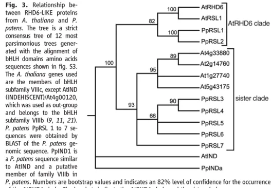
The most ancestral grade of land plants are the bryophytes—the earliest microfossils of land plants from the middle Ordovician (~475 million years ago) have bryophyte charac-teristics (12). Bryophytes do not have roots but possess tip-growing cells that are mor-phologically similar to root hairs and fulfill rooting functions. In mosses, caulonemal cells increase the surface area of the filamentous protonema tissue in contact with the substrate and rhizoids anchor the leafy gametophore to their growth substrate (13, 14); both cell types are hypothesized to be involved in nutrient acquisition (13). However, rhizoids and caulonema develop from the gameto-phyte of mosses, whereas root hairs develop from the sporophyte of modern vascular plants. Thus, according to the current view that land plants evolved by the intercalation of a sporo-phytic generation from a haplontic algal an-cestor followed by the progressive increase of size and complexity of the sporophyte in paral-lel to a reduction of the gametophyte (4, 15), neither rhizoids nor caulonema are homol-ogous to root hairs. To determine if the develop-mental mechanism that controls the development of root hairs in angiosperms also controls the development of nonhomologous tip-growing cells with a rooting function in bryophytes, we identified RHD6-LIKE genes from the moss Physcomitrella patens. We identified seven members of the AtRHD6 subfamily of bHLH genes from the publicly available P. patens genomic sequence (http://moss.nibb.ac.jp/), sug-gesting that these genes have been conserved through the land plant evolution. These were designated Physcomitrella patens RHD SIX-LIKE 1 to 7 (PpRSL1 to PpRSL7). To analyze the relationship between P. patens and A. thaliana RSL genes, we constructed trees by maximum parsimony. A strict consensus tree shows that AtRHD6, AtRSL1, and the two P. patens genes PpRSL1 and PpRSL2 are closely related and together form a monophyletic clade (AtRHD6 clade) that is sister to the clade com-prising all the other members of the subfamily (sister clade) (Fig. 3 and fig. S3). This indicates that the AtRHD6 clade evolved before the separation of the bryophytes and the vascular plants from a common ancestor.
To characterize the function of the RHD6-LIKE genes in moss, we constructed deletion mutants that lacked the function of PpRSL1 and PpRSL2 genes and determined whether they developed morphological defects. Three independent RNA null mutants with single insertions into the PpRSL1 and PpRSL2 genes were made. Double mutants with single in-sertions into both genes were also generated (fig. S4). The phenotypes of each of these mu-tants were then analyzed. A haploid protonema develops upon germination of a wild-type P.
Fig. 3. Relationship be-tween RHD6-LIKE proteins from A. thaliana and P. patens. The tree is a strict consensus tree of 12 most parsimonious trees gener-ated with the alignment of bHLH domains amino acids sequences shown in fig. S3. The A. thaliana genes used are the members of bHLH subfamily VIIIc, except AtIND (INDEHISCENT)/At4g00120, which was used as out-group and belongs to the bHLH subfamily VIIIb (9, 11, 21). P. patens PpRSL 1 to 7 se-quences were obtained by BLAST of the P. patens ge-nomic sequence. PpIND1 is a P. patens sequence similar to AtIND and a putative member of family VIIIb in
P. patens. Numbers are bootstrap values and indicates an 82% level of confidence for the occurrence of the AtRHD6 clade. The brackets indicate the AtRHD6 clade and the sister clade.
Fig. 4. PpRSL1 and PpRSL2 positively control the development of caulonemal cells and rhizoids in P. patens, and PpRSL1 and AtRHD6 have a conserved molecular function. (A and B) Eighteen-day-old protonema from WT, Pprsl1, and Pprsl2 single mutants, and Pprsl1 Pprsl2 double mutant, were grown from spores on 0.8% agar. (A) Whole protonema growing from a single spore. (B) Dissected filaments from protonema shown in (A). (C) Isolated 1-month-old gametophores. (D) Roots of the A. thaliana Atrhd6-3 mutant carrying the 35S::PpRSL1 transgene compared to WT and Atrhd6-3 roots. ca, caulonemal cell; ch, chloronemal cell; rh, rhizoid. Scale bars, 1 mm (A), 100 mm (B), 1 mm (C), and 500mm (D).
patens spore (13). This filamentous tissue comprises two cell types, the chloronema and the caulonema (Fig. 4, A and B). Chloronemal cells contain large chloroplasts and grow by a slow tip-growth mechanism (16). Caulonemal cells are more elongated, contain few smaller chloroplasts, grow by rapid tip growth, and are involved in the colonization of the substrate. Leafy gametophores usually develop from caulonema and are anchored to their substrate by tip-growing multicellular rhizoids that are morphologically similar to caulonema (Fig. 4C). The Pprsl1 and Pprsl2 single mutants have slightly smaller and greener protonema cultures than the wild type (WT), and this phenotype is much stronger in the Pprsl1 Pprsl2 double mutant, which produces small dark-green protonema (Fig. 4A). Pprsl1 and Pprsl2 single mutants produce fewer caulone-mal cells than the WT, indicating that the greener protonema phenotype is the result of a defect in the development of caulonemal cells (Fig. 4B). No caulonemal cells develop in the Pprsl1 Pprsl2 double mutant, and the protonema of this mutant consists of chlo-ronemal cells only (Fig. 4B). In wild-type plants gametophores develop from caulonema, but in the Pprsl1 Pprsl2 double mutants the gameto-phores develop from chloronema, as previous-ly observed in another caulonema-defective mutant (17). The gametophores of the Pprsl1 Pprsl2 double mutant develop few very short rhizoids (Fig. 4C). No other defective pheno-types were detected in the chloronema, in the leafy part of the gametophore, or in the spo-rophyte in the single or double mutants. This indicates that PpRSL1 and PpRSL2 together regulate the development of caulonemal cells and rhizoids in the moss gametophyte. The lack of a defect in chloronemal cells, which are the other tip-growing cells that develop in moss (16), in the Pprsl1 Pprsl2 double mutant shows that, as in A. thaliana, these genes are not general regulators of tip growth. Instead it suggests that they function specifically to regulate the development of cells with rooting functions such as caulonemal cells and rhi-zoids. To determine if protein function is con-served across the land plants, we performed a cross-species complementation experiment. Expression of PpRSL1 under the cauliflower mosaic virus (CaMV) 35S promoter in the Atrhd6-3 mutant resulted in the formation of wild-type root hairs (Fig. 4D). Thus, the moss PpRSL1 gene can substitute for loss of AtRHD6 function in A. thaliana. This indicates that the molecular function of PpRSL1 and AtRHD6 has been conserved since the di-vergence of seed plants and mosses from a common ancestor and suggests that the same molecular mechanism controls the develop-ment of A. thaliana root hairs and P. patens caulonema and rhizoids.
We have shown that closely related tran-scription factors control the development of
root hairs and rhizoids in the seed plant sporophyte and the bryophyte gametophyte, respectively. The demonstration of the exis-tence and function of these genes in plants derived from the earliest colonizers of the land (bryophytes) indicates that an ancient common mechanism controls the develop-ment of these two nonhomologous cell types. The RHD6-LIKE genes will have been impor-tant for the invasion of land by plants because they control the development of structures required for anchorage to the terrestrial substrate and nutrient acquisition. The observation that rhizoids have been found on some of the oldest land-plant fossils is consistent with this view (18–20).
Our demonstration that RHD6-related genes function in both bryophytes and angio-sperms in the development of rhizoids and root hairs, respectively, suggests a mechanism to explain the increased morphological and cellular diversity of the sporophyte in the land plants derived from bryophyte ancestors. Our results suggest that RHD6-LIKE genes func-tioned in the haploid generation (gametophyte) of these early land plants that had a bryophyte-like life cycle (18), where they controlled the formation of cells with a rooting function. Then, during the subsequent radiation of the land plants, these genes were deployed in the development of the diploid generation (sporo-phyte) of the nonbryophyte land plants, where they controlled the development of rhizoids and root hairs. Here we propose a general mod-el for the increase in morphological diversity of the land-plant sporophyte based on these findings. We suggest that some of the genes that controlled the development of the bryo-phyte haploid body were recruited by the diploid phase in their descendants, where they provided part of the genetic mechanism for the increased morphological and cellular diversity of the sporophyte. Thus, the recruit-ment of genes from haploid to diploid phases of the life cycle, in concert with the mod-ification of function of sporophyte-specific genes, such as LFY (5), is a mechanism that may account for the explosion in morpholog-ical diversity of the diploid stage of the life cycle (sporophyte) that occurred in the mid-dle Palaeozoic when green plants colonized the continental surfaces of the planet (3). The full extent of the recruitment of genes from the haploid to the diploid phases during the colonization of the land will be quantified through future comparative analysis of gene function in bryophytes and angiosperms. The discovery and description of more bryophyte fossils from the middle Paleozoic is necessary to unequivocally define the nature of early land-plant life histories. This is important be-cause although it is likely that the earliest land plants had bryophyte-like life cycles, there is still a possibility that their life cycles were unlike those of modern bryophytes.
Only through the combination of paleobotan-ical and developmental genetic approaches will we understand the mechanism by which the land plant body developed over 400 mil-lion years ago.
References and Notes
1. R. A. Berner, in Plants Invade the Land, P. Gensel, D. Edwards, Eds. (Columbia Univ. Press, New York, 2001), pp. 173–178.
2. P. Kenrick, P. R. Crane, Nature 389, 33 (1997). 3. P. Kenrick, P. Davis, Fossil Plants (The Natural History
Museum, London, 2004).
4. L. E. Graham, M. E. Cook, J. S. Busse, Proc. Natl. Acad. Sci. U.S.A. 97, 4535 (2000).
5. T. Tanahashi, N. Sumikawa, M. Kato, M. Hasebe, Development 132, 1727 (2005).
6. R. J. Carol, L. Dolan, Philos. Trans. R. Soc. London B Biol. Sci. 357, 815 (2002).
7. T. S. Gahoonia, D. Care, N. E. Nielsen, Plant Soil 191, 181 (1997).
8. J. D. Masucci, J. W. Schiefelbein, Plant Physiol. 106, 1335 (1994).
9. M. A. Heim et al., Mol. Biol. Evol. 20, 735 (2003). 10. J. Schiefelbein, Curr. Opin. Plant Biol. 6, 74 (2003). 11. P. C. Bailey et al., Plant Cell 15, 2497 (2003). 12. C. H. Wellman, P. L. Osterloff, U. Mohiuddin, Nature
425, 282 (2003).
13. J. G. Duckett, A. M. Schmid, R. Ligrone, in Bryology for the Twenty-First Century, J. W. Bates, N. W. Ashton, J. G. Duckett, Eds. (British Bryological Society, Leeds, UK, 1998), pp. 223–245.
14. K. Sakakibara et al., Development 130, 4835 (2003).
15. W. H. Blackwell, Bot. Rev. 69, 125 (2003). 16. B. Menand, G. Calder, L. Dolan, J. Exp. Bot. 58, 1843
(2007).
17. M. Thelander, T. Olsson, H. Ronne, J. Exp. Bot. 56, 653 (2005).
18. D. Edwards, J. G. Duckett, J. B. Richardson, Nature 374, 635 (1995).
19. H. Kerp, H. Hass, V. Mosbrugger, in Plants Invade the Land, P. Gensel, D. Edwards, Eds. (Columbia Univ. Press, New York, 2001), pp. 52–82.
20. H. Kerp, N. H. Trewin, H. Hass, Trans. R. Soc. Edinb. Earth Sci. 94, 411 (2004).
21. S. J. Liljegren et al., Cell 116, 843 (2004). 22. This research is funded by a grant from the Natural
Environment Research Council (Ne/c510732/1) and Human Frontier Science Program Organization (HFSPO) (to L.D.), and a grant-in-aid from the Biotechnology and Biological Sciences Research Council to the John Innes Centre. B.M. and S.J. were funded by the European Molecular Biology Organization (EMBO) and Marie Curie Fellowships (EMBO ALTF 89-2002 and Marie Curie HPMF-CT-2002-01935 to B.M.). E.R. was supported by a Marie Curie Fellowship. L.H. is funded by the Marie Curie TIPNET network. K.Y. is partially supported by a Joint Scholarship between the University of East Anglia and China Scholarship Council and HFSPO (RGP0012/2005-C). We are grateful to E. Moylan and J. Harrison for advice in using PAUP*. We also thank J. Doonan, N. Harberd, M. Pernas-Ochoa, S. Takeda, and Y. Yasumura for critical comments on the manuscript and N. Pires for help with BLAST of the P. patens genome. We thank J. Langdale and P. Kenrick for invaluable discussions and J. Duckett for teaching us moss morphology.