HAL Id: hal-01907453
https://hal.sorbonne-universite.fr/hal-01907453
Submitted on 29 Oct 2018HAL is a multi-disciplinary open access
archive for the deposit and dissemination of sci-entific research documents, whether they are pub-lished or not. The documents may come from teaching and research institutions in France or abroad, or from public or private research centers.
L’archive ouverte pluridisciplinaire HAL, est destinée au dépôt et à la diffusion de documents scientifiques de niveau recherche, publiés ou non, émanant des établissements d’enseignement et de recherche français ou étrangers, des laboratoires publics ou privés.
Precore G1896A mutation is associated with reduced
rates of HBsAg-seroclearance in treated HIV-hepatitis B
virus co-infected patients from Western Africa Running
title: Precore G1896A mutations in SSA
Anders Boyd, Raoul Moh, Sarah Maylin, Mariama Chekaraou, Nadia
Mahjoub, Delphine Gabillard, Xavier Anglaret, Serge Paul Eholié, Constance
Delaugerre, Christine Danel, et al.
To cite this version:
Anders Boyd, Raoul Moh, Sarah Maylin, Mariama Chekaraou, Nadia Mahjoub, et al.. Precore G1896A mutation is associated with reduced rates of HBsAg-seroclearance in treated HIV-hepatitis B virus co-infected patients from Western Africa Running title: Precore G1896A mutations in SSA. Journal of Viral Hepatitis, Wiley-Blackwell, 2018, 25 (10), pp.1121-1131. �10.1111/jvh.12914�. �hal-01907453�
Precore G1896A mutation is associated with reduced rates of HBsAg-seroclearance in
treated HIV-hepatitis B virus co-infected patients from Western Africa Running title: Precore G1896A mutations in SSA
Anders Boyd1, Raoul Moh2, Sarah Maylin3,4, Mariama Abdou Chekaraou5, Nadia Mahjoub3,
Delphine Gabillard6,7, Xavier Anglaret2,6,7, Serge Paul Eholié2,8,9, Constance Delaugerre3,4,10,
Christine Danel2,6,7, Fabien Zoulim5, Karine Lacombe11,12 for the ANRS 12240 VarBVA study
Institutional affiliations:
1Sorbonne Université, INSERM, Institut Pierre Louis d’Epidémiologie et de Santé Publique
IPLESP, F75012 Paris, France;
2Programme PAC-CI, ANRS Research Site, Treichville University Hospital, Abidjan, Côte
d’Ivoire;
3Laboratoire de Virologie, Hôpital Saint-Louis, AP-HP, Paris, France; 4Université Paris-Diderot, Paris, France;
5Centre de Recherche sur le Cancer de Lyon, Equipes 15 et 16, INSERM, Unité 1052, CNRS,
UMR 5286, Lyon, France;
6INSERM, U1219, Epidémiologie-Biostatistique, Bordeaux, France; 7University of Bordeaux, ISPED, Bordeaux, France;
8Department of Infectious and Tropical Diseases, Treichville University Teaching Hospital,
Abidjan, Côte d’Ivoire;
9Medical School, University Felix Houphouet Boigny, Abidjan, Côte d'Ivoire; 10INSERM U941, Paris, France;
12Sorbonne Université, INSERM, Institut Pierre Louis d’Epidémiologie et de Santé Publique
IPLESP, AP-HP, Hôpital Saint Antoine, F75012 Paris, France.
Corresponding author:
Dr. Anders Boyd
Services des Maladies Infectieuses et Tropicales; Hôpital Saint-Antoine 184 rue du Fbg. St. Antoine
75571 Paris Cedex 12; France Telephone: +33 1 71 97 05 17 Fax: +33 1 49 28 25 95 Email: [email protected]
Acknowledgements: We thank all patients who participated in the ANRS Trivacan and
Temprano trials. We also acknowledge the valuable contributions of the SMIT, CeDReS, CEPREF, USAC, CIRBA, CNTS, La Pierre Angulaire, Hôpital Général Abobo, Formation Sanitaire Anonkoua Kouté, Centre de santé El Rapha, the Programme PACCI team, as well as the INSERM exU593 and U897 teams. We are also grateful to Bristol-Myers Squibb for
providing Zerit and Videx; Gilead Sciences, for the donation of Truvada; and Merck Sharp & Dohme, for the donation of Stocrin.
This study was supported by funds from the Agence Nationale de Recherche sur le Sida et les Hépatites (ANRS 12240). The Trivacan and Temprano studies also received funding from the ANRS (ANRS 1269/ANRS 12104 and ANRS 12136, respectively). A.B. was awarded a post-doctoral fellowship from the ANRS and SIDACTION for some of the work presented in this manuscript.
ABSTRACT
The nucleotide substitution G1896A on the precore (pc) region has been implicated in virological and serological responses during treatment in hepatitis B virus (HBV)-infected patients.
Whether this mutation affects the therapeutic course of HIV-HBV co-infected patients, especially from Western Africa, is unknown. In this prospective cohort study, 86 antiretroviral (ARV)-naïve
HIV-HBV co-infected patients from Côte d’Ivoire, initiating ARV-treatment containing lamivudine (n=53) or tenofovir (n=33), had available baseline pc sequences. Association of the pcG1896A mutation with time-to-undetectable HBV-DNA, hepatitis B “e” antigen (HBeAg)-seroclearance (in HBeAg-positive patients), and hepatitis B surface antigen (HBsAg)-seroclearance was evaluated using Cox proportional hazards regression. At ARV-initiation, median HBV-DNA was 6.04 log10
copies/mL (IQR=3.70-7.93) with 97.7% harboring HBV genotype E. Baseline pcG1896A mutation was identified in 51 (59.3%) patients, who were more commonly HBeAg-negative (p<0.001) and had basal core promotor A1762T/ G1764A mutations (p<0.001). Patients were followed for a median 36 months (IQR=24-36). Cumulative proportion of undetectable HBV-DNA was significantly higher in patients with baseline mutation (pcG1896A=86.6% versus no pcG1896A=66.9%, p=0.04), but not after adjusting for baseline HBV-DNA levels and anti-HBV agent (p=0.2). No difference in cumulative proportion of HBeAg-seroclearance was observed between mutation groups (pcG1896A=57.1% versus no pcG1896A=54.3%, p=0.7). Significantly higher cumulative proportion of HBsAg-seroclearance was observed in patients without this mutation (pcG1896A=0% versus no pcG1896A=36.9%, p<0.001), even after adjusting for baseline HBsAg-quantification and anti-HBV agent (p<0.001). In conclusion, lacking the pcG1896A mutation before ARV-initiation appeared to increase HBsAg-seroclearance rates during treatment. The therapeutic implications of this mutation need further exploration in this setting.
Keywords: antiviral treatment; basal core promotor; genetic variability; immunosuppression;
INTRODUCTION
In sub-Saharan Africa (SSA), it is estimated that approximately 10% of HIV-infected patients have chronic hepatitis B virus (HBV) infection (1). HIV-HBV co-infection is known to increase the risk of severe liver disease and accelerate progression to liver-related death compared to either infection individually (2,3). Circulating HBV is a strong contributor of the increased risk in both liver-related and overall mortality (4,5), hence the importance of reducing serum HBV DNA viral loads through effective antiviral therapy (6,7).
There are, however, certain virological factors of the HBV genome that might impact response during therapy with an anti-HBV nucleoside/nucleotide analogue (NA). For example, patients harboring the precore (pc) G1896A mutation are less likely to achieve lower HBV DNA viral loads during treatment with lamivudine (LAM) (8). Pc mutant variants have been shown in vitro to increase replication yields of LAM-resistant strains, which are normally deficient in replicative capacity (9), and have been clinically associated with faster development of LAM-resistance (10). Importantly, the presence of pc mutations, even when existing as a minority quasi-species, hampers hepatitis B surface antigen (HBsAg) loss during treatment with the potent NA tenofovir (TDF) (11). The pcG1896A mutation would then appear to impose serious consequences on the therapeutic course of NA-treated patients.
Within the continent, data on the prevalence of pc mutations in both HBV mono-infected and HIV-HBV co-infected patients are certainly available (12–16). However, their implications with respect to virological and serological response during anti-HBV NA therapy remain to be elucidated. In the study herein, we used unique data from a cohort of antiretroviral treatment (ART)-naïve HIV-HBV co-infected patients from Côte d’Ivoire initiating treatment with an anti-HBV containing regimen. We first evaluated the distribution of mutations observed on the basal
core promoter (BCP) and pc regions before treatment initiation, while examining risk-factors of harboring the pcG1896A mutation. We then aimed to determine the effect of harboring this mutation on virological and serological response during LAM- or TDF-containing ART.
MATERIALS AND METHODS Study design and visits
The VarBVA study (17) is an observational cohort including patients from two prospective, randomized, open-label, multi-center trials in Abidjan, Côte d’Ivoire: Trivacan ANRS 1269 (NCT00158405), aimed at evaluating the benefits and risks of structured treatment interruption; and Temprano ANRS 12136 (NCT00495651), aimed at evaluating the benefits and risks of starting ART earlier than current World Health Organization (WHO) recommendations. Study randomization and follow-up procedures have been detailed elsewhere (18,19).
Inclusion criteria were as follows – both studies: age ≥18 years, HIV-1 or mixed HIV-1/2 infection, and ART-naïve (with the exception of short-course treatment for the prevention of mother-to-child HIV transmission); Trivacan: CD4 cell count between 150-350/mm3 or CD4
percentage between 12.5%-20.0%; Temprano: CD4 cell count <800/mm3 and no concurrent
criteria for ART-initiation according to most recent WHO guidelines. Non-inclusion criteria are summarized in the Supplementary methods. All participants gave written informed consent and approval of the study protocols were obtained by the Ministry of Health of Côte d’Ivoire and the French National Agency for Research on AIDS and Viral Hepatitis (ANRS, Paris, France).
For this study, we included patients testing HBsAg-positive at study inclusion (Mini Vidas® assay; Biomerieux, Marcy l’Etoile, France), confirmed by the HBsAg Qual II Architect assay (Abbott Laboratories, Rungis, France), and who started ART containing LAM and/or TDF at inclusion or any time during participation in the trial (Supplementary Methods). We defined the “baseline” visit at ART-initiation and “follow-up” visits at each yearly visit thereafter until the date of last follow-up (with available frozen sample), study termination, permanent treatment
discontinuation, or treatment switch. In the Temprano study, if the last follow-up visit was not on the yearly interval, the closest 6-month visit was used instead.
HBV-related parameters
Plasma alanine aminotransferase (ALT) and aspartate aminotransferase (AST) levels were quantified at baseline. From frozen samples stored at -80°C, HBV-DNA viral loads were
quantified at baseline and every follow-up visit using an in-house PCR-based assay (QuantiFast SYBR® Green PCR kit, Qiagen, Courtaboeuf, France; Light Cycler 480, Roche, Boulogne-Billancourt, France) with a detection limit of 12 copies/mL (17). Qualitative HBsAg was detected
using the HBsAg Qual II test (detection limit=0.03 IU/mL, Architect, Abbott Laboratories, Rungis, France) at baseline and during follow-up. Qualitative HBeAg and anti-HBe antibodies (anti-HBeAb) were detected using the Elecsys assay (Roche Diagnostics, Meylan, France) for the same visits. HBeAg-seroclearance was defined as HBeAg-loss from the previous visit in
HBeAg-positive patients. HBsAg-seroclearance was defined as loss of HBsAg from the previous visit for all patients.
HBeAg quantification (qHBeAg) and HBsAg quantification (qHBsAg) were quantified for HBeAg and HBsAg positive samples, respectively, using the Elecsys assay with the Modular E170 analyzer (Roche Diagnostics, Meylan, France). qHBeAg levels were provided as a
semi-quantitative result and were interpreted as the ratio of sample relative light units to a cut-off value. These units were converted to Paul Ehrlich Institute units (PEI U/mL) from a previously established protocol (20).
Detection of HBV mutations on viral genome sequences
Genotypic analysis was performed at baseline for all patients with an HBV DNA viral load >1000 copies/mL (17). Nucleotide (nt) sequences from the BCP and pc regions were examined by direct sequencing after nested-PCR amplification of the preC/C gene (nt 1743-2362). Individual sequences were aligned using the ClustalW full multiple alignment program with 1000
bootstraps in BioEdit (v7.0.5.3, Carlsbad, CA) and were compared to a consensus sequence from 454 genotype E and 500 genotype A preC/C nucleotide sequences retrieved from the HBVdb (21).
Amino acid sequences of the pol and S-genes were also examined after PCR amplification of the reverse transcriptase (rt) and surface antigen (s) encoding regions (between rt107-rt385 and s99-s226, respectively). Mutant variants were detected using similar methods as described above (17).
Phylogenetic analysis
HBV genotypes were determined via phylogenetic analysis conducted on aligned sequences of the pc region (nt 1814-2452), which were compared to several referent sequences of HBV genotypes A-H (GenBank accession numbers listed in Supplementary Table 1). The neighbor-joining method was employed with 1000 bootstrap replications using a Kimura-2 parameter substitution model that included transitions and transversions (d). A discrete gamma model with
mean equal to one was also used to incorporate rate variation among sites. The resulting phylogenetic tree was constructed using MEGA6 software (22) and is provided in
Supplementary Figure 1.
Statistical analysis
The distribution of specific mutations of the preC/C gene (13) at baseline was described. We chose to focus further analysis on the pcG1896A mutation due to its strong association with virological and serological responses (23). Determinants of harboring this mutation at inclusion were examined using logistic regression. Demographic, treatment, and clinical characteristics related to HIV and HBV-infection with a p≤0.1 in univariable analysis were retained and used to create a predictive, multivariable model. A backwards-stepwise selection process was then performed, removing any co-variable greater than this p-value threshold.
We then assessed the effect of harboring the pcG1896A mutation at inclusion on virological and serological parameters during treatment. The cumulative probability of time until undetectable HBV, time until HBeAg-seroclearance (for patients with positive HBeAg at baseline), and time until HBsAg-seroclearance was calculated using Kaplain-Meier curves. Survivor functions were stratified on mutation groups and compared using the log-rank test. Cox proportional hazards regression was also used to compare rates of these events, both unadjusted and adjusted for baseline quantified parameter (HBV DNA, qHBeAg or qHBsAg) and anti-HBV treatment (LAM versus TDF).
Additionally, qHBsAg levels were summarized for each study visit. Changes in antigen levels from baseline were modeled using a mixed-effect linear regression with a random-intercept to account for between-patient variability. The model included time and mutation group as
independent variables along with the interaction between the two, which was tested in order to determine differences in overall on-treatment antigen decline between mutation groups.
All analyses were performed using STATA (v12.1, College Station, TX, USA). All statistical tests were two-sided and a p-value of <0.05 was considered significant.
RESULTS
Description of the study population
Among the 259 HBsAg-positive patients enrolled in both trials, 173 were not included in analysis for the following reasons: did not have confirmed HBsAg-positive serology (n=31) or had
suspected acute HBV-infection (n=1), never initiated ART (n=30), had only one follow-up visit (n=16), discontinued ART <6 months due to pregnancy (n=3) or adverse event (n=2), had missing data on HBV viral loads (n=8) or missing data on BCP or pc genetic variability (n=82). In total, 86 patients were included in analysis. Demographic, HIV-related and HBV-related characteristics at baseline are summarized in Table 1. These characteristics are compared between source studies in Supplementary Table 2, demonstrating a significantly lower CD4+ T-cell count (p<0.001) and higher proportion with WHO clinical stage III/IV (p=0.007) among participants enrolled in the Trivacan versus Temprano study.
Basal core promoter/precore mutant variants at baseline
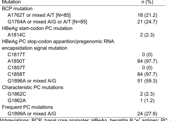
Table 2 describes specific BCP and pc mutations identified at baseline. The most common mutations were A1850T and C1858T nt substitutions on the pc region. Roughly two-thirds of
patients (59.3%) harbored the G1896A or mixed G1896G/A pc mutation and almost one-quarter of patients harbored the A1762T (21.2%) or G1764A (24.7%) BCP mutations. Almost all
patients with a BCP mutation at these nt positions also had the pcG1896A mutation (n=19/21, 90.5%).
As shown in Table 3, the pcG1896A mutation at baseline was associated with HBeAg-negative status (p<0.001), anti-HBe antibody positive status (p<0.001), lower HBV DNA levels (p=0.001), and presence of BCP mutations (p=0.004). Of note, median ALT/AST levels were not
significantly different in patients with versus without the pcG1896A mutation (32/39 IU/mL versus 25/40 IU/mL, respectively, p=0.10/0.6). Only two patients harbored a genotype other than E (genotype A) in whom no pcG1896A mutation was detected. In multivariable analysis, HBeAg-negative status (p<0.001) and presence of BCP mutations (p<0.001) were significantly and independently associated with baseline presence of pcG1896A mutations (Table 3), while considering the strong collinearity between associated variables.
Baseline pcG1896A mutation is not associated with virological response and antiviral resistance during treatment
Patients were followed for a median 36 (IQR=24-36) months. Of the 53 patients undergoing LAM-containing ART, 24 (45.3%) had per protocol treatment interruptions after a median 12 (IQR=11-37) months of follow-up. Median cumulative duration of treatment was 26 (IQR=19-34) months for all those undergoing LAM. All 33 TDF-treated patients underwent continuous ART for a median 24 (IQR=17-30) months.
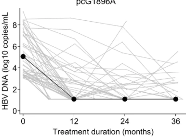
Individual and median HBV DNA viral loads during ART containing an anti-HBV agent are provided in Figure 1A, while stratified on baseline pcG1896A status. During follow-up,
undetectable HBV DNA was achieved in 66 patients after a median 12 (IQR=11-23) months of follow-up (cumulative proportion=78.9%). Time to undetectable HBV DNA was significantly faster for patients with versus without baseline pcG1896A (cumulative proportion= 86.6% versus 66.9%, respectively, log rank test p=0.04) (Figure 1B). However, this association was no longer significant after adjusting for baseline HBV DNA levels and anti-HBV agent (p=0.2). At the end of follow-up, 31 (36.1%) patients had detectable HBV DNA, with no significant difference between mutation groups (with pcG1896A=31.4% versus without pcG1896A=42.9%, p=0.3). Thirteen (41.9%) of these patients had an HBV DNA viral load >10,000 copies/mL, only one of whom developed the LAM-resistance rtV173L+rtL180M+rtM204V mutation during treatment (without the pcG1896A mutation at baseline).
Lack of baseline pcG1896A mutation is associated with HBsAg, but not HBeAg, quantification and serological response during treatment
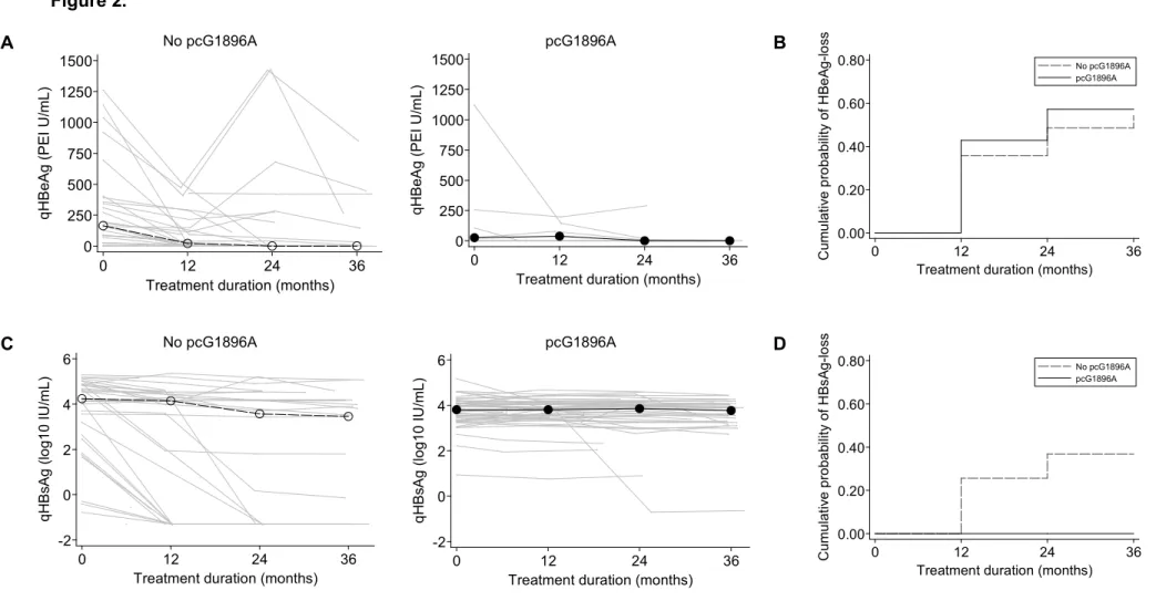
In the 35 HBeAg-positive patients at baseline, median qHBeAg levels decreased from 160.4 PEI U/mL (IQR=24.9-355.4) at treatment initiation to <0.05 PEI U/mL (IQR=<0.05-115.7) at the end of follow-up. At baseline, qHBeAg levels were lower in patients with versus without the
pcG1896A mutation (24.9 PEI U/mL, n=7 versus 164.6 PEI U/mL, n=27, respectively), yet this association was not significant (p=0.13). No significant differences between mutation groups were observed in change of qHBeAg levels from baseline (p=0.9), owing to the low antigen levels overall during anti-HBV treatment (Figure 2A). HBeAg-seroclearance occurred in 18 patients after a median 12 months (IQR=11-23) of follow-up (cumulative proportion=55.3%), with no significant difference in time to HBeAg-seroclearance between patients with versus without the pcG1896A mutation (cumulative proportion=57.1% versus 54.3%, respectively, p=0.7) (Figure 2B).
Overall, median qHBsAg levels decreased from 4.00 log10 IU/mL (IQR=3.36-4.36) at treatment
initiation to 3.79 log10 IU/mL (IQR=3.10-4.15) at the end of follow-up. qHBsAg levels at baseline
were significantly lower in patients with versus without the pcG1896A mutation (3.81 versus 4.24 log10 IU/mL, respectively, p=0.02). Over time, change in qHBsAg levels from baseline was
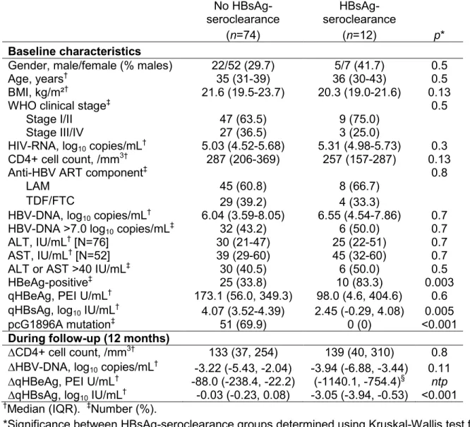
significantly faster in patients without the pcG1896A mutation (p<0.001) with substantial between-patient variability (Figure 2C). HBsAg-seroclearance occurred in 12 patients after a median 12 months (IQR=21) of follow-up (cumulative proportion=14.5%). Baseline and 12-month characteristics of patients are compared between those with versus without HBsAg-seroclearance in Table 4. Ten and two of these patients were positive and HBeAg-negative, respectively, with HBeAg-positive patients more likely to lose HBsAg (log-rank test
p=0.001). As shown in Figure 2D, only patients without the pcG1896A mutation exhibited
HBsAg-seroclearance (p<0.001). This significant association held when adjusting for baseline qHBsAg levels and anti-HBV treatment regimen (p<0.001).
Low incidence of pcG1896A mutation during treatment
Of the 35 patients without the pcG1896A mutation at baseline, two patients developed incident pcG1896A mutations during treatment (IR=2.5/100 person-years). The first patient had high HBV DNA levels at treatment initiation (7.57 log10 copies/mL). The patient was undergoing
LAM-containing ART with CD4+ T-cell guided therapeutic interruptions. The G1896G/A mutation was detected at the month-24 visit with an HBV DNA viral load at 228,000 copies/mL, which
gradually declined to 9260 copies/mL at the last study visit. The patient remained HBeAg-positive and HBsAg-HBeAg-positive during follow-up. The second patient also had high HBV DNA at treatment initiation (7.93 log10 copies/mL). After initiating continuous LAM-containing ART, HBV
DNA viral loads steadily decreased to 1074 copies/mL at month-24, when the G1896G/A mutation was detected, and finally to 810 copies/mL at the last study visit. The patient lost both
HBeAg and HBsAg during follow-up, while no apparent mutations on the “a” determinant of the
S-gene emerged. No LAM-resistance mutations were observed for either patient.
DISCUSSION
The pcG1896A mutation is frequently observed around the time of HBeAg-seroconversion, allowing abrogated HBeAg-production with persistent HBV DNA replication and possibly immune escape from HBeAg-directed immune responses (23). Since most patients in Western Africa are HBeAg-negative and infected with HBV genotypes prone to the G1896A nt substitution (24,25), it is assumed that a substantial proportion with detectable HBV DNA replication would be infected with this mutation.
At treatment initiation, we did indeed identify pcG1896A mutant variants in almost two-thirds of co-infected patients harboring genotype E, reflecting other studies in this region (26–28). The presence of this mutation was associated with widely-recognized determinants of more inactive forms of HBV infection, such as low HBV DNA replication and HBeAg-negative status. Since the G to A substitution at position pc1896 replaces tryptophan for a stop codon during translation of the HBeAg and thereby limits its production (23), it is unsurprising that this mutation was not frequently observed in HBeAg-positive individuals. BCP mutations at either nt 1762 or 1764 were almost always present with the pcG1896A mutation in our study and are also associated with reduced HBeAg production and HBeAg-negative status (29). Meanwhile, CD4+ T-cell concentrations failed to show an association with baseline pc mutation and would suggest that HIV-induced immunosuppression might not play an essential role on the genetic variability of the pc region (30).
We further our understanding on the therapeutic role of the pcG1896A mutation in genotype E infection, while clearly demonstrating that patients with this mutation at treatment initiation will likely have difficulty in clearing HBsAg during NA-based therapy. Reduced
HBsAg-seroclearance rates have been described with this mutation, as well as with increased genetic diversity of the pc encoding region, during TDF-treatment in HBV mono-infected patients (11). The importance of this mutation was further evidenced when deep-sequencing was applied, in which pcG1896A mutant strains detected as even minority variants (>1% of the HBV
quasispecies pool) were able to produce greater sensitivity in predicting HBsAg-seroclearance (11). Interestingly, these results originated from a study population with HBV genotypes A-D, high transaminase levels, and male and HBeAg-positive predominance. The fact that our study comprised mostly HBeAg-negative women with genotype E would suggest the robustness of this association across different HBV-infected populations.
The pathophysiological explanations of this result remain largely unknown. HBsAg-seroclearance is mainly viewed as the consequence of complex and appropriate immune responses during the course of infection (31). NA-based therapy does not have substantial immunomodulatory effects compared to other treatment options, such as pegylated-interferon (32). Regardless, higher rates of HBsAg-seroclearance are often observed during HBeAg-positive phases of HBV when patients are undergoing treatment with any antiviral agent (33). When replacing HBeAg-positive status with the lack of pcG1896A mutation, we were able to obtain a marginally higher sensitivity associated with loss of HBsAg in our study. Two HBeAg-negative individuals had HBsAg-seroclearance, both of whom did not harbor the pcG1896A mutation. From our data, it is difficult to determine if these individuals recently had HBeAg-seroclearance prior to study inclusion or if the pcG1896A was present as a minority quasi-species. As increased pc genetic diversity during HBeAg-seroconversion spans over several years (34), perhaps the presence of pc mutations might be a more accurate reflection of later
stages of HBV-infection when HBsAg-seroclearance is most difficult to clear. Whether this means that earlier treatment prior to pc mutant emergence should be considered remains to be determined.
Patients harboring the pcG1896A mutation at baseline did not have increased rates of HBeAg-seroclearance, which was somewhat unexpected (8). One reason could be the few patients with HBeAg-positive serology harboring this mutation at baseline, thereby reducing the power to establish any difference. On the other hand, qHBeAg production did seem to persist for some patients with the pc mutation. It could be speculated that the quasi-species make-up of pcG1896A mutant variants remained at consistent levels during follow-up. Substantial evolutionary changes on the pc encoding region over time are needed in order for HBeAg-seroconversion to occur, at least during the natural history of infection (34), which might not have been the case in our study. This might also explain why there was no significant
association with this mutation and qHBeAg at treatment initiation and during treatment, despite the well-established impact that the pcG1896A mutation has on HBeAg-production in clinical settings (35,36). This hypothesis could be further clarified by examining the specific viral subpopulations during treatment using next-generation sequencing technologies.
We did not observe any association with the pcG1896A mutation at baseline and virological response or LAM-resistance, which is in contrast to other studies evaluating the use of NA-based agents in HBV mono-infected patients (8,10). Several noteworthy features of our cohort could explain this finding. Persistent viremia observed in these patients was mostly due to insufficient follow-up, immunocompromised status, or for some, LAM-interruption (17). TDF is also highly effective in suppressing HBV DNA among co-infected patients, even when
pcG1896A mutations are present (37). These factors would have eclipsed any purported role of pcG1896A mutations on HBV suppression. In addition, HBV DNA viral loads and transaminase
levels were for the most part low at treatment initiation. These conditions might have been ideal to abate the emergence of LAM-resistance (38) and have appeared to be associated with low LAM-resistance rates in other treated, co-infected populations from SSA (39). Considering that only one patient in this study had LAM-resistance, there was an insufficient number of events to appropriately address this question.
As previously shown in treated co-infected patients (40), the pcG1896A mutation emerged infrequently during follow-up. It is debatable, however, whether this pc mutation was truly incident. Appearance of the G1896A nt substitution did not occur in concert with ALT increases, which is a common event in HBeAg-seroconversion when genetic alterations on the pc region are readily observed (34,41). Furthermore, mixed wildtype and pcG1896A was observed in the two patients identified with incident mutation. These patients potentially had minority variants at treatment initiation and displayed fluctuations in mutant populations detected with population sequencing during therapy. This is an important consideration given that one of the patients with incident pcG1896G/A mutation lost HBsAg. Had this mutant variant been detected by deep-sequencing, it would have reduced the sensitivity in predicting HBsAg-seroclearance.
Nevertheless, this patient had a unique progression of HBV infection with continuing HBV DNA replication despite negative HBsAg serology.
Other limitations of our study need to be addressed. First, we excluded patients with severely elevated transaminases or clinical signs of severe liver disease at study inclusion. This criterion probably resulted in the lack of liver-related clinical events observed during follow-up and thus the relationship between BCP or pc mutations and liver-related morbidity was unable to be assessed. Conversely, individuals with occult HBV infection, which is rather prevalent in SSA (16), were not included in our study. Although the low levels of HBV DNA often observed in these patients make it difficult to conduct sequencing, the few reports of occult infection in SSA
do not support high prevalence of the pcA1896G mutations during this infection “phase” (42). Second, almost all patients undergoing LAM were taking part in the Trivacan study, with most interrupting treatment during follow-up. ART-interruptions could have affected time to
undetectable HBV DNA (17) and some caution should be given when interpreting the
association between pc mutations and virological response. Third, almost all patients with the pcG1896A mutation also harbored mutations on the BCP. Although previous research has shown differential clinical outcomes for patients with various BCP and pc mutation combinations (43), this stratification was unfeasible in our analysis. Fourth, HBsAg was tested only once during study enrollment. Chronic HBV infection (i.e. minimum 6 months of HBsAg-positive serology) was not firmly established at study inclusion and those with HBsAg-seroclearance could have had acute HBV infection. Nevertheless, the vast majority of HBV-infected patients from SSA acquire HBV horizontally during childhood or adolescence (44), hence this scenario would be considered fairly unlikely.
Another consideration worth mentioning is the higher rate of HBeAg- and HBsAg-seroclearance from what would be expected among treated HBV mono-infected patients (33). Other studies in European and Asian co-infected cohorts have observed this phenomenon, which has been linked to accelerated ART-induced immunorestauration under severe immunosuppression (45,46). Individuals with versus without HBsAg-seroclearance did have somewhat lower CD4 T-cell count at baseline, yet the lack of difference in on-treatment CD4 increases would suggest that immunocompromised status might not explain most of the seroclearance events observed in our study.
In conclusion, the lack of pcG1896A mutation at baseline, as determined by population
sequencing, was strongly linked to HBsAg-seroclearance in this group of NA-treated co-infected patients. However, rates of HBeAg-seroclearance and virological response were no different
between patients with or without this pc mutation. In light of these findings, there could be a strong rationale to initiate NA-based therapy in HBV-infected patients from this setting before the pcG1896A mutation emerges. Nevertheless, future research would be needed to investigate the genetic diversity of the pc and BCP encoding regions of these strains and confirm its therapeutic and clinical utility.
REFERENCES
1. Matthews PC, Geretti AM, Goulder PJR, Klenerman P. Epidemiology and impact of HIV coinfection with hepatitis B and hepatitis C viruses in Sub-Saharan Africa. J Clin Virol. 2014 Sep;61(1):20–33.
2. Ioannou GN, Bryson CL, Weiss NS, Miller R, Scott JD, Boyko EJ. The prevalence of cirrhosis and hepatocellular carcinoma in patients with human immunodeficiency virus infection. Hepatology. 2013 Jan;57(1):249–57.
3. Falade-Nwulia O, Seaberg EC, Rinaldo CR, Badri S, Witt M, Thio CL. Comparative risk of liver-related mortality from chronic hepatitis B versus chronic hepatitis C virus infection. Clin Infect. 2012 Aug;55(4):507–13.
4. Chen C-J, Yang H-I, Iloeje UH, REVEAL-HBV Study Group. Hepatitis B virus DNA levels and outcomes in chronic hepatitis B. Hepatology. 2009 May;49(5 Suppl):S72-84.
5. Kouamé G-M, Boyd A, Moh R, Badje A, Gabillard D, Ouattara E, et al. Higher mortality despite early ART in HIV and hepatitis B virus coinfected patients with high HBV replication. Clin Infect Dis. 2018 Jan 6;66(1):112-120.
6. Hawkins C, Christian B, Ye J, Nagu T, Aris E, Chalamilla G, et al. Prevalence of hepatitis B co-infection and response to antiretroviral therapy among HIV-infected patients in
7. Papatheodoridis GV, Chan HL-Y, Hansen BE, Janssen HLA, Lampertico P. Risk of hepatocellular carcinoma in chronic hepatitis B: Assessment and modification with current antiviral therapy. J Hepatol. 2015 Apr;62(4):956–67.
8. Zoutendijk R, Sonneveld MJ, Reijnders JGP, van Vuuren AJ, Biesta P, Hansen BE, et al. Precore and core promoter mutants are associated with higher HBeAg seroconversion but low disease remission rates in HBV patients treated with nucleos(t)ide analogues. J Viral Hepat. 2013 May;20(5):322–7.
9. Chen RY m, Edwards R, Shaw T, Colledge D, Delaney WE, Isom H, et al. Effect of the G1896A precore mutation on drug sensitivity and replication yield of lamivudine-resistant HBV in vitro. Hepatology. 2003 Jan;37(1):27–35.
10. Thompson AJV, Ayres A, Yuen L, Bartholomeusz A, Bowden DS, Iser DM, et al.
Lamivudine resistance in patients with chronic hepatitis B: role of clinical and virological factors. J Gastroenterol Hepatol. 2007 Jul;22(7):1078–85.
11. Bayliss J, Yuen L, Rosenberg G, Wong D, Littlejohn M, Jackson K, et al. Deep sequencing shows that HBV basal core promoter and precore variants reduce the likelihood of HBsAg loss following tenofovir disoproxil fumarate therapy in HBeAg-positive chronic hepatitis B. Gut. 2017 Nov;66(11):2013–23.
12. Andersson MI, Maponga TG, Ijaz S, Barnes J, Theron GB, Meredith SA, et al. The epidemiology of hepatitis B virus infection in HIV-infected and HIV-uninfected pregnant women in the Western Cape, South Africa. Vaccine. 2013 Nov 12;31(47):5579–84. 13. Olinger CM, Venard V, Njayou M, Oyefolu AOB, Maïga I, Kemp AJ, et al. Phylogenetic
analysis of the precore/core gene of hepatitis B virus genotypes E and A in West Africa: new subtypes, mixed infections and recombinations. J Gen Virol. 2006 May;87(Pt 5):1163– 73.
14. Mayaphi SH, Martin DJ, Mphahlele MJ, Blackard JT, Bowyer SM. Variability of the preC/C region of hepatitis B virus genotype A from a South African cohort predominantly infected with HIV. J Med Virol. 2013 Nov;85(11):1883–92.
15. Baudi I, Iijima S, Chin’ombe N, Mtapuri-Zinyowera S, Murakami S, Isogawa M, et al. Molecular epidemiology of co-infection with hepatitis B virus and human immunodeficiency virus (HIV) among adult patients in Harare, Zimbabwe. J Med Virol. 2017 Feb;89(2):257– 66.
16. Bell TG, Makondo E, Martinson NA, Kramvis A. Hepatitis B virus infection in human immunodeficiency virus infected southern African adults: occult or overt--that is the question. PloS One. 2012;7(10):e45750.
17. Boyd A, Moh R, Gabillard D, le Carrou J, Danel C, Anglaret X, et al. Low risk of lamivudine-resistant HBV and hepatic flares in treated HIV-HBV-coinfected patients from Côte d’Ivoire. Antivir Ther. 2015;20(6):643–54.
18. Danel C, Moh R, Minga A, Anzian A, Ba-Gomis O, Kanga C, et al. CD4-guided structured antiretroviral treatment interruption strategy in HIV-infected adults in west Africa (Trivacan ANRS 1269 trial): a randomised trial. Lancet. 2006 Jun 17;367(9527):1981–9.
19. Danel C, Moh R, Chaix M-L, Gabillard D, Gnokoro J, Diby C-J, et al. Two-months-off, four-months-on antiretroviral regimen increases the risk of resistance, compared with
continuous therapy: a randomized trial involving West African adults. J Infect Dis. 2009 Jan 1;199(1):66–76.
20. Maylin S, Boyd A, Martinot-Peignoux M, Delaugerre C, Bagnard G, Lapalus M, et al. Quantification of hepatitis B e antigen between Elecsys HBeAg and Architect HBeAg assays among patients infected with hepatitis B virus. J Clin Virol. 2013 Apr;56(4):306–11. 21. Hayer J, Jadeau F, Deléage G, Kay A, Zoulim F, Combet C. HBVdb: a knowledge
22. Tamura K, Stecher G, Peterson D, Filipski A, Kumar S. MEGA6: Molecular Evolutionary Genetics Analysis Version 6.0. Mol Biol Evol. 2013 Dec;30(12):2725–9.
23. Chotiyaputta W, Lok ASF. Hepatitis B virus variants. Nat Rev Gastroenterol Hepatol. 2009 Aug;6(8):453–62.
24. Hadziyannis SJ. Natural history of chronic hepatitis B in Euro-Mediterranean and African countries. J Hepatol. 2011 Jul;55(1):183–91.
25. Tong S, Revill P. Overview of hepatitis B viral replication and genetic variability. J Hepatol. 2016 Apr;64(1 Suppl):S4-16.
26. Suzuki S, Sugauchi F, Orito E, Kato H, Usuda S, Siransy L, et al. Distribution of hepatitis B virus (HBV) genotypes among HBV carriers in the Cote d’Ivoire: complete genome
sequence and phylogenetic relatedness of HBV genotype E. J Med Virol. 2003 Apr;69(4):459–65.
27. Grant J, Agbaji O, Kramvis A, Yousif M, Auwal M ’azu, Penugonda S, et al. Hepatitis B virus sequencing and liver fibrosis evaluation in HIV/HBV co-infected Nigerians. Trop Med Int Health. 2017 Jun;22(6):744–54.
28. Candotti D, Opare-Sem O, Rezvan H, Sarkodie F, Allain J-P. Molecular and serological characterization of hepatitis B virus in deferred Ghanaian blood donors with and without elevated alanine aminotransferase. J Viral Hepat. 2006 Nov;13(11):715–24.
29. Jansen L, Welkers MRA, van Dort KA, Takkenberg RB, Lopatin U, Zaaijer HL, et al. Viral minority variants in the core promoter and precore region identified by deep sequencing are associated with response to peginterferon and adefovir in HBeAg negative chronic hepatitis B patients. Antiviral Res. 2017 Sep;145:87–95.
30. Audsley J, Littlejohn M, Yuen L, Sasadeusz J, Ayres A, Desmond C, et al. HBV mutations in untreated HIV-HBV co-infection using genomic length sequencing. Virology. 2010 Sep 30;405(2):539–47.
31. Yuan T, Jiang Y, Li M, Li W. Chronic hepatitis B surface antigen seroclearance-related immune factors. Hepatol Res. 2017 Jan;47(1):49–59.
32. Marcellin P, Ahn SH, Ma X, Caruntu FA, Tak WY, Elkashab M, et al. Combination of Tenofovir Disoproxil Fumarate and Peginterferon α-2a Increases Loss of Hepatitis B Surface Antigen in Patients With Chronic Hepatitis B. Gastroenterology. 2016 Jan;150(1):134–144.e10.
33. European Association for the Study of the Liver. EASL 2017 Clinical Practice Guidelines on the management of hepatitis B virus infection. J Hepatol. 2017 Aug;67(2):370–98.
34. Cheng Y, Guindon S, Rodrigo A, Wee LY, Inoue M, Thompson AJV, et al. Cumulative viral evolutionary changes in chronic hepatitis B virus infection precedes hepatitis B e antigen seroconversion. Gut. 2013 Sep;62(9):1347–55.
35. Thompson AJV, Nguyen T, Iser D, Ayres A, Jackson K, Littlejohn M, et al. Serum hepatitis B surface antigen and hepatitis B e antigen titers: disease phase influences correlation with viral load and intrahepatic hepatitis B virus markers. Hepatology. 2010 Jun;51(6):1933–44. 36. Maylin S, Boyd A, Lavocat F, Gozlan J, Lascoux-Combe C, Miailhes P, et al. Kinetics of
hepatitis B surface and envelope antigen and prediction of treatment response to tenofovir in antiretroviral-experienced HIV-hepatitis B virus-infected patients. AIDS. 2012 May 15;26(8):939–49.
37. Boyd A, Gozlan J, Maylin S, Delaugerre C, Peytavin G, Girard P-M, et al. Persistent viremia in human immunodeficiency virus/hepatitis B coinfected patients undergoing long-term tenofovir: virological and clinical implications. Hepatology. 2014 Aug;60(2):497–507. 38. Gish R, Jia J-D, Locarnini S, Zoulim F. Selection of chronic hepatitis B therapy with high
barrier to resistance. Lancet Infect Dis. 2012 Apr;12(4):341–53.
39. Thio CL, Smeaton L, Hollabaugh K, Saulynas M, Hwang H, Saravanan S, et al. Comparison of HBV-active HAART regimens in an HIV-HBV multinational cohort: outcomes through 144 weeks. AIDS. 2015 Jun 19;29(10):1173–82.
40. Boyd A, Lacombe K, Lavocat F, Miailhes P, Lascoux-Combe, Girard P-M, et al. Low incidence of precore W28* mutant variants in treated hepatitis B virus and human immunodeficiency virus co-infected patients. Antiviral Res. 2018 Jan;149:174-178. 41. Yuen M-F, Yuan H-J, Hui C-K, Wong DK-H, Wong W-M, Chan AO-O, et al. A large
population study of spontaneous HBeAg seroconversion and acute exacerbation of chronic hepatitis B infection: implications for antiviral therapy. Gut. 2003 Mar;52(3):416–9.
42. Makondo E, Bell TG, Kramvis A. Genotyping and molecular characterization of hepatitis B virus from human immunodeficiency virus-infected individuals in southern Africa. PloS One. 2012;7(9):e46345.
43. Ducancelle A, Pivert A, Bertrais S, Boursier J, Balan V, Veillon P, et al. Different
precore/core mutations of hepatitis B interact with, limit, or favor liver fibrosis severity. J Gastroenterol Hepatol. 2016 Oct;31(10):1750–6.
44. Lemoine M, Eholié S, Lacombe K. Reducing the neglected burden of viral hepatitis in Africa: strategies for a global approach. J Hepatol. 2015 Feb;62(2):469–76.
45. Miailhes P, Trabaud M-A, Pradat P, Lebouché B, Chevallier M, Chevallier P, et al. Impact of highly active antiretroviral therapy (HAART) on the natural history of hepatitis B virus (HBV) and HIV coinfection: relationship between prolonged efficacy of HAART and HBV surface and early antigen seroconversion. Clin Infect Dis. 2007 Sep 1;45(5):624–32. 46. Matthews GV, Ali RJ, Avihingsanon A, Amin J, Hammond R, Bowden S, et al. Quantitative
HBsAg and HBeAg predict hepatitis B seroconversion after initiation of HAART in HIV-HBV coinfected individuals. PloS One. 2013;8(4):e61297.
TABLES
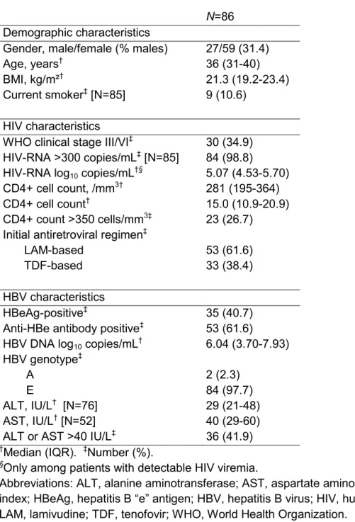
Table 1. Description of the study population at treatment initiation
N=86
Demographic characteristics
Gender, male/female (% males) 27/59 (31.4)
Age, years† 36 (31-40)
BMI, kg/m²† 21.3 (19.2-23.4)
Current smoker‡ [N=85] 9 (10.6)
HIV characteristics
WHO clinical stage III/VI‡ 30 (34.9)
HIV-RNA >300 copies/mL‡ [N=85] 84 (98.8)
HIV-RNA log10 copies/mL†§ 5.07 (4.53-5.70)
CD4+ cell count, /mm3† 281 (195-364)
CD4+ cell count† 15.0 (10.9-20.9)
CD4+ count >350 cells/mm3‡ 23 (26.7)
Initial antiretroviral regimen‡
LAM-based 53 (61.6)
TDF-based 33 (38.4)
HBV characteristics
HBeAg-positive‡ 35 (40.7)
Anti-HBe antibody positive‡ 53 (61.6)
HBV DNA log10 copies/mL† 6.04 (3.70-7.93)
HBV genotype‡
A 2 (2.3)
E 84 (97.7)
ALT, IU/L† [N=76] 29 (21-48)
AST, IU/L† [N=52] 40 (29-60)
ALT or AST >40 IU/L‡ 36 (41.9) †Median (IQR). ‡Number (%).
§Only among patients with detectable HIV viremia.
Abbreviations: ALT, alanine aminotransferase; AST, aspartate aminotransferase; BMI, body mass index; HBeAg, hepatitis B “e” antigen; HBV, hepatitis B virus; HIV, human immunodeficiency virus; LAM, lamivudine; TDF, tenofovir; WHO, World Health Organization.
Table 2. Distribution of specific basal core promotor (BCP) and precore (PC) mutations
Mutation n (%)
BCP mutation
A1762T or mixed A/T [N=85] 18 (21.2)
G1764A or mixed A/G or A/T [N=85] 21 (24.7) HBeAg start-codon PC mutation
A1814C 2 (2.3)
HBeAg PC stop-codon apparition/pregenomic RNA encapsidation signal mutation
C1817T 0 (0)
A1850T 84 (97.7)
C1857T 0 (0)
C1858T 84 (97.7)
G1896A or mixed A/G 51 (59.3)
Characteristic PC mutations
G1862C 2 (2.3)
G1862A 1 (1.2)
Frequent PC mutations
G1899A or mixed A/G 24 (27.9)
Table 3. Determinants of precore G1896A mutation at baseline
Univariable Multivariable†
OR (95%CI) p OR (95%CI) p
Age per year 1.03 (0.97-1.09) 0.4
Male versus female 1.00 (0.40-2.53) 0.9 BMI >25 kg/m² 2.63 (0.51-13.46) 0.2 WHO Stage III or IV 0.69 (0.28-1.68) 0.4 HIV RNA per log10 copies/mL 1.03 (0.63-1.68) 0.9
CD4 cell count
>500 mm3 0.67 (0.13-3.51) 0.6
>350 mm3 1.41 (0.52-3.79) 0.5
>250 mm3 2.12 (0.88-5.12) 0.10
HBeAg positive 0.04 (0.01-0.13) <0.001 0.02 (0.01-0.10) <0.001 anti-HBe antibody positive 36.80 (10.65-127.18) <0.001
HBV DNA
per log10 copies/mL 0.68 (0.54-0.86) 0.001
>7.0 log10 copies/mL 0.22 (0.09-0.55) 0.001
qHBsAg per log10 IU/mL 1.00 (0.69-1.46) 0.9
AST per ULN 1.39 (0.83-2.32) 0.2
ALT per ULN 1.23 (0.81-1.85) 0.3
AST/ALT > 40 IU/mL 1.39 (0.58-3.35) 0.5
BCP mutations 9.50 (2.04-44.19) 0.004 28.94 (3.43-478.91) <0.001
†In the multivariable model, variables on HBV-DNA viral load and anti-HBe antibody status were not
included as they were highly collinear with HBeAg-serology. Due to the limited numbers of patient groups, exact logistic regression was used to model parameter estimates.
Abbreviations: ALT, alanine aminotransferase; AST, aspartate aminotransferase; BCP, basal core promoter; BMI, body mass index; CI, confidence interval; HBeAg, hepatitis B “e” antigen; HBV, hepatitis B virus; HIV, human immunodeficiency virus; OR, odds ratio; qHBsAg, hepatitis B surface antigen quantification; WHO, World Health Organization.
Table 4. Determinants of HBsAg-seroclearance
seroclearance No HBsAg- seroclearance
(n=74) (n=12) p*
Baseline characteristics
Gender, male/female (% males) 22/52 (29.7) 5/7 (41.7) 0.5
Age, years† 35 (31-39) 36 (30-43) 0.5
BMI, kg/m²† 21.6 (19.5-23.7) 20.3 (19.0-21.6) 0.13
WHO clinical stage‡ 0.5
Stage I/II 47 (63.5) 9 (75.0)
Stage III/IV 27 (36.5) 3 (25.0)
HIV-RNA, log10 copies/mL† 5.03 (4.52-5.68) 5.31 (4.98-5.73) 0.3
CD4+ cell count, /mm3† 287 (206-369) 257 (157-287) 0.13
Anti-HBV ART component‡ 0.8
LAM 45 (60.8) 8 (66.7)
TDF/FTC 29 (39.2) 4 (33.3)
HBV-DNA, log10 copies/mL† 6.04 (3.59-8.05) 6.55 (4.54-7.86) 0.7
HBV-DNA >7.0 log10 copies/mL‡ 32 (43.2) 6 (50.0) 0.7
ALT, IU/mL† [N=76] 30 (21-47) 25 (22-51) 0.7
AST, IU/mL† [N=52] 39 (29-60) 45 (32-60) 0.7
ALT or AST >40 IU/mL‡ 30 (40.5) 6 (50.0) 0.5
HBeAg-positive‡ 25 (33.8) 10 (83.3) 0.003
qHBeAg, PEI U/mL† 173.1 (56.0, 349.3) 98.0 (4.6, 404.6) 0.6
qHBsAg, log10 IU/mL† 4.07 (3.52-4.39) 2.45 (-0.29, 4.08) 0.005
pcG1896A mutation‡ 51 (69.9) 0 (0) <0.001
During follow-up (12 months)
∆CD4+ cell count, /mm3† 133 (37, 254) 139 (40, 310) 0.8
∆HBV-DNA, log10 copies/mL† -3.22 (-5.43, -2.04) -3.94 (-6.88, -3.44) 0.11
∆qHBeAg, PEI U/mL† -88.0 (-238.4, -22.2) (-1140.1, -754.4)§ ntp
∆qHBsAg, log10 IU/mL† -0.03 (-0.23, 0.08) -3.05 (-3.94, -0.53) <0.001 †Median (IQR). ‡Number (%).
*Significance between HBsAg-seroclearance groups determined using Kruskal-Wallis test for continuous variables and Pearson χ² test or Fisher’s exact test for categorical variables. ntp – no test performed
§Only two values were available as the other 8 patients were at undetectable levels on the
FIGURE LEGENDS
Figure 1. HBV DNA viral loads during treatment and precore G1896A mutation
HBV-DNA viral loads (A) and cumulative probability of achieving undetectable HBV DNA viral
load (B) are compared between patients with and without the precore G1896A mutation at
treatment initiation. Individual levels of HBV DNA are expressed as gray lines, whereas median levels are given as dots. HBV DNA was imputed at detection thresholds (12 copies/mL) when undetectable.
Figure 2. HBV serological parameters during treatment and precore G1896A mutation
The following end-points were compared between patients harboring the precore G1896A mutation at treatment initiation: (A) hepatitis B “e” antigen quantification (qHBeAg), (B)
cumulative probability of hepatitis B “e” antigen seroclearance for patients with HBeAg-positive serology at baseline, (C) hepatitis B surface antigen quantification (qHBsAg), and (D) cumulative
probability of hepatitis B surface antigen seroclearance. Individual levels of qHBeAg and
qHBsAg are expressed as gray lines, whereas median levels are given as dots. Levels of these markers were imputed at detection thresholds (0.05 IU/mL or PEI U/mL) when their
Figure 1. A No pcG1896A pcG1896A B 0 2 4 6 8 H BV D N A ( log 10 c op ies /m L) 0 12 24 36
Treatment duration (months)
0 2 4 6 8 H BV D N A ( log 10 c op ies /m L 0 12 24 36
Treatment duration (months)
0.00 0.20 0.40 0.60 0.80 1.00 C um ul at iv e p ro ba bi lit y of V R 0 12 24 36
Treatment duration (months)
No pcG1896A pcG1896A
Figure 2. A No pcG1896A pcG1896A B C No pcG1896A pcG1896A D 0 250 500 750 1000 1250 1500 qH Be Ag (PE I U /m L) 0 12 24 36
Treatment duration (months)
0 250 500 750 1000 1250 1500 qH Be Ag (PE I U /m L) 0 12 24 36
Treatment duration (months)
0.00 0.20 0.40 0.60 0.80 C um ul at iv e pr ob ab ilit y of H BeA g-los s 0 12 24 36
Treatment duration (months)
No pcG1896A pcG1896A -2 0 2 4 6 qH Bs Ag (l og 10 IU /m L) 0 12 24 36
Treatment duration (months)
-2 0 2 4 6 qH Bs Ag (l og 10 IU /m L) 0 12 24 36
Treatment duration (months)
0.00 0.20 0.40 0.60 0.80 C um ul at iv e pr ob ab ilit y of H Bs Ag-los s 0 12 24 36
Treatment duration (months)
No pcG1896A pcG1896A
SUPPLMENTARY MATERIALS
Supplement to: Boyd A, Moh R, Maylin S, et al. Precore G1896A mutation is associated with reduced rates of HBsAg-seroclearance in treated HIV-hepatitis B virus co-infected patients from Western Africa.
TABLE OF CONTENTS
SUPPLEMENTARY METHODS ... 2
Non-inclusion criteria for the ANRS Trivican and Temprano studies ... 2
Antiretroviral treatment ... 2
SUPPLEMENTARY TABLES ... 3
Supplementary Table 1. GenBank referent hepatitis B virus sequences used in phylogenetic analysis ... 3
Sequence references ... 4
Supplementary Table 2. Patient characteristics between source studies at ART-initiation ... 7
SUPPLEMENTARY FIGURES ... 8
Supplementary Figure 1. Hepatitis B virus preC/C gene phylogenetic tree for baseline sequences ... 8
2
SUPPLEMENTARY METHODS
Non-inclusion criteria for the ANRS Trivican and Temprano studies
The study non-inclusion criteria were as follows – both studies: residence outside of Abidjan; unwillingness to participate; pregnancy; severe renal or hepatic disease; severe psychiatric disorder; or any ongoing severe clinical features of undiagnosed origin; Trivacan: severe hematological disorder or Karnofsky score <50; Temprano: breastfeeding, ongoing tuberculosis disease, or severe cardiac disorder.
Antiretroviral treatment
In the Trivacan trial, all patients started ART at inclusion, receiving zidovudine/lamivudine (LAM) in combination with either efavirenz or ritonavir-boosted lopinavir (“LAM-containing ART”). After a 6 to 18 months phase of continuous ART, those who fulfilled randomization criteria (CD4 >350 /mm3, plasma HIV-1 RNA <300 copies/mL) were randomized to one of three arms:
continuous-ART, CD4-guided ART interruptions (reintroduction when CD4 <250/mm3, interruption when
CD4 >350/mm3), or fixed-schedule ART interruptions (2-months-off and 4-months-on). Those
who did not reach randomization criteria underwent continuous-ART.
In the Temprano trial, patients were randomized at inclusion to either start ART immediately or defer ART until WHO ART-initiation criteria were met. In both strategies, the first-line ART regimen was TDF/emtricitabine (FTC) in combination with one other antiretroviral agent: efavirenz, zidovudine, or ritonavir-boosted lopinavir (“TDF/FTC-containing ART”).
3
SUPPLEMENTARY TABLES
Supplementary Table 1. GenBank referent hepatitis B virus sequences used in phylogenetic analysis
Genotype Subtype GenBank accession number
A A1 HM535200 (1); AF090842 (2). A2 AF090838 (2); AB014370 (3). A3 AB194950, AB194951 (4). A4 AM180623 (5). A5 FJ692594, FJ692596 (6). B - D00329, D00330 (7). C C1 AB697502, AB697510 (8). C2 AB642095, AB642097 (9). C3 X75656, X75665 (10). C4 AB048704, AB048705 (11). C5 EU410080, EU410081 (12). C6 GQ358157 (13). C7 EU670263 (12). C8 AP011104, AP011105 (14). C9 AP011108 (14). C10 AB540583 (15). C11 AB554019, AB554020 (16). C12 AB560662 (17); AB554025 (16). C13 AB644280, AB644281 (18). C14 AB644283, AB644284 (18). C15 AB644286 (18). C16 AB644287 (18). D - L27106 (19); AY090452 (20). D1 AF151735 (21); AF280817 (NR). D2 AB090268, AB090269 (22). D3 AJ132335 (NR); AJ131956 (23). D4 AB048701 (11); AJ627219 (NR). D5 AB033558 (7); DQ315779 (24). D6 AB493846, AB493848 (25). D7 FJ904436, FJ904439 (26). D8 FN594769, FN594771 (27). E - AB091255 (NR); DQ060830 (28); AB205191 (29); AB106564 (NR); FN594763, FN594749 (27). F - AB036905, AB036906 (30). G - AB056514, AB056515 (31). H - AB298362 (32); AB266536 (33). NR, no reference.
4 Sequence references:
1. Gulube Z, Chirara M, Kew M, Tanaka Y, Mizokami M, Kramvis A. Molecular
characterization of hepatitis B virus isolates from Zimbabwean blood donors. J Med Virol. 2011 Feb;83(2):235–44.
2. Stuyver L, De Gendt S, Cadranel JF, Van Geyt C, Van Reybroeck G, Dorent R, et al. Three cases of severe subfulminant hepatitis in heart-transplanted patients after nosocomial transmission of a mutant hepatitis B virus. Hepatology. 1999 Jun;29(6):1876–83. 3. Ogawa M, Yamaguchi T, Setiyono A, Ho T, Matsuda H, Furusawa S, et al. Some
characteristics of a cellular receptor for virulent infectious bursal disease virus by using flow cytometry. Arch Virol. 1998;143(12):2327–41.
4. Kurbanov F, Tanaka Y, Fujiwara K, Sugauchi F, Mbanya D, Zekeng L, et al. A new subtype (subgenotype) Ac (A3) of hepatitis B virus and recombination between genotypes A and E in Cameroon. J Gen Virol. 2005 Jul;86(Pt 7):2047–56.
5. Olinger CM, Venard V, Njayou M, Oyefolu AOB, Maïga I, Kemp AJ, et al. Phylogenetic analysis of the precore/core gene of hepatitis B virus genotypes E and A in West Africa: new subtypes, mixed infections and recombinations. J Gen Virol. 2006 May;87(Pt 5):1163– 73.
6. Andernach IE, Nolte C, Pape JW, Muller CP. Slave trade and hepatitis B virus genotypes and subgenotypes in Haiti and Africa. Emerg Infect Dis. 2009 Aug;15(8):1222–8.
7. Okamoto H, Tsuda F, Sakugawa H, Sastrosoewignjo RI, Imai M, Miyakawa Y, et al. Typing Hepatitis B Virus by Homology in Nucleotide Sequence: Comparison of Surface Antigen Subtypes. J Gen Virol. 1988 Oct 1;69(10):2575–83.
8. Fujisaki S, Yokomaku Y, Shiino T, Koibuchi T, Hattori J, Ibe S, et al. Outbreak of infections by hepatitis B virus genotype A and transmission of genetic drug resistance in patients coinfected with HIV-1 in Japan. J Clin Microbiol. 2011 Mar;49(3):1017–24.
9. Inoue J, Ueno Y, Kawamura K, Yamamoto T, Mano Y, Miura M, et al. Association between S21 substitution in the core protein of hepatitis B virus and fulminant hepatitis. J Clin Virol. 2012 Oct;55(2):147–52.
10. Norder H, Couroucé A-M, Magnius LO. Complete Genomes, Phylogenetic Relatedness, and Structural Proteins of Six Strains of the Hepatitis B Virus, Four of Which Represent Two New Genotypes. Virology. 1994 Feb;198(2):489–503.
11. Butterworth LA, Suzuki S, Ueda R, Kato H, Kimura Y, Ohno T, et al. A novel variant genotype C of hepatitis B virus identified in isolates from Australian Aborigines: complete genome sequence and phylogenetic relatedness. J Gen Virol. 2001 Apr 1;82(4):883–92.
5 12. Cavinta L, Sun J, May A, Yin J, von Meltzer M, Radtke M, et al. A new isolate of hepatitis B
virus from the Philippines possibly representing a new subgenotype C6. J Med Virol. 2009 Jun;81(6):983–7.
13. Thedja MD, Muljono DH, Nurainy N, Sukowati CHC, Verhoef J, Marzuki S.
Ethnogeographical structure of hepatitis B virus genotype distribution in Indonesia and discovery of a new subgenotype, B9. Arch Virol. 2011 May;156(5):855–68.
14. Mulyanto, Depamede SN, Surayah K, Tsuda F, Ichiyama K, Takahashi M, et al. A
nationwide molecular epidemiological study on hepatitis B virus in Indonesia: identification of two novel subgenotypes, B8 and C7. Arch Virol. 2009;154(7):1047–59.
15. Mulyantol, Depamede SN, Surayah K, Tjahyono AAH, Jirintai, Nagashima S, et al. Identification and characterization of novel hepatitis B virus subgenotype C10 in Nusa Tenggara, Indonesia. Arch Virol. 2010 May;155(5):705–15.
16. Mulyantol, Depamede SN, Wahyono A, Jirintai, Nagashima S, Takahashi M, et al. Analysis of the full-length genomes of novel hepatitis B virus subgenotypes C11 and C12 in Papua, Indonesia. J Med Virol. 2011 Jan;83(1):54–64.
17. Juniastuti, Utsumi T, Nugrahaputra VE, Amin M, Soetjipto, Hayashi Y, et al. Another novel subgenotype of hepatitis B virus genotype C from papuans of Highland origin. J Med Virol. 2011 Feb;83(2):225–34.
18. Mulyanto, Pancawardani P, Depamede SN, Wahyono A, Jirintai S, Nagashima S, et al. Identification of four novel subgenotypes (C13-C16) and two inter-genotypic recombinants (C12/G and C13/B3) of hepatitis B virus in Papua province, Indonesia. Virus Res. 2012 Jan;163(1):129–40.
19. Hasegawa K, Huang J, Rogers SA, Blum HE, Liang TJ. Enhanced replication of a hepatitis B virus mutant associated with an epidemic of fulminant hepatitis. J Virol. 1994;68(3):1951– 9.
20. Arauz-Ruiz P, Norder H, Robertson BH, Magnius LO. Genotype H: a new Amerindian genotype of hepatitis B virus revealed in Central America. J Gen Virol. 2002 Aug 1;83(8):2059–73.
21. Gerner P, Lausch E, Friedt M, Tratzmöller R, Spangenberg C, Wirth S. Hepatitis B virus core promoter mutations in children with multiple anti-HBe/HBeAg reactivations result in enhanced promoter activity. J Med Virol. 1999 Dec;59(4):415–23.
22. Duong TN, Horiike N, Michitaka K, Yan C, Mizokami M, Tanaka Y, et al. Comparison of genotypes C and D of the hepatitis B virus in Japan: a clinical and molecular biological study. J Med Virol. 2004 Apr;72(4):551–7.
6 23. Petzold DR, Tautz B, Wolf F, Drescher J. Infection chains and evolution rates of hepatitis B
virus in cardiac transplant recipients infected nosocomially. J Med Virol. 1999 May;58(1):1– 10.
24. Banerjee A, Kurbanov F, Datta S, Chandra PK, Tanaka Y, Mizokami M, et al. Phylogenetic relatedness and genetic diversity of hepatitis B virus isolates in Eastern India. J Med Virol. 2006 Sep;78(9):1164–74.
25. Utsumi T, Lusida MI, Yano Y, Nugrahaputra VE, Amin M, Juniastuti, et al. Complete genome sequence and phylogenetic relatedness of hepatitis B virus isolates in Papua, Indonesia. J Clin Microbiol. 2009 Jun;47(6):1842–7.
26. Meldal BHM, Moula NM, Barnes IHA, Boukef K, Allain J-P. A novel hepatitis B virus subgenotype, D7, in Tunisian blood donors. J Gen Virol. 2009 Jul;90(Pt 7):1622–8. 27. Abdou Chekaraou M, Brichler S, Mansour W, Le Gal F, Garba A, Dény P, et al. A novel
hepatitis B virus (HBV) subgenotype D (D8) strain, resulting from recombination between genotypes D and E, is circulating in Niger along with HBV/E strains. J Gen Virol. 2010 Jun;91(Pt 6):1609–20.
28. Kramvis A, Arakawa K, Yu MC, Nogueira R, Stram DO, Kew MC. Relationship of serological subtype, basic core promoter and precore mutations to
genotypes/subgenotypes of hepatitis B virus. J Med Virol. 2008 Jan;80(1):27–46. 29. Huy TTT, Ishikawa K, Ampofo W, Izumi T, Nakajima A, Ansah J, et al. Characteristics of
hepatitis B virus in Ghana: full length genome sequences indicate the endemicity of genotype E in West Africa. J Med Virol. 2006 Feb;78(2):178–84.
30. Nakano T, Lu L, Hu X, Mizokami M, Orito E, Shapiro C, et al. Characterization of hepatitis B virus genotypes among Yucpa Indians in Venezuela. J Gen Virol. 2001;82(Pt 2):359–65. 31. Kato H, Orito E, Sugauchi F, Ueda R, Gish R, Usuda S, et al. Determination of hepatitis B
virus genotype G by polymerase chain reaction with hemi-nested primers. J Virol Methods. 2001;98(2):153–9.
32. Suzuki F, Akuta N, Suzuki Y, Yatsuji H, Sezaki H, Arase Y, et al. Selection of a virus strain resistant to entecavir in a nucleoside-naive patient with hepatitis B of genotype H. J Clin Virol. 2007 Jun;39(2):149–52.
33. Kumagai I, Abe K, Oikawa T, Sato A, Sato S, Endo R, et al. A male patient with severe acute hepatitis who was domestically infected with a genotype H hepatitis B virus in Iwate, Japan. J Gastroenterol. 2007 Feb;42(2):168–75.
7
Supplementary Table 2. Patient characteristics between source studies at ART-initiation
Source study p¶ Temprano n=34 Trivacan n=52 Demographic characteristics
Gender, male/female (% males) 11/23 (32.4) 16/36 (30.8) 0.9
Age, years† 36 (30-40) 35 (31-39) 0.6
BMI, kg/m²† 21.0 (19.0-23.8) 22.0 (19.4-23.2) 0.5
Current smoker‡ [N=85] 6 (17.7) 3 (5.9) 0.15
HIV characteristics
WHO clinical stage III/VI‡ 6 (17.7) 24 (46.2) 0.007
HIV-RNA >300 copies/mL‡ [N=85] 34 (100) 50 (98.0) 0.9
HIV-RNA log10 copies/mL†§ 5.00 (4.48-5.72) 5.14 (4.64-5.68) 0.4
CD4+ cell count, /mm3† 348 (275-467) 226 (169-292) <0.001
CD4+ cell count† 19.9 (14.5-22.9) 12.9 (9.4-18.5) <0.001
CD4+ count >350 cells/mm3‡ 17 (50.0) 6 (11.5) <0.001
HBV characteristics
HBeAg-positive‡ 13 (38.2) 22 (42.3) 0.7
Anti-HBe antibody positive‡ 20 (58.8) 33 (63.5) 0.7
HBV DNA log10 copies/mL† 5.60 (3.59-8.20) 6.16 (3.71-7.80) 0.7
HBV genotype‡ 0.5 A 0 (0) 2 (3.9) E 34 (100) 50 (96.2) pcG1896A mutation‡ 20 (58.8) 31 (59.6) 0.9 ALT, IU/L† [N=76] 28 (20-47) 30 (21-51) 0.7 AST, IU/L† [N=52] -- 40 (29-60) ntp
ALT or AST >40 IU/L‡ 10 (29.4) 26 (50.0) 0.08 †Median (IQR). ‡Number (%).
§Only among patients with detectable HIV viremia.
¶Significance between source studies determined using Kruskal-Wallis test for continuous variables
and Pearson χ² test or Fisher’s exact test for categorical variables. ntp – no test performed
Abbreviations: ALT, alanine aminotransferase; AST, aspartate aminotransferase; BMI, body mass index; HBeAg, hepatitis B “e” antigen; HBV, hepatitis B virus; HIV, human immunodeficiency virus; WHO, World Health Organization.
8
SUPPLEMENTARY FIGURES
Supplementary Figure 1. Hepatitis B virus preC/C gene phylogenetic tree for baseline sequences
Individual hepatitis B virus (HBV) sequences from patient samples taken prior to antiviral therapy (hollow diamonds) are compared with complete preC/C sequences from HBV referent strains of genotypes A-H (GenBank ascension numbers provided in Supplementary Table 1). Since D7 and D8 HBV subtypes have close evolutionary distances with genotype E and these subtypes have been identified as possible genotype E/D recombinants (1), all patients with phylogenies close to these referent strains were considered to have HBV genotype E. In total, two patients harbored genotype A and 84 harbored genotype E.
Reference:
1. Abdou Chekaraou M, Brichler S, Mansour W, Le Gal F, Garba A, Dény P, et al. A novel hepatitis B virus (HBV) subgenotype D (D8) strain, resulting from recombination between
9 genotypes D and E, is circulating in Niger along with HBV/E strains. J Gen Virol. 2010 Jun;91(Pt 6):1609–20.