CASE REPORT
Hydrometrocolpos, postaxial polydactyly, and hypothalamic
hamartoma in a patient with confirmed
Pallister-Hall syndrome: a clinical overlap
with McKusick-Kaufman syndrome
Sebastian Kos&Katharina Roth&Dirk Korinth&
Georg Zeilinger&Georg Eich
Received: 12 February 2008 / Revised: 25 March 2008 / Accepted: 1 April 2008 / Published online: 14 May 2008 # Springer-Verlag 2008
Abstract We present a preterm-born girl with polydactyly of both hands and massive hydrometrocolpos, the latter due to vaginal atresia. This association led initially to the diagnosis of McKusick-Kaufman syndrome (MKKS). However, additional features, including characteristic ra-diographic findings of the hands and a large hypothalamic tumour, presumably a hamartoma, favoured the diagnosis of Pallister-Hall syndrome (PHS), which was then geneti-cally confirmed by detection of a GLI3 mutation (Q717X). This is the second genetically confirmed case revealing the previously described association of PHS with hydrometro-colpos due to vaginal atresia as a clinical overlap with MKKS.
Keywords Pallister-Hall . McKusick-Kaufman . Hydrometrocolpos . Child
Introduction
In 1980, Hall et al. [1] described a partly lethal syndrome (Pallister-Hall syndrome, PHS; MIM 146510) in six infants and neonates. The malformations included postaxial poly-dactyly, hypothalamic hamartoma, imperforate anus, intra-uterine growth retardation and several visceral anomalies, including renal agenesis or dysplasia, renal ectopia, congenital heart defects (persistent ductus arteriosus, ventricular septal defect, endocardial cushion defect, mitral and aortic valve defects and aortic coarctation), micro-phallus, cryptorchidism, and abnormal lung lobation. PHS is inherited with an autosomal-dominant trait with variable expressivity [2]. Mutations in the zinc-finger transcription factor gene (GLI3) on the chromosomal locus 7p13 were found to cause PHS in 2005 [3].
McKusick-Kaufman syndrome (MKKS), characterized by hydrometrocolpos due to vaginal atresia, imperforate anus, postaxial polydactyly and congenital heart defects, may present with clinical features similar to those of PHS. So far, only one case of PHS with hydrometrocolpos has been genetically proven; another one was suspected to represent PHS [4, 5]. The patient with genetically con-firmed PHS had a c.2567 C>A mutation. However, no hydronephrosis or congenital heart defect, as in our patient, was present (Table 1). Therefore, our patient further enlarges the spectrum of associated malformations in PHS.
Case report
This infant was born to nonconsanguineous healthy parents. The mother (G3,P3) was 40 years of age and the father was 36 years of age. The pregnancy was uneventful without
S. Kos (*)
Institute of Radiology, University Hospital Basel, Petersgraben 4,
4031 Basel, Switzerland e-mail: [email protected] K. Roth
Division of Pediatric Surgery, Kantonsspital Aarau, Aarau, Switzerland
D. Korinth
Institute of Medical Genetics, University Zürich, Zürich, Switzerland
G. Zeilinger
Department of Pediatrics, Kantonsspital Aarau, Aarau, Switzerland
G. Eich
Department of Radiology, Kantonsspital Aarau, Aarau, Switzerland
recorded drug exposure, infection, or unusual environmen-tal factors. On routine US at 34 weeks a large cystic mass was found in the fetal abdomen. Amniocentesis revealed a normal female karyotype 46,XX. The increasing size of the cyst on repeat examination led to early delivery by caesarean section at 36+0weeks. Birth weight was 2,700 g (25th to 50th centile) with a length of 44.5 cm (3rd to 10th centile) and a head circumference of 31 cm (3rd to 10th centile). The APGAR score was 3/6/7. The newborn was intubated due to respiratory insufficiency.
The initial babygram showed a large abdominal mass displacing the intestine and elevating the diaphragms (Fig. 1). Abdominal US revealed a fluid-filled and massively dilated vagina and uterus (hydrometrocolpos) that compressed and displaced the urinary bladder anteri-orly (Fig.2). The kidneys were small, the long axis being 3.0 cm on the left and 2.4 cm on the right. Dilatation of the ureters and collecting systems was present. Serum creati-nine (184μmol/l) and urea (9.7 mmol/l) were elevated. A bladder catheter was inserted through the single introital orifice. Percutaneous insertion of another catheter led to drainage of urine from the vagina. Contrast studies of the urinary bladder and vagina (Fig. 2) and vesicoscopy confirmed a fistula between the bladder neck and vagina.
Dysmorphic features at the age of 3 months included microcephaly, small nose (Fig. 3), high-arched palate, brachydactyly of the hands and feet, and a sacral dimple. Postaxial heptadactyly and brachydactyly of both hands were present. In addition, hypoplasia of the finger nails and single palmar creases were noted (Fig.3).
Cranial US, undertaken because of the suspicion of PHS, revealed a large hypothalamic mass (Fig.4). MRI (Fig.4) confirmed the presence and location of the mass. It was solitary, nonenhancing, and isointense to grey and white matter of the brain, consistent with a hamartoma.
Neurological, ophthalmological and otological examina-tions were unremarkable. Apart from isolated deficiency of growth hormone (GH), no hypopituitarism or adrenal insufficiency was found. Echocardiography showed mild tricuspid regurgitation, a small patent foramen ovale, and a
persistent ductus arteriosus with resulting left-to-right shunt.
Genetic analysis
Consecutive gene analysis was performed by the Institute for Molecular and Human Genetics of the Georgetown University Medical Center in cooperation with the team of L. Biesecker at the National Human Genome Research
Fig. 1 Initial babygram at birth following tracheal intubation shows a large lower abdominal mass that displaces the intestine superiorly and laterally and elevates the diaphragms. No calcifications are present Table 1 Summary of the features of PHS and MKKS compared with those of the patients reported by Unsinn et al. [4] and McCann et al. [5] and our patient
Features PHS MKKS Reference4 Reference5 Our patient
Hydrometrocolpos − + + + +
Postaxial polydactyly + + + + +
Hamartoma + − + + +
Nail dysplasia + − + + +
Congenital heart defects + + − − +
Hydronephrosis − + + − +
Facial dysmorphism + − + + +
Institute in Bethesda, MD, USA. PCR amplification of exon 14 of GLI3 gene and its flanking intronic region (forward np 11,602–11,622, and reverse np 12,065–12,084; forward np 11,611–11,630, and reverse np 12,019–12,041
primers) followed by direct DNA sequencing with forward primers (np 11,602–11,622 and np 11,611–11,630) con-firmed a mutation at c.2149C>T (Q717X). This mutation was detected only in the index patient and not in her parents, and most likely represented a de novo mutation. A germ-cell mutation mosaic cannot be ruled out, hitherto described in only one case [6].
Discussion
The association of hydrometrocolpos and postaxial poly-dactyly led to the initial differential diagnosis of Bardet-Biedel syndrome (BBS), MKKS and PHS (Table2) [7,8]. However, the peculiar bone morphology of the hands matched the original description of PHS and was less compatible with either MKKS or BBS.
The subsequent search for the hypothalamic hamartoma confirmed a hypothalamic and suprasellar lesion that was isoechoic and isointense to normal brain tissue, was nonenhancing and, therefore, compatible with a hamar-toma. Hypothalamic hamartoma is a rare usually isolated lesion, but a major feature of PHS. An association with BBS is very rare and not known with MKKS. Other features of our patient are consistent with PHS and inconsistent with either MKKS or BBS (Table 2).
Familial PHS is known to be inherited as an autosomal-dominant trait with variable expressivity. Linkage with mutations of the zinc-finger transcription factor (GLI3) on 7p13 has been demonstrated in some of these patients. GLI3 mutations were present in 95% of all patients with clinically diagnosed PHS [3]. Interestingly, mutations in the
Fig. 3 Facial and hand appearances. a Facial photograph. Note small nose, depressed nasal bridge and long philtrum. b Photograph of the right hand shows postaxial polydactyly with seven digits. Note hypoplastic finger nails and a single palmar crease. Radiographs of the left hand (c) and right hand (d) show seven digits and six metacarpals. The first and particularly the distally displaced fifth metacarpals are short with a proximal defect of the fifth. There is partial fusion of the left third and fourth metacarpals that appear V-shaped. The middle phalanges are short and oval V-shaped. Absent ossification of the distal phalanges is noted
Fig. 2 Imaging of the urogenital tract. a Sagittal US image of the lower abdomen shows the fluid-filled vagina and uterine cavity (asterisk). b Contrast medium injected into the bladder through the indwelling catheter demonstrates an anteriorly displaced and com-pressed urinary bladder and faint opacification of the dilated vagina
posteriorly (asterisk). c Contrast medium injected into the vagina through the vaginostomy catheter shows opacification of the urinary bladder via a small fistula (arrow) between the vagina and urethra. Note the lead marker at the urethral opening
GLI3 gene are also responsible for the Greig cephalopoly-syndactyly syndrome (GCPS) with two clear phenotype– phenotype correlations [3]. PHS is caused by frameshift/ nonsense and splicing mutations, but many types of alterations in the GLI3 gene cause GCPS, i.e.
trans-locations, large deletions, exonic deletions and duplications and small in-frame deletions, and missense, frameshift/ nonsense, and splicing mutations. Mutations in the first third of the GLI3 gene cause PHS, whereas mutations in the second and third thirds are responsible for GCPS.
Fig. 4 Hypothalamic hamartoma. a, d Cranial US images (a coronal, d sagittal) show a sharply marginated hypothalamic and suprasellar mass lesion (arrow) with a diameter of approximately 2.5 cm. The mass is homogeneous and isoechoic with the brain. Hydrocephalus is not present. b, c, e, f Cranial MR images (b coronal W, c axial
T2-W, e sagittal contrast-enhanced T2-W, f axial contrast-enhanced T1-W) show the hypothalamic mass (arrow) to be nonenhancing and isointense to grey and white matter in all sequences, consistent with a hamartoma
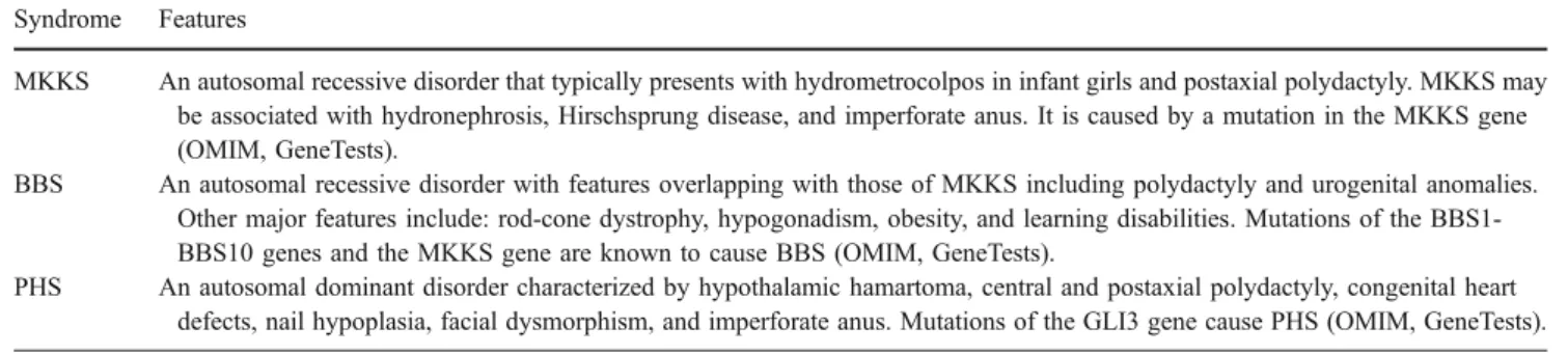
Table 2 Key features of MKKS, BBS and PHS Syndrome Features
MKKS An autosomal recessive disorder that typically presents with hydrometrocolpos in infant girls and postaxial polydactyly. MKKS may be associated with hydronephrosis, Hirschsprung disease, and imperforate anus. It is caused by a mutation in the MKKS gene (OMIM, GeneTests).
BBS An autosomal recessive disorder with features overlapping with those of MKKS including polydactyly and urogenital anomalies. Other major features include: rod-cone dystrophy, hypogonadism, obesity, and learning disabilities. Mutations of the BBS1-BBS10 genes and the MKKS gene are known to cause BBS (OMIM, GeneTests).
PHS An autosomal dominant disorder characterized by hypothalamic hamartoma, central and postaxial polydactyly, congenital heart defects, nail hypoplasia, facial dysmorphism, and imperforate anus. Mutations of the GLI3 gene cause PHS (OMIM, GeneTests).
PHS caused by the GLI3 mutation (Q717X) identified in our patient has been described in only one previous patient [5]. Corresponding clinical manifestations in our patient were hypothalamic hamartoma, insertional and postaxial polydactyly, and nail hypoplasia. However, our patient did not show a bifid epiglottis, dental hypoplasia, imperforate anus and (at the time of this report) no seizures, whereas the genitourinary malformations were not seen in the previous-ly described patient, suggesting that there is no genotype– phenotype correlation in PHS.
As the GLI3 mutation (Q717X) was detected in our patient, but not in her parents, it is most likely to have been a de novo mutation. As there has been only one previously described patient with germ-cell mosaic [6], its presence in the parents and the likelihood of recurrence in prospective offspring is very low (<1%).
In conclusion, we present the second genetically con-firmed association of PHS with hydrometrocolpos due to vaginal atresia. In addition we demonstrate that this association may also include congenital heart defects and hydronephrosis as described above, which were not detected in the patient described by McCann et al. [5]. Therefore, hydrometrocolpos due to vaginal atresia is an overlapping feature of PHS and MKKS.
References
1. Hall JG, Pallister PD, Clarren SK et al (1980) Congenital hypothalamic hamartoblastoma, hypopituitarism, imperforate anus and postaxial polydactyly – a new syndrome? Part I: clinical, causal, and pathogenetic considerations. Am J Med Genet 7:47–74
2. Topf KF, Kletter GB, Kelch RP et al (1993) Autosomal dominant transmission of the Pallister-Hall syndrome. J Pediatr 123:943–946 3. Johnston JJ, Olivos-Glander I, Killoran C et al (2005) Molecular and clinical analyses of Greig cephalopolysyndactyly and Pallister-Hall syndromes: robust phenotype prediction from the type and position of GLI3 mutations. Am J Hum Genet 76:609–622 4. Unsinn KM, Neu N, Krejci A et al (1995) Pallister-Hall syndrome
and McKusick-Kaufmann syndrome: one entity? J Med Genet 32:125–128
5. McCann E, Fryer AE, Craigie R et al (2006) Genitourinary malformations as a feature of the Pallister-Hall syndrome. Clin Dysmorphol 15:75–79
6. Ng D, Johnston JJ, Turner JT et al (2004) Gonadal mosaicism in severe Pallister-Hall syndrome. Am J Med Genet A 124:296–302 7. GeneTests (2008) Medical genetics information resource (online
database). Copyright University of Washington, Seattle. Updated weekly.http://www.genetests.org. Accessed 14 April 2008 8. Online Mendelian Inheritance in Man, OMIM (2008)
McKusick-Nathans Institute of Genetic Medicine, Johns Hopkins University (Baltimore, MD) and National Center for Biotechnology Informa-tion, National Library of Medicine (Bethesda, MD). http://www. ncbi.nlm.nih.gov/omim/. Accessed 17 Apr 2008